Article

Influence of Plant Organs and Functional Traits on the Structure of Bacterial and Fungal Communities in Three Acer Species

1
School of Ecology, Northeast Forestry University, Harbin 150040, China
2
Key Laboratory of Sustainable Forest Ecosystem Management—Ministry of Education, Northeast Forestry University, Harbin 150040, China
3
Northeast Asia Biodiversity Research Center, Northeast Forestry University, Harbin 150040, China
*
Author to whom correspondence should be addressed.
Forests 2025, 16(6), 875; https://doi.org/10.3390/f16060875
Submission received: 20 April 2025 / Revised: 13 May 2025 / Accepted: 18 May 2025 / Published: 22 May 2025
(This article belongs to the Section Forest Ecology and Management)

Abstract

:
Plants and the microorganisms living on their surfaces are an inseparable community that interacts with plant functional traits and influences plant growth, but the differences in microorganisms between plant organs and their relationship with plant functional traits have not been deeply explored. In this study, we used high-throughput sequencing to investigate the variation of microorganisms across different plant organs (leaves, twigs, trunks, and roots) of three species (Acer pictum subsp. mono, Acer tegmentosum, and Acer ukurunduense) in the Xiaoxing’an Mountains in Northeastern China and explored the relationship between microorganisms and plant functional traits. The results indicate that rhizosphere microorganisms have a high diversity. Plant organs explained 52.1% and 32.3% of the variations in bacterial and fungal community structures, respectively. The core microbiome consists of the phyla Proteobacteria and Actinobacteria in bacteria and the phyla Ascomycota and Basidiomycota in fungi. Plant functional traits such as specific leaf area and non-structural carbohydrates, as well as soil total carbon and total phosphorus content, were significantly correlated with microbial community composition. The results highlight that host plant organ characteristics are key drivers of variation in plant-associated microbial communities. By elucidating the regulatory role of host traits in microbiome assembly, our findings provide new mechanistic insights into plant–microbe interactions and ecological coexistence strategies.

1. Introduction

Plant microorganisms consist of a complex and diverse microbial community, including bacteria, archaea, fungi, and viruses, which inhabit the surface and interior of the plant host [1,2,3]. Diverse microbial communities are tightly associated with their host plants, influencing plant health and ecological functioning by regulating the plant’s ability to utilize nutrients [4] or by providing protection against severe external biotic or abiotic stresses [5,6,7]. Plant microorganisms have demonstrated that they are affected by many factors, such as climate and geographic location (such as evergreens in tropical areas and deciduous plants in temperate areas), and host genotypic diversity significantly impacts the structure of the phyllosphere microbial community [8,9], and changes in host habitat drive variations in the structure of the rhizosphere microbial community [10,11].
The plant surface is not a homogeneous transitional habitat, as significant variations in resource availability and nutrient supply exist between different plant organs, such as leaves, twigs, trunks, and roots [12,13]. Consequently, the diversity, structure, and function of the microbial community associated with the same plant exhibit considerable variability across different organs [14,15]. For instance, a recent study indicated that the plant organ was the primary factor driving the diversity and community structure of fungi in Betula platyphylla Sukaczev and Corylus avellana L., while the host species and geographical location had relatively minor effects [16]. Additionally, the role of habitat filtering due to organ-specific differences has been highlighted as a key factor influencing microbial community composition in Agave species and Cacti species [17,18]. On the other hand, below-ground microbial communities of plants generally showed higher diversity and more complex community structures than above-ground microbial communities [19,20]. All of these indicate that the differences in the composition of bacterial and fungal microbial communities primarily occur due to habitat differentiation [21] and highlight the significance of differences among plant organs in shaping microbial community diversity [22,23,24]. Furthermore, environmental factors, as well as the plant’s own physiological traits and structural characteristics, also influence microbial community composition [25,26,27,28]. For example, the lighting environment can affect the synthesis of secondary metabolites, such as flavonols and gibberellins, in plants, thereby impacting the composition of microbial communities [29]. Consequently, variations in forest canopy structure, such as forest gaps and non-forest gaps, and plant size, may induce differentiation in microbial community composition due to changes in light exposure across these environments [30]. However, whether the extent to which microorganisms within individual plants vary in different organs is consistent across gap environments and plant sizes remains underexplored.
Plant functional traits reflect the plant’s resource utilization strategy and exert a significant influence on microbial community structure [31]. Leaf traits, such as specific leaf area (SLA) and leaf area (LA), play a crucial role in the colonization of phyllosphere microorganisms [32]. Also, leaf total phosphorus content was found to be strongly correlated with the abundance of phyllosphere fungi, with higher phosphorus levels leading to a marked increase in the abundance of Chytridiomycota [33]. These findings suggest that phyllosphere microbial communities are related to the plant’s ability to obtain resources. Plants with acquisition-type resource strategies tend to have higher nutrient contents, thereby providing more resources for microorganisms, which benefits the survival and proliferation of copiotrophic microbial species. For twigs, a significant positive correlation was also observed between twig length (TL) and twig diameter (TD) and fungal diversity, indicating that a larger living space facilitates microbial colonization and community enrichment [34]. For trunks, a study demonstrated that the total phenolic content (TPC) plays a crucial role in driving variations in fungal community composition [35]. Plant microbial communities are also influenced by soil physicochemical properties, with soil nitrogen and phosphorus being key limiting factors for microbial growth [4,36]. Soil pH significantly affects the abundance of specific microbial groups, such as Acidobacteria, and serves as an important driver of the co-evolutionary dynamics between plant hosts and their symbiotic microorganisms [37]. However, the relationship between microbial variations among plant organs and plant functional traits is unclear.
In this study, we investigated how plant functional traits influence the microbial variations of plant organs, forest gap environment, and plant size. We selected three representative broad-leaved tree species (Acer pictum subsp. mono (Maxim.) Ohashi, Acer tegmentosum Maxim, and Acer ukurunduense Trautv. & C. A. Mey.) from the climax broad-leaved Korean pine (Pinus koraiensis Siebold & Zucc.) forest in the eastern mountainous region of Northeast China. The study aimed to determine the diversity of microbial communities on the below-ground and above-ground organs (leaves, twigs, trunks, and roots) of host trees in different environments (forest gaps and non-forest gaps) and at varying plant size stages (large trees and small trees). Furthermore, we assessed the functional traits of plant organs and the physical and chemical properties of the rhizosphere soil. First, we hypothesized that plant organs are the most important factor affecting plant microbial variation, while tree species, forest gap, and plant size have relatively low explanatory power because the microenvironmental variation between organs is greater than other conditions; therefore, the microbial variation is greater (H1). Secondly, we hypothesized that plant functional traits related to the economic spectrum, such as total phosphorus, would be significantly correlated with the proportion of copiotrophic and oligotrophic microbial groups because the acquisition strategy could provide more appropriate nutrients for copiotrophic microorganisms, which is beneficial to their survival (H2). This research contributes to elucidating the interactions between plant and microbial communities, thereby enriching the understanding of host–microbe relationships and microbial ecological function.

2. Materials and Methods

2.1. Study Site

The study site is located in the Liangshui National Nature Reserve in Heilongjiang Province (47°10′50″ N, 128°53′20″ E) in Northeastern China. The region’s topography is complex and varied, with elevations ranging from 280 to 707 m and an average altitude of 430 m, characterized as a typical low mountainous and hilly area. The area experiences a temperate continental monsoon climate, with precipitation predominantly occurring in the summer months. The average annual precipitation is 694 mm, and winters are cold and prolonged, with an average annual temperature of 1.52 °C. The dominant vegetation type in the region is a temperate mixed coniferous and broad-leaved forest dominated by Korean pine, and it is accompanied by a variety of broad-leaved plants, such as those from the maple family. Dark brown soil is the dominant soil type in this area [38].

2.2. Sample Collection

Three broad-leaved dominant species, A. pictum subsp. mono, A. tegmentosum, and A. ukurunduense, were selected for sampling in the broad-leaved Korean pine forest on sunny and windless days from August 2022. Three experimental conditions were established: large trees in forest gaps, small trees in forest gaps, and small trees in the non-forest gaps. We ensured that the selected sample trees were of similar height and that the altitudes and aspects of the sampling locations were similar, in order to exclude the effect of terrain factors. For each experimental condition, 5 individuals of each tree species were selected as biological replicates, with a minimum distance of 20 m between each sample tree. Samples were collected from four organs (leaves, twigs, trunks, and rhizosphere) of each sample tree, representing the microenvironment of each organ. Four distinct sample types were collected: leaves, current-year twigs (with twig tips extending to the first bud scale scar as new growth) [39], bark tissue, and rhizosphere soil. In this experiment, we used the rhizosphere soil samples from the root surface to represent the root organ microenvironment. Detailed characteristics of the sample trees are provided in Table S1.

2.3. Collection of Microbial Samples

All samples were collected with sterile gloves, and all laboratory tools were sterilized before each sampling event to prevent cross-contamination between different sample types. A sufficient quantity of fresh, intact, and undamaged leaves, as well as current-year twigs, was carefully selected from each tree and placed into sterile bags. Bark tissue was scraped using a sterilized scraper and transferred into a sterile bag. All samples of leaves, twigs, and trunks of the above-ground plant parts were treated in the same way. The specific steps were as follows: A 5 g aliquot of each sample was weighed and placed into sterile centrifuge tubes; then, sterile phosphate-buffered saline (PBS) was added to each tube at a 1:10 ratio, and the mixture was subjected to thorough shaking using both an ultrasonic cleaner and a vortex mixer. This process was repeated three times to ensure the complete suspension of microbial communities. The resulting microbial suspension was subsequently filtered through a 0.22 µm sterile filter membrane. The filter membrane was then transferred to a sterile centrifuge tube and stored at −80 °C for further microbial analysis.
For rhizosphere soil samples, we used a sterilized shovel to dig out the plant root system in the shade environment of tree canopies, starting from the main root and moving downward until reaching the terminal root system with intact fine roots. Then, sterile gloves were worn by the experimenters to maintain aseptic conditions, the roots were gently shaken to remove loose soil, and the rhizosphere soil adhering to the root surface was carefully brushed off using a sterilized soft-bristle brush. The soil collected from between the roots was placed into 50 mL sterile centrifuge tubes and immediately stored at −80 °C to preserve microbial DNA. For each sample, two aliquots were prepared: one for the determination of plant functional traits and the other for microbial sequencing. All samples were shipped on dry ice to Majorbio Corporation (Shanghai, China) for DNA extraction and microbial sequencing analysis. Microbial samples were collected from the leaf, twig, bark, and rhizosphere soil, which represent the microbial communities of the leaf, twig, trunk, and root organ microenvironments, respectively.

2.4. DNA Extraction and PCR Amplification

DNA extraction was performed using the E.Z.N.A.® Soil DNA Kit (Omega Bio-tek, Norcross, GA, USA), and DNA quality was assessed using 1% agarose gel electrophoresis. Prior to the assay, samples were thawed on ice, thoroughly mixed, and centrifuged. A suitable volume of the samples was then used for the assay, which was conducted with a voltage setting of 5 V·cm−1 for 20 min. DNA concentration and purity were determined using a NanoDrop 2000 spectrophotometer (Thermo Scientific, Wilmington, DE, USA). For amplification of the V3–V4 region, PCR primers were selected as follows: for bacteria, 338F (5′-ACTCCTACGGGGAGGCAGCAG-3′) and 806R (5′-GGACTACHVGGGGTWTCTAAT-3′); for fungi, ITS1F (5′-CTTGGTCATTTAGAGGAAGTAA-3′) and ITS2R (5′-GCTGCGTTCTTCATCGATGC-3′). Bacterial PCR amplification was carried out using the TransGen AP221-02: TransStart FastPfu DNA Polymerase system, which contained 4 µL of FastPfu Buffer (5×), 2 µL of 2.5 mM dNTPs, 0.8 µL of each primer (forward and reverse, 5 µM), 0.4 µL of FastPfu Polymerase, 0.2 µL of BSA, and 10 ng of template DNA. Fungal PCR amplification was performed using the Takara rTaq DNA Polymerase system, consisting of 2 µL of Buffer (10×), 2 µL of 2.5 mM dNTPs, 0.8 µL of each primer (forward and reverse, 5 µM), 0.2 µL of rTaq Polymerase, 0.2 µL of BSA, and 10 ng of template DNA. PCR products were purified using the AxyPrep DNA Gel Extraction Kit (Axygen Biosciences, Union City, CA, USA), followed by detection via 2% agarose gel electrophoresis. Quantification of the purified PCR products was conducted using the Quantus™ Fluorometer (Promega Corporation, Madison, WI, USA). Sequencing was performed on an Illumina MiSeq PE300 platform (Illumina, San Diego, CA, USA).

2.5. Preprocessing of Sequencing Data

The paired-end sequences obtained from Illumina sequencing were merged and quality controlled (QC). Filtering was performed using FLASH software (version 1.2.11) to eliminate sequences shorter than 200 bp and to remove chloroplast and mitochondrial sequences. A 10 bp sliding window was set, and the average base quality value within the window was calculated. If the average value was less than 20, low-quality bases were truncated from the end of the sequence. At the same time, reads containing N bases were filtered out, and sequences with end quality values less than 20 were removed. For sequences with overlapping regions exceeding 10 bp, if their mismatch density did not exceed 0.2, they were merged. Chimeras were removed after quality control and sequence assembly, and representative sequences were obtained. The remaining sequences were clustered into operational taxonomic units (OTUs) at a 97% similarity threshold using UPARSE software (version 7.0.1090), and chimeric sequences were removed. OTU taxonomic classification was performed by comparing the sequences to the SILVA 16S rRNA database for bacteria (Release 138; http://www.arb-silva.de accessed on 16 December 2022) and the UNITE ITS database for fungi (Release 8.0; http://unite.ut.ee/index.php accessed on 16 December 2022). To mitigate the impact of sequencing depth, all samples were rarefied to a uniform sequence number, ensuring that the average sequence coverage for each sample exceeded 99%. The resulting sample dilution curve is presented in Figure S1. The bacterial community identified in this study was classified into 35 phyla, 91 classes, 199 orders, 328 families, 612 genera, and 1380 species. The fungal community identified in this study was classified into 13 phyla, 52 classes, 152 orders, 362 families, 806 genera, and 1327 species.

2.6. Measurement of Plant Functional Traits

Leaf thickness (LT) was determined, using vernier calipers with an accuracy of 0.01 mm, at the anterior, middle, and terminal positions of the non-dominant leaf veins on the leaf blade, and the mean LT value was recorded. Leaf area (LA) was measured by scanning the leaf blades using a portable scanner (Canon LiDE400, Tokyo, Japan) in combination with leaf area calculation software (with an accuracy of 0.01 cm2). Saturated fresh mass of the leaves (g) was determined using an electronic balance (accuracy of 0.0001 g). Leaves were then dried at 65 °C to constant weight to obtain the dry mass, and specific leaf area (SLA, cm2·g−1) was calculated as the ratio of LA to dry mass. Leaf dry matter content (LDMC, g·g−1) was defined as the ratio of leaf dry mass to saturated fresh mass [40]. Twig length (TL) was measured as the full length of the whole twig using a graduated ruler with an accuracy of 1 mm. Twig diameter (TD) was measured as the diameter of the transverse section of the twig, using a vernier caliper with an accuracy of 0.01 mm. Non-structural carbohydrates (the sum of soluble sugars and starch, NSC) of plant tissues were determined via the phenol–sulfuric acid method [41]. Soil water content (SWC) was measured via the drying method and soil pH was determined by a potentiometric method. Determination of chemical traits among organs was achieved by the same method; total carbon content (TC) was determined by a multi N/C 2100S carbon and nitrogen analyzer (Analytik Jena AG, Jena, Germany); total nitrogen content (TN) was determined by an AA3 continuous flow analyzer after high-temperature digestion; and total phosphorus content (TP) was determined by a molybdenum–antimony colorimetric method [42]. Information on plant functional traits is summarized in Table S2.

2.7. Statistical Analyses

Permutational multivariate analysis of variance (PERMANOVA) was used to assess the degree to which different experimental conditions explained the variations in microbial communities. One-way analysis of variance (ANOVA) was employed to examine the differences in α-diversity indices (Chao1, Shannon, Simpson) among various organs. Differences in bacterial and fungal β-diversity among organs were analyzed using non-metric multidimensional scaling (NMDS) based on the Bray–Curtis distance algorithm, and significance was tested using the analysis of similarities (ANOSIM) method. A Venn diagram was used to show the number of shared and unique OTUs among different organs of the host plant. The composition of microbial communities at different taxonomic levels (phylum and genus) was statistically analyzed and presented using community bar charts. Linear discriminant analysis effect size (LEfSe) (LDA score > 4, p < 0.05) was used to identify microbial taxa with significant differences in abundance at the phylum-to-genera level among organs. One-way ANOVA was used to analyze the variability in relative abundance of dominant species (phylum and genus) among organs. One-way ANOVA was used to analyze differences in shared functional traits among plant organs. Redundancy analysis (RDA) was used to analyze the relationship between environmental factors, plant functional traits, and microbial communities. A correlation heatmap was used to analyze the magnitudes of correlation between microbial communities in different organs, plant functional traits, and environmental factors. All the above analyses and mappings were performed on the Majorbio BioCloud platform (https://cloud.majorbio.com accessed on 18 August 2024).

3. Results

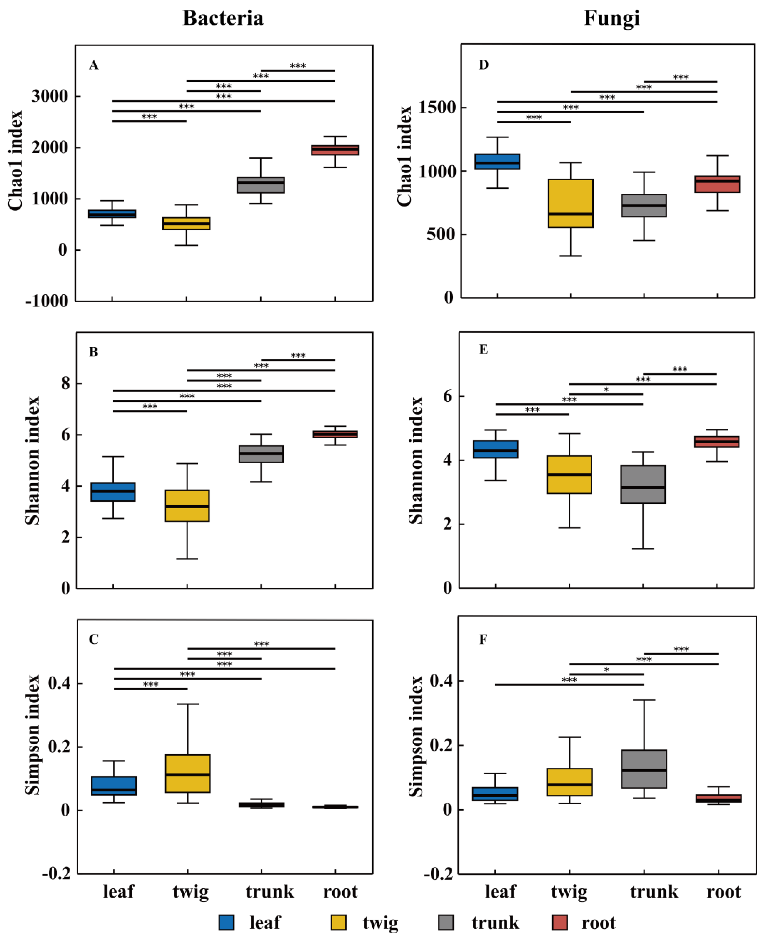
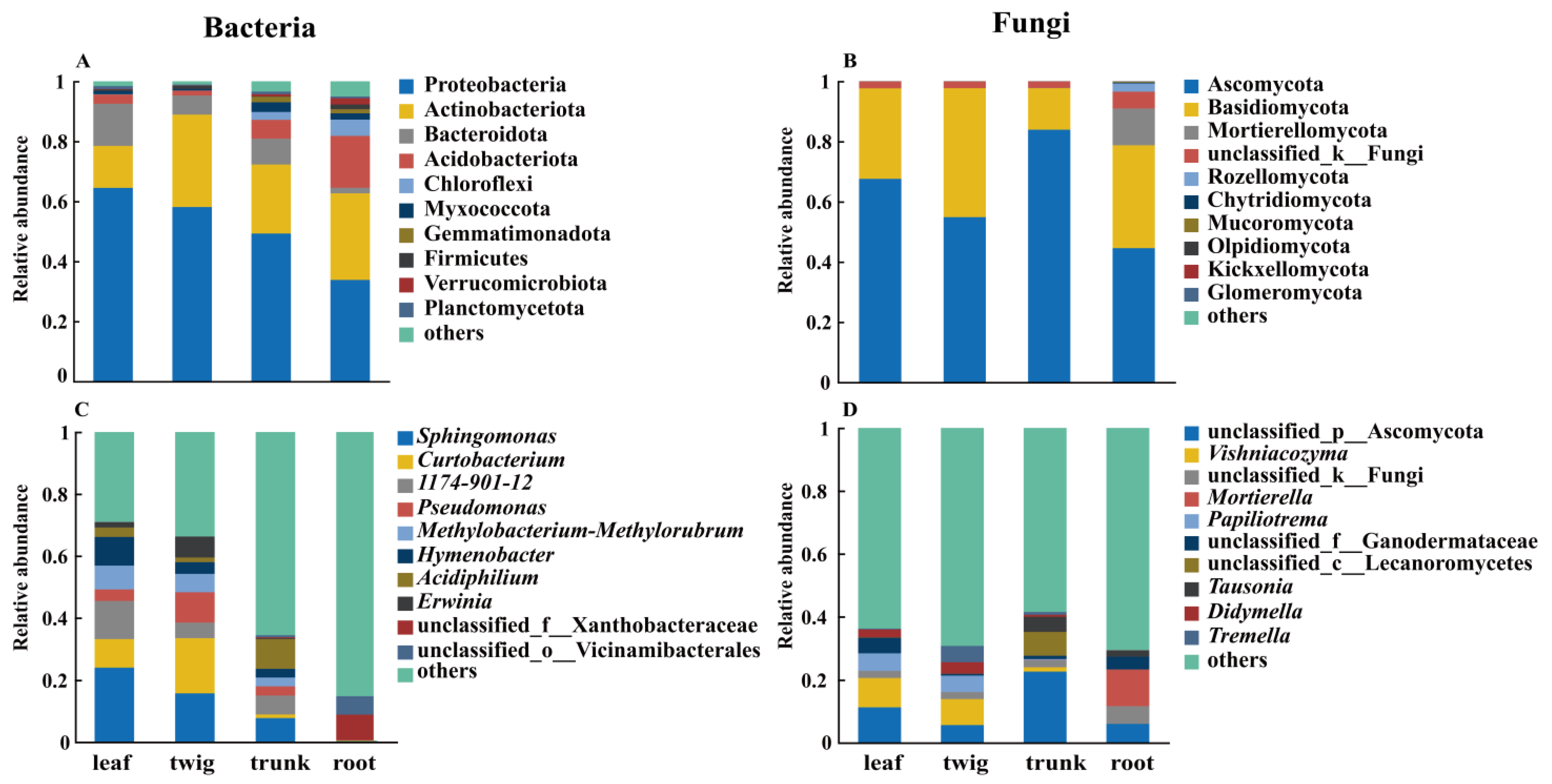
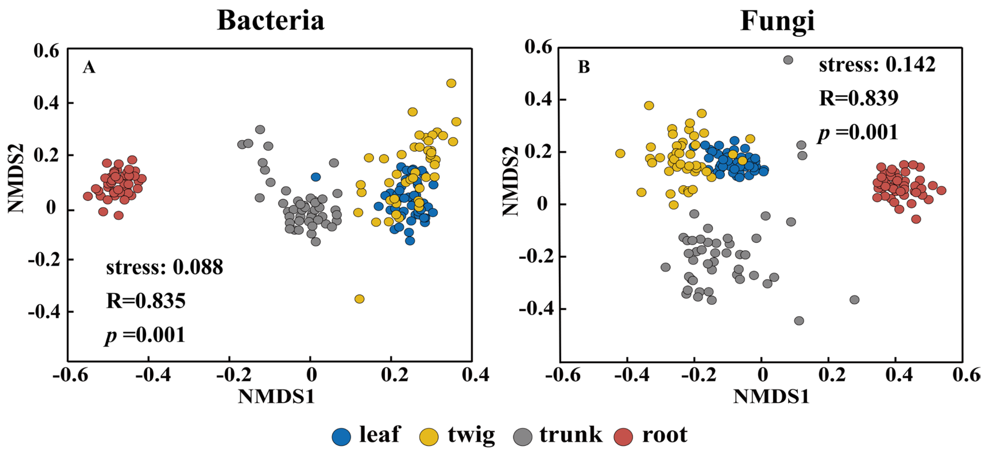
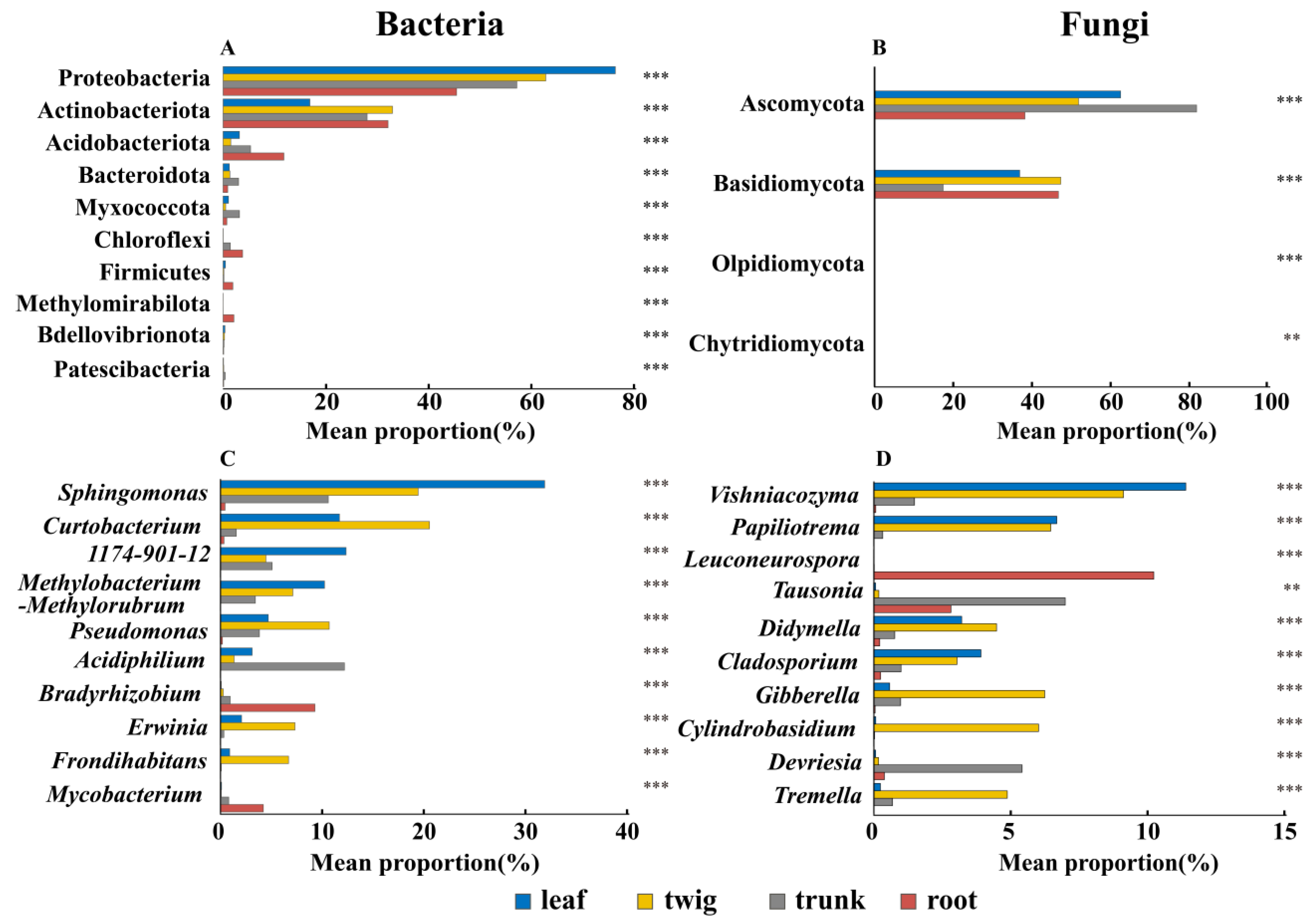
3.1. Differences in the Composition of Microbial Communities Among Different Organs

Plant organs had a more significant effect on the microbial community structure than tree species, forest gaps, and tree size. Specifically, plant organs accounted for 52.1% of the bacterial variation and 32.3% of the fungal variation (Table 1). Organ differences were an important influence on microbial community variation within plant individuals, followed by tree species. However, tree size and forest gaps had no significant effect on the structural variation of bacterial communities and explained only a minor proportion of fungal community variation (Table 1).
Microbial α-diversity varied significantly among the four plant organs (Figure 1). For bacteria, both Chao1 and Shannon indices in the rhizosphere were significantly higher than those in other plant organs (p < 0.001). Conversely, the Chao1 and Shannon indices for bacterial communities in the twigs were significantly lower than those in the other organs, while the Simpson index for twig bacteria was significantly higher (p < 0.001). No significant difference in the Simpson index was observed between the trunk and rhizosphere bacterial communities. For fungi, the Chao1 index of phyllosphere fungi was significantly higher than that of other organs, with values ranked as follows: leaf > root > twig > trunk. The Shannon index for fungi in the trunk was significantly lower than in other organs, while the Simpson index for trunk fungi was significantly higher than those of other organs.
Common microbial communities exist in different plant organs. The Venn diagram reveals that the numbers of unique OTUs in the tree trunks and rhizosphere were relatively high for both bacteria and fungi (Figure 2). Specifically, rhizosphere-specific bacterial OTUs accounted for 11.57%, and fungal OTUs accounted for 27.1%. The number of bacterial OTUs shared between organs amounted to 759 (18.64%), and the number of fungal OTUs shared amounted to 945 (27.1%). Furthermore, the proportion of common OTUs in above-ground parts (leaves, twigs, and trunks) was also notably high. Among these, bacterial common OTUs constituted 16.33%, and fungal common OTUs constituted 18.61%.
At the phylum level, Proteobacteria dominated the microbial composition in all plant organs, accounting for the highest relative abundance across all samples. In the phyllosphere, Bacteroidota (14.05%) ranked second in relative abundance, following Proteobacteria (64.47%). In the rhizosphere bacterial composition, the relative abundance of Actinobacteriota (28.87%) was lower than that of Proteobacteria (33.79%) but higher than Acidobacteriota (17.30%). Phyla such as Verrucomicrobiota, Firmicutes, and Planctomycetota showed consistently low relative abundances across all organ samples (Figure 3A). At the genus level, the microbial community composition varied significantly among plant organs. In the phyllosphere, Sphingomonas (23.99%) was the most abundant genus. In the twigs, Curtobacterium (17.72%) predominated, while in the trunk, Acidiphilium (9.5%) were dominant genera. In the rhizosphere, norank_f__Xanthobacteraceae (8.2%) also occupied a prominent position in the microbial community composition.
At the phylum level, Ascomycota and Basidiomycota were the most abundant, together accounting for approximately 78% to 97% of the total relative abundance within each organ’s fungal community composition (Figure 3B). At the genus level, the fungal communities in the phyllosphere and trunk were dominated by unclassified_p__Ascomycota (11.23% and 22.6%, respectively). In twigs, Vishniacozyma was the most abundant genus (8.26%). In the rhizosphere, Mortierella (11.68%) was the most dominant fungal genus.
NMDS analysis revealed significant differences (p < 0.05) in bacterial and fungal community compositions across plant organs (Figure 4). The microbial communities in the trunk and rhizosphere exhibited distinct differences from those in other organs, indicating substantial variability. Notably, pronounced distinctions were observed between below-ground (rhizosphere) and above-ground (trunk, twig, and leaf) microbial communities.

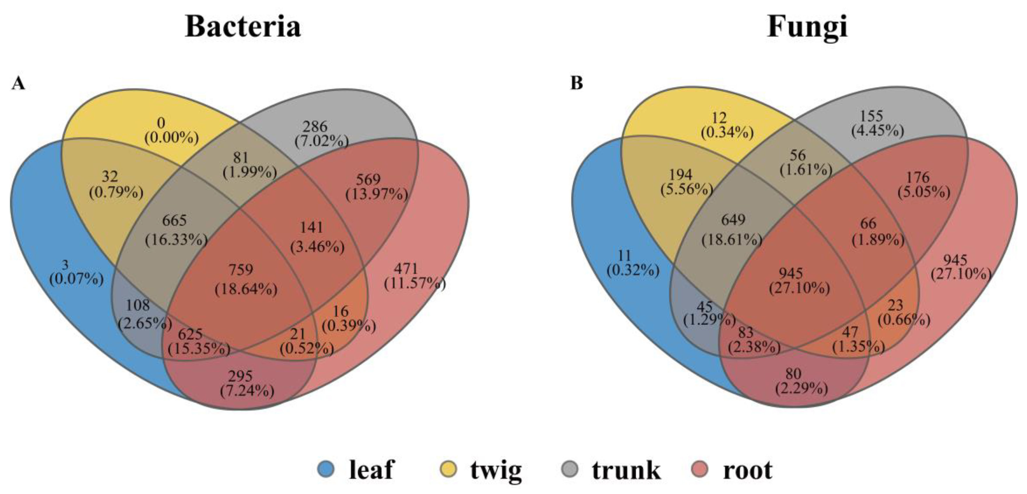
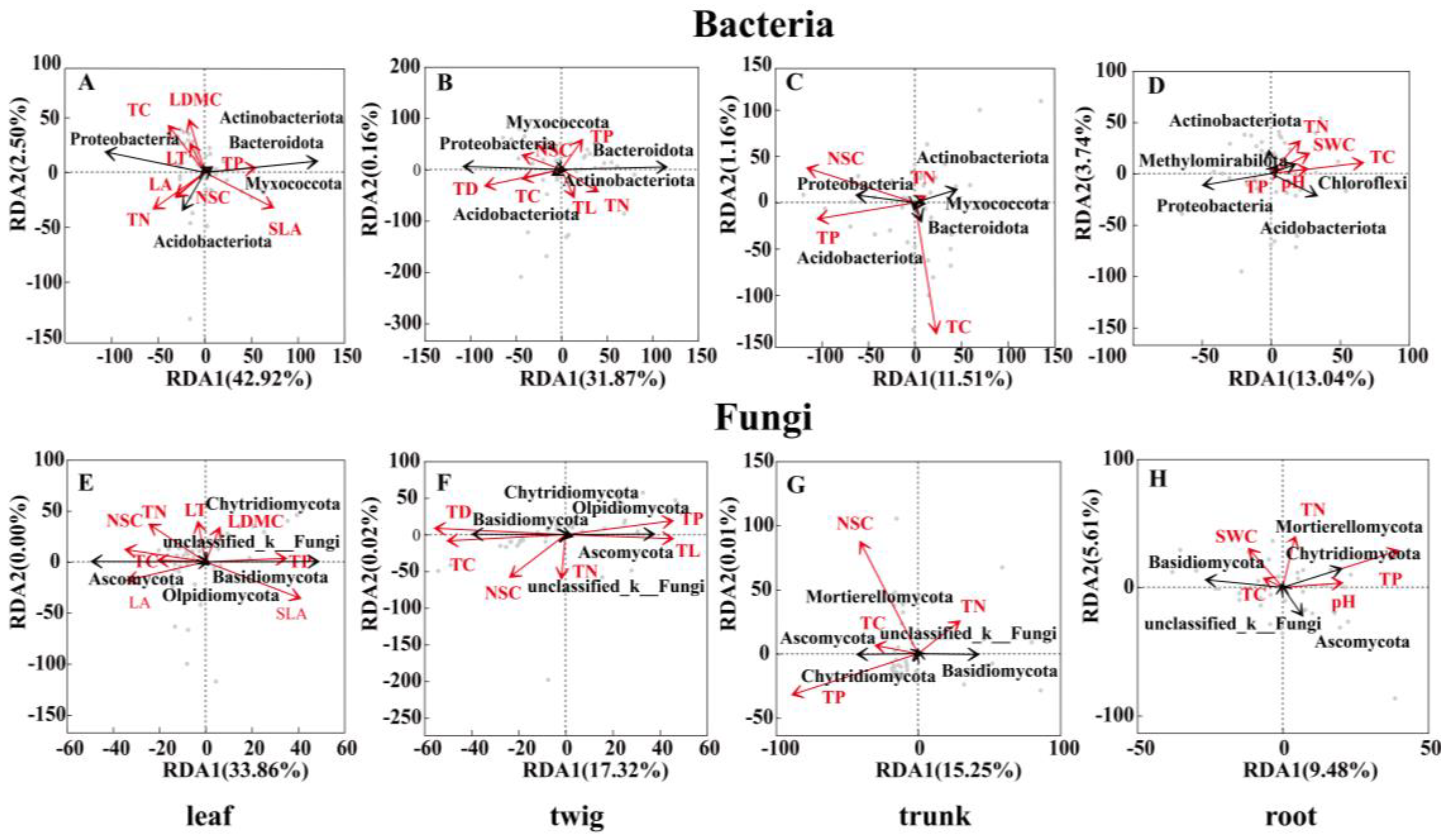
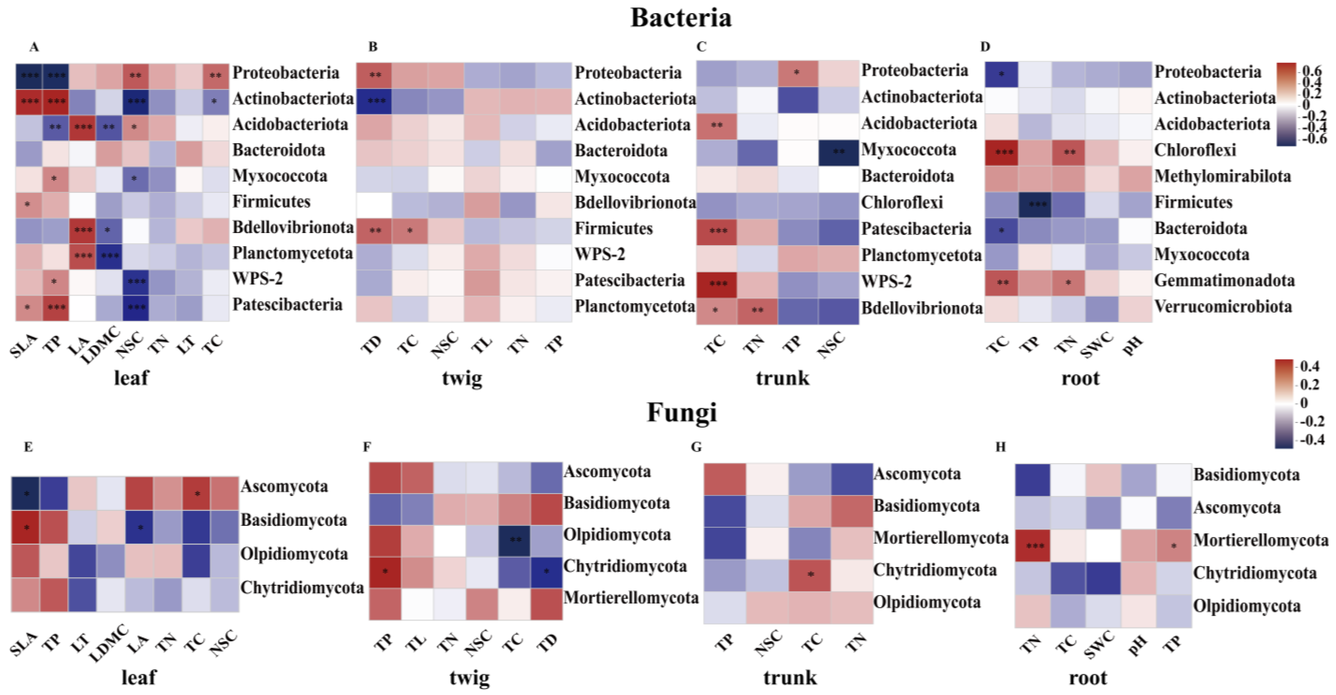
3.2. Core Microbiota and Their Relationship with the Environment and Plant Functional Traits

LEfSe analysis identified microbial taxa with significant differences across different plant organ habitats (Figure 5). The results reveal distinct microbial marker taxa in the bacterial community for each organ: 8 marker taxa in the phyllosphere, 8 in the twigs, 12 in the trunk, and 23 in the rhizosphere. Specifically, Proteobacteria and Bacteroidota were significantly enriched in the phyllosphere, while Microbacteriaceae was the predominant bacterial taxon in the twigs. In the trunk, Acetobacterales represented the dominant bacterial group, and in the rhizosphere, Acidobacteriota and Chloroflexi were significantly enriched.
For fungi, LEfSe analysis identified 19 phyllosphere fungal marker taxa, 14 twig marker taxa, 6 trunk marker taxa, and 8 rhizosphere marker taxa. In the phyllosphere, the majority of fungal taxa were from Ascomycota (Eurotiomycetes) and Basidiomycota (Polyporales). In the twigs, Basidiomycota (Tremellales) was the most abundant group, while Ascomycota was significantly enriched in the trunk. The fungal community in the rhizosphere was predominantly composed of Ascomycota (Leotiomycetes) and Basidiomycota (Agaricomycetes).
Inter-group comparisons revealed significant differences (p < 0.001) in microbial relative abundances across organs at multiple taxonomic levels. Proteobacteria (76.44%) and Sphingomonas (31.93%) showed significantly higher relative abundances in the phyllosphere compared to other organs (p < 0.001). Actinobacteriota (33.01%) and Curtobacterium (20.60%) were significantly enriched in twigs (p < 0.001). Bacteroidota (3.02%) and Acidiphilium (12.23%) in the trunk had significantly higher relative abundances than those in other organs (p < 0.001). Acidobacteriota (11.84%) and Bradyrhizobium (9.33%) exhibited significantly higher abundances in the rhizosphere than in other organs (Figure 6A,C).
For fungi, Ascomycota and Basidiomycota were the dominant phyla in all organ habitats. Basidiomycota reached its highest relative abundance in twigs (47.41%), significantly exceeding levels in other organs (Figure 6B, p < 0.001). Ascomycota in the trunk had the highest relative abundance (81.93%), significantly exceeding other organs (p < 0.001). At the genus level, the relative abundances of Vishniacozyma (11.41%) and Papiliotrema (6.7%) in the phyllosphere were significantly higher than their abundances in the trunk and rhizosphere habitats (p < 0.001). Gibberella (6.25%) was significantly enriched in twigs (p < 0.001). Tausonia (7%) showed significantly higher abundance in the trunk than in the phyllosphere and twigs (p < 0.001). Leuconeurospora (10.24%) showed a higher enrichment degree in the rhizosphere (Figure 6D, p < 0.001).
RDA revealed significant effects of environmental factors and plant functional traits on microbial composition across plant organs. In the phyllosphere, microbial composition was strongly associated with SLA and NSC. The first two RDA axes explained 42.92% and 2.5% of bacterial community variation, respectively, cumulatively accounting for 45.42% (Figure 7A). In twigs, TD was the primary factor influencing microbial composition (Figure 7B). In the trunk, TC and NSC had the most significant impact on microbial composition (Figure 7C), whereas rhizosphere bacterial composition was primarily driven by TC (Figure 7D). For fungi, phyllosphere community composition correlated with SLA and TC (Figure 7E). TD significantly influenced fungal composition in twigs (Figure 7F). In the trunk, microbial composition was most strongly affected by NSC and TP (Figure 7G), while rhizosphere fungal communities were predominantly influenced by TP (Figure 7H).
Correlation analysis identified significant associations between bacterial phyla and plant functional traits/environmental factors, including soil physicochemical properties (Figure 8). In the phyllosphere, SLA showed a significant negative correlation with Proteobacteria (p < 0.001) and a positive correlation with Actinobacteriota (p < 0.001). In twigs, TC positively correlated with Firmicutes (p < 0.05), whereas trunk TC correlated with WPS-2 (p < 0.001). Additionally, TP was significantly negatively correlated with Firmicutes (p < 0.001). For fungi, in the phyllosphere, SLA was negatively correlated with Ascomycota (p < 0.05), whereas twig TC showed a negative correlation with Olpidiomycota (p < 0.01). In the trunk, TC positively correlated with Chytridiomycota (p < 0.05). Rhizosphere soil TN and TP both positively correlated with Mortierellomycota (p < 0.05) (Figure 8).

4. Discussion

4.1. Organ Differences Determine Microbial Diversity and Community Structure

Consistent with Hypothesis 1, plant organs exerted the strongest influence on microbial variation. Rhizosphere bacterial abundance and diversity were significantly higher than in above-ground plant parts (Figure 1). This difference is mainly attributed to the microenvironmental differences caused by host plants and plant organ types [15]. Specifically, niche differentiation mechanisms drive significant variations in microbial community characteristics among different plant organs. Above-ground plant parts, such as leaves and twigs, are exposed to atmospheric conditions where water stress and light radiation limit microbial colonization, resulting in lower microbial abundance in these environments [43]. The soil environment offers a stable microbial habitat, offering protection from harsher environmental stresses compared to the above-ground environment [44]. Additionally, at the microscale, microenvironment of the root system, nutrient inputs from plant litter decomposition and deposition in the root microenvironment supply abundant nutrients for rhizosphere microorganisms, while the regulatory influence of soil microorganism predators further enhances the diversity and abundance of rhizosphere bacterial communities [3,45]. Plant-associated microorganisms predominantly originate from soil dispersal or airborne deposition [4,44]. Bacterial community composition is most sensitive to host surface microenvironments, driven primarily by plant genotype and organ-specific traits, whereas tree size has minimal influence [15,46]. Tree species shape bacterial communities through modulation of plant functional traits and physiological processes, thereby regulating microbial colonization [47]. Bacterial communities exhibit low sensitivity to light gradients, likely explaining the negligible impact of canopy environmental variation on their structure [17]. This aligns with Cregger et al. [19], who demonstrated that habitat heterogeneity is a key driver of bacterial diversity and community composition.
Likewise, plant organs significantly influence fungal distribution, but distinct patterns emerged between fungi and bacteria. Notably, the fungal Chao1 index was higher in phyllosphere environments, potentially driven by airborne spore dispersal facilitating community assembly. Katsoula et al. [48] showed that summer leaf nutrient enrichment promotes spore deposition and colonization, enhancing phyllosphere fungal proliferation. Furthermore, fungi and bacteria exhibit notable differences in their distribution and ecological functions. Fungi generally demonstrate greater tolerance to dry environments and external stressors compared to bacteria [49]. And the interaction between bacteria and fungi also affects the difference in their respective abundances. For example, fungi have a certain inhibitory effect on bacteria, because their extensive hyphal networks enable more efficient nutrient uptake from host plants through leaf stomata, which enhances fungal colonization in the phyllosphere [35,50]. Trunk fungal communities exhibited the lowest Shannon diversity indices among plant organs. This discrepancy can potentially be attributed to the seasonal expansion and contraction of the bark. Additionally, the filtering effect of the environment on the trunk surface as the tree grows and develops has been proposed as a possible cause of the decrease in trunk fungal diversity [13,34]. In contrast, the selective nature of root exudates further influences the composition of rhizosphere fungi, which mostly form symbioses with plants that inhibit their diversity differentiation; they tend to have simpler network structures than phyllosphere fungi [15,51]. Collectively, these microbial adaptations to their respective environments have shaped the distinct distribution patterns of microbial communities in both the phyllosphere and rhizosphere.

4.2. The Structural Differences and Changes of the Core Microbial Community

Core microbial groups are present across various plant organs, although their abundance varies among different organs. Proteobacteria and Actinobacteriota dominated at the phylum level. Proteobacteria are considered the marker taxa for the phyllosphere microbiome. The rapidly fluctuating leaf environment exposes leaf-associated microbes to more severe environmental stresses compared to other habitats [52,53]. Among the Proteobacteria, Sphingomonas of the Alphaproteobacteria, as an oligotrophic bacterium, exhibits relatively strong tolerance and can effectively absorb nutrients from the host surface, thereby growing rapidly and being better able to adapt to the harsh environment of above-ground organs [24]. At the same time, during the growth period, leaves usually have a higher nutrient content and exhibit a “rapid investment-reward” strategy that facilitates nutrient accumulation, thus providing favorable conditions for bacterial colonization [51]. However, the small surface area of twigs presents challenges in retaining adequate moisture for plant utilization, which favors the survival of drought-resistant Actinobacteria while limiting the colonization of acidophilic taxa such as Acidobacteria. Furthermore, twigs, which primarily function as structural supports for trees, possess a higher organic carbon content that facilitates the rapid growth and reproduction of Proteobacteria. These findings are consistent with some research [54,55]. A study indicated that Acetobacteraceae (belongs to Alphaproteobacteria) might represent a specialized group within the trunk microbiome, requiring extended periods for growth and, therefore, being better suited to colonize tissues with slow growth such as bark or older branches [22]. However, there are studies showing Firmicutes as the dominant group in the trunk [56]. This discrepancy may be attributed to variations in drought and salinity stress intensities experienced by the host plants. Further research is required to clarify the underlying mechanisms driving these differences. Acidobacteriota exhibited a marked enrichment in rhizosphere microorganisms, with significantly higher abundance compared to other organs (Figure 6A). This is related to the characteristics of microorganisms. Acidobacteriota are well adapted to acidic soil environments, and the acidic pH in the study area may further support the dominance of Acidobacteriota in the rhizosphere [57,58].
In fungi, Ascomycota and Basidiomycota are the dominant phyla across all samples. Ascomycota holds a particularly prominent position in the composition of plant fungal communities, a finding that has been consistently reported in numerous studies [24,59]. In the phyllosphere, fungi are primarily dominated by Dothideomycetes (belongs to Ascomycota) and Eurotiomycetes (belongs to Ascomycota) [60]. Similarly, a study of olive trees in the Mediterranean region observed that Davidiellaceae (belongs to Dothideomycetes) exhibits strong tolerance to stressors such as ultraviolet radiation on the leaf surface [10]. Furthermore, Dothideomycetes play a crucial role in leaf abscission and the decomposition of plant litter, which facilitates their colonization of the phyllosphere [46]. For genera, Tremella and Gibberella exhibit higher richness in twigs, a pattern commonly observed in bark tissue in previous reports [61]. This may reflect a transitional dispersal of fungal hyphae within the environment, leading to community differentiation [49]. In tree trunks, Ascomycota is the most abundant fungal phylum, with Dothideomycetes, a class within Ascomycota, demonstrating a distinct adaptation to high ultraviolet radiation and nutrient-poor environments [35]. The competitive advantage of Dothideomycetes in these conditions likely accounts for the higher proportion of Ascomycota fungi in the tree trunk [54]. Plants further influence the complexity of plant–microbe interaction networks by selecting for fungal taxa that are better adapted to specific host and environmental conditions, driven by both host-specific and filtering effects of different plant organs’ microenvironments [20,62].

4.3. The Relationship Between Plant Functional Traits and Microbial Community

Consistent with Hypothesis 2, plant functional traits significantly affected the microbial composition among organs. Moreover, plant leaf functional traits play a crucial role in driving microbial colonization and influencing community functional transitions. For instance, SLA serves as a key indicator of plant growth strategy and affects the distribution of microorganisms, while NSC reflects the availability of nutrients in the environment, influencing microbial colonization and abundance [45]. In the phyllosphere, Actinobacteriota were significantly positively correlated with SLA but negatively with NSC (Figure 8A). This pattern suggests that metabolic activities of Actinobacteriota rely on higher leaf growth rates and lower carbon storage. Plants with higher SLA typically exhibit lower NSC content and adopt a fast-growing “acquisition” strategy for resource investment [63,64]. In contrast, Proteobacteria demonstrated inverse relationships: negative with SLA and positive with NSC. This suggests that Proteobacteria may maintain their metabolic activity and ecological niche advantage by accessing plant-derived carbohydrates, with NSC serving as a rich carbon source to support their colonization and growth [4,15]. Moreover, Gammaproteobacteria (belongs to Proteobacteria), as copiotrophs, are also more adaptable to environments with higher nutrient utilization efficiency. These findings suggest that plant–microbe interactions co-regulate plant nutrient resource utilization strategies [8]. Additionally, most bacterial phyla exhibited a negative correlation with NSC. This may be attributed to the transient accumulation of NSC during the early stages of drought and high-temperature stress in plants, which leads to an increase in the concentration of secondary metabolites secreted by the plant [63]. In this context, more nutrient resources are diverted toward defense traits, thus limiting microbial activity [43]. Larger twigs offer enhanced mechanical support and facilitate the accumulation of microbial communities that can decompose complex carbon sources, such as those of Proteobacteria [54]. TP was shown to have a significant correlation with most bacterial phyla in the above-ground parts of the plant (leaves, twigs, and trunks), suggesting that plant phosphorus content may influence microbial composition by regulating nutrient availability [9]. Moreover, soil pH was widely recognized as an important factor driving variation in bacterial community structure and function [37]. However, no significant relationship was observed between soil pH and bacterial community structure in this study. This lack of correlation may be due to the low heterogeneity of soil environments within the same woodland type, where a convergence effect maintains the microbial community in a relatively stable equilibrium state [65].
Plant functional traits play a critical role in shaping the fungal community composition [16,66]. For instance, SLA and LA were significantly correlated with the composition of fungal communities on leaf surfaces (Figure 8E). This significant negative correlation between SLA and Ascomycota may be attributed to the fact that under higher SLA conditions, leaves exhibit enhanced resource acquisition capabilities, which may not favor the growth of saprophytic fungi that thrive in nutrient-poor environments [17]. In contrast, SLA was positively correlated with Basidiomycota, likely because these fungi can form symbiotic relationships with photosynthetic autotrophic algae to acquire nutrients. Additionally, larger SLA values allow for greater light capture, which may benefit the growth and colonization of Basidiomycota [67,68]. In twigs, Chytridiomycota showed a significant positive correlation with TP, which aligns with findings from Wang et al. [69]; increased phosphorus availability likely promotes the growth, reproduction, and development of Chytridiomycota, although the precise mechanisms underlying this relationship remain to be fully elucidated. Moreover, the low TP environment in the trunk imposes limitations on fungal colonization. However, colonizing fungi can activate insoluble phosphorus to meet their metabolic needs, thus adapting to the phosphorus-deficient conditions of the trunk habitat [70,71]. Furthermore, NSC significantly influences the differentiation of fungal community structure on tree trunks. The structural role of the trunk leads plants to allocate more resources to the development of defense traits. NSC, primarily stored as starch in the trunk, is less accessible for direct fungal decomposition. Additionally, the highly lignified trunk structure tends to favor the colonization of fungi, such as Basidiomycota, which possess a strong capacity to decompose complex carbon sources [72]. Moreover, various properties of plant rhizosphere soils influence the composition of fungal communities. For example, TN was significantly correlated with Mortierellomycota (Figure 8H). Mortierellomycota are predominantly distributed in soil habitats, where their strong environmental resilience enables them to thrive under extreme conditions. This fungal group also plays a vital role in soil nutrient cycling, contributing to ecosystem functioning through decomposition and nutrient turnover [73]. In this study, no significant relationship was observed between soil pH, SWC, and fungal community composition. Sweeney et al. [11] concluded that root traits account for the majority of variation in fungal communities, while phytosome immune mechanisms mediated by root-secreted substances also serve as significant drivers of this variation. Interactions within the plant–environment–microbe continuum and ecological adaptive strategies collectively influence the composition of rhizosphere fungal communities. Furthermore, the specific regulatory mechanisms governing rhizosphere fungi in broad-leaf red pine forests warrant further investigation and validation [11,15]. Under resource-limited conditions, plants will optimize the microenvironment by adjusting their functional traits (SLA, NSC, TP, etc.) and selectively promote the colonization of microorganisms. This adaptive mechanism enhances plant resilience by improving nutrient acquisition efficiency and stress tolerance [47,51].

5. Conclusions

This study highlights the significant influence of host plant organ differences on the variation of bacterial and fungal communities. Core microbial communities can be formed across different plant organs, with their composition closely linked to variations in the microenvironment (plant functional traits and soil physicochemical properties). These findings highlight how plant organs drive microbial community differentiation along a gradient from the above- to below-ground plant systems. The results provide new evidence for organ-specific microbial differentiation, advancing our understanding of niche partitioning within plant-associated microbiomes and improving our understanding of the mechanisms underlying plant–microbe interactions at the plant organ level. Moreover, in natural ecosystems, understanding how plant traits shape microbial communities may inform restoration strategies by selecting plant species, offering new perspectives for plant protection and the development of microbial resources.

Supplementary Materials

The following supporting information can be downloaded at: https://www.mdpi.com/article/10.3390/f16060875/s1, Figure S1: Dilution curves of different organs of bacteria and fungi; Table S1: Main characteristics of three maple species in mixed broad-leaved Korean pine forest; Table S2: Individual traits of each organ.

Author Contributions

Z.L. and G.J. designed the experiment, J.G. and L.W. performed the study data collection and analyzed the results, J.G. wrote the first draft, Z.L. contributed to discussing content and reviewing the article. All authors have read and agreed to the published version of the manuscript.

Funding

This study was financially supported by the Natural Science Foundation of Heilongjiang Province of China (TD2023C006), the Fundamental Research Funds for the Central Universities (2572022DS13) and Specialized Technical Service Project of Liangshui National Nature Reserve (HFW250160003).

Data Availability Statement

Data are available from the corresponding author upon reasonable request.

Conflicts of Interest

The authors declare no conflicts of interest.

References

  1. Baldrian, P.; Banin, E. Forest microbiome: Diversity, complexity and dynamics. FEMS Microbiol. Rev. 2016, 41, 109–130. [Google Scholar] [CrossRef] [PubMed]
  2. Garbeva, P.; Elsas, J.D.V.; Veen, J.A.V. Rhizosphere microbial community and its response to plant species and soil history. Plant Soil 2008, 302, 19–32. [Google Scholar] [CrossRef]
  3. Gao, Z.L.; Karlsson, I.; Geisen, S.; Kowalchuk, G.; Jousset, A. Protists: Puppet masters of the rhizosphere microbiome. Trends Plant Sci. 2019, 24, 165–176. [Google Scholar] [CrossRef]
  4. Osburn, E.D.; McBride, S.G.; Bahram, M.; Strickland, M.S. Global patterns in the growth potential of soil bacterial communities. Nat. Commun. 2024, 15, 6881. [Google Scholar] [CrossRef]
  5. Khan, M.F.; Chowdhary, S.; Koksch, B.; Murphy, C.D. Biodegradation of amphipathic fluorinated peptides reveals a new bacterial defluorinating activity and a new source of natural organofluorine compounds. Environ. Sci. Technol. 2023, 57, 9762–9772. [Google Scholar] [CrossRef]
  6. Hacquard, S.; Schadt, C.W. Towards a holistic understanding of the beneficial interactions across the Populus microbiome. New Phytol. 2014, 205, 1424–1430. [Google Scholar] [CrossRef]
  7. Khan, M.F.; Liao, J.; Liu, Z.; Chugh, G. Bacterial cytochrome P450 involvement in the biodegradation of fluorinated pyrethroids. J. Xenobiot. 2025, 15, 58. [Google Scholar] [CrossRef]
  8. Lajoie, G.; Maglione, R.; Kembel, S.W. Adaptive matching between phyllosphere bacteria and their tree hosts in a neotropical forest. Microbiome 2020, 8, 70. [Google Scholar] [CrossRef]
  9. Zhang, J.Y.; Liu, W.D.; Bu, J.S.; Lin, Y.B.; Bai, Y. Host genetics regulate the plant microbiome. Curr. Opin. Microbiol. 2023, 72, 102268. [Google Scholar] [CrossRef]
  10. Gomes, T.; Pereira, J.A.; Benhadi, J.; Lino-Neto, T.; Baptista, P. Endophytic and epiphytic phyllosphere fungal communities are shaped by different environmental factors in a Mediterranean ecosystem. Microb. Ecol. 2018, 76, 668–679. [Google Scholar] [CrossRef]
  11. Sweeney, C.J.; de Vries, F.T.; van Dongen, B.E.; Bardgett, R.D. Root traits explain rhizosphere fungal community composition among temperate grassland plant species. New Phytol. 2020, 229, 1492–1507. [Google Scholar] [CrossRef] [PubMed]
  12. Tardif, S.; Yergeau, É.; Tremblay, J.; Legendre, P.; Whyte, L.G.; Greer, C.W. The willow microbiome is influenced by soil petroleum-hydrocarbon concentration with plant compartment-specific effects. Front. Microbiol. 2016, 7, 1363. [Google Scholar] [CrossRef] [PubMed]
  13. Dreyling, L.; Schmitt, I.; Dal Grande, F. Tree size drives diversity and community structure of microbial communities on the bark of beech (Fagus sylvatica). Front. For. Glob. Change 2022, 5, 858382. [Google Scholar] [CrossRef]
  14. Martins, F.; Pereira, J.A.; Bota, P.; Bento, A.; Baptista, P. Fungal endophyte communities in above- and belowground olive tree organs and the effect of season and geographic location on their structures. Fungal Ecol. 2016, 20, 193–201. [Google Scholar] [CrossRef]
  15. Li, Y.; Tian, D.S.; Pan, J.X.; Zhou, B.J.; Zhang, R.Y.; Song, L.; Wang, J.S.; Niu, S.L. Different patterns and drivers of fungal communities between phyllosphere and rhizosphere in alpine grasslands. Funct. Ecol. 2023, 37, 523–535. [Google Scholar] [CrossRef]
  16. Küngas, K.; Bahram, M.; Põldmaa, K. Host tree organ is the primary driver of endophytic fungal community structure in a hemiboreal forest. FEMS Microbiol. Ecol. 2019, 96, 199. [Google Scholar] [CrossRef]
  17. Coleman-Derr, D.; Desgarennes, D.; Fonseca-Garcia, C.; Gross, S.; Clingenpeel, S.; Woyke, T.; North, G.; Visel, A.; Partida-Martinez, L.P.; Tringe, S.G. Plant compartment and biogeography affect microbiome composition in cultivated and native Agave species. New Phytol. 2015, 209, 798–811. [Google Scholar] [CrossRef]
  18. Fonseca-García, C.; Coleman-Derr, D.; Garrido, E.; Visel, A.; Tringe, S.G.; Partida-Martínez, L.P. The Cacti microbiome: Interplay between habitat-filtering and host-specificity. Front. Microbiol. 2016, 7, 150. [Google Scholar] [CrossRef]
  19. Jia, T.; Yao, Y.S.; Guo, T.Y.; Wang, R.H.; Chai, B.F. Effects of plant and soil characteristics on phyllosphere and rhizosphere fungal communities during plant development in a Copper Tailings Dam. Front. Microbiol. 2020, 11, 556002. [Google Scholar] [CrossRef]
  20. Xiong, C.; Zhu, Y.G.; Wang, J.T.; Singh, B.; Han, L.L.; Shen, J.P.; Li, P.P.; Wang, G.B.; Wu, C.F.; Ge, A.H.; et al. Host selection shapes crop microbiome assembly and network complexity. New Phytol. 2020, 229, 1091–1104. [Google Scholar] [CrossRef]
  21. Cregger, M.A.; Veach, A.M.; Yang, Z.K.; Crouch, M.J.; Vilgalys, R.; Tuskan, G.A.; Schadt, C.W. The Populus holobiont: Dissecting the effects of plant niches and genotype on the microbiome. Microbiome 2018, 6, 31. [Google Scholar] [CrossRef]
  22. Leff, J.W.; Del Tredici, P.; Friedman, W.E.; Fierer, N. Spatial structuring of bacterial communities within individual Ginkgo biloba trees. Environ. Microbiol. 2014, 17, 2352–2361. [Google Scholar] [CrossRef]
  23. Lee, N.L.Y.; Huang, D.; Quek, Z.B.R.; Lee, J.N.; Wainwright, B.J. Distinct fungal communities associated with different organs of the mangrove Sonneratia alba in the Malay Peninsula. IMA Fungus 2020, 11, 71–78. [Google Scholar] [CrossRef]
  24. Guo, Q.X.; Liu, L.; Liu, J.T.; Korpelainen, H.; Li, C.Y. Plant sex affects plant-microbiome assemblies of dioecious Populus cathayana trees under different soil nitrogen conditions. Microbiome 2022, 10, 191. [Google Scholar] [CrossRef]
  25. Grady, K.L.; Sorensen, J.W.; Stopnisek, N.; Guittar, J.; Shade, A. Assembly and seasonality of core phyllosphere microbiota on perennial biofuel crops. Nat. Commun. 2019, 10, 4135. [Google Scholar] [CrossRef]
  26. Zhang, X.P.; Zhong, Z.K.; Gai, X.; Ying, J.F.; Li, W.F.; Du, X.H.; Bian, F.Y.; Yang, C.B. Leaf-associated shifts in bacterial and fungal communities in response to chicken rearing under moso bamboo forests in subtropical China. Forests 2019, 10, 216. [Google Scholar] [CrossRef]
  27. Li, Y.; Zhang, Z.Y.; Liu, W.Y.; Ke, M.J.; Qu, Q.; Zhou, Z.G.; Lu, T.; Qian, H.F. Phyllosphere bacterial assemblage is affected by plant genotypes and growth stages. Microbiol. Res. 2021, 248, 126743. [Google Scholar] [CrossRef]
  28. Dinnage, R.; Simonsen, A.K.; Barrett, L.G.; Cardillo, M.; Raisbeck-Brown, N.; Thrall, P.H.; Prober, S.M.; Shefferson, R. Larger plants promote a greater diversity of symbiotic nitrogen-fixing soil bacteria associated with an Australian endemic legume. J. Ecol. 2018, 107, 977–991. [Google Scholar] [CrossRef]
  29. Johnson, N.C.; Wilson, G.W.T.; Bowker, M.A.; Wilson, J.A.; Miller, R.M. Resource limitation is a driver of local adaptation in mycorrhizal symbioses. Proc. Natl. Acad. Sci. USA 2010, 107, 2093–2098. [Google Scholar] [CrossRef]
  30. Zheng, C.Y.; Ji, B.M.; Zhang, J.; Zhang, F.S.; Bever, J.D. Shading decreases plant carbon preferential allocation towards the most beneficial mycorrhizal mutualist. New Phytol. 2014, 205, 361–368. [Google Scholar] [CrossRef]
  31. Shen, Y.; Umaña, M.N.; Li, W.B.; Fang, M.; Chen, Y.X.; Lu, H.P.; Yu, S.X. Coordination of leaf, stem and root traits in determining seedling mortality in a subtropical forest. For. Ecol. Manag. 2019, 446, 285–292. [Google Scholar] [CrossRef]
  32. Wang, X.; Yang, T.; Mao, Z.K.; Lin, F.; Ye, J.; Fang, S.; Dai, G.H.; Hu, J.R.; Hao, Z.Q.; Wang, X.G. Community structure of phyllosphere fungi associated with dominant tree species in a broad-leaved Korean pine forest of Changbai Mountain, Northeast China. Chin. J. Appl. Ecol. 2022, 33, 2405–2412. [Google Scholar] [CrossRef]
  33. Kembel, S.W.; Mueller, R.C. Plant traits and taxonomy drive host associations in tropical phyllosphere fungal communities. Botany 2014, 92, 303–311. [Google Scholar] [CrossRef]
  34. Lamit, L.J.; Lau, M.K.; Sthultz, C.M.; Wooley, S.C.; Whitham, T.G.; Gehring, C.A. Tree genotype and genetically based growth traits structure twig endophyte communities. Am. J. Bot. 2014, 101, 467–478. [Google Scholar] [CrossRef]
  35. Pellitier, P.T.; Zak, D.R.; Salley, S.O. Environmental filtering structures fungal endophyte communities in tree bark. Mol. Ecol. 2019, 28, 5188–5198. [Google Scholar] [CrossRef]
  36. Lin, D.M.; Shen, R.; Lin, J.N.; Zhu, G.Y.; Yang, Y.C.; Fanin, N. Relationships between rhizosphere microbial communities, soil abiotic properties and root trait variation within a pine species. J. Ecol. 2024, 112, 1275–1286. [Google Scholar] [CrossRef]
  37. Chen, Y.; Xi, J.J.; Xiao, M.; Wang, S.L.; Chen, W.J.; Liu, F.Q.; Shao, Y.Z.; Yuan, Z.L. Soil fungal communities show more specificity than bacteria for plant species composition in a temperate forest in China. BMC Microbiol. 2022, 22, 208. [Google Scholar] [CrossRef]
  38. Liu, Z.L.; Jin, G.Z.; Qi, Y. Estimate of leaf area index in an old-growth mixed broadleaved-Korean pine forest in northeastern China. PLoS ONE 2012, 7, e32155. [Google Scholar] [CrossRef]
  39. Zhang, Z.Y.; Jin, G.Z.; Liu, Z.L. Effects of needle age on leaf traits and their correlations of Pinus koraiensis across different regions. Chin. J. Plant Ecol. 2021, 45, 253–264. [Google Scholar] [CrossRef]
  40. Liu, Z.L.; Hikosaka, K.; Li, F.R.; Jin, G.Z. Variations in leaf economics spectrum traits for an evergreen coniferous species: Tree size dominates over environment factors. Funct. Ecol. 2020, 34, 458–467. [Google Scholar] [CrossRef]
  41. Liu, J.F.; Kang, F.F.; Yu, A.H.; Yang, W.J.; Chang, E.M.; Jiang, Z.P. Responses of foliar carbohydrates and nutrient status of two distinctive cypress species to shading and nitrogen addition. Glob. Ecol. Conserv. 2018, 16, e00452. [Google Scholar] [CrossRef]
  42. Allen, S.E. Chemical Analysis of Ecological Materials; Blackwell Science: Oxford, UK, 1989. [Google Scholar]
  43. Roesch, L.F.W.; Fulthorpe, R.R.; Riva, A.; Casella, G.; Hadwin, A.K.M.; Kent, A.D.; Daroub, S.H.; Camargo, F.A.O.; Farmerie, W.G.; Triplett, E.W. Pyrosequencing enumerates and contrasts soil microbial diversity. ISME J. 2007, 1, 283–290. [Google Scholar] [CrossRef]
  44. Wagg, C.; Bender, S.F.; Widmer, F.; van der Heijden, M.G.A. Soil biodiversity and soil community composition determine ecosystem multifunctionality. Proc. Natl. Acad. Sci. USA 2014, 111, 5266–5270. [Google Scholar] [CrossRef]
  45. Zhang, S.N.; Wang, Y.; Sun, L.T.; Qiu, C.; Ding, Y.Q.; Gu, H.L.; Wang, L.J.; Wang, Z.S.; Ding, Z.T. Organic mulching positively regulates the soil microbial communities and ecosystem functions in tea plantation. BMC Microbiol. 2020, 20, 103. [Google Scholar] [CrossRef]
  46. Xiong, C.; Singh, B.K.; He, J.Z.; Han, Y.L.; Li, P.P.; Wan, L.H.; Meng, G.Z.; Liu, S.Y.; Wang, J.T.; Wu, C.F.; et al. Plant developmental stage drives the differentiation in ecological role of the maize microbiome. Microbiome 2021, 9, 171. [Google Scholar] [CrossRef]
  47. Wagner, M.R.; Lundberg, D.S.; del Rio, T.G.; Tringe, S.G.; Dangl, J.L.; Mitchell-Olds, T. Host genotype and age shape the leaf and root microbiomes of a wild perennial plant. Nat. Commun. 2016, 7, 12151. [Google Scholar] [CrossRef]
  48. Katsoula, A.; Vasileiadis, S.; Karamanoli, K.; Vokou, D.; Karpouzas, D.G. Factors structuring the epiphytic archaeal and fungal communities in a semi-arid Mediterranean ecosystem. Microb. Ecol. 2021, 82, 638–651. [Google Scholar] [CrossRef]
  49. Andrews, J.H.; Harris, R.F. The ecology and biogeography of microorganisms on plant surfaces. Annu. Rev. Phytopathol. 2000, 38, 145–180. [Google Scholar] [CrossRef]
  50. Ibrahim, M.; Sieber, T.N.; Schlegel, M. Communities of fungal endophytes in leaves of Fraxinus ornus are highly diverse. Fungal Ecol. 2017, 29, 10–19. [Google Scholar] [CrossRef]
  51. Vandenkoornhuyse, P.; Quaiser, A.; Duhamel, M.; Le Van, A.; Dufresne, A. The importance of the microbiome of the plant holobiont. New Phytol. 2015, 206, 1196–1206. [Google Scholar] [CrossRef]
  52. Edwards, J.; Johnson, C.; Santos-Medellín, C.; Lurie, E.; Podishetty, N.K.; Bhatnagar, S.; Eisen, J.A.; Sundaresan, V. Structure, variation, and assembly of the root-associated microbiomes of rice. Proc. Natl. Acad. Sci. USA 2015, 112, 911–920. [Google Scholar] [CrossRef] [PubMed]
  53. Mina, D.; Pereira, J.A.; Lino-Neto, T.; Baptista, P. Epiphytic and endophytic bacteria on olive tree phyllosphere: Exploring tissue and cultivar effect. Microb. Ecol. 2020, 80, 145–157. [Google Scholar] [CrossRef] [PubMed]
  54. Ren, F.; Kovalchuk, A.; Mukrimin, M.; Liu, M.X.; Zeng, Z.; Ghimire, R.P.; Kivimäenpää, M.; Holopainen, J.K.; Sun, H.; Asiegbu, F.O. Tissue microbiome of Norway spruce affected by Heterobasidion-induced wood decay. Microb. Ecol. 2018, 77, 640–650. [Google Scholar] [CrossRef]
  55. Bulgarelli, D.; Schlaeppi, K.; Spaepen, S.; van Themaat, E.V.L.; Schulze-Lefert, P. Structure and Functions of the Bacterial Microbiota of Plants. Annu. Rev. Plant Biol. 2013, 64, 807–838. [Google Scholar] [CrossRef]
  56. Yang, D.; Yue, H.T.; Wu, J.Y.; Zhao, L.Y.; Xing, X.X.; Guo, F.; Yang, J. Diversity and biological function of endophytic bacteria in Populus euphratica leaves and phloem. Acta Microbiol. Sin. 2022, 62, 213–226. [Google Scholar] [CrossRef]
  57. Yu, Y.; Liu, H.; Kuang, C.T.; Gong, M.M.; Dong, L.; Cao, H. Structural and functional differentiation of soil bacterial sub-communities under different gardens. Soils 2023, 55, 1035–1043. [Google Scholar] [CrossRef]
  58. Du, X.F.; Deng, Y.; Li, S.Z.; Escalas, A.; Feng, K.; He, Q.; Wang, Z.J.; Wu, Y.N.; Wang, D.R.; Peng, X.; et al. Steeper spatial scaling patterns of subsoil microbiota are shaped by deterministic assembly process. Mol. Ecol. 2020, 30, 1072–1085. [Google Scholar] [CrossRef]
  59. Tardy, V.; Chabbi, A.; Charrier, X.; de Berranger, C.; Reignier, T.; Dequiedt, S.; Faivre-Primot, C.; Terrat, S.; Ranjard, L.; Maron, P.A. Land use history shifts In Situ fungal and bacterial successions following wheat straw input into the soil. PLoS ONE 2015, 10, e0130672. [Google Scholar] [CrossRef]
  60. Wang, L.; Liu, Z.L.; Bres, C.; Jin, G.Z.; Fanin, N. Coniferous Tree species identity and leaf Aging alter the composition of phyllosphere communities through changes in leaf traits. Microb. Ecol. 2024, 87, 126. [Google Scholar] [CrossRef]
  61. Aguirre-von-Wobeser, E.; Alonso-Sánchez, A.; Méndez-Bravo, A.; Villanueva Espino, L.A.; Reverchon, F. Barks from avocado trees of different geographic locations have consistent microbial communities. Arch. Microbiol. 2021, 203, 4593–4607. [Google Scholar] [CrossRef]
  62. Guo, L.D.; Li, X.C.; He, C.; Sun, X.; Yao, H. Host identity is more important in structuring bacterial epiphytes than endophytes in a tropical mangrove forest. FEMS Microbiol. Ecol. 2020, 96, fiaa038. [Google Scholar] [CrossRef]
  63. Hartmann, H.; Trumbore, S. Understanding the roles of nonstructural carbohydrates in forest trees-from what we can measure to what we want to know. New Phytol. 2016, 211, 386–403. [Google Scholar] [CrossRef]
  64. Peng, Z.T.; Jin, G.Z.; Liu, Z.L. Leaf trait variations and relationships of three Acer species in different tree sizes and canopy conditions in Xiao Hinggan Mountains of Northeast China. Chin. J. Plant Ecol. 2024, 48, 730–743. [Google Scholar] [CrossRef]
  65. Balestrini, R.; Coince, A.; Cordier, T.; Lengellé, J.; Defossez, E.; Vacher, C.; Robin, C.; Buée, M.; Marçais, B. Leaf and root-associated fungal assemblages do not follow similar elevational diversity patterns. PLoS ONE 2014, 9, e100668. [Google Scholar] [CrossRef]
  66. Zhu, T.; Yao, J.; Liu, H.; Zhou, C.H.; Liu, Y.Z.; Wang, Z.W.; Quan, Z.X.; Li, B.; Yang, J.; Huang, W.C.; et al. Cross-phytogroup assessment of foliar epiphytic mycobiomes. Environ. Microbiol. 2021, 23, 6210–6222. [Google Scholar] [CrossRef]
  67. Kembel, S.W.; O’Connor, T.K.; Arnold, H.K.; Hubbell, S.P.; Wright, S.J.; Green, J.L. Relationships between phyllosphere bacterial communities and plant functional traits in a neotropical forest. Proc. Natl. Acad. Sci. USA 2014, 111, 13715–13720. [Google Scholar] [CrossRef]
  68. Bowsher, A.W.; Benucci, G.M.N.; Bonito, G.; Shade, A. Seasonal dynamics of core fungi in the switchgrass phyllosphere, and co-occurrence with leaf bacteria. Phytobiomes J. 2021, 5, 60–68. [Google Scholar] [CrossRef]
  69. Wang, Y.; Ji, H.F.; Hu, Y.X.; Wang, R.; Rui, J.P.; Guo, S.L. Different selectivity in fungal communities between manure and mineral fertilizers: A study in an alkaline soil after 30 years fertilization. Front. Microbiol. 2018, 9, 2613. [Google Scholar] [CrossRef]
  70. Cao, H.; Chen, R.; Wang, L.; Jiang, L.; Yang, F.; Zheng, S.; Wang, G.; Lin, X. Soil pH, total phosphorus, climate and distance are the major factors influencing microbial activity at a regional spatial scale. Sci. Rep. 2016, 6, 25815. [Google Scholar] [CrossRef]
  71. Mou, G.F.; Bau, T. Modicella guangxiensis (Mortierellomycota, Mortierellaceae), a new species from south-western karst areas of China. Biodivers. Data J. 2024, 12, e115044. [Google Scholar] [CrossRef]
  72. Zhao, B.Y.; Chen, J.J.; Zou, Y.J.; Dai, Z.X.; Xing, P.; Wu, Q.L.L. Co-occurrence pattern of bacteria and fungi on the leaves of the invasive aquatic plant Alternanthera philoxeroides. FEMS Microbiol. Ecol. 2023, 99, fiad022. [Google Scholar] [CrossRef] [PubMed]
  73. Adomako, M.O.; Roiloa, S.; Yu, F.H. Potential roles of soil microorganisms in regulating the effect of soil nutrient heterogeneity on plant performance. Microorganisms 2022, 10, 2399. [Google Scholar] [CrossRef]
Figure 1. Analysis of α-diversity index of bacteria (AC) and fungi (DF) among different organs. * Indicates significant difference. * p < 0.05; *** p < 0.001.
Forests 16 00875 g001
Figure 2. Venn diagram for bacterial (A) and fungal (B) OTU numbers in different organs; overlapping portions represent the number of shared OTU numbers, and non-overlapping portions represent the number of unique OTU numbers.
Forests 16 00875 g002
Figure 3. Relative abundances of bacteria (A,C) and fungi (B,D) among different organs at phyla and genera levels (top 10 species abundances).
Forests 16 00875 g003
Figure 4. NMDS analysis of bacteria and fungi in different organ classifications. Stress indicates the test results of NMDS analysis; stress < 0.2 indicates a large difference between groups; a larger R-value indicates greater inter-group differences than intra-group differences; p < 0.05 indicates the difference between groups was significant.
Forests 16 00875 g004
Figure 5. Multistage differential discriminant analysis of bacteria (A) and fungi (B). Different color nodes represent the microbial groups where species are significantly enriched in the corresponding group and have a significant effect on the difference between the groups; light yellow nodes indicate no significant difference between groups.
Forests 16 00875 g005
Figure 6. Differences in relative abundance of bacterial and fungal core microbial species at the phylum level (A,B) and genus level (C,D) among organs. * Indicates significant difference, ** p < 0.01; *** p < 0.001.
Forests 16 00875 g006
Figure 7. Relationships between bacterial (AD) and fungal (EH) communities among different organs with plant functional traits and environmental factors. Black arrows indicate the top 5 species in terms of abundance; red arrows indicate quantitative environmental factors; grey circles represent sample points; the length of the arrow of an environmental factor can represent the degree of the environmental factor’s influence on species; the angle between the arrows of environmental factors represents positive and negative correlation (Acute Angle: positive correlation; Obtuse Angle: negative correlation; Right Angle: no correlation). SLA, specific leaf area; LDMC, leaf dry matter content; LT, leaf thickness; LA, leaf area; TD, twig diameter; TL, twig length; pH, soil pH; SWC, soil water content; NSC, non-structural carbohydrates; TC, total carbon content; TN, total nitrogen content; TP, total phosphorus content.
Forests 16 00875 g007
Figure 8. Analysis of the correlations between bacterial (AD) and fungal (EH) communities in different organs at the plant level and plant functional traits as well as environmental factors. * Indicates significant difference, * p < 0.05; ** p < 0.01; *** p < 0.001; the depth of the color indicates the magnitude of the correlation, while red indicates a positive correlation and blue indicates a negative correlation. SLA, specific leaf area; LDMC, leaf dry matter content; LT, leaf thickness; LA, leaf area; TD, twig diameter; TL, twig length; pH, soil pH; SWC, soil water content; NSC, non-structural carbohydrates; TC, total carbon content; TN, total nitrogen content; TP, total phosphorus content.
Forests 16 00875 g008
Table 1. PERMANOVA analysis of the effects of various factors on microbial community structure.
TaxaTreatmentdfSums of SqsFR2p
BacteriaOrgan329.68063.7180.5210.001
Species21.5932.5440.0280.009
Tree size10.4681.4730.0080.131
Forest gap10.4241.3330.0070.200
FungiOrgan323.08527.9500.3230.001
Species22.0432.6010.0290.001
Tree size10.8382.1090.0120.007
Forest gap10.7611.9140.0110.018
Note: df, degree of freedom; Sums Of Sqs, total variance; F, statistical test F value; R2, explanatory degree of each factor to sample differences; p, significant difference test, p < 0.05 indicates that the result is statistically significant.
Disclaimer/Publisher’s Note: The statements, opinions and data contained in all publications are solely those of the individual author(s) and contributor(s) and not of MDPI and/or the editor(s). MDPI and/or the editor(s) disclaim responsibility for any injury to people or property resulting from any ideas, methods, instructions or products referred to in the content.

