Article

Nutrient Attraction and Secondary Metabolites Induce Eogystia hippophaecola (Lepidoptera: Cossidae) Larvae Transfer from Sea Buckthorn Trunks to Roots

by
Yurong Li
1,
Yuying Shao
1,
Jing Tao
1,
Sanhe Liu
2,
Xiangbo Lin
2 and
Shixiang Zong
1,*
1
Key Laboratory of Beijing for the Control of Forest Pests, Beijing Forestry University, Beijing 100083, China
2
Junggar Banner Forestry and Grassland Business Development Centre, Ordos 017000, China
*
Author to whom correspondence should be addressed.
Forests 2025, 16(5), 829; https://doi.org/10.3390/f16050829
Submission received: 11 March 2025 / Revised: 24 April 2025 / Accepted: 29 April 2025 / Published: 16 May 2025
(This article belongs to the Section Forest Ecophysiology and Biology)

Abstract

Eogystia hippophaecola (Hua, Chou, Fang & Chen, 1990) is a major borer pest of sea buckthorn (Hippophae rhamnoides L.), and during its four-year growth period, its feeding site shifts progressively from the trunks to the roots, establishing a complex mutualistic relationship with the host. The factors driving the host-shifting behavior of E. hippophaecola larvae remain unclear. In this study, we compared the nutrient composition of the roots and trunks, measured the activity of metabolizing enzymes in E. hippophaecola, and analyzed secondary metabolites in both the roots and trunks following infestation using LC-MS. Sugars, proteins, fats, and free amino acids were more abundant in the roots, and larvae feeding on this part exhibited a stronger resistance. Non-targeted metabolomics identified 8493 metabolites in total. The trunks exhibited 394 DEMs compared to the roots (223 up-regulated and 171 down-regulated). Lipids and lipid-like molecules represented more than two-thirds of the total, primarily involved in steroid biosynthesis, linoleic acid metabolism, and glycerophospholipid metabolism. The significant down-regulation of key compounds, such as lecithin and linoleate, suggests that resistance metabolism is less pronounced in the trunks compared to the roots. In summary, the host-shifting behavior of E. hippophaecola larvae is primarily driven by a combination of the host’s nutritional conditions and defense mechanisms.

1. Introduction

Plant–insect interactions are complex and multifaceted, occurring through various mechanisms such as symbiosis, competition, antagonism, and synergy [1,2]. Various metabolites from plants and insects interact to regulate this complex network. Primary metabolites, including proteins, sugars, lipids, water, minerals, vitamins, and amino acids, are essential for plant growth and development [3,4]. Proper, nutritionally balanced food intake is crucial for completing an insect’s life cycle. Plant nutrient levels and the evolution of insect digestive enzymes significantly influence feeding adaptations [5,6]. Plant primary metabolites can also indirectly affect insect feeding behavior. For example, high sugar levels can negatively impact the activity and survival of female and male Aedes aegypti mosquitoes [7,8]. Host plants have developed induced defense mechanisms to protect against environmental threats (e.g., insects, pathogens) by regulating nutrient levels and producing secondary metabolites that can either inhibit or enhance insect feeding [9,10,11]. Plant secondary metabolites, including phenylpropanoids, quinones, flavonoids, tannins, terpenoids, steroids, and alkaloids, fall into seven major categories and can adversely affect phytophagous insects by luring, repelling, poisoning, or deterring feeding [12,13]. Research on terpenoids, flavonoids, and alkaloids is more prevalent in studies of secondary metabolites related to insect resistance in forest trees.
In response to plant defense mechanisms, phytophagous insects metabolize toxic secondary metabolites ingested from plants via selective feeding and their own detoxifying and protective enzymes [14,15]. Plant secondary metabolites significantly activate cytochrome P450 enzymes in phytophagous insects, detoxifying a wide range of flavonoids, tannins, phenols, and terpenoids [16,17]. Glutathione-S-transferases function as detoxification and antioxidant enzymes by catalyzing glutathione production, preventing the covalent binding of insect cell biomolecules to exogenous chemicals [18]. Acetylcholinesterase catalyzes the hydrolysis of acetylcholine, a neurotransmitter, to maintain normal nerve impulse transmission and contribute to detoxification [19,20]. Insect protective enzymes, including superoxide dismutase (SOD), catalase (CAT), and peroxidase (POD), help maintain insect vitality by eliminating reactive oxygen species (ROS), thus restoring the balance of free radicals in the body [20,21]. Plants and insects form a mutually adaptive equilibrium, reflecting the feeding adaptation mechanism of phytophagous insects [22,23]. Consequently, phytophagous insects often shift their feeding sites in response to local plant defense mechanisms, nutrient distribution, and adaptations in their feeding behavior. This phenomenon is referred to as “host shifting” [24]. Current research on plant–insect interactions primarily focuses on biology, physiology, ecology, biochemistry, and genomics [10,25,26].
Sea buckthorn (Hippophae rhamnoides L.) is a small, deciduous shrub or sub-tree in the Eleagnaceae family. It is highly resistant to drought, barren soils, salinity, and alkalinity, demonstrating strong vitality and significant ecological and economic value. As a result, it plays a key role as an economic forest plant in arid and semi-arid regions [27,28]. Eogystia hippophaecola (Hua, Chou, Fang & Chen 1990) is a member of the Lepidoptera order and Cossidae family [29]. It has become a significant drilling pest in sea buckthorn forests across China in recent years. In Liaoning, China, E. hippophaecola completes a four-year life cycle, with larvae going through 16 instars [30]. Its control is challenging due to a low number of natural enemies, a prolonged damage period, and irregular age distribution of the damage. Larval feeding can cause severe damage to sea buckthorn, often leading to trunk desiccation or xylem hollowing. This impedes growth and may result in the death of the entire plant, posing a significant threat to both the ecological and economic value of sea buckthorn forests [31]. Previous studies have shown [30] that newly hatched larvae of E. hippophaecola initially infest the phloem of sea buckthorn trunks. By the onset of winter in the same year, approximately 70.5% of the larvae migrate through the phloem and shallow xylem to the base of the trunk and the subcortical region of the roots; fewer penetrate deeper into the trunk xylem. Larvae infesting roots often excavate them, ultimately killing the plant. However, the underlying mechanisms driving the host-shifting behavior of E. hippophaecola larvae remain unclear. Therefore, we conducted a study on the feeding preferences of E. hippophaecola larvae. The results revealed no significant differences in their preference for male versus female sea buckthorn plants, nor did their feeding induce notable changes in plant volatile organic compounds (VOCs) [32]. However, we found that the larval feeding significantly altered the nutritional composition of various sea buckthorn tissues [33]. Combined with the observed host-shifting behavior following larval infestation of the stems, we hypothesize that shifts in plant nutritional content may be a primary driver. Furthermore, since plants typically undergo secondary metabolic responses after herbivore attack, we further analyzed the nutritional profiles and secondary metabolites in different infested tissues (trunks and roots) of sea buckthorn. At the same time, we measured the activities of digestive, detoxification, and antioxidant enzymes in E. hippophaecola feeding on different parts of the plant. These findings lay the foundation for future investigations into the mechanisms driving host transfer behavior in the larvae.

2. Materials and Methods

2.1. Test Plants and Insects

Damaged sea buckthorn tissues and larvae of E. hippophaecola were collected from the sea buckthorn forest in Jianping, Liaoning, China (42°1′ N, 119°37′ E), in June 2018. Six 5–8-year-old sea buckthorn trees, spaced at least 50 m apart, were randomly selected from the forest interior, avoiding forest edges and roads. Trunk and root borings were made in each tree. We collected 400 g of combined phloem and xylem tissues from both trunks and roots of unaffected sections of each sampled tree. These samples were immediately frozen in liquid nitrogen, divided into two equal parts, and stored in a foam box with dry ice before transportation to the laboratory. Fresh larval frass was collected from the insect-infested regions of these plants, specifically from the larval feeding tunnels (borer galleries).
Sterile forceps were used to collect larvae separately from infected sea buckthorn roots and trunk boreholes. The specimens were subsequently transported to the laboratory for age classification, which was determined by measuring head capsule width [30]. Fifty larvae, aged 6 to 9 instars and at their peak feeding stage, were randomly selected from the roots and trunks. The selected larvae were surface-sterilized with 70% ethanol solution (30 s), followed by two rinses with sterile water (1 min per rinse) to ensure surface cleanliness. The larval midgut was dissected under ice-cold conditions. Midgut tissue samples were collected individually, placed into 1.5 mL centrifuge tubes, and immediately stored in an ultra-low-temperature freezer at −80 °C. The activities of digestive, detoxification, and protective enzymes in these larvae were measured within 48 h of collection.
For plant tissues, the root tissues of infested sea buckthorn served as the control group, while the trunk tissues were designated as the experimental group. For insect samples, larvae feeding on roots were assigned to the control group, whereas those feeding on trunks comprised the experimental group.

2.2. Determination of Nutrients in Sea Buckthorn Roots and Trunks

2.2.1. Determination of Nutrients and Mineral Elements

A total of 200 g each of infected sea buckthorn root and trunk tissues, as well as 200 g of frass produced by E. hippophaecola larvae, was collected for quantitative determination of nutritional components. Moisture content was determined using the difference method at drying temperatures between 101 °C and 105 °C [34]. Ash content was determined by adding a magnesium acetate solution (24 g/L) to the samples for 10 min, followed by evaporation to dryness in a water bath. The residue was then weighed after charring and cooling [35].
Fat content was determined by Soxhlet extraction using anhydrous ether under reflux for 6–10 h, followed by solvent evaporation in a water bath and gravimetric measurement [36]. Protein content was determined using the Kjeldahl method. Briefly, a measured amount of plant tissue or larval frass was digested at 420 °C for 1 h in the presence of 0.4 g copper sulfate (CuSO4), 6 g potassium sulfate (K2SO4), and 20 mL concentrated sulfuric acid (H2SO4). After cooling, the digested sample was distilled using a Kjeldahl distillation apparatus, and the liberated ammonia was titrated with 0.100 mol/L hydrochloric acid (HCl) standard solution to quantify nitrogen content [37].
Crude fiber content was analyzed by acid–base hydrolysis as follows: Samples were defatted three times with 30 mL petroleum ether, followed by agitation with 100 mL hydrochloric acid (HCl) for 5 min and two washes with 100 mL distilled water. Subsequently, acid digestion was performed using 150 mL sulfuric acid (H2SO4), and the residue was filtered. A second defatting step was then conducted. Alkaline digestion was carried out by adding 150 mL potassium hydroxide (KOH) solution, followed by a second filtration. The remaining residue was dried, ashed, and weighed. Blank measurements were included for calibration [38]. Total sugar content was assessed using anthrone colorimetry. A standard curve was constructed using a gradient concentration series of glucose standard solutions. The samples were mixed with phenol-concentrated sulfuric acid reagent using a vortex mixer, followed by incubation in a water bath at 30 °C for 20 min. The absorbance was measured at 490 nm, and the total sugar content was calculated based on the standard curve [39]. The Fehling’s reagent method was employed to quantify reducing sugars [40]. Briefly, 5 mL each of Fehling’s solution A (alkaline copper tartrate) and solution B (sodium potassium tartrate) were mixed in a conical flask, followed by the addition of 10 mL distilled water and three glass beads. Approximately 9 mL of glucose standard solution was initially dispensed from a burette, and the mixture was heated to boiling point within 2 min. Subsequently, the titration was completed by dropwise addition (1 drop per 2 s) of the glucose standard solution while maintaining boiling until the blue color of the solution just disappeared. The volume of glucose solution consumed was recorded to calculate the reducing sugar content in the sample.
Amino acids were analyzed using High-Performance Liquid Chromatography (HPLC). The tryptophan content was determined using an alkaline hydrolysis–HPLC method based on the classical protocol described by Yust et al. [41], with optimization of specific parameters. Samples were hydrolyzed with 4.3 M NaOH at 110 °C for 20 h, then adjusted to pH 4.3 with 4 M HCl. The hydrolysates were analyzed by reverse-phase HPLC using a C18 column (4.6 × 250 mm, 5 μm) with UV detection at 280 nm. The mobile phase consisted of a phosphate buffer–acetonitrile gradient system that was optimized to achieve improved peak shape and resolution. The determination of other free amino acids was comprehensively referenced to the methods established by Robert L. [42], Peng Zhou [43], and Yuchen Zhu [44]. The pretreatment involved using 0.02 M hydrochloric acid, followed by sequential reactions with 6%–8% sulfosalicylic acid solution, 1 M triethylamine acetonitrile solution, 0.1 M phenyl isothiocyanate acetonitrile solution, and n-hexane to obtain hydrolysates. Chromatographic separation was performed on a C18 column with UV detection at 254 nm. Mobile phase A contained 0.1 mol/L sodium acetate in acetonitrile (3:97, v/v), while mobile phase B consisted of acetonitrile/water (4:1, v/v), thereby ensuring analytical accuracy.
The elemental contents (Fe, Cu, Mn, Zn, K, Ca, Na, Mg, and P) were determined using inductively coupled plasma optical emission spectrometry (ICP-OES) following nitric acid digestion [45]. The instrumental parameters were optimized as follows: RF power, 1550 W; auxiliary gas flow rate, 0.8 L/min; coolant gas flow rate, 14 L/min; peristaltic pump speed, 40 rpm; sampling depth, 5 mm; nebulizer temperature, 2.7 °C; and nebulizer gas flow rate, 1.122 L/min.
The nutrient content of the root and trunk was compared, and larval nutrient uptake was measured by analyzing moisture, ash, fat, protein, crude fiber, sugar, essential and non-essential amino acids, as well as macronutrients and micronutrients. All samples were sent to Qingdao Kechuang Quality Inspection Co., Ltd., Qingdao, China (No. STI-20180815-053N) for analysis.

2.2.2. Data Analysis

All statistical analyses were performed using SPSS Statistics (version 26, IBM Corp., Armonk, NY, USA). Data were tested for homogeneity of variance using Levene’s test. Based on the results, one-way ANOVA was performed to assess group differences. Post hoc comparisons were conducted using Tukey’s HSD test when equal variances were assumed, or the Games–Howell test when the assumption of homogeneity was violated. Figures were illustrated in Origin Pro 2022 (Origin Lab Corporation, Northampton, MA, USA).

2.3. Determination of Enzyme Activities in the Larval Midgut

2.3.1. Determination of Digestive Enzyme Activity in the Midgut of Larvae

The digestive enzyme activities in larvae from different parts of sea buckthorn were measured using kits (C016/A054/ A140-1-1/A183/A080-2/) from the Nanjing Jiancheng Institute of Biological Engineering. All operational steps were rigorously executed following the standardized protocols specified in the reagent kit manufacturer’s documentation. Subsequent to midgut tissue pretreatment, the activities of intestinal amylase, lipase, pectinase, cellulase, and trypsin in the larvae from the roots and trunks of sea buckthorn were measured. Total protein concentration was measured using Caomler’s Brilliant Blue method. Amylase and lipase activities were assessed using the iodine–amylase colorimetric assay, while pectinase and cellulase activities were evaluated using the DNS colorimetric assay [46,47]. Finally, trypsin activity was measured using a double-antibody sandwich assay [48].

2.3.2. Determination of Larval Intestinal Detoxification Enzyme Activity

The activity of the detoxification enzyme system in the midgut of larvae was assessed using kits (A004/A024/H677-1-2) from the Nanjing Jiancheng Institute of Bioengineering. This assessment aimed to elucidate the physiological differences in the defense responses of larvae to sea buckthorn across various feeding sites. The GST kit assessed enzyme activity by detecting changes in GSH concentration before and after the GST-catalyzed reaction during a specified time period (U/mg prot) [49]. The AChE kit employed a colorimetric assay. AChE hydrolyzes acetylcholine to produce choline, which reacts with a sulfhydryl chromogen to form a yellow TNB compound [50]. The intensity of this compound is proportional to AChE activity (U/mg prot). CYP450 activity is evaluated using a double-antibody sandwich assay [51].

2.3.3. Measurement of Intestinal Protective Enzyme Activity in Larvae

The antioxidant function of E. hippophaecolus larvae at various damaged sites was evaluated by measuring the activities of catalase (CAT), superoxide dismutase (SOD), and peroxidase (POD) using kits (A007-2/A001-1/A084-1) from the Nanjing Jiancheng Institute of Bioengineering. CAT activity was determined using the ammonium molybdate method, where H2O2 reacts with ammonium molybdate to form a light yellow complex. Results were expressed as CAT activity (U·mg prot−1) [52]. SOD activity (U·g−1) was determined using the WST-1 method, which measures SOD’s ability to catalyze the disproportionation of O2 [53]. POD activity (U·mg prot−1) was measured using a colorimetric method, in which POD catalyzes a peroxide reaction to assess enzyme activity [54].

2.3.4. Data Analysis

Differences in enzyme activities or contents between insect midguts were assessed using independent-sample t-tests. Prior to the t-tests, Levene’s test was used to examine the homogeneity of variances. When the assumption of equal variances was met, a standard independent-sample t-test was applied; otherwise, Welch’s t-test was used to account for unequal variances by adjusting the degrees of freedom. All statistical analyses were conducted using SPSS v.26. (IBM Corp., Armonk, NY, USA), and significance was considered at p < 0.05. Figures were illustrated in Origin Pro 2022 (Origin Lab, USA).

2.4. Non-Targeted Metabolomics Analysis

2.4.1. Ultra-High-Performance Liquid Chromatography/Mass Spectrometry (UPLC-MS) Analysis

For each of the six affected sea buckthorn plants, 200 g of the undecayed phloem and xylem of both the roots and trunks was collected. From each sample, 80 mg aliquots were precisely weighed and combined with 40 μL of L-2-chlorophenylalanine (0.3 mg/mL, dissolved in methanol), 20 μL of C-17 (0.01 mg/mL, dissolved in methanol), and 1 mL of a methanol–water mixture (7:3, v/v). The extracts were pre-cooled to 20 °C for 2 min and then ground using a grinder (60 Hz for 2 min). This was followed by sonication in an ice-water bath for 30 min and centrifugation at 13,000 rpm for 15 min at 4 °C after standing at −20 °C for 30 min. The supernatant was then filtered through a 0.22 μm organic phase pinhole filter, and subsequently analyzed using LC-MS (Waters ACQUITY UPLC ultra-high-performance liquid tandem Xevo G2-XS QTof high-resolution mass spectrometer) (Waters, Milford, MA, USA). The chromatographic conditions were as follows: an ACQUITY UPLC BEH C18 column at a temperature of 45 °C, with mobile phase A being water and mobile phase B being acetonitrile, a flow rate of 0.4 mL/min, and an injection volume of 2 μL. The mass spectrometry conditions included ESI, with signal acquisition conducted in both positive and negative ion scanning modes [55].

2.4.2. Analysis of Untargeted Metabolomics Raw Data

The raw data underwent several preprocessing steps, including baseline filtering, peak identification, integration, retention time correction, peak alignment, and normalization, all performed using Progenesis QI software (version 2.4, Waters Corporation, Milford, CO, USA). This process resulted in a data matrix that includes retention times, mass-to-charge ratios, and peak intensities. The data matrices were imported into SIMCA software (version 14.0, Umetrics, Umeå, Sweden). Unsupervised principal component analysis (PCA) was initially conducted to examine the overall sample distribution and evaluate the stability of the analysis. Subsequently, supervised orthogonal partial least squares discriminant analysis (OPLS-DA) was employed to differentiate the metabolic profiles between groups. Significant differences in metabolic profiles between the groups were identified. Variables with variable importance in projection (VIP) values exceeding 1 were considered significant in the OPLS-DA analyses. To prevent model overfitting, we employed seven-fold cross-validation and 200-response permutation testing (RPT) to assess the model quality [56].

2.4.3. Metabolic Pathway Annotation and Enrichment Pathway Analysis

Differential metabolites were identified based on a combination of statistically significant variable influence on projection (VIP) values from the OPLS-DA model and p-values from a two-tailed Student’s t-test of the normalized peak areas. Metabolites with VIP values greater than 1.0 and p-values less than 0.05 were considered differential metabolites [57]. Metabolite identification was performed by querying against public databases including the Human Metabolome Database (HMDB; http://www.hmdb.ca/, accessed on 15 May 2023) and LIPID MAPS (http://www.lipidmaps.org/, accessed on 15 May 2023), supplemented with our in-house metabolite database.
Pathway enrichment analysis was performed using the MBRole pathway analysis tool (v2.0; http://csbg.cnb.csic.es/mbrole2/index.php/, accessed on 13 June 2022), utilizing KEGG IDs for the differential metabolites. Significant metabolic pathway enrichment results were obtained (p < 0.05). The KEGG pathway mapper was used to visualize the differential metabolic pathways, with metabolites color-coded to indicate up- or down-regulation. In the metabolic pathway maps, metabolites highlighted in red represent up-regulated metabolites (experimentally detected), while those in blue represent down-regulated metabolites.

3. Results

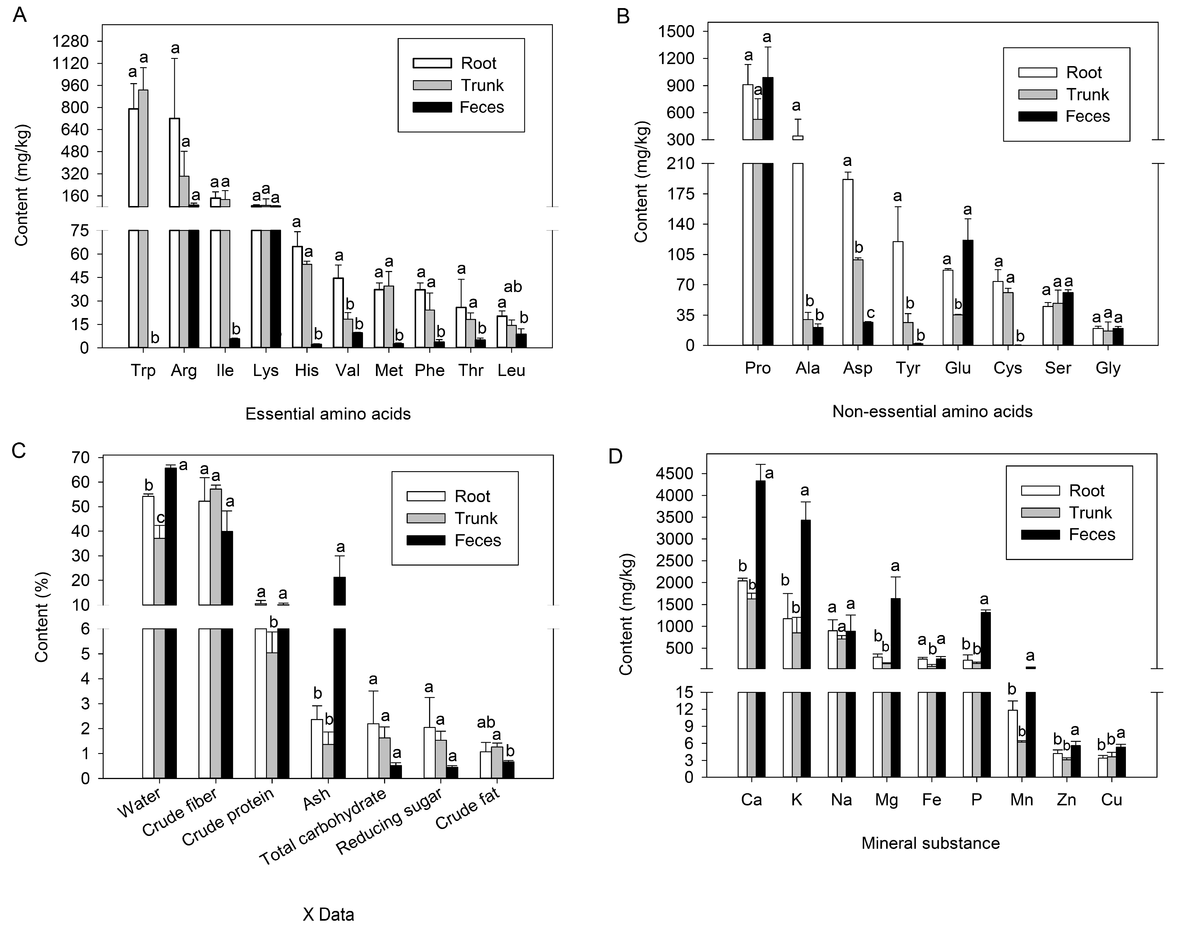
3.1. Nutrient Content of E. hippophaecola Larval Host Plants and Feces

To investigate the nutritional profile of different parts of the E. hippophaecola larval host, we analyzed the nutrient composition of free amino acids (both essential and non-essential), sugars, lipids, and proteins in the roots and trunks of sea buckthorn. We also assessed the excretion of these substances by larvae following digestion and absorption. Among the 10 essential insect amino acids (Figure 1A), we observed that tryptophan (Trp) content was highest in sea buckthorn, with 790 mg/kg in the roots and 930 mg/kg in the trunks (df = 2, F = 37.642, p < 0.001). However, it was undetectable in larval feces. The contents of arginine (Arg) (df = 2, F = 8.312, p = 0.019) and lysine (Lys) (df = 2, F = 0.031, p = 0.970) were among the highest in all three samples, with no significant differences observed. Valine (Val) (df = 2, F = 56.183, p < 0.001) content was significantly higher in sea buckthorn roots (44.52 mg/kg) compared to the trunks (18.45 mg/kg), but no significant difference was found between the trunks and larval feces (9.54 mg/kg). The remaining six essential amino acids were significantly more abundant in the host plant than in the larvae’s excreted feces. No significant differences were observed between different parts of the host plant, although the roots generally contained slightly higher levels than the trunks.
Regarding non-essential amino acids (Figure 1B), proline (Pro) (df = 2, F = 2.566, p = 0.157), serine (Ser) (df = 2, F = 4.198, p = 0.072), and glycine (Gly) (df = 2, F = 0.229, p = 0.802) showed no significant differences between sea buckthorn roots, trunks, and larval feces. However, their concentrations were slightly higher in larval feces compared to the roots and trunks of the host plant. Alanine (Ala) (df = 2, F = 17.492, p = 0.003) and tyrosine (Tyr) (df = 2, F = 40.501, p < 0.001) levels were significantly higher in sea buckthorn roots than in both the trunks and larval feces. No significant differences were observed between the latter two, and their concentrations gradually decreased across all three samples. Aspartic acid (Asp) (df = 2, F = 1577.57, p < 0.001) concentration was significantly higher in sea buckthorn roots (191.71 mg/kg) compared to trunks (98.79 mg/kg), and higher in trunks than in feces (26.76 mg/kg). Glutamic acid (Glu) levels (df = 2, F = 27.275, p = 0.001) were significantly higher in sea buckthorn roots (86.93 mg/kg) and larval feces (121.45 mg/kg) than in sea buckthorn trunks (35.38 mg/kg). In contrast, cystine (Cys) levels (df = 2, F = 130.302, p < 0.001) were significantly higher in the host plants (73.99 mg/kg in roots and 60.90 mg/kg in trunks) than in larval feces (0.53 mg/kg). Overall, non-essential amino acids were more abundant in sea buckthorn roots than in trunks and exhibited a more diversified distribution in larval feces compared to essential amino acids.
Figure 1C illustrates the compound fractions in sea buckthorn roots, trunks, and E. hippophaecola larval feces, offering a visual representation of the nutrient conditions in the different host parts. Both sea buckthorn roots and larval feces contained more than 50% water (df = 2, F = 61.817, p < 0.001), significantly higher than the water content in sea buckthorn trunks. The ash content (df = 2, F = 14.820, p = 0.005) in larval feces was 21.27%, significantly higher than 2.37% in sea buckthorn roots and 1.37% in trunks. Crude fiber content (df = 2, F = 4.321, p = 0.069) in sea buckthorn roots and trunks was 52.2% and 57.17%, respectively, both higher than 39.87% in larval feces, with no significant differences among the three. The total protein content (df = 2, F = 40.618, p < 0.001) exceeded 10% in both sea buckthorn roots and larval feces, significantly higher than 5.04% in sea buckthorn trunks. The total sugar (df = 2, F = 3.390, p = 0.103) and reducing sugar (df = 2, F = 3.755, p = 0.088) contents in sea buckthorn roots, trunks, and larval feces decreased sequentially, with no significant differences among them. Total fat (df = 2, F = 4.941, p = 0.054) content was low in all three, around 1%, but significantly higher in sea buckthorn trunks than in larval feces. Generally, compound fractions were less abundant in the feces after digestion and absorption in the larval gut compared to the host plant. Additionally, the nutrient content in sea buckthorn roots was slightly higher than in the trunks.
Figure 1D presents a comparison of mineral element contents in sea buckthorn roots, trunks, and larval feces, along with the inorganic salt ion levels in each. The metal element content in the feces of E. hippophaecola larvae was significantly higher than in the roots and trunks of the host plant, except for Na (df = 2, F = 0.575, p = 0.591) and Fe (df = 2, F = 13.342, p = 0.006). In contrast, the metal element levels in different parts of the sea buckthorn plant were relatively uniform, except for Fe. The Fe concentration in the roots was 246.5 mg/kg, three times higher than in the trunks (85.13 mg/kg) and comparable to that in the feces of E. hippophaecola larvae (251.33 mg/kg).

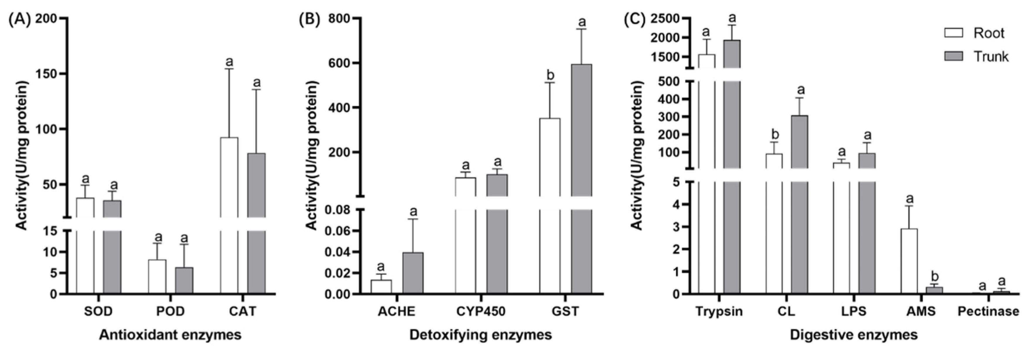
3.2. Effects of Different Feeding Sites on the Digestive and Metabolic Enzyme System of E. hippophaecola Larvae

Nutrient variation between different plant parts (e.g., roots and trunks) prompted us to investigate how E. hippophaecola larvae respond by measuring digestive enzyme activities (Figure 2C) in the midguts of larvae collected from various plant parts. The results indicated that larvae feeding on roots had lower enzyme activity in their midguts compared to those feeding on trunks, except for amylase (AMS) activity (Welch’s t-test, t = 7.289, df = 7.380, p < 0.001), which was significantly higher in roots (2.93 U/mg) than in trunks (0.32 U/mg). The activities of trypsin, lipase (LPS) (independent-sample t-test, t = −1.755, df = 5, p = 0.140), and pectinase (independent-sample t-test, t = −1.167, df = 4, p = 0.308) were similar across both groups, though no significant differences were found. Furthermore, cellulase (CL) (independent-sample t-test, t = −3.567, df = 5, p = 0.016) activity was significantly lower in larvae feeding on roots (91.35 U/mg) compared to those feeding on trunks (308.37 U/mg).
Insect feeding preferences are influenced not only by the nutritional quality of the host plant but also by secondary metabolites and interactions with other organisms. We assessed the activities of antioxidant and detoxification enzymes in E. hippophaecola larvae feeding on different plant parts. The activities of three antioxidant enzymes—SOD (independent-sample t-test, t = 0.394, df = 9, p = 0.702), POD (independent-sample t-test, t = 0.675, df = 9, p = 0.516), and CAT (independent-sample t-test, t = 0.392, df = 9, p = 0.704)—did not differ significantly between larvae feeding on different plant parts. However, antioxidant enzyme activity was generally higher in larvae feeding on roots than in those feeding on trunks. The results for three detoxification enzymes showed minor differences. GST activity (independent-sample t-test, t = −3.158, df = 15, p = 0.006) was significantly higher in larvae feeding on trunks (594.98 U/mg) compared to those feeding on roots (352.36 U/mg). Although no significant differences were observed for ACHE (Welch’s t-test, t = −1.854, df = 4.211, p = 0.134) and CYP450 (independent-sample t-test, t = −1.252, df = 16, p = 0.229) activity, larvae feeding on trunks exhibited slightly higher values than those feeding on roots.

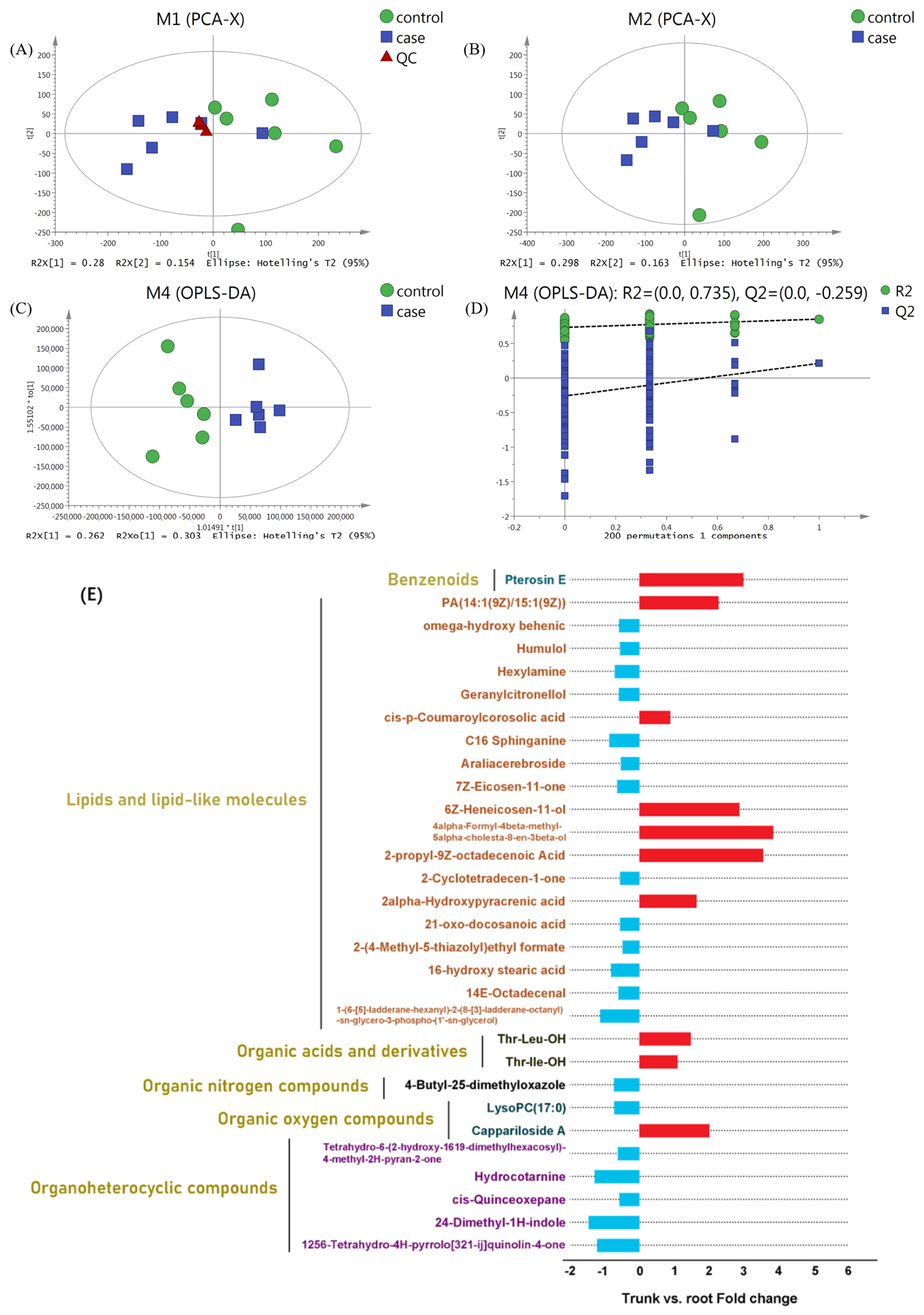
3.3. Metabolic Profile of Sea Buckthorn Roots and Trunks After Being Fed on

Secondary metabolites in plants can influence insect feeding behavior. To investigate whether the damage caused by E. hippophaecola larvae on sea buckthorn is associated with the secondary metabolic response, we analyzed the metabolic profiles of the affected roots and trunks. A total of 8493 metabolites were analyzed and identified through non-targeted metabolomics. Principal component analysis (PCA) (Figure 3A,B) revealed that the quality control (QC) samples clustered together, confirming the reliability of the analytical method. Using roots as the control, the sea buckthorn root and trunk samples could be broadly classified into two distinct groups. To obtain more reliable metabolite data distinguishing between root and trunk samples, we used orthogonal partial least squares discriminant analysis (OPLS-DA), identifying 394 differential metabolites (DEMs). Of these, 223 metabolites were up-regulated in trunks, while 171 were down-regulated (VIP > 1, p < 0.05).
All DEMs were ranked by VIP values, yielding the top 30 metabolites (10 up-regulated and 20 down-regulated) contributing to the classification. These 30 metabolites were classified into six major groups: nearly two-thirds were lipids and lipid-like molecules (six up-regulated and thirteen down-regulated), five were organic acids and derivatives (all down-regulated), and the remaining four groups included six species of secondary metabolites, with four up-regulated and two down-regulated. Overall, the top 30 DEMs primarily targeted plant defense mechanisms, protecting the plant from herbivores, pathogens, and environmental stressors. Several up-regulated DEMs directly activate the plant’s defense system in response to biotic and abiotic stresses. As an example, 6Z-Heneicosen-11-ol (up-regulated 2.905-fold) can act as a warning signal to neighboring plants, prompting them to enhance their defense responses. Pterosin E (3.025-fold up-regulated) and 2alpha-Hydroxypyracrenic acid (1.674-fold up-regulated) may function as natural insecticides or growth promoters, potentially reducing the need for chemical pesticides. Down-regulated DEMs, such as Geranylcitronellol (0.622-fold), Araliacerebroside (0.563-fold), Humulol (0.594-fold), 2-Cyclotetradecen-1-one (0.591-fold), Hexylamine (0.591-fold), and Pseudomonas aeruginosa (0.591-fold), also contribute to plant defense mechanisms. Additionally, compounds such as 4-Butyl-2,5-dimethyloxazole (0.761-fold down-regulated), Hexylamine (0.737-fold down-regulated), and 14E-Octadecenal (0.637-fold down-regulated), along with other terpenoids, amines, and azoles, exhibit insect resistance. Other compounds, including 2-(4-Methyl-5-thiazolyl) ethyl formate (0.516-fold down-regulated) and 2,4-Dimethyl-1H-indole (1.496-fold down-regulated), also act indirectly by attracting natural enemies of herbivores.

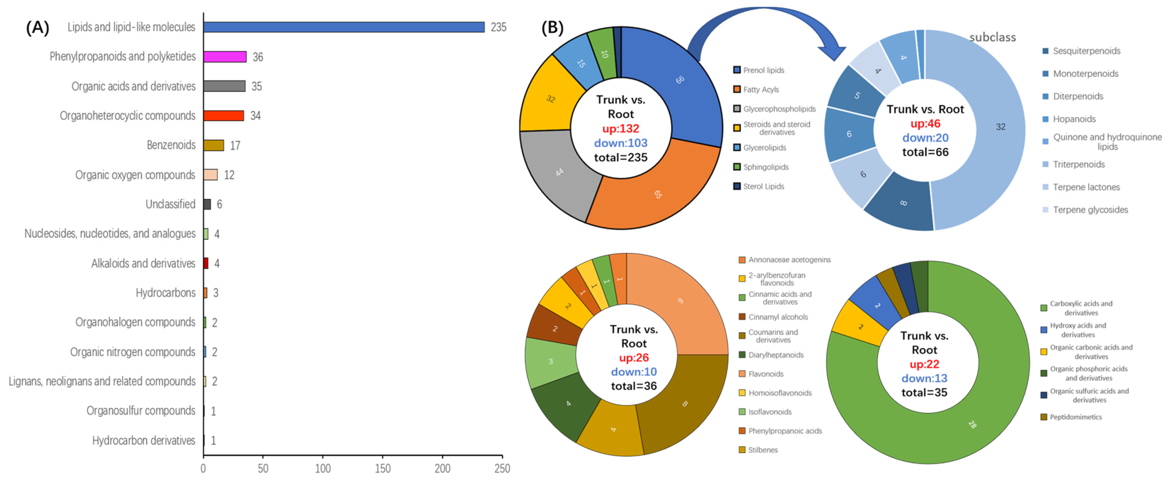
3.4. Analysis of the Expression Pattern of DEMs

As shown in Figure 4, we conducted a preliminary analysis of 394 differentially expressed metabolites (DEMs) produced by sea buckthorn roots and trunks after infestation by E. hippophaecola larvae. The differentially expressed metabolites in the affected sea buckthorn roots and trunks were classified into 15 major groups. The majority of these metabolites were lipids and lipid-like molecules (235 species), which may contribute to host transfer by E. hippophaecola larvae. These lipids and lipid-like molecules were further categorized and analyzed using pie charts. Compared to the roots, the main DEMs in the sea buckthorn trunks included 132 up-regulated and 103 down-regulated metabolites, such as 66 isoprenoids (prenol lipids), 65 fatty acyls, 44 glycerophospholipids, and 32 steroids and steroid derivatives. Triterpenoids accounted for half (32 species) of the isoprenoids, potentially influencing E. hippophaecola larval host transfer.
Phenylpropanoids and polyketides represented the second largest category, with a total of 36 metabolites (26 up-regulated and 10 down-regulated). Of these, nine flavonoids and eight coumarins and derivatives accounted for nearly half. Organic acids and derivatives totaled 35 metabolites (22 up-regulated and 13 down-regulated), including 28 carboxylic acids and their derivatives. Among them, 27 were amino acids, peptides, and analogs, indicating a significant alteration in the amino acid metabolism following infestation of the sea buckthorn roots and trunks.

3.5. Metabolic Pathway Analysis of DEMs

Metabolic pathway enrichment analysis was conducted on all differentially expressed metabolites (DEMs), resulting in the identification of 26 enriched pathways. Figure 5 displays a bubble plot of the top 20 enriched DEM pathways. Significant metabolic differences (p < 0.05) were observed between the sea buckthorn trunks and roots in three pathways: steroid biosynthesis, linoleic acid metabolism, and glycerophospholipid metabolism, all related to lipid metabolism. Of the total pathways, 10 were involved in lipid metabolism, 4 in amino acid metabolism, 3 in energy metabolism, and the others were related to the metabolism of alkaloids, phenylpropanoic acid, and organic acids. Overall, most enriched metabolic pathways were associated with plant defense mechanisms against stress and phytonutrient synthesis.

3.6. Metabolic Regulatory Network of Damaged Sea Buckthorn Roots and Trunks

Figure 6 shows the network of major metabolic pathways significantly altered in the roots and trunks of buckthorn following infestation by E. hippophaecola larvae. This network illustrates the major metabolic pathways significantly altered in the roots and trunks of buckthorn after infestation by E. hippophaecola larvae. Linoleic acid metabolism, significantly enriched in the network, radiates outward from the center, accompanied by glycerophospholipid metabolism, arachidonic acid metabolism, alpha-linolenic acid metabolism, biosynthesis of unsaturated fatty acids, and glycine, serine, and threonine metabolism, collectively forming a metabolic regulatory network. Within this network, down-regulation of lecithin and linoleate leads to up-regulation of 9,10,13-TriHOME in the linoleic acid metabolism pathway. Additionally, linoleate is regulated by upstream linoleoyl-CoA in the biosynthesis of the unsaturated fatty acid pathway. The down-regulation of lecithin affects the other three pathways, contributing to the down-regulation of betaine in the glycine, serine, and threonine metabolism pathway through the regulation of the glycerophospholipid metabolism pathway. Overall, the expression of resistance metabolism was weaker in the trunks of sea buckthorn compared to the roots when infested by E. hippophaecola larvae.

4. Discussion

This study identified nutritional differences in various parts of the sea buckthorn plant, the host of E. hippophaecola larvae. These variations influenced larval digestive enzymes and may have contributed to shifts in feeding sites between the roots and trunks. Metabolic pathways in different parts of sea buckthorn showed distinct responses after feeding, resulting in variations in the regulation of secondary metabolites. These variations were associated with changes in the antioxidant and detoxification enzyme activities of the larvae, depending on the feeding site. Such metabolic shifts may contribute to the host transfer behavior of E. hippophaecola larvae.
The nutrient content of host plants affects the feeding preferences and adaptations of herbivorous insects. Specifically, the levels of sugars, proteins, and fats directly affect the growth, development, and reproduction of these insects [58,59,60]. Studies show significant differences in the duration of developmental stages, survival rates, and egg production in Carposina sasakii larvae when feeding on host plants with varying nutrient conditions [61,62]. In contrast, Spodoptera litura larvae feeding on asparagus and calamus preferred stems and leaves, respectively [63,64]. Sugars are essential for insect function, and higher levels of reducing sugars in plants can trigger insect damage [65]. In contrast, locusts feeding on the roots of graminaceous plants exhibit higher amylase activity compared to those feeding on the leaves [66]. Our results indicated that the reducing sugar content in sea buckthorn roots was 1.34 times higher than in trunks and 4.46 times higher than in larval feces. The total sugar content was 1.33 times higher than in the trunks and 4.23 times higher than in larval feces. Additionally, the amylase activity of larvae feeding on roots was significantly higher (9 times) than that of larvae feeding on trunks. This suggests that the demand for saccharides in E. hippophaecola larvae may partly explain their preference for feeding on roots with higher sugar content.
Moreover, differences in protein content across various parts of the host plant can directly influence insect nutrient acquisition. For instance, Plutella xylostella larvae prefer feeding on young, protein-rich leaves rather than older, fibrous ones to achieve higher growth and survival rates [67]. Feeding on plants with higher nitrogen content also accelerates the metabolic rate and reproductive cycle of phytophagous insects, potentially providing them with a competitive advantage in the ecosystem [68,69]. In this research, sea buckthorn roots contained more proteins; however, the trypsin enzyme activity in larvae feeding on roots was notably lower than in those feeding on the trunks. This discrepancy contradicts general principles of nutrient ecology, suggesting that the higher protein content in roots may not be entirely conducive to larval digestion, potentially due to plant resistance. For example, Bt proteins cause cell rupture and lethality in Ostrinia furnacalis larvae by binding to their gut receptors [70]. Protein inhibitors in wheat can block the digestive enzymes of the wheat aphid, impairing its feeding and growth [71]. Additionally, peas enhance insect resistance by synthesizing specific antimicrobials [72]. In addition, metallic elements can influence insect feeding behavior to a certain extent. Studies on desert locusts (Schistocerca gregaria) have demonstrated that trace iron (Fe) modulates oxidative stress responses in these insects [73]. Conversely, elevated sodium (Na) levels have been shown to negatively impact grasshopper (Melanoplus differentialis) survival, morphological development, and locomotor performance [74]. This suggests that inorganic salt ions, which typically act as insect inhibitors in plants, accumulate in the insect’s body and are excreted, potentially stimulating further feeding. This could also explain the higher levels of lipase, pectinase, and cellulase enzymes in E. hippophaecola larvae feeding on sea buckthorn trunks compared to those feeding on the roots. Although sea buckthorn roots may offer slightly better nutrient conditions and be preferred by E. hippophaecola larvae, nutrient availability may be lower in the roots compared to the trunks. It is supported by the enzyme activities of antioxidant and detoxification enzymes in E. hippophaecola larvae from different feeding sites. Insects feeding on different parts of the same host plant may experience varying levels of oxidative stress due to plant defenses, leading to differential activation of the antioxidant system. For example, Plutella xylostella larvae experience varying levels of oxidative stress when feeding on the leaves and stems of cruciferous plants, resulting in differential expression of antioxidant enzymes [75]. In this study, the activities of SOD, POD, and CAT enzymes in larvae feeding on sea buckthorn roots were slightly higher than in those feeding on the trunks. This suggests that sea buckthorn roots, compared to trunks, produce secondary metabolites and reactive oxygen species more intensively in response to insect feeding, likely to prevent further infestation by E. hippophaecola larvae. Conversely, a different pattern emerges in the regulation of detoxification enzyme activities in E. hippophaecola larvae. In general, this follows the pattern of higher detoxification enzyme activity in trunk-feeding insects compared to root-feeding ones. The detoxifying enzyme system in insects is crucial for their adaptation to host plants, as it metabolizes and detoxifies harmful substances, thereby enhancing their ability to utilize the host plants. According to chemical ecology theory, chemical stress on different parts of the host plant leads to varied detoxification enzyme activities in insects, directly influencing their host plant choice [76]. In other words, higher detoxification enzyme activities at a specific site suggest that detoxification enzymes efficiently metabolize harmful plant components, reducing poisoning risks, and making that site more likely to be chosen for feeding. This difference in antioxidant and detoxification enzyme activities may also explain why many E. hippophaecola larvae prefer feeding on sea buckthorn trunks despite their poorer nutritional conditions.
Essential amino acids from host plants are crucial for insect protein synthesis and growth. Plutella xylostella [67] and Spodoptera exigua [77] larvae prefer host plants with higher levels of essential amino acids. Spodoptera exigua larvae increased feeding when exposed to higher concentrations of valine in kale, indicating sensitivity to this amino acid [78]. This study found that valine concentrations were significantly higher in sea buckthorn roots compared to the trunks. It was the only amino acid showing a significant difference, potentially serving as a nutritional signal influencing the feeding behavior of E. hippopharcola larvae. Arginine and lysine did not show significant differences between the host plant and insect feces, suggesting these amino acids primarily influence plant resistance to insects. The larvae significantly absorbed and utilized tryptophan from both sea buckthorn roots and trunks, despite no significant difference in content. Some aphid species prefer feeding on foods rich in tryptophan to enhance growth and reproductive success [79]. This implies that tryptophan may serve as a nutritional target for pest control, based on the feeding preferences of E. hippophaecola larvae. Furthermore, high tryptophan levels appear to trigger a stronger plant defense response. In an untargeted metabolomic analysis of affected sea buckthorn, 2,4-Dimethyl-1H-indole, a tryptophan derivative, was significantly down-regulated by 1.496-fold in the expression in sea buckthorn branches compared to roots. It can activate the jasmonic acid pathway in plants, generating reactive oxygen species that inhibit insects from feeding. Additionally, it may attract natural predators, such as parasitic wasps, making it a potential natural insecticide [80]. Another tryptophan derivative, tryptamine, has been demonstrated to kill locusts when accumulated to high concentrations [81]. However, the complex enzymatic pathways and regulatory networks involved warrant further investigation.
Although non-essential amino acids (NEAAs) are less critical than essential amino acids for fulfilling insects’ nutritional needs, certain NEAAs can affect the palatability and nutritional quality of host plants, influencing the feeding preferences of phytophagous insects. Under starvation stress, Bicyclus anynana larvae tend to feed on sugar solutions containing glutamate [82]. Results from the “Styropor method” assay on Spodoptera littoralis larvae demonstrated that a mixture of 0.125 M L-glutamic acid and 0.125 M sucrose exhibited a phagostimulatory effect [83]. The growth and development of Chilo partellus were enhanced when fed maize seedlings with higher aspartic acid content [84]. Helicoverpa armigera reared on alanine-rich plants exhibited faster growth and higher survival rates [85]. In this experiment, the concentrations of glutamic acid and aspartic acid were significantly higher in the roots of sea buckthorn than in the trunk, suggesting that the roots are more nutritionally attractive to E. hippophaecola larvae. NEAAs may play a crucial role as metabolic intermediates or regulatory molecules in plant defense responses during plant–insect interactions. Tyrosine, a key component in the synthesis of plant defense compounds, can be converted into phenylalanine-like substances toxic to insects, influencing their feeding preferences by altering plant taste and odor characteristics. Certain aphids and Plutella xylostella prefer plants with low tyrosine content [86,87]. Heliothis virescens consumes artificial diets with progressively higher tyrosine concentrations, leading to a significant decrease in growth and increased mortality [88]. The tyrosine concentration in sea buckthorn roots was significantly higher than in the trunk and larval feces, indicating that tyrosine may play a key role in the transfer of E. hippophaecola larvae from the host roots to other tissues. In future control strategies for E. hippophaecola larvae, tyrosine and other key substances in its metabolic pathway could be targeted to reduce chemical pesticide use and promote eco-friendly control methods.
Plant–insect interactions are dynamic and adaptive processes for both parties, with secondary metabolites playing a crucial role in mediating the transition between attack and defense, and even in facilitating mutual benefits. Manduca sexta has evolved detoxification mechanisms to counteract nicotine produced by Tobacco spp. [89]. Chrysomela spp. have evolved tolerance by altering the metabolic pathways that process phenolic compounds in Salix spp. (willow) plants [90]. Pseudomyrmex spp. have adapted to and benefit from tannic acid-based chemical defenses produced by Acacia spp., while also protecting these plants from herbivores and competitors [91]. In this research, the roots and trunks of sea buckthorn infested by E. hippophaecola showed distinct metabolic pathways, with a notable down-regulation of nutrient synthesis and metabolic responses in the trunks compared to the roots. These findings suggest differences in the defense responses between the roots and trunks of sea buckthorn to E. hippophaecola larvae infestation, potentially influencing the larvae’s feeding site shift. The linoleic acid metabolic pathway, including linoleate and lecithin, which play a role in inducing plant resistance to insect feeding, was significantly down-regulated, potentially inhibiting plant defense mechanisms. Ethyl linoleate, the esterified derivative of linoleate, demonstrates significant insecticidal potential against Plutella xylostella [92]. Furthermore, ethyl linoleate isolated from Thapsia garganica exhibits both strong toxicity and repellent effects against adult Tribolium castaneum [93]. These all result in a weaker defense response in the trunks compared to the roots, potentially attracting E. hippophaecola larvae to migrate back to the trunks.
Recent advancements in synthetic biology and biotechnology have led to the increasing application of RNA interference (RNAi) and gene-editing technologies in pest control research [94]. A variety of plant-derived metabolites have already been commercialized for agricultural applications. For example, azadirachtin, derived from Azadirachta indica (neem), has shown exceptional efficacy in pest management [95]. Furthermore, breakthroughs like the de novo synthesis of cembratrien-ol—an insect repellent derived from natural products—through the mevalonate (MVA) pathway in engineered yeast (Saccharomyces cerevisiae) have established a foundation for the development of next-generation plant-based insecticidal agents [96]. These advancements offer a strategic framework for the future development of environmentally friendly control methods targeting E. hippophaecola larvae, utilizing natural compounds derived from sea buckthorn.

5. Conclusions

The feeding behavior of E. hippophaecola larvae on H. rhamnoides (sea buckthorn) trunks and roots significantly altered both the nutritional profiles and secondary metabolites. Comparative analysis showed that infested roots had significantly higher concentrations of sugars, proteins, lipids, and amino acids compared to damaged trunks, creating a nutritional gradient that led to larval migration from the trunks to the roots. Concurrently, the down-regulation of lecithin—a pivotal metabolite in the secondary metabolic network—triggered subsequent decreases in arachidonate and betaine within the resistance metabolism of trunks compared to roots, indicating weakened defense responses. This metabolic shift enhanced the adaptive capacity of trunk-feeding larvae, ultimately driving partial recolonization of trunks. Consequently, elucidating the mechanistic basis of this host-shifting behavior in E. hippophaecola larvae holds significant potential for developing targeted biological control strategies.

Author Contributions

Conceptualization, Y.L. and S.Z.; methodology, Y.L. and Y.S.; validation, Y.L., Y.S., J.T., S.L., X.L. and S.Z.; formal analysis, Y.L.; resources, J.T. and S.Z.; data curation, Y.L. and Y.S.; writing—original draft preparation, Y.L.; writing—review and editing, Y.L.; project administration, J.T. and S.Z. All authors have read and agreed to the published version of the manuscript.

Funding

This research was funded by the Research and Application of an Integrated Green Control Technology System for Major Insect Pest Outbreaks in Seabuckthorn Forests (grant number YF20240009).

Data Availability Statement

The data are not publicly available due to their use in an ongoing project.

Acknowledgments

We are very grateful to the staff of the Junggar Banner Forestry and Grassland Business Centre for their great assistance during the fieldwork.

Conflicts of Interest

The authors declare no conflicts of interest.

References

  1. Calatayud, P.A.; Sauvion, N.; Thiery, D. Plant-Insect Interactions. Oxf. Bibliogr. 2018, 2, 268–272. [Google Scholar]
  2. Schuler, M.A. P450s in Plant-Insect Interactions. Biochim. Biophys. Acta-Proteins Proteom. 2011, 1814, 36–45. [Google Scholar] [CrossRef] [PubMed]
  3. Pu, M.-W. Evaluation of Host Plant Quality and Host Selection Behavior of Spodoptera litura (Fabricius). Master’s Thesis, Yangzhou University, Yangzhou, China, 2007. [Google Scholar]
  4. Singer, M.; Stireman, J. How Foraging Tactics Determine Host-Plant Use by a Polyphagous Caterpillar. Oecologia 2001, 129, 98–105. [Google Scholar] [CrossRef] [PubMed]
  5. Alexandra, F.; Unsicker, S.B.; Specht, J.; Köhler, G.; Weisser, W.W. Being a Generalist Herbivore in a Diverse World: How Do Diets from Different Grasslands Influence Food Plant Selection and Fitness of the Grasshopper Chorthippus parallelus? Ecol. Entomol. 2010, 35, 126–138. [Google Scholar]
  6. Jacek, O.; Reich, P.B.; Zytkowiak, R.; Karolewski, P.; Tjoelker, M.G. Needle Nutrients in Geographically Diverse Pinus sylvestris L. Populations. Ann. For. Sci. 2002, 59, 1–18. [Google Scholar]
  7. Forde, U.I.; Bose, E.A.; Hart, C.; Lahondère, C. Temperature and Sugar Feeding Effects on the Activity of a Laboratory Strain of Aedes aegypti. Insects 2019, 10, 347. [Google Scholar] [CrossRef]
  8. Floriane, A.; Terhzaz, S.; Terry, S.; McFarlane, M.; Gestuveo, R.J.; Szemiel, A.M.; Varjak, M.; McDonald, A.; Kohl, A.; Pondeville, E. Sugar Feeding Protects against Arboviral Infection by Enhancing Gut Immunity in the Mosquito Vector Aedes aegypti. PLoS Pathog. 2021, 17, e1009870. [Google Scholar]
  9. Joel, F.-H.; Zagrobelny, M.; Bak, S. Plant Defense against Insect Herbivores. Int. J. Mol. Sci. 2013, 14, 10242–10297. [Google Scholar]
  10. Huang, X. Adaptive Mechanisms of Oedaleus asiaticus Bey-Bienko(Orthoptera: Acrididae) to Food Plants. Ph.D. Thesis, Chinese Academy of Agricultural Sciences, Beijing, China, 2018. [Google Scholar]
  11. Mamunur, A.M.R.; Ruhul, A. Insect Plant Interaction with Reference to Secondary Metabolites: A Review. Agric. Rev. 2021, 42, 427–433. [Google Scholar]
  12. Wink, M. Plant Secondary Metabolites Modulate Insect Behavior-Steps toward Addiction? Front. Physiol. 2018, 9, 364. [Google Scholar] [CrossRef]
  13. Jameel, M.A.-K.; Rashmi, R.; Toppo, V.; Chole, P.B.; Banadka, A.; Sudheer, W.N.; Nagella, P.; Shehata, W.F.; Al-Mssallem, M.Q.; Alessa, F.M.; et al. Plant Secondary Metabolites: The Weapons for Biotic Stress Management. Metabolites 2023, 13, 716. [Google Scholar] [CrossRef] [PubMed]
  14. Erb, M.; Robert, C.A.M. Sequestration of Plant Secondary Metabolites by Insect Herbivores: Molecular Mechanisms and Ecological Consequences. Curr. Opin. Insect Sci. 2016, 14, 8–11. [Google Scholar] [CrossRef] [PubMed]
  15. Hall, C.R.; Carroll, A.R.; Kitching, R.L. A Meta-Analysis of the Effects of Galling Insects on Host Plant Secondary Metabolites. Arthropod-Plant Interact. 2017, 11, 463–473. [Google Scholar] [CrossRef]
  16. Ralf, N.; Bass, C.; Feyereisen, R.; Vontas, J. The Role of Cytochrome P450s in Insect Toxicology and Resistance. Annu. Rev. Entomol. 2022, 67, 105–124. [Google Scholar]
  17. Zhang, X.M.; Shi, Z.Y.; Yang, C.Q.; Li, J.; Liu, J.Z.; Zhang, A.B. Gut Transcriptome Analysis of P450 Genes and Cytochrome P450 Reductase in Three Moth Species Feeding on Almaghasla, and Adel Abdel-Sabour Rezk. Front. Ecol. Evol. 2022, 10, 948043. [Google Scholar] [CrossRef]
  18. Shen, S.; Chien, Y.; Chien, C. Induction of Glutathione S-Transferases Activities in Drosophila melanogaster Exposed to Phenol. Arch. Insect Biochem. Physiol. 2003, 53, 80–91. [Google Scholar] [CrossRef]
  19. Zou, C.-S. Study on Enzymatic Properties and Biochemical Toxicology of the Enzymes from Dendrolimus superans (Butler). Ph.D. Thesis, Northeast Forestry University, Harbin, China, 2013. [Google Scholar]
  20. Wan, N.F.; Ji, X.Y.; Yin, Y.Y.; Zhang, H.; Jiang, J.X. The Impact of Co-Infection by a Nucleopolyhedrovirus and the Endoparasitoid Microplitis pallidipes Szepligeti on the Protective Enzymes in Hemolymph of Spodoptera exigua. Int. J. Pest Manag. 2017, 63, 341–348. [Google Scholar] [CrossRef]
  21. Liu, X.-L. Effect of Several Pesticides on the Activities of Detoxifying Enzymes and Protective Enzymes in Clostera anastomosis. Master’s Thesis, Northeast Forestry University, Harbin, China, 2015. [Google Scholar]
  22. Hernández-Vera, G.; Tosevski, I.; Caldara, R.; Emerson, B.C. Evolution of Host Plant Use and Diversification in a Species Complex of Parasitic weevils (Coleoptera: Curculionidae). Peerj 2019, 7, e6625. [Google Scholar] [CrossRef]
  23. Nishida, R. Evolutionary Interactions Between Insects and Plants Mediated by Phytochemicals. Kaken. 1998. Available online: https://kaken.nii.ac.jp/grant/KAKENHI-PROJECT-10460049/ (accessed on 28 April 2025).
  24. Agosta, S.J. On Ecological Fitting, Plant–Insect Associations, Herbivore Host Shifts, and Host Plant Selection. Oikos 2006, 114, 556–565. [Google Scholar] [CrossRef]
  25. Dyer, L.A.; Philbin, C.S.; Ochsenrider, K.M.; Richards, L.A.; Massad, T.J.; Smilanich, A.M.; Forister, M.L.; Parchman, T.L.; Galland, L.M.; Hurtado, P.J.; et al. Modern Approaches to Study Plant–Insect Interactions in Chemical Ecology. Nat. Rev. Chem. 2018, 2, 50–64. [Google Scholar] [CrossRef]
  26. Zebelo, S.A.; Maffei, M.E. Role of Early Signalling Events in Plant–Insect Interactions. J. Exp. Bot. 2014, 66, 435–448. [Google Scholar] [CrossRef] [PubMed]
  27. Xiong, B.; Dong, Y.; Jun, Y.; Ming, Z.; Yong, Z.; Jianbin, D. The Wild Plant Resources and Utilization of Hippophae in China. Chin. Wild Plant Resour. 2004, 23, 25–26. [Google Scholar]
  28. Qiong, W.U.; Kun, S.; Hui, Z.; Wen, C.; Xue, S.U.; Xue-Lin, C. Study on Phenotypic Diversity of Natural Populations of Hippophae rhamnoides Subsp. Sinensis in Shanxi Province. J. Northwest Norm. Univ. 2007, 43, 78–84. [Google Scholar]
  29. Yakovlev, R.V. A New Species and New Records of the Genus Meharia chretien 1915 (Lepidoptera, Cossidae) from the Middle East and Central Asia. Zool. Zhurnal 2018, 97, 1127–1131. [Google Scholar] [CrossRef]
  30. Zong, S. Studies on the Bio-Ecological Characteristics of Seabuckthorn Carpenter Moth: Holcocerus hippophaecolus (Lcpidoptera: Cossidac). Ph.D. Thesis, Beijing Forestry University, Beijing, China, 2006. [Google Scholar]
  31. Luo, Y. Integrated Management of Holcocerus hippophaecolus (Lepidoptera:Cossidae). Sci. Silvae Sin. 2007, 43, 146–150. [Google Scholar]
  32. Wang, R.; Zong, S.X.; Yu, L.F.; Lu, P.F.; Luo, Y.Q. Rhythms of Volatile Release from Female and Male Sea Buckthorn Plants and Electrophysiological Response of Sea Buckthorn Carpenter Moths. J. Plant Interact. 2014, 9, 763–774. [Google Scholar] [CrossRef]
  33. Zong, S.X.; Wang, R.; Cao, C.J.; Wang, T.; Luo, Y.Q. Impact of Chlorophorus caragana Damage on Nutrient Contents of Caragana korshinskii. J. Plant Interact. 2014, 9, 488–493. [Google Scholar] [CrossRef]
  34. Li, T.S.C.; Beveridge, T.H.J. Sea Buckthorn (Hippophae rhamnoides L.): Production and Utilization; NRC Research Press: Ottawa, ON, Canada, 2003. [Google Scholar]
  35. Hurlbert, S.H. Pseudoreplication and the Design of Ecological Field Experiments. Ecol. Monogr. 1984, 54, 187–211. [Google Scholar] [CrossRef]
  36. Min, D.B.; Ellefson, W.C. Fat Analysis. In Food Analysis; Nielsen, S.S., Ed.; Springer: Berlin/Heidelberg, Germany, 2010; Volume 117. [Google Scholar]
  37. Purificación, S.-P.; Micha, T.; Navas, M.J.; Asuero, A.G.; Wybraniec, A. An Overview of the Kjeldahl Method of Nitrogen Determination. Part I. Early History, Chemistry of the Procedure, and Titrimetric Finish. Crit. Rev. Anal. Chem. 2013, 43, 178–223. [Google Scholar]
  38. Ida, A.R.; Setyawan, A.R.; Suadi; Jayanti, A.D. Comparison of Nutritional Composition in Red and Green Strains of Kappaphycus alvarezii Cultivated in Gorontalo Province, Indonesia. E3S Web Conf. 2020, 147, 03029. [Google Scholar]
  39. Chow, P.S.; Landhäusser, S.M. A Method for Routine Measurements of Total Sugar and Starch Content in Woody Plant Tissues. Tree Physiol. 2004, 24, 1129–1136. [Google Scholar] [CrossRef] [PubMed]
  40. BeMiller, J.N. Carbohydrate Analysis. In Food Analysis; Nielsen, S.S., Ed.; Springer International Publishing: Cham, Switzerland, 2017; pp. 333–360. [Google Scholar]
  41. Yust, M.; Pedroche, J.; Girón-Calle, J.; Vioque, J.; Millán, F.; Alaiz, M. Determination of Tryptophan by High-Performance Liquid Chromatography of Alkaline Hydrolysates with Spectrophotometric Detection. Food Chem. 2004, 85, 317–320. [Google Scholar] [CrossRef]
  42. Heinrikson, R.L.; Meredith, S.C. Amino Acid Analysis by Reverse-Phase High-Performance Liquid Chromatography: Precolumn Derivatization with Phenylisothiocyanate. Anal. Biochem. 1984, 136, 65–74. [Google Scholar] [CrossRef] [PubMed]
  43. Peng, Z.; Zhao, F.; Chen, M.; Ye, N.; Lin, Q.; Ouyang, L.; Cai, X.; Meng, P.; Gong, X.; Wang, Y. Determination of 21 Free Amino Acids in 5 Types of Tea by Ultra-High Performance Liquid Chromatography Coupled with Tandem Mass Spectrometry (Uhplc–Ms/Ms) Using a Modified 6-Aminoquinolyl-N-Hydroxysuccinimidyl Carbamate (Aqc) Method. J. Food Compos. Anal. 2019, 81, 46–54. [Google Scholar]
  44. Zhu, Y.; Luo, Y.; Wang, P.; Zhao, M.; Li, L.; Hu, X.; Chen, F. Simultaneous Determination of Free Amino Acids in Pu-Erh Tea and Their Changes during Fermentation. Food Chem. 2016, 194, 643–649. [Google Scholar] [CrossRef]
  45. Khan, S.R.; Sharma, B.; Chawla, P.A.; Bhatia, R. Inductively Coupled Plasma Optical Emission Spectrometry (Icp-Oes): A Powerful Analytical Technique for Elemental Analysis. Food Anal. Methods 2022, 15, 666–688. [Google Scholar] [CrossRef]
  46. Cara, J.B.; Moyano, F.J.; Cárdenas, S.; Fernández-Díaz, C.; Yúfera, M. Assessment of Digestive Enzyme Activities during Larval Development of White Bream. J. Fish Biol. 2003, 63, 48–58. [Google Scholar] [CrossRef]
  47. Lakshmesha, K.K.; Lakshmidevi, N.; Mallikarjuna, S.A. Changes in Pectinase and Cellulase Activity of Colletotrichum capsici Mutants and Their Effect on Anthracnose Disease on Capsicum Fruit. Arch. Pflanzenschutz 2005, 38, 267–279. [Google Scholar] [CrossRef]
  48. Lemieux, H.; Blier, P.U. Trypsin Activity Measurement in Fish and Mammals: Comparison of Four Different Methods. J. Aquat. Food Prod. Technol. 2007, 4, 16. [Google Scholar] [CrossRef]
  49. Shen, H.; Huo, X.; Liu, K.; Li, X.; Gong, W.; Zhang, H.; Xu, Y.; Wang, M.; Li, X.; Zhang, J.; et al. Genetic Variation in Gstm1 Is Associated with Susceptibility to Noise-Induced Hearing Loss in a Chinese Population. J. Occup. Environ. Med. 2012, 54, 1157–1162. [Google Scholar] [CrossRef]
  50. Liu, D.M.; Xu, B.J.; Dong, C. Recent Advances in Colorimetric Strategies for Acetylcholinesterase Assay and Their Applications. Trac-Trends Anal. Chem. 2021, 142, 116320. [Google Scholar] [CrossRef]
  51. Turpeinen, M.; Jouko, U.; Jorma, J.; Olavi, P. Multiple P450 Substrates in a Single Run: Rapid and Comprehensive in Vitro Interaction Assay. Eur. J. Pharm. Sci. 2005, 24, 123–132. [Google Scholar] [CrossRef] [PubMed]
  52. Zhang, J.Q.; Shen, M.; Zhu, C.C.; Yu, F.X.; Liu, Z.Q.; Ally, N.; Sun, S.C.; Li, K.; Liu, H.L. 3-Nitropropionic Acid Induces Ovarian Oxidative Stress and Impairs Follicle in Mouse. PLoS ONE 2014, 9, e86589. [Google Scholar] [CrossRef] [PubMed]
  53. Deng, Y.; Wang, W.; Yu, P.F.; Xi, Z.J.; Xu, L.J.; Li, X.L.; He, N.Y. Comparison of Taurine, Gaba, Glu, and Asp as Scavengers of Malondialdehyde in Vitro and in Vivo. Nanoscale Res. Lett. 2013, 8, 190. [Google Scholar] [CrossRef]
  54. Li, H.X.; Xiao, Y.; Cao, L.L.; Yan, X.; Li, C.; Shi, H.Y.; Wang, J.W.; Ye, Y.H. Cerebroside C Increases Tolerance to Chilling Injury and Alters Lipid Composition in Wheat Roots. PLoS ONE 2013, 8, e73380. [Google Scholar] [CrossRef]
  55. Zhang, Y.; Lai, J.L.; Ji, X.H.; Luo, X.G. Unraveling Response Mechanism of Photosynthetic Metabolism and Respiratory Metabolism to Uranium-Exposure in Vicia faba. J. Hazard. Mater. 2020, 398, 122997. [Google Scholar] [CrossRef]
  56. Lai, J.L.; Deng, Z.X.; Ji, X.H.; Luo, X.G. Absorption and Interaction Mechanisms of Uranium & Cadmium in Purple Sweet Potato(Ipomoea batatas L.). J. Hazard. Mater. 2020, 400, 123264. [Google Scholar]
  57. Li, X.N.; Qu, C.S.; Bian, Y.R.; Gu, C.G.; Jiang, X.; Song, Y. New Insights into the Responses of Soil Microorganisms to Polycyclic Aromatic Hydrocarbon Stress by Combining Enzyme Activity and Sequencing Analysis with Metabolomics. Environ. Pollut. 2019, 255, 113312. [Google Scholar] [CrossRef]
  58. Babic, B.; Poisson, A.; Darwish, S.; Lacasse, J.; Merkx-Jacques, M.; Despland, E.; Bede, J.C. Influence of Dietary Nutritional Composition on Caterpillar Salivary Enzyme Activity. J. Insect Physiol. 2008, 54, 286–296. [Google Scholar] [CrossRef]
  59. Li, Y.; Yao, Y.; Peng, Z. Effect of Palm Tree Leaves with Different Growth Phases on Midgut Digestive Enzyme Activity of Brontispa longissima. Jiangsu Agric. Sci. 2017, 45, 94–97. [Google Scholar]
  60. Wang, H.; Zhou, W.L.; Li, Z.; Niklas, K.J.; Sun, S.C. Plant Volatiles Mediate Evolutionary Interactions between Plants and Tephritid Flies and Are Evolutionarily More Labile Than Non-Volatile Defenses. J. Anim. Ecol. 2021, 90, 846–858. [Google Scholar] [CrossRef] [PubMed]
  61. Gou, C.-Q.; Sun, P.; Liu, D.-C.; Dilinuer, A.; Feng, H.-Z. Effects of Lygus pratensis (Hemiptera: Miridae) Infestation on the Nutrient Contents and Protective Enzyme Activities in Host Plants. Acta Entomol. Sin. 2018, 61, 976–983. [Google Scholar]
  62. Li, D.X.; Lei, X.H.; Li, Z.; Gao, L.W.; Shen, Z.R. Effects of Different Host Plants on the Development and Reproduction of the Peach Fruit Borer, Carposina sasakii Matsumura (Lepidoptera: Carposinidae). Acta Entomol. Sin. 2012, 55, 554–560. [Google Scholar]
  63. Nutchaya, K.; Wiwattanawanichakun, P.; Phankaen, P.; Saiyaitong, C.; Koul, O.; Nobsathian, S.; Bullangpoti, V.; Dunkhunthod, B. Phenolic Secondary Metabolites from Acorus Calamus (Acorales: Acoraceae) Rhizomes: The Feeding Deterrents for Spodoptera litura (Lepidoptera: Noctuidae). J. Econ. Entomol. 2023, 116, 1613–1620. [Google Scholar]
  64. Ye, L.-M.; Di, X.-Y.; Yan, B.; Liu, J.-F.; Wang, X.-Q.; Yang, M.-F. Population Parameters and Feeding Preference of Spodoptera litura (Lepidoptera: Noctuidae) on Different Asparagus Officinalis Tissues. Insects 2022, 13, 1149. [Google Scholar] [CrossRef]
  65. Moghaddam, B.; Reza, M.; Van den Ende, W. Sugars and Plant Innate Immunity. J. Exp. Bot. 2012, 63, 3989–3998. [Google Scholar] [CrossRef]
  66. Tarai, N.; Doumandji, S. Feeding Preferences of Gregarious Nymphs and Adults of the Desert Locust, Schistocerca gregaria Forskal (Orthoptera, Cyrtacanthacridinae) in Different Habitats at Biskra Oasis, Algeria. Adv. Environ. Biol. 2009, 3, 308–313. [Google Scholar]
  67. Campos, W.G.; Schoereder, J.H.; Sperber, C.F. Does the Age of the Host Plant Modulate Migratory Activity of Plutella xylostella? Entomol. Sci. 2004, 7, 323–329. [Google Scholar] [CrossRef]
  68. Polacco, J.C.T.; Christopher, D. Ecological Aspects of Nitrogen Metabolism in Plants; John Wiley & Sons: Hoboken, NJ, USA, 2011. [Google Scholar]
  69. Gu, H.; Wang, H.; Liu, M.; Shangguan, Z.; Shi, H.; Xu, W.; Ren, F.; Zhu, J.; He, J.-S. Leaf N:P Stoichiometry Overrides the Effect of Individual Nutrient Content on Insect Herbivore Population Dynamics in a Tibetan Alpine Grassland. Agric. Ecosyst. Environ. 2022, 336, 108032. [Google Scholar] [CrossRef]
  70. Xu, L.; Ferry, N.; Wang, Z.; Zhang, J.; Edwards, M.G.; Gatehouse, A.M.R.; He, K. A Proteomic Approach to Study the Mechanism of Tolerance to Bt Toxins in Ostrinia furnacalis Larvae Selected for Resistance to Cry1ab. Transgenic Res. 2013, 22, 1155–1166. [Google Scholar] [CrossRef]
  71. Prashant, P.; Bandani, A.R.; Fitches, E.; Gatehouse, J.A. Protein Digestion in Cereal Aphids (Sitobion avenae) as a Target for Plant Defence by Endogenous Proteinase Inhibitors. J. Insect Physiol. 2011, 57, 881–891. [Google Scholar]
  72. Shimul, D.; Porter, L.D.; Ma, Y.; Coyne, C.J.; Chaves-Cordoba, B.; Naidu, R.A. Resistance in Lentil (Lens Culinaris) Genetic Resources to the Pea Aphid (Acyrthosiphon pisum). Entomol. Exp. Appl. 2022, 170, 755–769. [Google Scholar]
  73. David, R.; Dorrah, M.A.; Mohamed, A.A.; Abdelfattah, E.A.; Bassal, T.T.M. Assessment of Oxidative Stress and Activities of Antioxidant Enzymes Depicts the Negative Systemic Effect of Iron-Containing Fertilizers and Plant Phenolic Compounds in the Desert Locust. Environ. Sci. Pollut. Res. 2016, 23, 21989–22000. [Google Scholar]
  74. Peterson, T.N.; Welti, E.A.R.; Kaspari, M. Dietary Sodium Levels Affect Grasshopper Growth and Performance. Ecosphere 2021, 12, e03392. [Google Scholar] [CrossRef]
  75. Collins, R.M.; Afzal, M.; Ward, D.A.; Prescott, M.C.; Sait, S.M.; Rees, H.H.; Tomsett, A.B. Differential Proteomic Analysis of Arabidopsis Thaliana Genotypes Exhibiting Resistance or Susceptibility to the Insect Herbivore, Plutella xylostella. PLoS ONE 2010, 5, e10103. [Google Scholar] [CrossRef]
  76. Ernest, S. Chemical Ecology; Elsevier: Amsterdam, The Netherlands, 2012. [Google Scholar]
  77. Showler, A.T. Spodoptera exigua Oviposition and Larval Feeding Preferences for Pigweed, Amaranthus Hybridus, over Squaring Cotton, Gossypium hirsutum, and a Comparison of Free Amino Acids in Each Host Plant. J. Chem. Ecol. 2001, 27, 2013–2028. [Google Scholar] [CrossRef]
  78. Luo, Q.; Duan, F.; Song, W. Transcriptomics Integrated with Metabolomics Reveals the Defense Response of Insect-Resistant Zea Mays Infested with Spodoptera exigua. Heliyon 2025, 11, e42565. [Google Scholar] [CrossRef]
  79. Leroy, P.D.; Sabri, W.; Verheggen, F.; Thonart, C.; Haubruge, E. Aphid-Host Plant Interactions:Does Aphid Honeydew Exactly Reflect the Host Plant Amino Acid Composition? Arthropod-Plant Inte 2011, 5, 193–199. [Google Scholar] [CrossRef]
  80. Qin, D.; Wang, Q.; Li, H.; Jiang, X.; Fang, K.; Wang, Q.; Li, B.; Pan, C.; Wu, H. Identification of Key Metabolites Based on Non-Targeted Metabolomics and Chemometrics Analyses Provides Insights into Bitterness in Kucha [Camellia kucha (Chang Et Wang) Chang]. Food Res. Int. 2020, 138, 109789. [Google Scholar] [CrossRef]
  81. Tong, X.; Wang, Y.; Yang, P.; Wang, C.; Kang, L. Tryptamine Accumulation Caused by Deletion of Mrmao-1 in Metarhizium Genome Significantly Enhances Insecticidal Virulence. PLoS Genet. 2020, 16, e1008675. [Google Scholar] [CrossRef]
  82. Tobias, K.; Fischer, K. Larval Starvation Reduces Responsiveness to Feeding Stimuli and Does Not Affect Feeding Preferences in a Butterfly. J. Insect Physiol. 2012, 58, 1028–1035. [Google Scholar]
  83. Ascher, K.R.S.; Meisner, J.; Flowers, H.M. Effects of Amino Acids on the Feeding Behavior of the Larva of the Egyptian Cotton Leafworm, Spodoptera littoralis Boisd. Phytoparasitica 1976, 4, 85–91. [Google Scholar] [CrossRef]
  84. Yogesh, Y.; Dhillon, M.K.; Tanwar, A.K.; Kumar, S. Amino and Fatty Acids Contributing to Antibiosis against Chilo partellus (Swinhoe) in Maize. Arthropod-Plant Interact. 2021, 15, 721–736. [Google Scholar]
  85. Hong, Z.J.; Zhu, W.C. Feeding Selectivity of Helicoverpa armigera Larvae on Amino Acids, Sugars, and Secondary Substances of Cotton. Plant Prot. 2001, 27, 3–5. [Google Scholar]
  86. Hopkins, D.P.; Cameron, D.D.; Butlin, R.K. The Chemical Signatures Underlying Host Plant Discrimination by Aphids. Sci. Rep. 2017, 7, 8498. [Google Scholar] [CrossRef]
  87. Robin, A.H.K.; Hossain, M.R.; Park, J.-I.; Kim, H.R.; Nou, I.-S. Glucosinolate Profiles in Cabbage Genotypes Influence the Preferential Feeding of Diamondback Moth (Plutella xylostella). Front. Plant Sci. 2017, 8, 1244. [Google Scholar] [CrossRef]
  88. Coley, P.D.; Lokvam, J.; Rudolph, K.; Bromberg, K.; Sackett, T.E.; Wright, L.; Brenes-Arguedas, T.; Dvorett, D.; Ring, S.; Clark, A.; et al. Divergent Defensive Strategies of Young Leaves in Two Species of Inga. Ecology 2005, 86, 2633–2643. [Google Scholar] [CrossRef]
  89. Kester, K.M.; Peterson, S.C.; Hanson, F.; Jackson, D.M.; Severson, R.F. The Roles of Nicotine and Natural Enemies in Determining Larval Feeding Site Distributions of Manduca sexta L. And Manduca quinquemaculata (Haworth) on Tobacco. Chemoecology 2002, 12, 1–10. [Google Scholar] [CrossRef]
  90. Topp, W. Survival in a Hostile Environment. Evaluation of the Developmental Success of the Oligophagous Leaf Beetle Chrysomela vigintipunctata (Scop). In Vertical Food Web Interactions: Evolutionary Patterns and Driving Forces; Dettner, K., Bauer, G., Völkl, W., Eds.; Springer: Berlin/Heidelberg, Germany, 1997; pp. 147–169. [Google Scholar]
  91. Coley, P.D.E.; Ghabash, M.-J.; Kidner, G.; Nicholls, C.A.; Pennington, J.A.; Mills, R.T.; Soule, A.G.; Lemes, A.J.; Stone, M.R.; Kursar, G.N.; et al. Macroevolutionary Patterns in Overexpression of Tyrosine: An Anti-Herbivore Defence in a Speciose Tropical Tree Genus, Inga (Fabaceae). J. Ecol. 2019, 107, 1620–1632. [Google Scholar] [CrossRef]
  92. Jun, P.; Chen, Z.; Chen, X.; Zheng, R.; Lu, S.; Seyab, M.; Yang, F.; Li, Q.; Tang, Q. Insecticidal Potential of a Consolida ajacis Extract and Its Major Compound (Ethyl Linoleate) against the Diamondback Moth, Plutella xylostella. Pestic. Biochem. Physiol. 2023, 195, 105557. [Google Scholar]
  93. Eya, K.; Mustapha, M.B.; Chaieb, I.; Ascrizzi, R.; Flamini, G.; Harrath, A.H.; Jannet, H.B.; Zardi-Bergaoui, A. Chemical Composition and Insecticidal Activity against Tribolium castaneum of Thapsia garganica L. Seed Essential Oil. Chem. Biodivers. 2023, 20, e202200646. [Google Scholar]
  94. Rubén, M.F.; Petek, M.; Gerasymenko, I.; Juteršek, M.; Baebler, Š.; Kallam, K.; Giménez, E.M.; Gondolf, J.; Nordmann, A.; Gruden, K.; et al. Insect Pest Management in the Age of Synthetic Biology. Plant Biotechnol. J. 2022, 20, 25–36. [Google Scholar]
  95. Giovanni, B.; Canale, A.; Toniolo, C.; Higuchi, A.; Murugan, K.; Pavela, R.; Nicoletti, M. Neem (Azadirachta indica): Towards the Ideal Insecticide? Nat. Prod. Res. 2016, 31, 369–386. [Google Scholar]
  96. Zhang, Y.; Bian, S.; Liu, X.; Fang, N.; Wang, C.; Liu, Y.; Du, Y.; Timko, M.P.; Zhang, Z.; Zhang, H. Synthesis of Cembratriene-Ol and Cembratriene-Diol in Yeast via the Mva Pathway. Microb. Cell Factories 2021, 20, 29. [Google Scholar] [CrossRef] [PubMed]
Figure 1. Changes in the content of various nutrients and minerals in the roots and trunks of affected sea buckthorn, as well as in the insect feces of E. hippophaecola larvae. (A) Essential amino acids. (B) Non-essential amino acids. (C) Nutrient fractions and ash. (D) Mineral substances. Columns of different colors represent different positions (white: roots; gray: trunks; black: feces). Values (mean ± SE) with different letters are significantly different (p < 0.05) as determined by Tukey’s HSD test following analysis of variance.
Forests 16 00829 g001
Figure 2. Changes in enzyme activities in the roots and trunks. (A) Antioxidant enzymes. (B) Detoxifying enzymes. (C) Digestive enzymes. Columns of different colors represent different positions: white for roots, gray for trunks. Values (mean ± SE) with different letters are significantly different (p < 0.05), as determined by Tukey’s multiple comparisons test following analysis of variance.
Forests 16 00829 g002
Figure 3. Metabolic profile of sea buckthorn roots and trunks under feeding exposure. Principal component analysis (A,B) and OPLS-DA discriminant analysis (C,D). Top 30 metabolites based on VIP values (E).
Forests 16 00829 g003
Figure 4. Analysis of the expression patterns of differentially expressed metabolites (DEMs). (A) Classification of differentially expressed metabolites (DEMs). (B) Expression patterns of differentially expressed metabolites (DEMs).
Forests 16 00829 g004
Figure 5. Metabolic pathway analysis of differentially expressed metabolites (DEMs).
Forests 16 00829 g005
Figure 6. Metabolic regulatory network of damaged sea buckthorn roots and trunks. Red metabolites represent up-regulation; blue metabolites represent down-regulation.
Forests 16 00829 g006
Disclaimer/Publisher’s Note: The statements, opinions and data contained in all publications are solely those of the individual author(s) and contributor(s) and not of MDPI and/or the editor(s). MDPI and/or the editor(s) disclaim responsibility for any injury to people or property resulting from any ideas, methods, instructions or products referred to in the content.

