Article

Standardized Protocol for Somatic Embryogenesis from Vegetative Organs in Hybrid Sweetgum (L. styraciflua × L. formosana)

1
State Key Laboratory of Efficient Production of Forest Resources, National Engineering Research Center of Tree Breeding and Ecological Restoration, Key Laboratory of Genetics and Breeding in Forest Trees and Ornamental Plants, Ministry of Education, The Tree and Ornamental Plant Breeding and Biotechnology Laboratory of National Forestry and Grassland Administration, College of Biological Sciences and Biotechnology, Beijing Forestry University, Beijing 100083, China
2
Henan Province Key Laboratory of Germplasm Innovation and Utilization of Eco-Economic Woody Plant, Pingdingshan University, Pingdingshan 467000, China
3
Guangxi Bagui Forest and Flowers Seedlings Co., Ltd., Nanning 530000, China
*
Authors to whom correspondence should be addressed.
These authors contributed equally to this work.
Forests 2025, 16(4), 670; https://doi.org/10.3390/f16040670
Submission received: 10 March 2025 / Revised: 8 April 2025 / Accepted: 9 April 2025 / Published: 11 April 2025

Abstract

:
Embryos propagated from vegetative organs can maintain the excellent characteristics of the ortet tree and can make full use of the advantages of somatic embryogenesis technology in the large-scale clonal propagation of forest trees. However, in forest trees, a major obstacle to reproducing seedlings through somatic embryogenesis is the challenge of inducing somatic embryos using vegetative organs as explants. In this study, we have successfully developed a procedure to induce somatic embryogenesis (SE) in adult hybrid sweetgum trees for the first time. Leaves, petioles, and stem segments isolated from test-tube seedlings of three genotypes of hybrid sweetgum trees were used as explants to induce SE. The induction of SE was significantly influenced by genotype, explant type, and medium composition. The highest induction and proliferation efficiencies were achieved using a modified Blaydes’ medium supplemented with 1.0 mg/L 2,4-D and 0.5 mg/L 6-BA. Mature somatic embryos were obtained in media without plant growth regulators (PGRs). Among the three genotypes, only FX-12 failed to induce somatic embryos in all the explants. Petiole explants of FX-2 yielded 22 somatic embryos per gram. In FX-54, somatic embryos were induced from both leaf and petiole explants. The PGR concentration in the germination medium significantly affected the efficiency of somatic embryo germination, with the best germination results observed in modified Blaydes’ medium containing 0.5 mg/L 6-BA. This procedure resulted in over 60% of somatic embryos developing normally into plantlets. This study develops an SE system using vegetative organs as explants for the first time, providing technical support for large-scale asexual propagation and molecular breeding in hybrid sweetgum.

1. Introduction

Chinese sweetgum (Liquidambar formosana Hance) is a hardwood broadleaf tree, mostly distributed in temperate and subtropical regions of China, where it is commonly used for landscaping and medicinal purposes [1]. American sweetgum (Liquidambar styraciflua L.) is a hardwood broadleaf tree native to the southern region of the United States, where it is an economically important species. It was introduced into China about half a century ago and is considered an important ornamental tree and a major tree species for afforestation in eastern China [2]. Hybrid sweetgum (L. styraciflua × L. formosana), obtained by crossing Chinese sweetgum and American sweetgum, exhibits a faster growth rate, higher wood density, and greater biomass production potential than the parental species and has a strong hybrid advantage [3,4]. Therefore, it is commercially and scientifically important to carry out breeding in hybrid sweetgum.
Conventional breeding methods pose significant challenges for forest trees due to their prolonged growth cycles and intricate genetic backgrounds, rendering the process both difficult and time-consuming. While molecular breeding methods hold considerable promise, the successful implementation of this technology is contingent upon the availability of effective in vitro regeneration systems.
Somatic embryogenesis (SE) represents a critical technique for in vitro plant regeneration. This approach is characterized by high reproductive efficiency and facilitates the large-scale asexual propagation of forest tree species. Furthermore, its integration with cryopreservation technology significantly enhances the preservation of forest germplasm resources. Additionally, somatic embryos serve as an ideal material for genetic transformation, thereby supporting molecular-assisted breeding in forest trees. Lastly, SE constitutes a valuable biological model system for investigating plant cell totipotency and elucidating the cellular and molecular mechanisms underlying zygotic embryogenesis and development [5,6]. When applied to forest tree varieties, this technology exhibits significant advantages, thereby highlighting its important application prospects in forest tree genetic breeding and improvement [7,8,9].
The successful induction of SE is influenced by multiple factors, including the type and developmental stage of explants, genotype, composition of the basal medium, added plant growth regulators (PGRs), culture conditions, stress treatments, and heavy metals [10]. The addition of PGRs to induce callus formation is widely recognized as the most effective and prevalent method. Furthermore, PGRs are essential for maintaining the proliferation stage of callus. Commonly utilized PGRs include auxins and cytokinins, such as 2,4-dichlorophenoxyacetic acid (2,4-D), 1-naphthaleneacetic acid (NAA), and 6-benzylaminopurine (6-BA). These compounds play a critical role in regulating cell dedifferentiation and sustaining callus proliferation [11]. However, during the maturation of somatic embryos, it is necessary to remove PGRs from the culture medium to initiate the polar transport of endogenous indole-3-acetic acid (IAA) within embryogenic callus, which is a pivotal step for embryo maturation [8]. During the germination of somatic embryos into seedlings, cold treatment is commonly employed to enhance both the germination rate and growth of somatic embryos. Additionally, 6-BA has been shown to promote the germination of somatic embryos across various species.
Selecting appropriate explant types and stages is critical for the successful induction of embryogenic callus and subsequent embryo development. In many woody plants, SE has been achieved using both immature and mature zygotic embryos as explants [8]. The further the explant deviates from the zygotic embryo stage, the more its SE potential diminishes. Once the shoot apical meristem forms, the ability to obtain embryogenic tissue is significantly reduced, indicating that SE potential decreases with increasing explant age [12]. Despite this, SE induction using mature tissues has been successfully demonstrated in certain woody species. For instance, somatic embryos were obtained from shoot tips and leaves in Eucalyptus globulus Labill. and the hybrid E. saligna Smith × E. maidenii across three genotypes [13]. In adult oak trees (Quercus robur L.), SE was induced using leaves and shoot tips derived from axillary bud proliferation cultures of young branches [14]. Additionally, SE was successfully induced in Cyphomandra betacea (Cav.) Sendt. (tamarillo), using leaves from in vitro seedlings as explants [15]. Based on these observations, we hypothesize that SE can be successfully induced using vegetative organs from in vitro seedlings with superior phenotypes of hybrid sweetgum as explants.
The initial report on the induction of somatic embryos in hybrid sweetgum was published by Vendrame et al. [16], who successfully developed an asexual propagation system using SE with immature zygotic embryos as explants. Dai et al. enhanced the efficiency of embryo induction through modifications to the culture method and medium composition, thereby developing a high-frequency SE system for hybrid sweetgum [17]. Merkle et al. substantially enhanced the germination and transformation efficiency of mature somatic embryos of hybrid sweetgum through an 8-week cold pretreatment, achieving a success efficiency of 100% [18]. Lu and Merkle utilized the RITA® bioreactor to significantly increase the production efficiency of somatic embryo-derived plants, resulting in notable improvements in both the germination and survival efficiency of somatic embryos [4]. In China, Qi et al. have also successfully developed an SE system for inducing somatic embryos from immature zygotic embryos of hybrid sweetgum [19]. Zhao et al. successfully induced SE in the leaves of hybrid sweetgum by adding brassinolide into the induction medium, but the selected plants were seedlings regenerated from somatic embryos [20].
To date, an efficient SE system utilizing immature zygotic embryos as explants has been successfully developed for hybrid sweetgum. However, this approach has certain limitations, including the seasonal constraints on explant collection and the unverified genetic quality of the regenerated plants. While cryopreservation technology can assess the genetic value of these plants, it incurs additional time and financial costs. Inducing somatic embryos from vegetative organs could address these limitations and enable large-scale asexual propagation, thereby providing critical technical support for clonal forestry and molecular design breeding in hybrid sweetgum. Consequently, developing an SE method using vegetative organs as explants is both important and urgent.
The primary objective of this study was to develop a robust protocol for inducing SE in mature tissues of hybrid sweetgum. In this investigation, leaves, petioles, and stem segments from in vitro seedlings of three genotypes of hybrid sweetgum were utilized as explants. In the callus induction stage, the influence of explant type, genotype, and PGR concentration on callus induction was investigated. Additionally, the effects of different callus sources, genotypes, and PGRs on callus proliferation were examined. The impact of genotypes and varying callus sources on somatic embryo maturation was also studied. In the somatic embryo germination stage, the effects of genotypes, different cotyledonary embryo sources, and 6-BA concentration on somatic embryo germination were analyzed. A further objective of this study was to develop a somatic embryogenesis system using vegetative organs as explants, thereby accelerating the breeding process in hybrid sweetgum and enhancing its economic value.

2. Materials and Methods

2.1. Development of In Vitro Plantlets from Mature Hybrid Sweetgum Leaves

Three three-year-old hybrid sweetgum greenhouse seedlings with superior phenotypes were selected from Shenzhou Lvpeng Agricultural Science & Technology Co., Ltd. (Tongzhou, Beijing, China), designated as FX-2, FX-12, and FX-54. Young and tender leaves were collected from these three genotypes of hybrid sweetgum. The leaves were washed thoroughly with detergent for 10 min, transferred to a beaker, rinsed under running water for 1 h, and subsequently placed in a laminar flow hood for surface sterilization. The sterilization protocol involved soaking the leaves in 75% ethanol for 1 min, followed by a single rinse with sterile distilled water. Next, the leaves were immersed in 2% sodium hypochlorite solution for 13 min, rinsed three times with sterile distilled water, and dried on sterile filter paper.
The sterilized leaves were excised using tissue culture scissors and cultured in 90 mm Petri dishes. The method employed for regenerating test-tube seedlings from the leaves was consistent with the procedure described by Zhang et al. [21]. The cultures were maintained in a tissue culture room under a 16 h photoperiod provided by cold white fluorescent lamps at a temperature of (25 ± 2 °C). After an 8-month cultivation period, the sterile test-tube plantlets of the three genotypes were successfully obtained.

2.2. Induction of Callus

In this study, three types of explants—leaves, petioles, and stem segments—were utilized to induce callus formation in each genotype. These explants were sourced from 8-month-old in vitro plantlets. Under aseptic conditions, the explants were excised using tissue culture scissors. The leaves were further sectioned into small pieces with a scalpel and placed on the induction medium with their front surfaces in contact with the medium. The stem segments were cut to uniform lengths and positioned on the induction medium, with their cut surfaces contacting the medium. The petioles were directly placed onto the induction medium.
The basic medium employed was modified Blaydes’ medium [22], supplemented with vitamins, as described by Gresshoff and Doy [23], Brown’s minor salts [24], Murashige and Skoog iron [25], and 0.1 g/L inositol. The induction medium consisted of the modified Blaydes’ medium supplemented with 2,4-D (1.0, 2.0, or 3.0 mg/L), 6-BA (0.25 or 0.50 mg/L), 1.0 g/L casein hydrolysate (CH), 40 g/L sucrose, and 2.6 g/L gellan gum (phytogel® Sigma, St. Louis, MO, USA). Detailed combinations of hormone treatments are shown in Table 1. The pH was adjusted to 5.6, and the medium was sterilized at 121 °C and 90 kPa for 15 min. Each 90 × 15 mm plastic Petri dish received 25 mL of sterilized medium. Ten explants were cultured per Petri dish, with three replicates per treatment, resulting in a total of 30 explants per treatment. The cultures were maintained under dark conditions at 25 °C. After one month, the callus induction efficiency was calculated for each combination of explant type, genotype, and hormone concentration. Both non-embryogenic and embryogenic callus were included in this calculation: callus induction efficiency (%) = (number of explants forming callus/total number of explants) × 100.

2.3. Proliferation of Callus

During the callus induction stage, based on prior laboratory experience, the callus derived from stem segments was preliminarily interpreted as a non-embryogenic callus. Additionally, considering the higher consumption of tissue culture seedlings when using stem segments as explants, the stem-induced callus was not utilized in subsequent experiments. Under the stereomicroscope, the induced callus from the leaves and petioles were carefully transferred to the proliferation medium using sterilized tweezers. The proliferation medium consisted of the modified Blaydes’ medium supplemented with varying concentrations of 2,4-D and 6-BA (0.5 + 0.25 mg/L or 1.0 + 0.5 mg/L) or NAA and 6-BA (0.5 + 0.25 mg/L or 1.0 + 0.5 mg/L), along with 1.0 g/L CH, 40 g/L sucrose and 2.6 g/L gellan gum. Detailed combinations of hormone treatments are shown in Table 2. The pH was adjusted to 5.6, and the medium was sterilized at 121 °C and 90 kPa for 15 min. Approximately 1 g of callus was cultured in each Petri dish, with three replicates per treatment. The cultures were maintained under dark conditions at 25 °C. After 30 days of culture, the fresh weight of the callus treated with different hormone concentrations was measured. To evaluate the effect of different hormone concentrations on callus proliferation, we used the callus proliferation coefficient, computed as follows: (fresh weight of callus cultured for 30 days)/(initial fresh weight of callus).

2.4. Maturation of Somatic Embryos

The proliferated callus were carefully transferred to the maturation medium using sterilized tweezers. The maturation medium consisted of the modified Blaydes’ medium supplemented with 1 g/L activated carbon (AC), 40 g/L sucrose, and 2.6 g/L gellan gum. No PGRs or CH were added. The pH was adjusted to 5.6, and the medium was sterilized at 121 °C under 90 kPa for 15 min. Approximately 1 g of callus was cultured in each Petri dish, with three replicates per treatment. The cultures were maintained under dark conditions at 25 °C. After one month, the number of somatic embryos that matured from the 1 g of callus was quantified. The somatic embryo maturation efficiency was evaluated based on the number of matured somatic embryos per gram of callus.

2.5. Somatic Embryo Germination

The cotyledonal embryos were transferred to a germination medium. The germination medium consisted of the modified Blaydes’ medium supplemented with 6-BA (0, 0.1, and 0.5 mg/L), 40 g/L sucrose, and 2.6 g/L gellan gum. The pH was adjusted to 5.6, and the medium was sterilized at 121 °C under 90 kPa for 15 min. Ten cotyledonal embryos were cultured in each tissue culture bottle, with three replicates for each treatment. The cultures were maintained under cold white fluorescent lamps with a photoperiod of 16 h per day at 25 °C. One month later, the germination efficiency of somatic embryos was evaluated as follows: somatic embryo germination efficiency (%) = (number of cotyledonal embryos that developed into seedlings/total number of cotyledonal embryos inoculated) × 100. The comprehensive procedure for inducing somatic embryogenesis is illustrated in Figure 1.

2.6. Statistical Analysis

In this study, the experimental design used was a completed randomized block design. All statistical analyses were conducted based on three independent replicates. The results are presented as the means ± the standard error and were analyzed using one-way ANOVA and two-way ANOVA, followed by Duncan’s test. Differences with p < 0.05 were considered statistically significant. The percentage data were subjected to arcsine transformation prior to analysis. All the experimental data were statistically analyzed using IBM SPSS Statistics version 26.0 (SPSS, Chicago, IL, USA) and Microsoft Excel 2019. All graphs were drawn using GraphPad Prism version 10.0.3. The photographs were processed to have a uniform background using Adobe Photoshop 2019 and combined using Adobe Illustrator 2019.

3. Results

3.1. Different Explant Types Induced Callus in Three Genotypes of Hybrid Sweetgum

Leaves, petioles, and stem segments from the sterile in vitro seedlings of the hybrid sweetgum species FX-2, FX-12, and FX-54 were utilized as explants. 2,4-D and 6-BA were employed as PGRs to induce dedifferentiation of the explants. Various concentrations of 2,4-D and 6-BA were combined into six treatments (Table 1). After a one-month culture, the responses of the different explants from the distinct genotypes of the hybrid sweetgum to varying hormone concentrations were assessed. The optimal callus induction medium was determined based on a comprehensive evaluation of both the phenotypic characteristics and callus induction efficiency of the callus.
After one month of culture, phenotypic differences were observed among the callus induced by the different explants. The phenotypic states of the induced callus were systematically observed and recorded. Based on these observations, we observed that the callus phenotypic states induced by the different explants exhibited variation, yet no significant differences were detected among the various genotypes. Taking genotype FX-2 as an example, its callus phenotypes can be categorized into four main types: white ice crystal-like callus (Figure 2a), compact callus (Figure 2b), spherical callus (Figure 2c), and loose callus (Figure 2d). These phenotypes may occur individually or in combination. Specifically, a leaf-derived callus typically exhibits white ice crystal-like, spherical, or loose phenotypes, while a petiole-derived callus predominantly shows white ice crystal-like and loose phenotypes. A stem segment-induced callus is generally in a compact state. According to laboratory experience, an embryogenic callus tends to exhibit a loose phenotype, and a spherical callus can also transition to a loose state after repeated subcultures. In contrast, compact and white ice crystal-like callus are typically non-embryogenic callus and are usually discarded during selective subculture.
In the FX-2, FX-12, and FX-54 genotypes, the impact of explant type and hormone treatment on callus induction was assessed by calculating the callus induction efficiency. An analysis of variance revealed that, across all three genotypes, variations in hormone concentration had a highly significant effect on the callus induction efficiency, whereas differences in the explant type did not significantly influence this efficiency (Supplementary Tables S1–S3).
Among the three genotypes, irrespective of whether the explants were leaves, petioles, or stem segments, the highest callus induction efficiency was observed in treatment 2, which involved supplementing the modified Blaydes’ basic medium with 1.0 mg/L 2,4-D and 0.5 mg/L 6-BA. Conversely, the lowest induction efficiency was noted in treatment 6, where the medium was supplemented with 3.0 mg/L 2,4-D and 0.5 mg/L 6-BA. Specifically, for genotype FX-2, the callus induction efficiency for leaves ranged from 56.67% to 90%, for petioles from 46.67% to 96.67%, and for stem segments from 56.67% to 96.67% (Table 3).
In the FX-12, the callus induction efficiency for leaves, petioles, and stem segments ranged from 53.33% to 80%, 50% to 86.67%, and 53.33% to 70%, respectively (Table 3). In the FX-54, the callus induction efficiency for leaves ranged from 50% to 93.33%, for petioles from 60% to 90%, and for stem segments from 60% to 80% (Table 3). These results indicate that a combination of higher concentrations of 2,4-D and lower concentrations of 6-BA is most effective for callus induction. However, excessively high auxin concentrations inhibit rather than promote callus formation. Based on a comprehensive evaluation of both the phenotypic state and callus induction efficiency, the optimal medium for callus induction is the modified Blaydes’ medium supplemented with 1.0 mg/L 2,4-D, 0.5 mg/L 6-BA, 1 g/L CH, 40 g/L sucrose, and 2.6 g/L gellan gum.

3.2. Effect of PGRs on Callus Proliferation

Based on the phenotypic characteristics of the induced callus, it was observed that the callus derived from stem segments exhibited a compact structure without loose or granular features, indicating it was a non-embryogenic callus. Additionally, the use of stem segments as explants resulted in the consumption of test-tube seedlings. Consequently, the callus derived from stem segments was not utilized in subsequent experiments. The callus induced from leaves and petioles were designated as Callus I and Callus II, respectively, and these designations were maintained in subsequent experiments. One month post-induction, the callus was subcultured for proliferation. Under a stereomicroscope, the callus was carefully transferred to the proliferation medium using forceps and subjected to various hormone concentration treatments (Table 2). After one month of culture, the proliferation coefficient of the callus under the different treatments was calculated, and the phenotypic state of the proliferated callus was evaluated to identify the optimal medium for callus proliferation.
In FX-2, various hormone treatments exerted a highly significant influence on the proliferation of callus. In contrast, the type of callus did not significantly affect its proliferation (Supplementary Table S4). Specifically, for Callus I, the highest proliferation coefficient was 2.12 under treatment 2, while the lowest was 1.58 under treatment 3 (Table 4). For Callus II, the highest proliferation coefficient was 2.10 under treatment 2, and the lowest was 1.63 under treatment 3 (Table 4).
In FX-12, the effects of various hormone treatments on callus proliferation were found to be highly significant. In contrast to FX-2, the type of callus in FX-12 had a significant influence on the proliferation coefficient (Supplementary Table S5). Specifically, Callus I exhibited the highest proliferation coefficient of 2.31 under treatment 2 and the lowest coefficient of 1.75 under treatment 3 (Table 4). Similarly, Callus II demonstrated its highest proliferation coefficient of 2.06 under treatment 2 and its lowest coefficient of 1.6 under treatment 3 (Table 4).
In FX-54, the effects of various hormone treatments and callus types on callus proliferation were found to be highly significant (Supplementary Table S6). Both Callus I and Callus II exhibited the highest proliferation coefficients under treatment 2, with values of 2.58 and 2.38, respectively, and the lowest coefficients under treatment 3, at 1.66 and 1.45, respectively (Table 4). After one month of proliferation culture, both Callus I and Callus II displayed loose and granular phenotypic characteristics. In summary, the optimal medium for callus proliferation is the modified Blaydes’ medium supplemented with 1.0 mg/L 2,4-D, 0.5 mg/L 6-BA, 1 g/L CH, 40 g/L sucrose, and 2.6 g/L gellan gum.

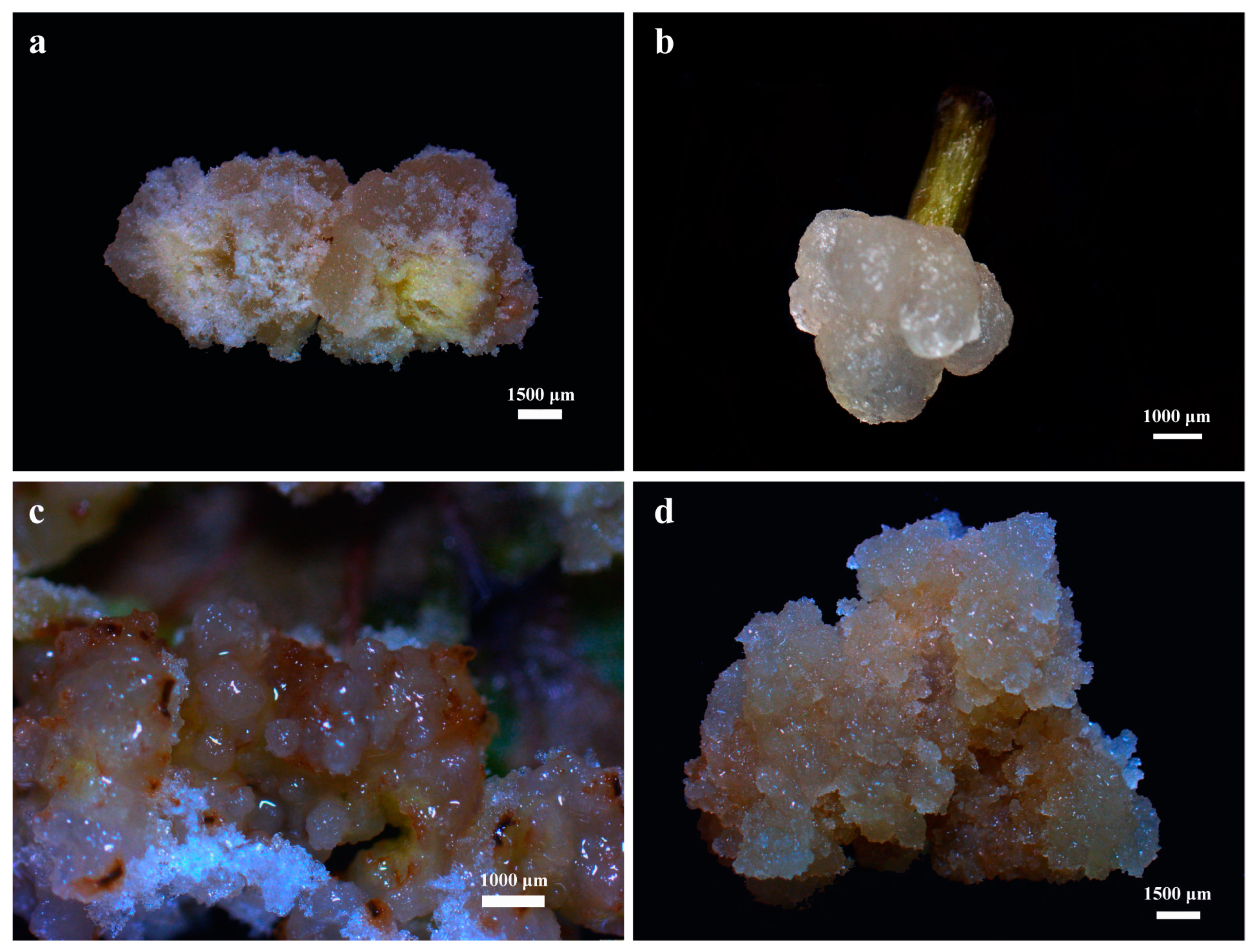
3.3. Somatic Embryo Maturation

A callus that had proliferated for one month underwent pre-maturation treatment, which entails transferring the callus to a liquid maturation medium for a 7-day suspension culture. Following the pre-maturation treatment, the callus was carefully filtered and subsequently transferred using forceps to a solid maturation medium for somatic embryo maturation. The solid maturation medium consists of the modified Blaydes’ medium devoid of PGRs and CH, supplemented with 1 g/L AC, 40 g/L sucrose, and 2.6 g/L gellan gum. The liquid maturation medium is identical to the solid maturation medium but excludes gellan gum. After one month of culture, the presence of somatic embryos was assessed and their numbers were quantified.
Following one month of culture, somatic embryos at various developmental stages were observed. Taking the somatic embryos induced from the petioles of the FX-2 genotype as an example, we observed the development of a globular embryo (Figure 3a), heart-shaped embryo (Figure 3b), torpedo-shaped embryo (Figure 3c), and cotyledonal embryo (Figure 3d).
Analysis of variance indicated that both genotype and callus type significantly influenced somatic embryo maturation (Supplementary Table S7). Specifically, in the FX-2 genotype, Callus II successfully induced somatic embryos, yielding 22 somatic embryos/g, whereas Callus I failed to induce somatic embryo formation (Table 5). In contrast, neither Callus I nor Callus II induced somatic embryo formation in the FX-12 genotype (Table 5). Notably, both Callus I and Callus II successfully induced somatic embryo formation in the FX-54 genotype, with Callus I producing 17 somatic embryos/g and Callus II producing 13 somatic embryos/g (Table 5). These findings underscore the critical role of genotype and callus type in the successful induction of somatic embryo formation.

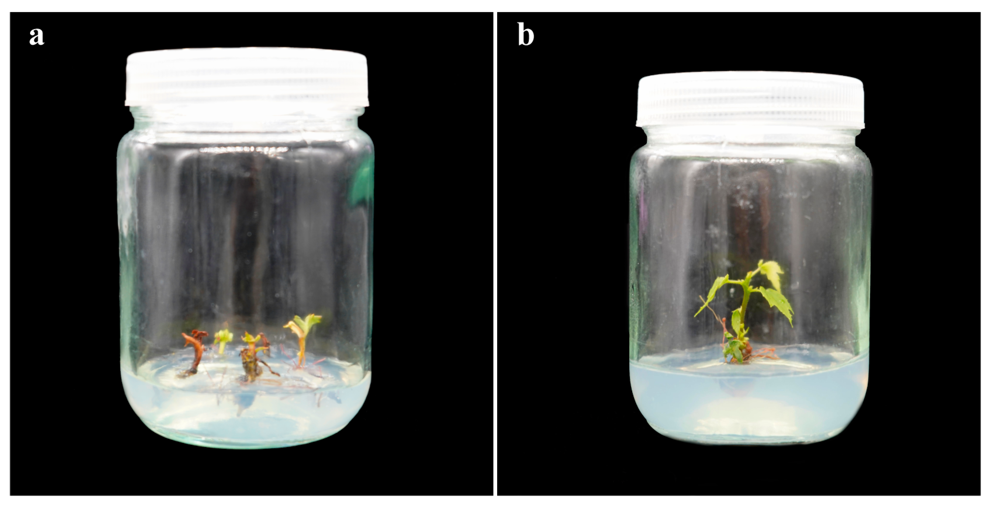
3.4. Effect of 6-BA on Somatic Embryo Germination into Seedlings

The cotyledonal embryos induced from leaves and petioles, characterized by distinct cotyledons, were designated as CE 1 and CE 2, respectively. Subsequently, CE 1 and CE 2 were transferred to germination mediums supplemented with varying concentrations of 6-BA. Notably, in FX-2, only CE 2 was subjected to somatic embryo germination experiments. After one month of culture, the germination of somatic embryos was assessed, and the germination efficiency was calculated to identify the optimal germination medium.
Variance analysis revealed that the concentration of 6-BA had a highly significant effect on somatic embryo germination in FX-2 (Supplementary Table S8). In contrast, in FX-54, while the concentration of 6-BA also had a highly significant effect on somatic embryo germination, the type of cotyledonal embryo did not significantly influence the germination efficiency (Supplementary Table S9).
During the induction of SE in hybrid sweetgum using immature zygotic embryos as explants, no PGRs were required at the somatic embryo germination stage. However, this study revealed that the absence of PGRs during the germination phase adversely impacted the germination of the somatic embryos. Research findings indicated that both the cotyledonal embryos of FX-2 and FX-54 could germinate into seedlings without the addition of plant hormones, but the germination efficiency was significantly lower, and the quality of the seedlings was suboptimal (Figure 4a). Conversely, the inclusion of an appropriate concentration of 6-BA markedly enhanced both the somatic embryo germination efficiency and the overall quality of the seedlings (Figure 4b).
This study evaluated three concentrations of 6-BA at 0, 0.1, and 0.5 mg/L. Statistical analysis revealed that for FX-2, the somatic embryo germination efficiency of CE 2 was lowest, at 13.33%, in the absence of 6-BA. The addition of 0.5 mg/L 6-BA resulted in the highest germination efficiency of 60%. Compared to the control group, the 0.5 mg/L concentration of 6-BA had a highly significant effect on the germination efficiency, whereas 0.1 mg/L did not show a significant difference (Figure 5a). Similar trends were observed in FX-54, where both CE 1 and CE 2 exhibited a peak germination efficiency of 76.67% and 73.33%, respectively, with the addition of 0.5 mg/L 6-BA. At 0.1 mg/L 6-BA, the germination efficiency was 40% and 43.33%, respectively. Without 6-BA, the germination efficiency was the lowest at 16.67% and 13.33%, respectively. Both 0.1 mg/L and 0.5 mg/L 6-BA showed highly significant effects compared to the control (Figure 5b). These findings suggest that an appropriate concentration of 6-BA can significantly enhance the efficiency of somatic embryo germination into seedlings. In conclusion, the optimal medium for somatic embryo germination is the modified Blaydes’ medium supplemented with 0.5 mg/L 6-BA, 40 g/L sucrose, and 2.6 g/L gellan gum.
At this stage, a somatic embryogenesis system utilizing vegetative organs in hybrid sweetgum as explants has been successfully developed. The complete induction process is illustrated in Figure 6.

4. Discussion

To date, research on the induction of SE from explants of mature trees within the Liquidambar species remains limited. Notably, successful induction has only been reported in Liquidambar styraciflua L., using male inflorescences as explants [26]. Immature zygotic embryos of hybrid sweetgum were utilized as explants for the induction of SE and the subsequent acquisition of regenerated plants [16,19]. This study represents the first report of SE induction from leaf and petiole explants in hybrid sweetgum. The findings indicate that the primary factors influencing SE induction in hybrid sweetgum are genotype, explant type, and medium composition.
In the majority of angiosperms and gymnosperms with documented SE systems, exogenous PGRs are recognized not only as pivotal factors for inducing SE but also for promoting the proliferation of embryogenic tissues [27]. 2,4-D is the most frequently utilized exogenous auxin in plant tissue culture, whereas 6-BA is the most commonly employed cytokinin [28]. In hybrid sweetgum, previous studies have demonstrated that 2,4-D and 6-BA effectively initiate SE from immature zygotic embryos [16,19]. Consequently, this study evaluated the effects of 2,4-D or NAA in combination with 6-BA on callus induction and proliferation. The findings revealed that a medium supplemented with 1.0 mg/L 2,4-D and 0.5 mg/L 6-BA was optimal for callus induction and proliferation, while a higher concentration of 2,4-D (3.0 mg/L) inhibited callus formation. Similar phenomena were also observed in Betula platyphylla. The concentrations of 2,4-D and 6-BA significantly influenced the induction efficiency of embryogenic callus. The highest induction frequency of 16.8% was achieved when 2 mg/L 2,4-D and 0.2 mg/L 6-BA were added to the 1/2 MS medium. Higher concentrations of 2,4-D (4 mg/L) inhibited SE [29]. In certain tree species, SE can occur without exogenous hormones. For instance, in Akebia trifoliata (Thunb.) Koidz (Lardizabalaceae), immature zygotic embryos served as explants, and somatic embryos were directly induced in hormone-free media. Furthermore, primary somatic embryos could generate secondary somatic embryos in hormone-free media with an efficiency as high as 95.8%. This study demonstrated that the addition of 2,4-D to the medium would inhibit somatic embryo induction [30]. One limitation of this study is the absence of an investigation into whether somatic embryos can be successfully induced without the addition of exogenous plant hormones. Therefore, future experiments should examine the impact of omitting PGRs on somatic embryo formation. During the maturation process of somatic embryos, PGRs are not required in the medium. Removing PGRs initiates polar auxin transport and the development of auxin gradients within the embryogenic callus [31]. The development of auxin polar transport is a critical step in the formation of meristems during embryo development [32]. In this study, somatic embryos were successfully obtained in a medium devoid of PGRs. However, the current yield of somatic embryos from vegetative organs remains low, indicating that further optimization of the somatic embryo system is necessary. Previous studies have demonstrated that somatic embryos derived from immature zygotic embryos can germinate in a medium devoid of PGRs [16,19]. However, our current study yielded contrasting results. In the absence of PGRs, somatic embryos failed to develop into normal seedlings; some perished, while others exhibited root development without subsequent elongation. This discrepancy may be attributed to differences in explant types. In this study, the addition of 0.5 mg/L 6-BA to the basal medium significantly improved somatic embryo germination. Similar observations have been reported in other species, such as Carica papaya L., where 6-BA enhances somatic embryo germination efficiency [33]. Although the maximum concentration of 6-BA used in this experiment was 0.5 mg/L, which achieved optimal germination efficiency, further investigations with higher concentrations are warranted to determine their effects on germination.
In woody plants, genotype is recognized as a critical determinant in in vitro asexual reproduction, particularly in SE. The findings presented herein indicate that only explants from genotypes FX-2 and FX-54 were successfully induced to undergo SE, whereas those from FX-12 failed to respond. Notably, FX-54 exhibited the capability of transforming somatic embryos into seedlings, suggesting it may possess superior embryogenic potential compared to the other two genotypes. In Betula platyphylla, an evaluation of SE across 10 genotypes revealed significant variability in callus and embryogenic callus induction frequencies, with the highest induction rate reaching 5.6% [29]. In Quercus robur L., among the five genotypes tested, only three were successfully induced to SE [14]. In Eucalyptus globulus Labill. and the hybrid E. saligna Smith × E. maidenii, Corredoira et al. mitigated the impact of genotypes on SE by optimizing culture conditions [13], resulting in somatic embryo formation from shoot tips and leaves of three genotypes, albeit with varying efficiencies. SE is genotype-dependent, and different genotypes may necessitate tailored culture conditions for optimal results.
In woody plants, inducing SE using mature tissues poses significant challenges. In Betula platyphylla, seven types of explants were evaluated: axillary buds, flower buds, young leaf blades, young petioles, young stems, mature seeds, and immature zygotic embryos. Research findings indicated that only mature and immature zygotic embryos could generate embryogenic callus, with the latter exhibiting a higher induction frequency [29]. For certain species, developing in vitro micropropagation systems from adult trees to produce explants has successfully facilitated SE induction [13,14,15]. This study adopted a similar strategy, achieving successful SE induction. However, no experiments have been conducted to determine if explants directly harvested from adult trees can induce SE effectively. Future studies should address this gap. In this study, explants were sourced from tissue cultures of test-tube seedlings rather than directly from wild-growing trees. This approach offers several advantages. Once sterile test-tube seedlings are developed, explants do not require additional sterilization, thereby reducing contamination during callus induction and minimizing mortality due to improper disinfection procedures. Additionally, it ensures a consistent supply of materials at the same developmental stage throughout the year.
In this study, vegetative organs were utilized as explants to successfully induce somatic embryogenesis in hybrid sweetgum, thereby providing robust technical support for molecular-assisted breeding in hybrid sweetgum. Subsequent research could integrate genomics, transcriptomics, or epigenomics to elucidate the underlying molecular mechanisms of somatic embryogenesis. Additionally, follow-up investigations may employ alternative explants (e.g., root or floral tissues) to validate the protocol developed herein, offering a practical reference for other woody plant species. Ultimately, given the ongoing decline in biodiversity, somatic embryogenesis protocols developed using vegetative organs as explants are particularly suited for the propagation of endangered plant species.

5. Conclusions

In conclusion, this study presents the first reliable protocol for SE induction from adult hybrid sweetgum trees. The combination of 2,4-D and 6-BA has been shown to positively influence callus induction and proliferation. Somatic embryos can be successfully obtained in a basic medium devoid of PGRs, although the yield remains relatively low. During the somatic embryo germination stage, the addition of 0.5 mg/L 6-BA significantly enhances germination efficiency. This initial development of SE induction from vegetative organs in hybrid sweetgum will accelerate its breeding process and enhance its commercial potential.

Supplementary Materials

The following supporting information can be downloaded at: https://www.mdpi.com/article/10.3390/f16040670/s1, Table S1. Variance analysis of callus induction efficiency under combined treatment of FX-2 explants and different hormone concentrations; Table S2. Variance analysis of callus induction efficiency under combined treatment of FX-12 explants and different hormone concentrations; Table S3. Variance analysis of callus induction efficiency under combined treatment of FX-54 explants and different hormone concentrations; Table S4. FX-2 callus type and different concentration of hormone combination treatment callus proliferation coefficient ANOVA table; Table S5. FX-12 callus type and different concentration of hormone combination treatment callus proliferation coefficient ANOVA table; Table S6. FX-54 callus type and different concentration of hormone combination treatment callus proliferation coefficient ANOVA table; Table S7. Variance analysis of somatic embryo maturation efficiency for different genotypes and callus types; Table S8. Variance analysis of embryo germination efficiency under different 6-BA concentrations in FX-2; Table S9. Variance analysis of embryo germination efficiency under different 6-BA concentrations in FX-54.

Author Contributions

Conceptualization, J.Z. (Jinfeng Zhang) and J.Z. (Jian Zhao); methodology, H.L.; software, H.L.; validation, F.B., Y.L., D.Z., Z.P. and L.C.; formal analysis, J.K.; investigation, Y.F.; resources, S.Q.; data curation, Y.F.; writing—original draft preparation, H.L.; writing—review and editing, Y.F.; visualization, H.L.; supervision, Y.F.; project administration, J.Z. (Jian Zhao) and J.Z. (Jinfeng Zhang); funding acquisition, J.Z. (Jinfeng Zhang). All authors have read and agreed to the published version of the manuscript.

Funding

This paper was supported by the National Key R&D Program of China (2023YFD2200602), the National Natural Science Foundation of China (32271836), the Fundamental Research Funds for the Central Universities (2019ZY39), and the Henan Province Science and Technology Research Project (232102110219).

Data Availability Statement

The data presented in this study are available on request from the corresponding author.

Acknowledgments

We would like to show our special thanks to Shenzhou Lvpeng Agricultural Science & Technology Co., Ltd. (Tongzhou, Beijing, China) for all of its support in this study.

Conflicts of Interest

The authors declare no conflicts of interest. Dingju Zhan and Zhenwu Pang are employed by Guangxi Bagui Forest and Flowers Seedlings Co., Ltd., and their employer’s company was not involved in this study. Therefore, there is no relevance between this study and their company. All authors declare that the research was conducted in the absence of any commercial or financial relationships that could be construed as a potential conflict of interest. The funders had no role in the design of the study; in the collection, analyses, or interpretation of data; in the writing of the manuscript; or in the decision to publish the results.

References

  1. Fan, Y.; Li, H.; Li, Y.; Bao, F.; Zhan, D.; Pang, Z.; Zhao, J.; Zhang, J. Construction of the First High-Density Genetic Linkage Map and QTL Mapping of Shikimic Acid Content in Liquidambar. Forests 2024, 15, 1662. [Google Scholar] [CrossRef]
  2. Lewis, J.; Will, R.; Zhai, L.; Murray, B.; Blazier, M.; Joshi, O. Productivity of hybrid sweetgum (Liquidambar formosana x styraciflua) plantations compared to native sweetgum (L. Styraciflua) in the Western Gulf Region of south-central U.S. New For. 2024, 55, 1753–1769. [Google Scholar] [CrossRef]
  3. Merkle, S. The ups and downs of developing hybrid sweetgum varieties for the U.S. bioenergy and pulp and paper industries: A 20-year case study. In The Fifth International Conference of the IUFRO Unit 2.09.02: Somatic Embryogenesis and Other Vegetative Propagation Technologies; University of Coimbra: Coimbra, Portugal, 2018. [Google Scholar]
  4. Lu, S.; Merkle, S.A. Enhancing hybrid Liquidambar somatic seedling production using a temporary immersion bioreactor. Trees 2021, 35, 503–512. [Google Scholar] [CrossRef]
  5. Kang, H.; Lee, C.; Kwon, S.; Park, J.; Kang, K.; Shim, D. Comparative transcriptome analysis during developmental stages of direct somatic embryogenesis in Tilia amurensis Rupr. Sci. Rep.-Uk 2021, 11, 6359. [Google Scholar] [CrossRef] [PubMed]
  6. Karami, O.; Philipsen, C.; Rahimi, A.; Nurillah, A.R.; Boutilier, K.; Offringa, R. Endogenous auxin maintains embryonic cell identity and promotes somatic embryo development in Arabidopsis. Plant J. 2023, 113, 7–22. [Google Scholar] [CrossRef] [PubMed]
  7. Berenguer, E.; Carneros, E.; Pérez-Pérez, Y.; Gil, C.; Martínez, A.; Testillano, P.S. Small molecule inhibitors of mammalian GSK-3β promotein vitro plant cell reprogramming and somatic embryogenesis in crop and forest species. J. Exp. Bot. 2021, 72, 7808–7825. [Google Scholar] [CrossRef] [PubMed]
  8. Chen, Y.; Hu, Y.; Wang, R.; Feng, K.; Di, J.; Feng, T.; Cao, F. Transcriptome and physiological analysis highlights the hormone, phenylpropanoid, and photosynthesis effects on early somatic embryogenesis in Ginkgo biloba. Ind. Crop. Prod. 2023, 203, 117176. [Google Scholar] [CrossRef]
  9. Li, Z.; Zhang, D.; Shi, P.; Htwe, Y.M.; Yu, Q.; Huang, L.; Zhou, H.; Liu, L.; Wang, Y. Cell wall lignification may be necessary for somatic embryogenesis of areca palm (Areca catechu). Sci. Hortic.-Amst. 2023, 307, 111538. [Google Scholar] [CrossRef]
  10. Spinoso-Castillo, J.L.; Bello-Bello, J.J. In Vitro Stress-Mediated Somatic Embryogenesis in Plants; Springer: New York, NY, USA, 2022; Volume 2527, pp. 223–235. [Google Scholar]
  11. Su, Y.H.; Tang, L.P.; Zhao, X.Y.; Zhang, X.S. Plant cell totipotency: Insights into cellular reprogramming. J. Integr. Plant Biol. 2021, 63, 228–243. [Google Scholar] [CrossRef]
  12. Cai, Y.; Tang, L.; Chen, H.; Li, Y.; Liu, R.; Chen, J. Somatic embryogenesis in Rosa chinensis cv. ‘Old Blush’. Plant Cell Tissue Organ Cult. 2022, 149, 645–656. [Google Scholar] [CrossRef]
  13. Corredoira, E.; Ballester, A.; Ibarra, M.; Vieitez, A.M. Induction of somatic embryogenesis in explants of shoot cultures established from adult Eucalyptus globulus and E. saligna × E. maidenii trees. Tree Physiol. 2015, 35, 678–690. [Google Scholar] [CrossRef]
  14. San-José, M.C.; Corredoira, E.; Martínez, M.T.; Vidal, N.; Valladares, S.; Mallón, R.; Vieitez, A.M. Shoot apex explants for induction of somatic embryogenesis in mature Quercus robur L. trees. Plant Cell Rep. 2010, 29, 661–671. [Google Scholar] [CrossRef] [PubMed]
  15. Correia, S.; Lopes, M.L.; Canhoto, J.M. Somatic embryogenesis induction system for cloning an adult Cyphomandra betacea (Cav.) Sendt. (tamarillo). Trees 2011, 25, 1009–1020. [Google Scholar] [CrossRef]
  16. Vendrame, W.; Holliday, C.; Merkle, S. Clonal propagation of hybrid sweetgum (Liquidambar styraciflua × L. formosana) by somatic embryogenesis. Plant Cell Rep. 2001, 20, 691–695. [Google Scholar] [CrossRef]
  17. Dai, J.; Vendrame, W.A.; Merkle, S.A. Enhancing the productivity of hybrid yellow-poplar and hybrid sweetgum embryogenic cultures. Vitr. Cell. Dev. Biol.-Plant 2004, 40, 376–383. [Google Scholar] [CrossRef]
  18. Merkle, S.; Montello, P.; Kormanik, T.; Le, H. Propagation of novel hybrid sweetgum phenotypes for ornamental use via somatic embryogenesis. Propag. Ornam. Plants 2010, 10, 220–226. [Google Scholar]
  19. Qi, S.; Zhao, R.; Yan, J.; Fan, Y.; Huang, C.; Li, H.; Chen, S.; Zhang, T.; Kong, L.; Zhao, J.; et al. Global Transcriptome and Coexpression Network Analyses Reveal New Insights Into Somatic Embryogenesis in Hybrid Sweetgum (Liquidambar styraciflua × Liquidambar formosana). Front. Plant Sci. 2021, 12, 751866. [Google Scholar] [CrossRef]
  20. Zhao, R.; Qi, S.; Cui, Y.; Gao, Y.; Jiang, S.; Zhao, J.; Zhang, J.; Kong, L. Transcriptomic and physiological analysis identifies a gene network module highly associated with brassinosteroid regulation in hybrid sweetgum tissues differing in the capability of somatic embryogenesis. Hortic. Res.-Engl. 2022, 9, uhab047. [Google Scholar] [CrossRef]
  21. Zhang, Y.; Wang, Z.; Qi, S.; Wang, X.; Zhao, J.; Zhang, J.; Li, B.; Zhang, Y.; Liu, X.; Yuan, W. In Vitro Tetraploid Induction from Leaf and Petiole Explants of Hybrid Sweetgum (Liquidambar styraciflua × Liquidambar formosana). Forests 2017, 8, 264. [Google Scholar] [CrossRef]
  22. Witham, F.H.; Blaydes, B.F.; Devlin, R.M. Experiments in Plant Physiology; Van Nostrand Reinhold: New York, NY, USA, 1971. [Google Scholar]
  23. Gresshoff, P.M.; Doy, C.H. Development and differentiation of haploid Lycopersicon esculentum (tomato). Planta 1972, 107, 161–170. [Google Scholar] [CrossRef] [PubMed]
  24. Sommer, H.E.; Brown, C.L. Embryogenesis in Tissue Cultures of Sweetgum. For. Sci. 1980, 26, 257–260. [Google Scholar]
  25. Murashige, T.; Skoog, F. A Revised Medium for Rapid Growth and Bio Agsays with Tohaoco Tissue Cultures. Physiol. Plant. 1962, 15, 473–497. [Google Scholar] [CrossRef]
  26. Merkle, S.A.; Battle, P.J. Enhancement of embryogenic culture initiation from tissues of mature sweetgum trees. Plant Cell Rep. 2000, 19, 268–273. [Google Scholar] [CrossRef]
  27. Hazubska-Przybył, T.; Ratajczak, E.; Obarska, A.; Pers-Kamczyc, E. Different Roles of Auxins in Somatic Embryogenesis Efficiency in Two Picea Species. Int. J. Mol. Sci. 2020, 21, 3394. [Google Scholar] [CrossRef]
  28. Martínez, M.; Corredoira, E. Recent Advances in Plant Somatic Embryogenesis: Where We Stand and Where to Go. Int. J. Mol. Sci. 2024, 25, 8912. [Google Scholar] [CrossRef]
  29. Yang, J.; Yang, D.; Lü, W.; Zhang, X.; Ma, M.; Liu, G.; Jiang, J.; Li, C. Somatic embryogenesis and plant regeneration in Betula platyphalla. J. For. Res. 2021, 32, 937–944. [Google Scholar] [CrossRef]
  30. Zou, S.; Yao, X.; Zhong, C.; Li, D.; Wang, Z.; Huang, H. Recurrent somatic embryogenesis and development of somatic embryos in Akebia trifoliata (Thunb.) Koidz (Lardizabalaceae). Plant Cell Tissue Organ Cult. (PCTOC) 2019, 139, 493–504. [Google Scholar] [CrossRef]
  31. Yuan, J.; Chao, Y.; Han, L. Uncovering a Phenomenon of Active Hormone Transcriptional Regulation during Early Somatic Embryogenesis in Medicago sativa. Int. J. Mol. Sci. 2022, 23, 8633. [Google Scholar] [CrossRef]
  32. Wójcik, A.M.; Wójcikowska, B.; Gaj, M.D. Current Perspectives on the Auxin-Mediated Genetic Network that Controls the Induction of Somatic Embryogenesis in Plants. Int. J. Mol. Sci. 2020, 21, 1333. [Google Scholar] [CrossRef]
  33. Solórzano-Cascante, P.; Sánchez-Chiang, N.; Jiménez, V.M. Explant Type, Culture System, 6-Benzyladenine, Meta-Topolin and Encapsulation Affect Indirect Somatic Embryogenesis and Regeneration in Carica papaya L. Front. Plant Sci. 2018, 9, 1769. [Google Scholar] [CrossRef] [PubMed]
Figure 1. The comprehensive procedure for inducing somatic embryogenesis.
Forests 16 00670 g001
Figure 2. Phenotypic status of callus induced by different explants of genotype FX-2. (a) Petiole-induced white ice crystal-like callus, bar = 1.5 mm. (b) Stem segment-induced compact callus, bar = 1 mm. (c) Leaf-induced spherical callus, bar = 1 mm. (d) Petiole-induced loose callus, bar = 1.5 mm.
Forests 16 00670 g002
Figure 3. Somatic embryos at various developmental stages were induced from petiole explants of the FX-2 genotype. (a) Globular embryo, bar = 1 mm. (b) Heart-shaped embryo, bar = 1 mm. (c) Torpedo-shaped embryo, bar = 1 mm. (d) Cotyledonal embryo, bar = 1 mm.
Forests 16 00670 g003
Figure 4. Cotyledonal embryos induced from leaf explants of genotype FX-54 exhibited germination of somatic embryos in both germination mediums without 6-BA and in medium supplemented with 0.5 mg/L 6-BA. (a) Without 6-BA treatment for somatic embryo germination. (b) Somatic embryo germination was treated with 0.5 mg/L 6-BA.
Forests 16 00670 g004
Figure 5. Germination efficiency of somatic embryos treated with different concentrations of 6-BA. (a) Effect of 6-BA at different concentrations in FX-2 on somatic embryo germination of CE 2. (b) Effect of different concentrations of 6-BA on somatic embryo germination in FX-54. CE 1 represents leaf-induced cotyledonal embryo, and CE 2 represents petiole petiole-induced cotyledonal embryo. Statistical differences between treatments are shown as “**” (p < 0.01) and “***” (p < 0.001). Statistical insignificance between treatments is shown as “ns”.
Forests 16 00670 g005
Figure 6. Induction process of somatic embryogenesis with vegetative organs in hybrid sweetgum as explants. (a) Leaf explant, bar = 1.5 mm. (b) Petiole explant, bar = 1.5 mm. (c) Stem explant, bar = 0.5 mm. (d) Leaf-induced callus, bar = 1 mm. (e) Petiole-induced callus, bar = 1 mm. (f) Stem-induced callus, bar = 1 mm. (g) Leaf-induced callus proliferation for one month, bar = 1 mm. (h) Petiole-induced callus proliferation for one month, bar = 1 mm. (i) Petiole-induced somatic embryo, bar = 1 mm. (j) Leaf-induced somatic embryo. (k) Petiole-induced cotyledon embryo germination into seedlings. (l) Leaf-induced cotyledon embryo germination into seedlings.
Forests 16 00670 g006
Table 1. Combinations of 2,4-D and 6-BA treatments at different concentrations in callus induction.
Treatment2,4-D(mg/L)6-BA(mg/L)
11.00.25
21.00.50
32.00.25
42.00.50
53.00.25
63.00.50
Table 2. Combinations of hormone treatments with different concentrations in callus proliferation.
Treatment2,4-D(mg/L)NAA(mg/L)6-BA(mg/L)
10.5-0.25
21.0-0.5
3-0.50.25
4-1.00.5
Table 3. Efficiency of callus induction in different explants treated with varying hormone concentrations.
GenotypeTreatmentCallus Induction Efficiency (%)
LeafPetioleStem Segment
FX-21.0 mg/L 2,4-D + 0.25 mg/L 6-BA80 ± 10 bcdeBCD86.67 ± 5.77 bcABC86.67 ± 5.77 bcABC
1.0 mg/L 2,4-D + 0.5 mg/L 6-BA90 ± 10 abAB96.67 ± 5.77 aA96.67 ± 5.77 aA
2.0 mg/L 2,4-D + 0.25 mg/L 6-BA86.67 ± 5.77 bcABC80 ± 10 bcdeBCD86.67 ± 5.77 bcABC
2.0 mg/L 2,4-D + 0.5 mg/L 6-BA83.33 ± 5.77 bcdBC76.67 ± 5.77 cdefBCD86.67 ± 5.77 bcABC
3.0 mg/L 2,4-D + 0.25 mg/L 6-BA70 ± 10 cdefCDE66.67 ± 5.77 defgCDE63.33 ± 5.77 efgCDE
3.0 mg/L 2,4-D + 0.5 mg/L 6-BA56.67 ± 5.77 fgDE46.67 ± 5.77 gE56.67 ± 5.77 fgDE
FX-121.0 mg/L 2,4-D + 0.25 mg/L 6-BA53.33 ± 5.77 deC50 ± 10 eC53.33 ± 5.77 deC
1.0 mg/L 2,4-D + 0.5 mg/L 6-BA80 ± 10 abAB86.67 ± 5.77 aA70 ± 10 abcdeABC
2.0 mg/L 2,4-D + 0.25 mg/L 6-BA76.67 ± 15.28 abcABC63.33 ± 15.28 bcdeABC66.67 ± 5.77 bcdeABC
2.0 mg/L 2,4-D + 0.5 mg/L 6-BA70 ± 10 abcdeABC60 ± 10 cdeBC63.33 ± 5.77 bcdeABC
3.0 mg/L 2,4-D + 0.25 mg/L 6-BA73.33 ± 5.77 abcdABC50 ± 10 eC60 ± 10 cdeBC
3.0 mg/L 2,4-D + 0.5 mg/L 6-BA63.33 ± 11.55 bcdeABC66.67 ± 15.28 bcdeABC56.67 ± 15.28 deBC
FX-541.0 mg/L 2,4-D + 0.25 mg/L 6-BA66.67 ± 5.77 bcdCD76.67 ± 5.77 bcBC60 ± 10 cdCD
1.0 mg/L 2,4-D + 0.5 mg/L 6-BA93.33 ± 5.77 aA90 ± 10 aAB80 ± 10 bBC
2.0 mg/L 2,4-D + 0.25 mg/L 6-BA76.67 ± 5.77 bcBC73.33 ± 5.77 bcCD70 ± 10 bcCD
2.0 mg/L 2,4-D + 0.5 mg/L 6-BA66.67 ± 5.77 bcdCD76.67 ± 5.77 bcBC73.33 ± 5.77 bcCD
3.0 mg/L 2,4-D + 0.25 mg/L 6-BA63.33 ± 5.77 bcdCD66.67 ± 5.77 bcdCD60 ± 10 cdCD
3.0 mg/L 2,4-D + 0.5 mg/L 6-BA50 ± 10 dD60 ± 10 cdCD63.33 ± 5.77 bcdCD
Note: Through Duncan’s multiple comparisons, lowercase letters represent significant differences in callus induction rates among treatments (p < 0.05); uppercase letters represent extremely significant differences in callus induction rates among treatments (p < 0.01).
Table 4. Callus proliferation coefficient under combination treatment of FX-2 callus types and different hormone concentrations.
GenotypeTreatmentCallus Proliferation Coefficient
Callus ICallus II
FX-20.5 mg/L 2,4-D + 0.25 mg/L 6-BA1.88 ± 0.06 bB1.84 ± 0.05 bBC
1.0 mg/L 2,4-D + 0.5 mg/L 6-BA2.12 ± 0.01 aA2.10 ± 0.13 aA
0.5 mg/L NAA + 0.25 mg/L 6-BA1.58 ± 0.07 cD1.63 ± 0.04 cD
1.0 mg/L NAA + 0.5 mg/L 6-BA1.69 ± 0.10 cCD1.65 ± 0.03 cCD
FX-120.5 mg/L 2,4-D + 0.25 mg/L 6-BA1.92 ± 0.04 bcBC1.88 ± 0.03 cdBC
1.0 mg/L 2,4-D + 0.5 mg/L 6-BA2.31 ± 0.19 aA2.06 ± 0.06 bB
0.5 mg/L NAA + 0.25 mg/L 6-BA1.75 ± 0.02 dCD1.60 ± 0.04 eD
1.0 mg/L NAA + 0.5 mg/L 6-BA1.92 ± 0.07 bcBC1.81 ± 0.04 cdCD
FX-540.5 mg/L 2,4-D + 0.25 mg/L 6-BA1.93 ± 0.04 cC1.84 ± 0.07 dCD
1.0 mg/L 2,4-D + 0.5 mg/L 6-BA2.58 ± 0.04 aA2.38 ± 0.05 bB
0.5 mg/L NAA + 0.25 mg/L 6-BA1.66 ± 0.04 eE1.45 ± 0.05 fF
1.0 mg/L NAA + 0.5 mg/L 6-BA1.82 ± 0.04 dCD1.78 ± 0.07 dD
Note: Through Duncan’s multiple comparisons, lowercase letters represent significant differences in callus induction rates among treatments (p < 0.05); uppercase letters represent extremely significant differences in callus induction rates among treatments (p < 0.01). Callus I represents the callus induced by leaves, and Callus II represents the callus induced by petioles.
Table 5. Number of somatic embryo maturation in different genotypes and callus types.
Callus TypeThe Number of Mature Embryos/g
GenotypeCallus ICallus II
FX-20 ± 0 cC22 ± 5 aA
FX-120 ± 0 cC0 ± 0 cC
FX-5417 ± 3 bAB13 ± 5 bB
Note: Through Duncan’s multiple comparisons, lowercase letters represent significant differences in callus induction rates among treatments (p < 0.05); uppercase letters represent extremely significant differences in callus induction rates among treatments (p < 0.01). Callus I represents the callus induced by leaves, and Callus II represents the callus induced by petioles.
Disclaimer/Publisher’s Note: The statements, opinions and data contained in all publications are solely those of the individual author(s) and contributor(s) and not of MDPI and/or the editor(s). MDPI and/or the editor(s) disclaim responsibility for any injury to people or property resulting from any ideas, methods, instructions or products referred to in the content.

