Article

The Impact of Pollinating Fig Wasps’ Entry on Fig Development and the Hormonal Regulation of Sex Differentiation in Ficus hispida

1
College of Forestry, Southwest Forestry University, Kunming 650224, China
2
CAS Key Laboratory of Tropical Forest Ecology, Xishuangbanna Tropical Botanical Garden, Chinese Academy of Sciences, Menglun 666303, China
3
Key Laboratory of Southwest Mountain Forest Resources Conservation and Utilization, Ministry of Education, Kunming 650224, China
*
Authors to whom correspondence should be addressed.
These authors contributed equally to this work.
Forests 2025, 16(2), 286; https://doi.org/10.3390/f16020286
Submission received: 17 December 2024 / Revised: 30 January 2025 / Accepted: 4 February 2025 / Published: 7 February 2025
(This article belongs to the Section Forest Ecophysiology and Biology)

Abstract

:
Ficus trees (Moraceae) play a vital role in sustaining the stability of tropical and subtropical rainforests. The obligate mutualism between Ficus species and their pollinating fig wasps renders them an exemplary model for investigating insect–plant coevolution. In this study, we employed Ficus hispida Linn. f., an ecologically significant fig species in tropical rainforests, to conduct a wasp-introduction controlled experiment in the field. This method enabled us to precisely delineate the developmental stages of figs. We collected samples at specific intervals and examined the impact of pollinating fig wasp entry on the hormonal metabolism of male and female figs using liquid chromatography–tandem mass spectrometry analysis. The findings demonstrate that pollinator entry significantly decreases fig abscission. Moreover, it substantially altered the developmental indices of the figs. Unpollinated figs exhibit elevated levels of abscisic acid (ABA), which increases the likelihood of fig abortion and reduces the probability of pollinator entry into senescent figs. Following pollinator entry, indole-3-acetic acid (IAA) levels rise in both male and female figs. Male figs show higher concentrations of 1-aminocyclopropane-1-carboxylic acid (ACC), jasmonic acid (JA), and salicylic acid (SA), whereas these changes are less pronounced in female figs. Additionally, pollinated male figs display increased levels of cytokinins (CKs) and other hormones compared to female figs, suggesting a coordinated hormonal response to the stress induced by pollinator oviposition and gall development. Our findings suggest that the entry of pollinators likely triggers the transition from the female to the interfloral phase, with hormonal regulation playing a crucial role in the reproductive dimorphism of figs. This research can offer novel insights into the mechanisms underlying fig–wasp mutualism.

1. Introduction

The coevolutionary dynamics between plants and insects have the potential to substantially impact the rate of species evolution and enhance biodiversity in natural ecosystems [1]. This phenomenon predominantly occurs between plants and phytophagous insects or between plants and pollinators [2]. Coevolutionary relationships are typically characterized by one-to-one interactions among species, which can arise from predator–prey interactions, parasitic associations, or reciprocal symbiosis [3]. Previous studies have demonstrated that plants possess not only defensive mechanisms, known as ‘resistance’, against predators but also the ability to ‘tolerate’ tissue damage [4,5,6]. The observed phenotypic traits and species interactions in contemporary ecosystems result from long-term associations and adaptations among diverse organisms [7]. Investigating mutualistic mechanisms between co-evolving species can enhance our understanding of organismal adaptive strategies and ecosystem stability [8].
Compared to other ecosystems, tropical rainforest ecosystems display the highest biodiversity and complex species interactions, with Ficus spp. occupying a central role [9,10]. The research species, Ficus hispida Linn. f., belonging to Moraceae, is a dioecious small tree widely distributed in tropical regions of Asia; like some other Ficus species, it serves as an essential food source for numerous frugivorous animals and provides habitats for parasitic and epiphytic plants, thereby significantly contributing to the maintenance of tropical rainforest landscapes [9,11]. Additionally, it is commonly used as a traditional Chinese medicine [9]. The developmental phases of the fig can be categorized into the pre-female phase, female phase (receptive phase or receptivity), interfloral phase, male phase (only for male fig), and post-floral phase. Ceratosolen solmsi marchali Mayr is the exclusive pollinator for this species (Figure 1). The ecological and industrial significance of Ficus plants is increasingly recognized, and many countries cultivate various Ficus species as valuable cash crops [12]. Successful pollination by fig wasps is essential for both fig production and the sustainability of Ficus populations. Given that Ficus flowers are enclosed within the syconium, effective pollination depends on fig wasps entering the figs at the optimal time and completing the pollination process [8,10].
Figs, also known as syconia, possess bracts at their apex through which only species-specific fig wasps can access the fig cavity via the ostiole. Once inside the fig, pollinating fig wasps engage in both pollination and oviposition activities. Consequently, some flowers produce seeds while others develop galls to support wasp offspring [13,14]. During a coevolutionary period of 60–90 million years, the Ficus tree and its pollinator have established one of nature’s most quintessential symbiotic relationships through reciprocal adaptations in physiology, morphology, and chemical attraction [14,15]. In the fig–fig wasp system, the reproduction of the fig tree is contingent upon seed production, while wasp reproduction relies on gall formation to generate offspring. However, both symbiotic species rely on female flower resources for successful reproduction. The seeds ensure effective fig propagation, whereas the wasps’ offspring are vectors for pollen transfer between figs [13]. Ficus spp. can be classified into monoecious and dioecious species. In monoecious figs, the abundance of pollinators may affect the reproductive allocation of female flowers; however, regardless of the number of fig wasps entering, it is certain that some flowers will develop into galls while others will develop into seeds. In dioecious figs, despite the presence of both ovipositing and pollinating behaviors in pollinators, no matter whether pollinators enter male or female figs, only seeds are ultimately produced in female figs, while gall formation exclusively occurs in male figs [13,16,17,18].
The regulation of developmental fate in flowers in figs has been a subject of inquiry for researchers, yet current explanations remain largely hypothetical. In response to diverse selective pressures, fig trees may have evolved the dual function of attracting pollinating fig wasps for successful pollination and inducing defense responses to deter excessive oviposition by these pollinators. Reproductive functions in female and male trees are distinct, potentially leading to trade-offs between reproductive and defensive traits among individuals of different sexes. These are regulated through multiple pathways encompassing morphological, physiological, hormonal, and genetic mechanisms [19,20,21].
Currently, the regulation of flower reproduction in male and female figs is primarily informed by morphological and physiological studies [22,23,24], with limited studies investigating hormonal regulation. Phytohormones are pivotal in regulating plant growth, development, and reproduction. Plants synthesize various hormones, including jasmonic acid (JA), salicylic acid (SA), gibberellic acid (GA), cytokinins (CKs), abscisic acid (ABA), and auxin [25]. These phytohormones modulate responses to both biotic and abiotic stresses, such as tissue damage [26]. The transition from vegetative to reproductive growth is characterized by shifts in hormone types and concentrations [27]. CKs, GA, and auxin are particularly important for regulating floral meristem size and organ initiation [28]. Parthenocarpy in Ficus carica Linn. is controlled through the suppression of ABA and ethylene biosynthesis and GA catabolism. ABA accumulation promotes anthocyanin biosynthesis, influencing fig color and nutritional quality [29,30]. In certain species, flower or fruit sex determination depends on specific hormone ratios. Elevated levels of growth hormones stimulate pollen production in male flowers, while increased auxin and GA support ovary and seed development in female flowers [31,32]. Hormone levels can fluctuate based on resource availability, especially in dioecious plants [33]. The differentiation of female flowers within male and female figs and its relationship to fig wasp behavior remain poorly understood [34,35,36]. We hypothesize that multiple hormonal interactions may regulate this complex process.
In this study, through ecological and metabolomics analysis, the following questions are anticipated to be addressed: (1) How do hormonal metabolisms in figs differ across various developmental stages? (2) Are there sex-specific variations in hormonal metabolisms between male and female figs? (3) Does the entry of fig wasps into the fig induce changes in hormonal regulation for both sexes?

2. Materials and Methods

2.1. Experimental Tree and Location

In this study, we selected a dioecious fig species (F. hispida) and its exclusive pollinator (Ceretosolen solmsi marchali) from Xishuangbanna as our research subjects. Through a controlled experiment in the field and metabolomics analysis, we investigated the gender-specific hormonal regulation disparities in fig development.
We conducted a preliminary survey of 30 F. hispida trees in the Xishuangbanna Tropical Botanical Garden (XTBG) and documented the reproductive phenology of each tree. After approximately two months of field investigation, 6 suitable trees (3 ♀ and 3 ♂) were selected for conducting the controlled experiment. The Xishuangbanna Tropical Botanical Garden, located in Menglun, Mengla County, Yunnan Province, Southwest China (101°15′ E, 21°55′ N, altitude 555 m), is an institution affiliated with the Chinese Academy of Sciences, situated adjacent to Southeast Asia. This area represents a typical tropical rainforest ecosystem.

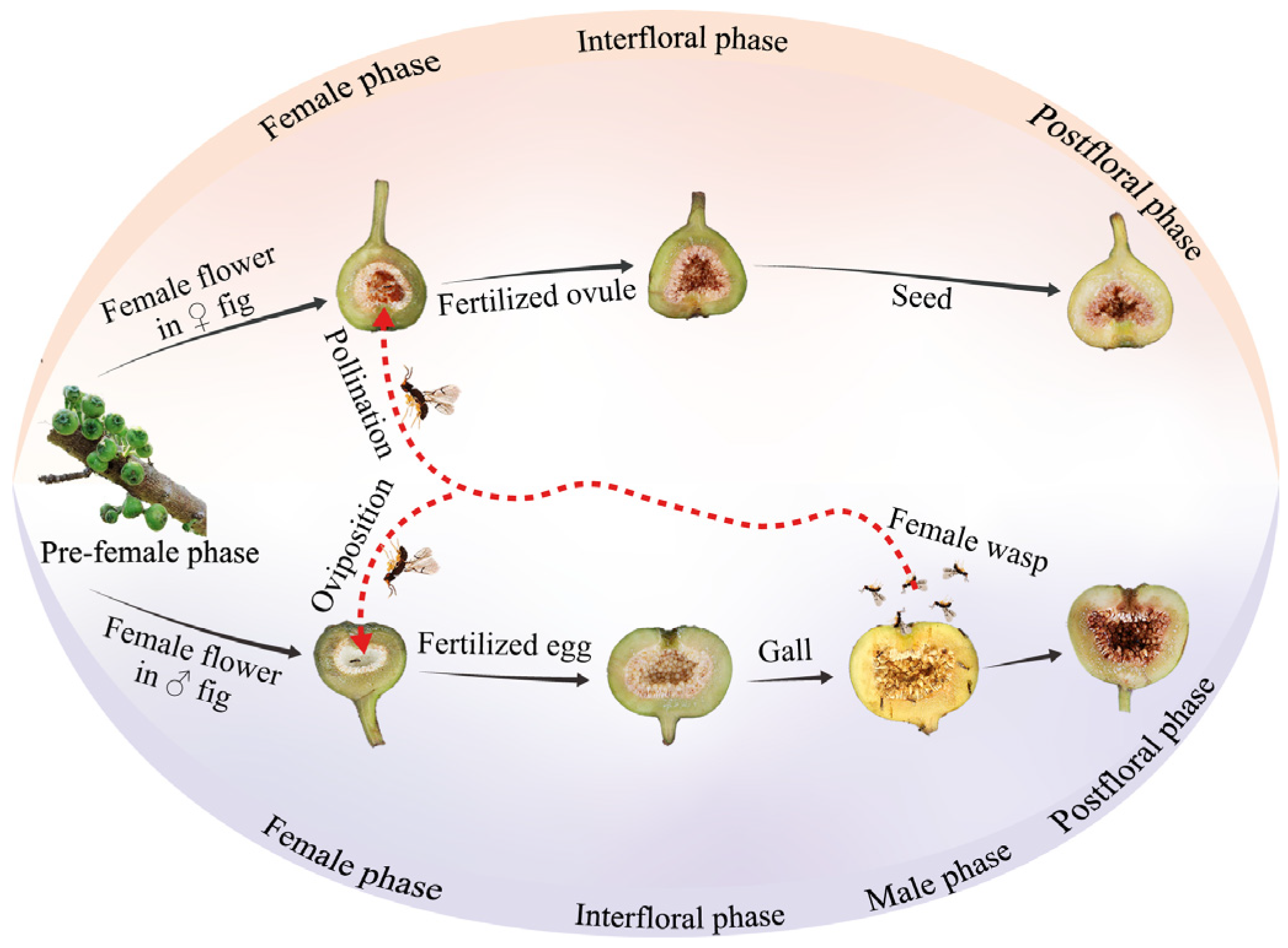
2.2. General Biology of Ficus hispida and Collection of Pollinating Fig Wasps

The pollinating fig wasps for the controlled experiment were obtained as follows: on the day preceding the field experiment, F. hispida figs in close proximity to the male phase were selected and individually enclosed in clean netting bags. Typically, the natural emergence of pollinating fig wasps occurs around 6–8 a.m. the following morning, after which they should be promptly taken to the field for a controlled experiment.

2.3. Identification of Female Phase

We conducted weekly monitoring of the reproductive phenology for the six experimental trees. Upon initiating F. hispida fig growth, we used netting bags to isolate the figs and prevent interference from fig wasps and other insects (Figure 2A,B). When the figs approached receptivity, as indicated by loosening bracts, an active pollinating fig wasp was introduced onto the fig wall. If the wasp climbed toward the bract within five minutes and exhibited entry behavior into the bract ostiole, the fig was considered receptive (Figure 2C). In such cases, we carefully removed the pollinating wasp using tweezers and designated that day as the first day of the female phase, followed by re-enclosing the fig in a bag. If the fig was not deemed receptive, it remained isolated within the bag. This procedure was repeated daily until the fig became receptive.

2.4. Introducing Pollinating Fig Wasps and Documenting Fig Abscission Rates

On the first day of receptivity, two pollinating fig wasps were introduced into the fig (sample size = 30 for each tree), ensuring no interference between wasps by waiting for the complete entry of the first wasp into the bract before placing the second one on the fig wall. The figs that received wasp introductions were marked and subsequently isolated using netting bags to facilitate normal development. Additionally, 30 figs were left on each tree without introducing any fig wasps; only the figs’ developmental phase was recorded. The number of abscission figs was recorded daily until all the figs had dropped off, allowing for the calculation of the daily cumulative fig abscission rate (the ratio of abscission fig number to total fig number) [37].

2.5. Collections of Plant Material

To acquire figs at specific developmental phases for subsequent analysis, this study employed controlled experiments in XTBG to collect experimental materials. Experiments were conducted on both female and male figs in the dry–hot season, on days that were both sunny and windless, with and without the introduction of pollinating fig wasps, following the methods described in Section 2.3 and Section 2.4. Four treatment groups were established: female figs with introduced fig wasps (F+), male figs with introduced fig wasps (M+), female figs without introduced fig wasps (F−), and male figs without introduced fig wasps (M−). One week after the onset of receptivity, samples were collected by dissecting 3–5 figs to extract female flower tissue (approximately 0.5 g), which were then placed into 2 mL centrifuge tubes and immediately frozen in liquid nitrogen for 72 h before being transferred to a −80 °C freezer for storage prior to endogenous hormone examination. Three female trees and three male trees were sampled, respectively, so each treatment included three biological replicates.

2.6. The Isolation and Purification of Endogenous Hormones

The research is based on the latest plant hormone library detection, which can simultaneously detect 34 substances of 9 major types of plant hormones at one time. Initially, a total of 34 distinct plant hormone standard solutions were prepared by diluting each with methanol to a concentration of 1 mg/mL. These individual standards were subsequently combined to create a standard curve stock solution, which was further diluted with methanol to the required concentrations to produce the standard curve working solution. All solutions were stored at −20 °C. Next, IAA (Indole-3-acetic acid) and ABA internal standards were accurately weighed and dissolved in methanol to form 1 mg/mL single-standard stock solutions. Then, 10 mL of these single-standard stock solutions were mixed to prepare a mixed internal standard stock solution containing 0.5 μg/mL IAA-D5 and 1 μg/mL ABA-D6, which was also stored at −20 °C. Finally, 15 µL of the standard curve working solution and 0.5 µL of the mixed internal standard stock solution were combined in a vial, followed by 134.5 µL of QC sample to prepare the final standard curve working solution. By correlating the unknown sample’s response with the standard curve, its concentration can be accurately determined, thereby ensuring the reliability and consistency of the analytical results in LC-MS/MS analysis.
The lyophilized flower sample in centrifuge tubes was ground to a fine powder. Subsequently, 1 mL of methanol was added and thoroughly mixed with the internal standard solution. Manual blending was performed, followed by ultrasonication for 10 min. The mixture was transferred to a water bath and agitated at room temperature for 4 h. Subsequently, the sample was centrifuged at 12,000 rpm at 4 °C for 10 min. The supernatant was collected, and 0.5 mL of methanol was added to the residual material. The resulting mixture was shaken in the water bath for an additional 2 h. The supernatant was then collected once more. Both supernatants were combined, subjected to another centrifugation at 12,000 rpm at 4 °C for 10 min, and subsequently filtered through a 0.22 μm filter. The filtrate was placed in a sample vial for preparation prior to LC-MS/MS analysis [38].

2.7. Determination of Endogenous Hormones

The mass spectrometry analysis was conducted using an ExionLC 2.0 UPLC system (AB Sciex, Framingham, MA, USA) equipped with an Acquity UPLC® CSH C18 (1.7 μm, 2.1 × 150 mm, Waters, Milford, MA, USA) column. The temperature of the column was set at 40 °C. The sample injection volume was 2 µL. The eluents consisted of 0.05% formic acid with 2 mM ammonium formate water (eluent A) and 0.05% formic acid in methanol (eluent B). The flow rate was set at 0.25 mL/min. Different gradient elutions were used to separate compound mixtures by gradually increasing the concentration of one or more mobile phase components while decreasing others. An elution gradient set was performed as follows: 0~2 min, 10% B; 2~4 min, 10%~30% B; 4~19 min, 30%~95% B; 19~19.10 min, 95%~10% B; and 19.10~22 min, 10% B [39,40]. The phytohormones were quantified using an electrospray ionization (ESI) source coupled with an AB SCIEX 6500+ Qtrap mass spectrometer (AB SCIEX OS V2.0.1.48692 software, Framingham, MA, USA) and analyzed by multiple reaction monitoring (MRM).

2.8. Data Analysis

We employed General Linear Models (GLMs) to evaluate the impact of wasps’ entry on fig abscission rates. Additionally, GLMs were utilized to examine the effects of wasps’ entry on fig developmental indices, including fig length, weight, wall thickness, and diameter. A p-value of less than 0.05 indicates a significant difference. All analyses were conducted using R 4.2.2 software.
Principal component analysis (PCA) was utilized to investigate the overall distribution among samples and to evaluate the stability consistency throughout the analysis process. Subsequently, Orthogonal Partial Least Squares Discriminant Analysis (OPLS-DA) was applied to elucidate the comprehensive differences in metabolic profiles across the different groups, thus identifying the differential metabolites. Metabolites with a Variable Importance in Projection (VIP) > 1, log2 |FC| > 0.5, and p-value < 0.5 were considered differential metabolites. Metabolite correlation assessment, heatmap analysis, Venn diagram comparison, and Kyoto Encyclopedia of Genes and Genomes (KEGG) enrichment analysis were performed using the Omicshare 6.4.5 toolkit (https://www.omicshare.com/tools/, accessed on 23 December 2021).

3. Results

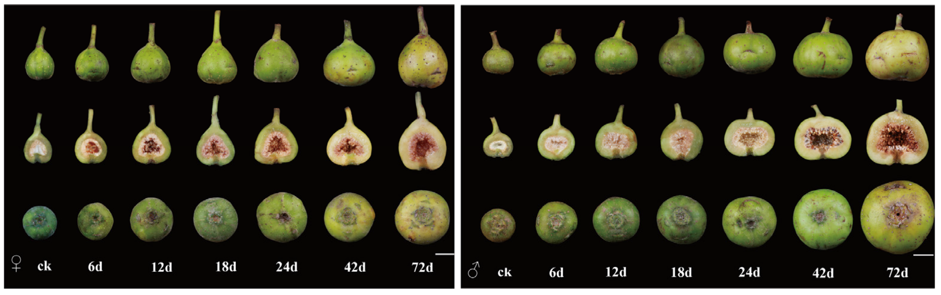
3.1. Developmental Processes and Morphological Characteristics of Male and Female Figs Following Wasp Entry

The fig typically requires 2–3 months from the onset of fruiting to reach maturity. Upon entering the figs, the pollinating fig wasp displays both oviposition and pollination behaviors in both female and male figs. However, seeds are exclusively produced in female figs, whereas the gall flowers necessary for reproducing the wasp’s offspring are solely found in male figs. Regarding fig morphology, female figs are generally oblate conical in shape and covered with fine, downy hair, while male figs are flat and smooth, featuring a larger fig cavity, which is hypothesized to provide ample space for the growth and mating of the wasp’s offspring (Figure 3).

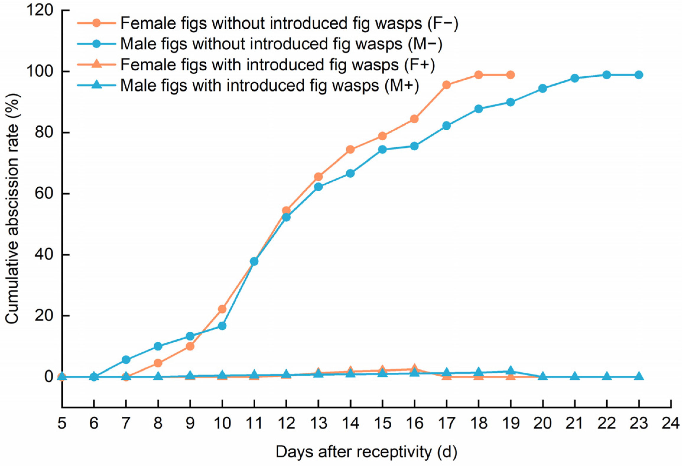
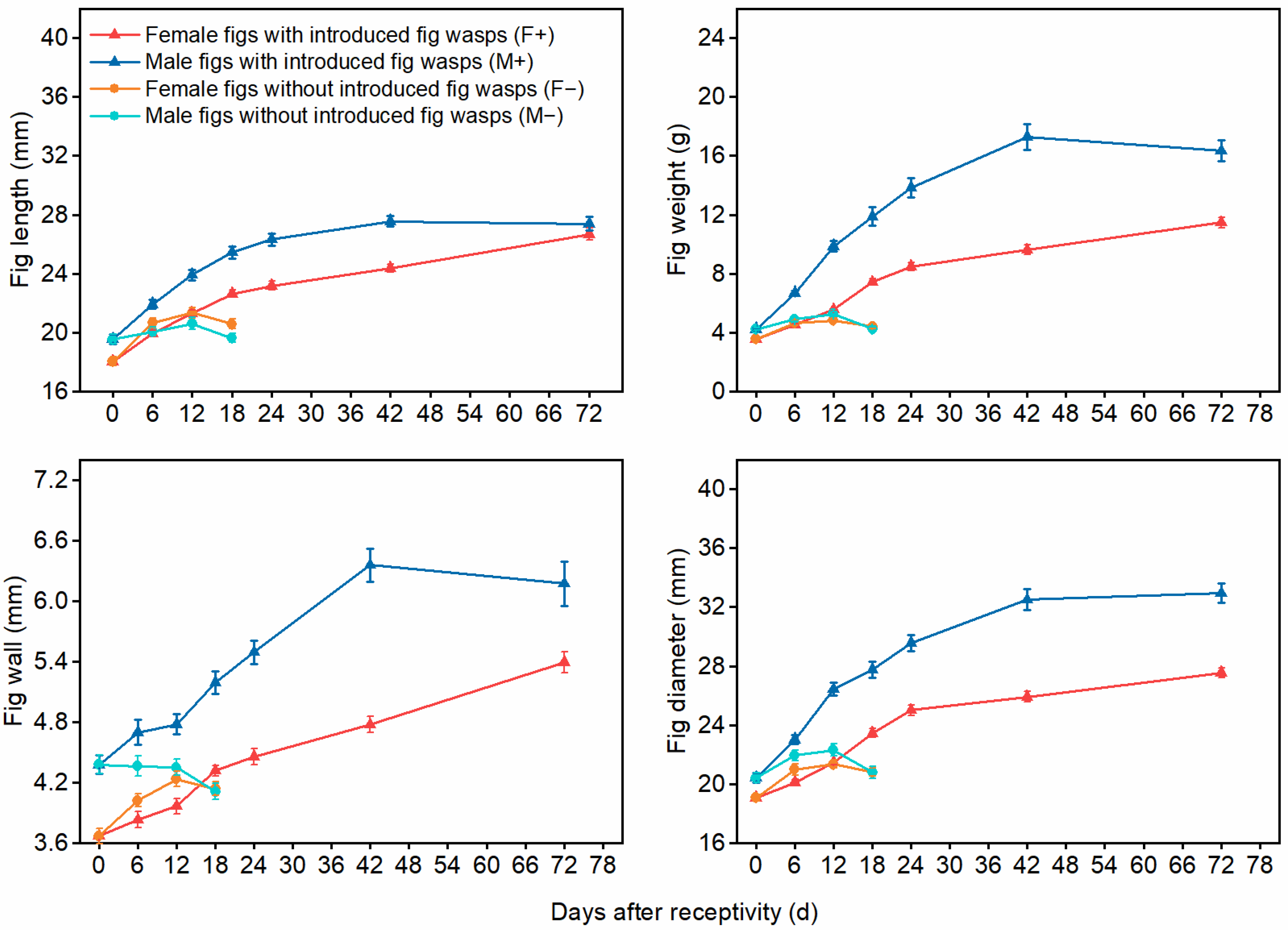
3.2. The Impact of Wasp Entry on Fig Abscission Rates and Developmental Indices for Male and Female Figs

The entry of fig wasps significantly influences the abscission rates of figs. For both male and female figs without wasp entry, the abscission rates exhibit a pronounced upward trend as the duration after receptivity increases (GLM, female: β ± SE = 6.53 ± 0.57, p < 0.01; male: β ± SE = 6.17 ± 0.42, p < 0.01). If no fig wasps enter the figs approximately 10 days after the onset of receptivity, the rate of fig abscission sharply increases. By the 19th day for female figs and the 23rd day for male figs, complete abscission occurs. The abscission trends for both male and female figs are largely consistent. In contrast, the abscission rates of male and female figs that have been entered by wasps remain consistently low at approximately 0% (Figure 4).
The impact of wasp entry on figs is evident not only in abscission rates but also in various developmental indices. Following wasps’ entry, both female and male figs exhibit significant increases in length (GLM, female: β ± SE = 0.81 ± 0.03, p < 0.01; male: β ± SE = 0.63 ± 0.01, p < 0.01), weight (GLM, female: β ± SE = 0.11 ± 0.02, p < 0.01; male: β ± SE = 0.10 ± 0.01, p < 0.01), wall thickness (GLM, female: β ± SE = 0.08 ± 0.01, p < 0.01; male: β ± SE = 0.06 ± 0.01, p < 0.01), and diameter (GLM, female: β ± SE = 0.78 ± 0.03, p < 0.01; male: β ± SE = 0.56 ± 0.02, p < 0.01). These developmental changes are significantly correlated with the duration of wasp presence. In contrast, in the absence of wasp entry, even with prolonged receptivity, there is no significant change in fig growth indices (p > 0.01) (Figure 5).

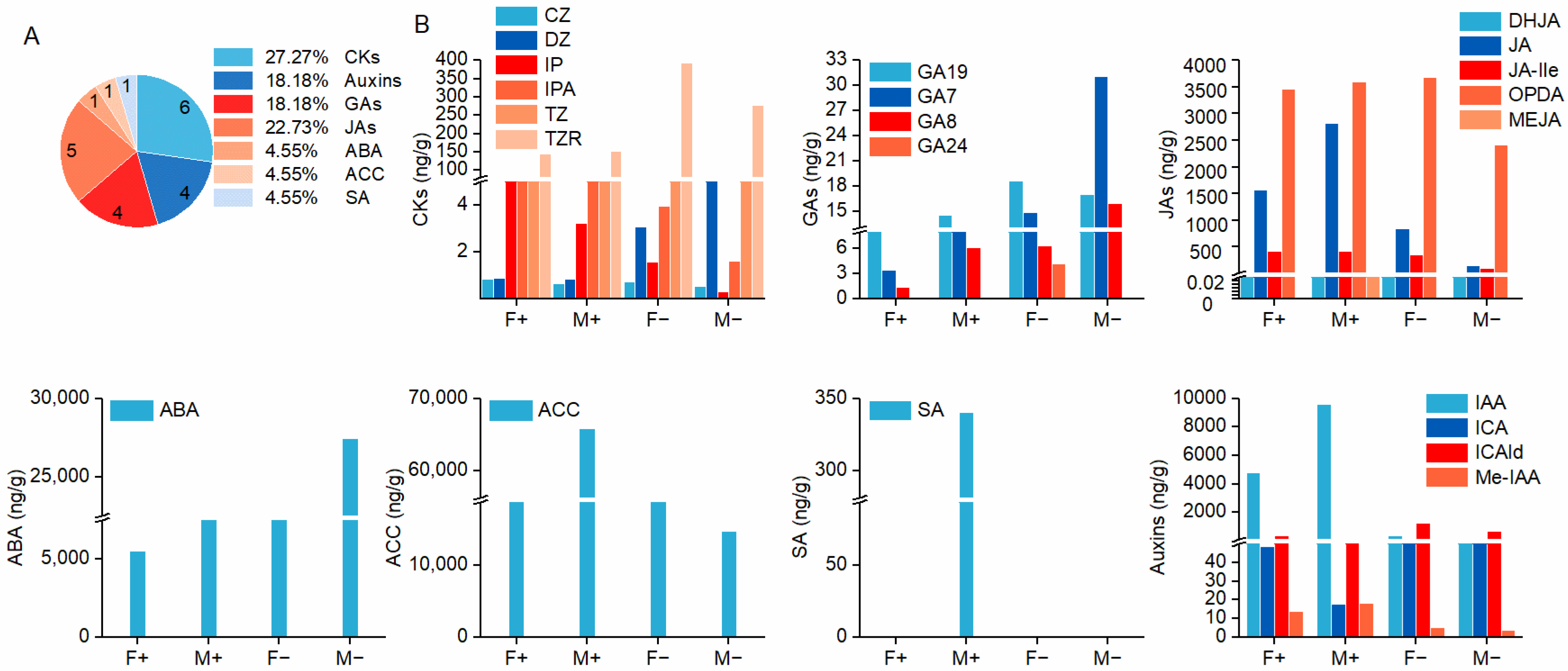
3.3. The Classification of Endogenous Phytohormones

In the four treatment groups, a total of 7 classes comprising 23 plant hormones were identified, including 6 types of cytokinins (CKs), 4 types of auxins, 4 types of gibberellins (GAs), 5 types of jasmonates (JAs), 1 type of abscisic acid (ABA), 1 type of 1-aminocyclopropane-1-carboxylic acid (ACC), and 1 type of salicylic acid (SA) (Figure 6A). Notably, jasmonic acid methyl ester (MEJA) and SA were exclusively detected in the pollinated male figs (M+), whereas gibberellin 24 (GA24) was uniquely presented in the unpollinated female figs (F−) (Figure 6B).
In the pollinated state, the concentrations of various hormones in male figs are higher than those in female figs, with the exception of CKs. In the unpollinated state, the concentrations of various hormones in female figs are higher than those in male figs, except for GAs and ABA. During the developmental process, ACC and ABA are two hormones with high concentrations, suggesting they may play a significant regulatory role in the growth and development of figs. However, the regulatory patterns of these two hormones during the developmental phase differ between male and female figs: in the pollinated state, the content of ACC in both male and female figs increases significantly, particularly in male figs, where the ACC concentration peaks at 6.6 × 10⁴ ± 6734.58 ng/g (Mean ± SE), which is 1.26 times higher than in female figs (5.2 × 10⁴ ± 5208.55 ng/g). In the unpollinated state, the ABA content is higher, with male figs having an ABA concentration of 2.7 × 10⁴ ± 2092.61 ng/g, which is 2.44 times higher than in female figs (1.1 × 10⁴ ± 629.54 ng/g). Additionally, the ACC content in female figs remains relatively high in both pollinated and unpollinated states (Figure 6B).

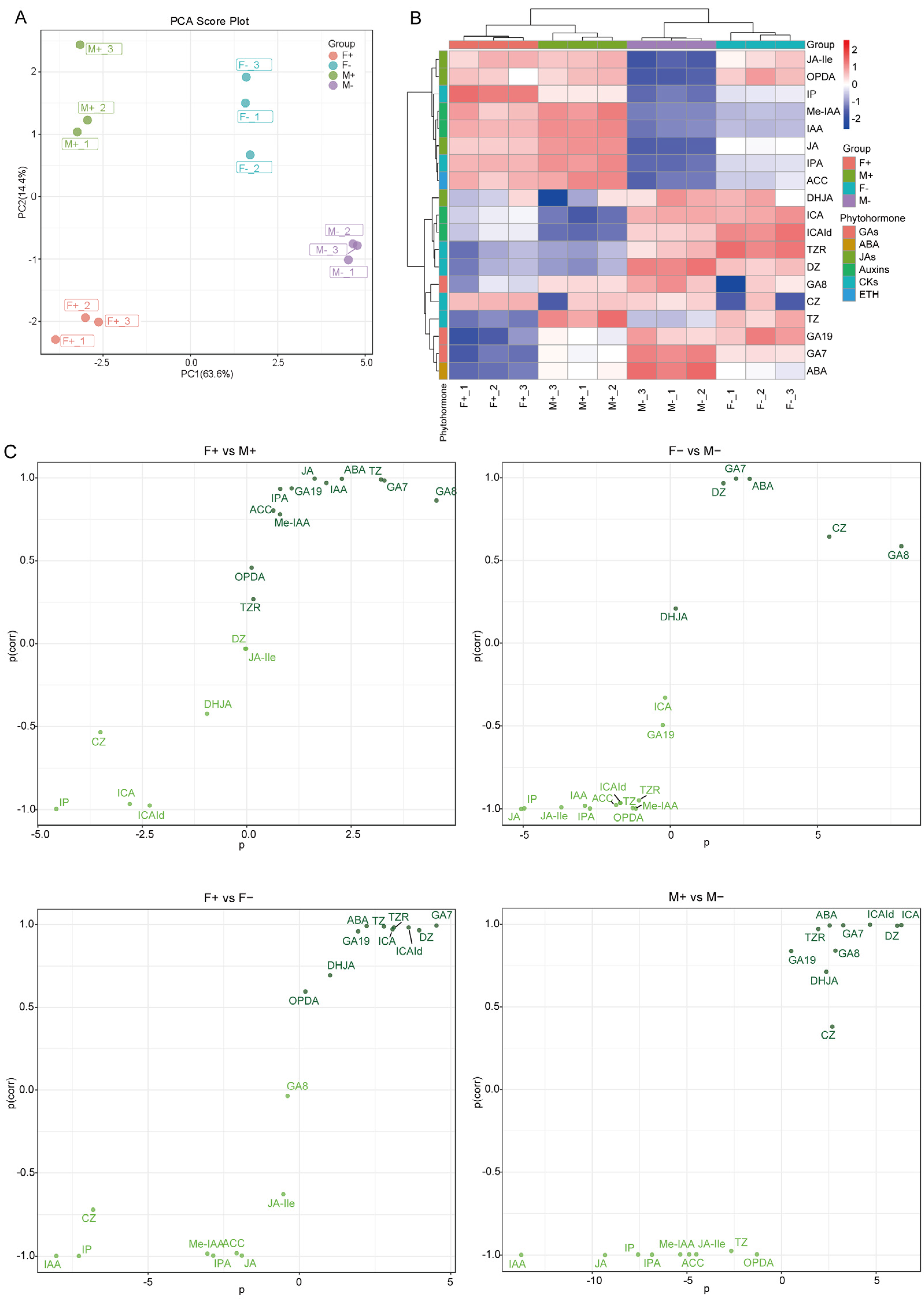
3.4. Multivariate Statistical Analysis of Hormone Metabolites in Male and Female Figs

To assess the fundamental characteristics and variations in hormone metabolites across treatment groups, we performed principal component analysis (PCA) and cluster analysis using 19 hormones from six distinct classes that were identified. The PCA results indicated that PC1 and PC2 explained 63.6% and 14.4% of the total variance, respectively. Furthermore, the samples within each group displayed a clustered distribution, suggesting high homogeneity among replicate samples and confirming the reliability of the data (Figure 7A). The clustering heat map analysis revealed that F+ and M+ were grouped together, as were F− and M−. This indicates that the metabolism of plant hormones in figs, entered by pollinators, exhibits similar accumulation characteristics between male and female figs. Similarly, in the absence of pollinators, the metabolism of these hormones also showed comparable accumulation patterns (Figure 7B).
The OPLS-DA model was utilized to identify the metabolites influencing comparison group classification. The findings indicated substantial differences in the composition of hormone metabolites across the F+, M+, F−, and M− samples (Q2 > 0.9). The distance of metabolites from the origin correlated positively with their contribution to sample differentiation. Notably, JAs and auxins significantly contributed to distinguishing figs with pollinators from those without. Furthermore, GAs were essential in differentiating female figs from male figs (Figure 7C).

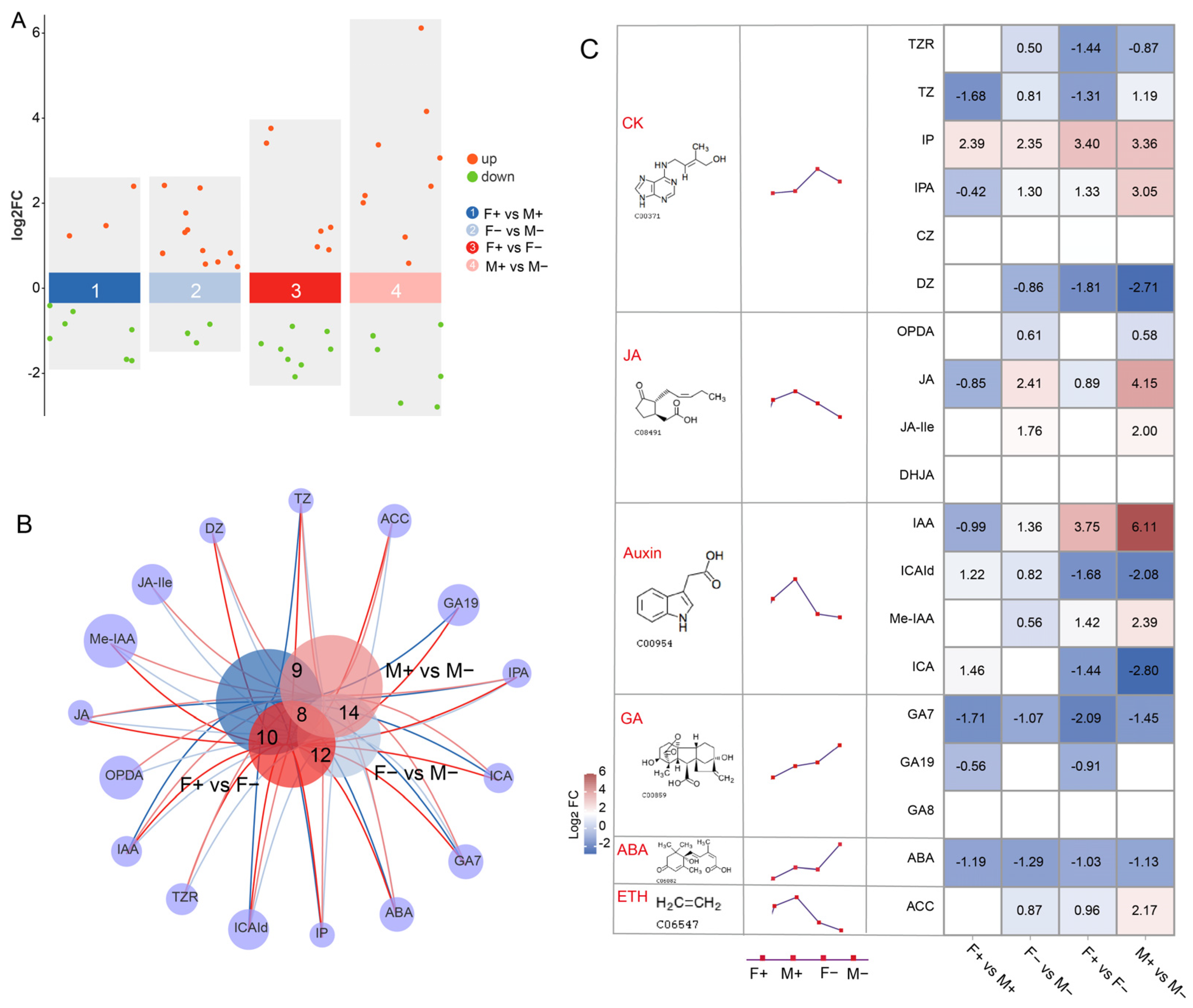
3.5. Comparative Analysis of Hormone Metabolites in Male and Female Figs

Differential metabolites were identified using the criteria of VIP > 1.0, log2 |FC| > 0.5, and p < 0.05. Specifically, 10, 14, 14, and 15 differential metabolites were detected in the comparisons of F+ vs. M+, F− vs. M−, F+ vs. F−, and M+ vs. M−, respectively (Figure 8A). The Venn diagram illustrated that a total of eight hormones overlapped among the four comparison groups, with CKs and auxins being the predominant overlapping hormones (Figure 8B). Compared to F+, M+ up-regulated three metabolites and down-regulated seven metabolites, whereas F− up-regulated six metabolites and down-regulated eight metabolites. The metabolites up-regulated by M+ were primarily CKs and auxins, while those down-regulated were GAs and ABA. In contrast, the up-regulated metabolites in F− were mainly associated with ACC and JAs, while the down-regulated metabolites included CKs, auxins, and GAs. Compared to M+, M− up-regulated nine metabolites and down-regulated six metabolites, with a particular emphasis on up-regulating CKs, auxins, JAs, and ACC while down-regulating GAs and ABA. The comparison between F− and M− revealed the up-regulation of 11 metabolites and the down-regulation of 3 metabolites. Specifically, the up-regulated metabolites were primarily CKs, JAs, and auxins, while the down-regulated metabolites were GAs and ABA (Figure 8C).

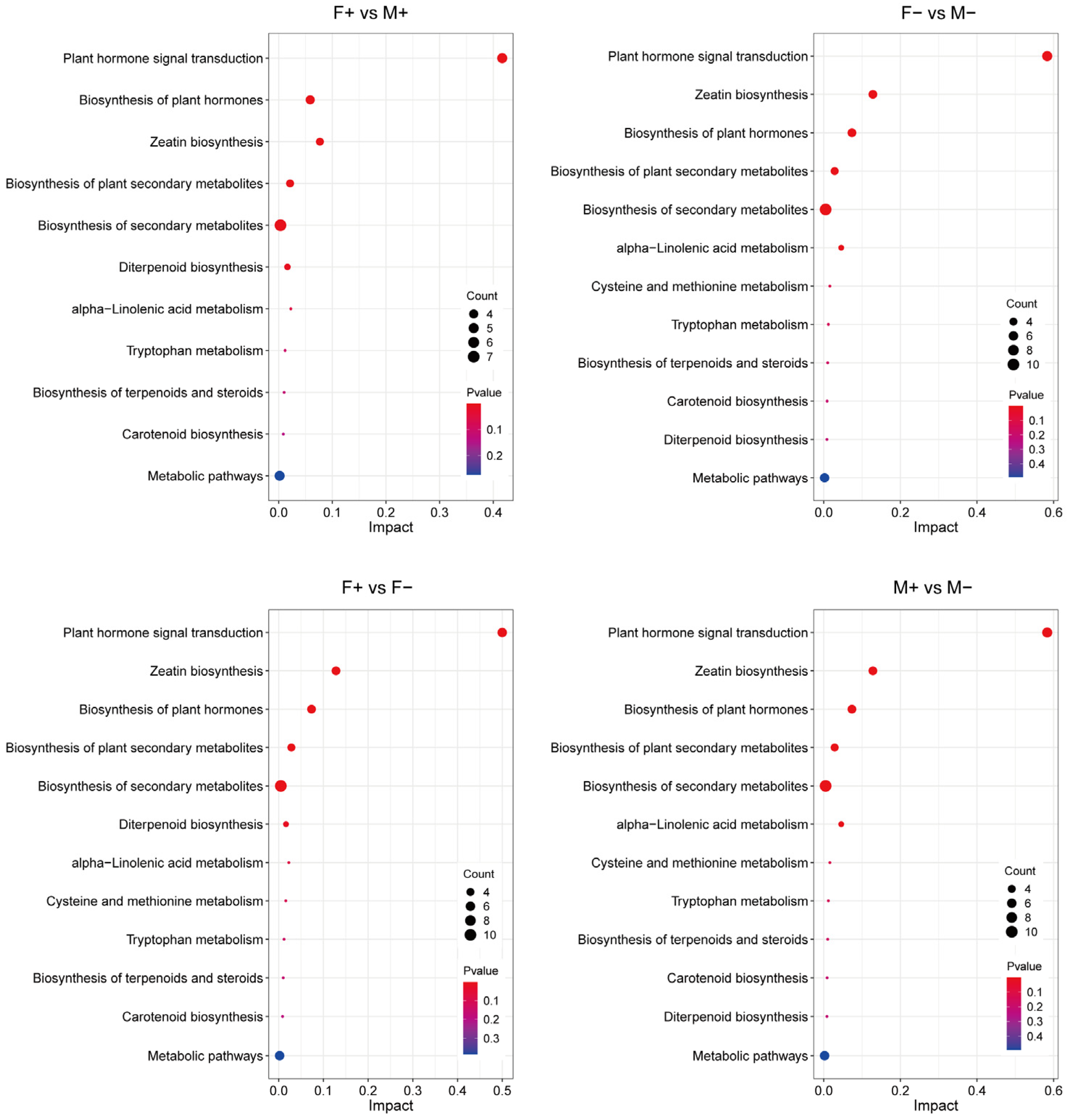
3.6. Analysis of KEGG Metabolic Pathways in Male and Female Figs

We conducted KEGG pathway enrichment analysis on the significantly different metabolites across different comparison groups, which revealed a high degree of similarity among the differential metabolite pathways: F+ vs. M+, F− vs. M−, F+ vs. F−, and M+ vs. M−. The key KEGG pathways are primarily involved in phytohormone signaling, phytohormone biosynthesis, zeatin biosynthesis, secondary metabolite biosynthesis, and the diterpene biosynthesis pathway (Figure 9).

4. Discussion

In this study, we integrated field ecological experiments with metabolomic analysis to compare the effects of pollinating fig wasp entry on the developmental processes and hormonal regulation in male and female figs of F. hispida. Our study aimed to elucidate the gender responses to pollinator entry and to investigate the role of hormonal regulation in the sexual functional differentiation of dioecious Ficus species. The results demonstrated significant differences in fig abscission rates, developmental processes, and hormone metabolism between figs with and without wasp entry and distinct variations in hormone levels and types between male and female figs. Male fig development is primarily regulated by ACC and IAA, whereas female fig development is predominantly influenced by IAA. Additionally, ABA and ACC may act synergistically to regulate the abscission process in figs. This study provides evidence for understanding the role of fig wasp entry in mediating fig development and promoting the functional differentiation of fig sexes.
Previous studies have demonstrated that ABA, an endogenous plant hormone, has been extensively investigated and found to accelerate the senescence and abscission of flowers, leaves, and fruits across numerous plant species [41]. For instance, during the ripening stage of tomato fruits, in addition to chlorophyll degradation and lycopene accumulation, there is also a significant increase in ABA content despite the fruit size remaining relatively stable [42]. Similarly, in F. carica figs, both ABA and ethylene levels show substantial increases during ripening [43]. Moreover, ABA typically interacts synergistically with ethylene to promote ripening while inhibiting tissue growth [25]. Our research indicates that figs without wasp entry are primarily regulated by the synergistic interaction between ABA and ACC, which modulates metabolism and signal transduction within the figs. In unpollinated figs, ABA accumulates rapidly, thereby slowing down fig development and increasing abscission rates. This mechanism effectively prevents fig wasps from entering the figs during the late receptive phase. This is critical because wasp entry at this stage would result in a significant decline in egg-laying and pollination efficiency, thereby negatively impacting the stability of the fig–wasp mutualistic system [44]. We hypothesize that ABA not only inhibits the development of female flowers but also prolongs the receptive phase. Consequently, the difference in ABA content between female and male figs partially explains the variation in the duration of the receptivity between female and male figs. However, whether this phenomenon is widespread across Ficus species requires further investigation [44,45]. At the gene regulatory level, the senescence of various floral organs, including petals, sepals, receptacles, stamens, and carpels, is characterized by elevated expression of RhPR10.1, a pathogenesis-related protein. Ethylene exposure has been shown to further enhance the expression of RhPR10.1 [46]. However, the mechanism by which figs respond to pollinating fig wasps through hormone-signaling genes remains poorly understood. Future research should employ physiological and molecular approaches to thoroughly investigate the changes in the expression patterns of specific genes during floral organ senescence.
Plant hormones play a crucial role in regulating plant growth and development, and they are also influenced by other biological factors that interact with the plant, such as microorganisms and insects [26]. In flowering plants, pollinators differentially influence the adaptability of female and male individuals, thereby serving as key drivers of sexual dimorphism [47]. This effect may be mediated by hormonal regulation. Research has demonstrated that plant hormones modulate gene expression programs associated with sexual differentiation through complex signal transduction pathways, ultimately determining the sex phenotype of plants [48,49]. For instance, the exogenous application of BA (Benzylaminopurine) can effectively induce the formation of female floral primordia and promote the development of female flowers in Plukenetia volubilis Linneo (Euphorbiaceae), particularly during early flower development stages [50]. Moreover, auxin biosynthesis and signaling pathway genes, such as GH3, AUX/IAA, ARP, and SAUR, exhibit significantly higher expression levels in female buds compared to male buds in papaya [51]. Furthermore, regulating female flower development in Cucurbita pepo L. appears to rely on the interplay between auxin and ethylene [52]. Furthermore, changes in hormone levels within a plant can alter the ratio of male to female flowers and may even induce asexual reproduction or parthenocarpy, significantly enhancing sexual diversity in plants [53]. In F. hispida, after pollinating fig wasps enter male figs to lay eggs, the concentration and diversity of hormones in male figs increase compared to female figs. This phenomenon may be attributed to the enhanced synthesis and metabolic regulation of multiple hormones supporting gall flower development and differentiation, leading to an increased variety of hormones in male figs. Alternatively, it could result from the damage caused by gall induction, which disrupts the normal physiological processes of female flowers and impedes the flow of nutrients essential for their development. In contrast, following pollination by fig wasps, the IAA levels in female figs significantly increase. This increase may be attributed to the higher IAA concentrations required during the early fruit set stage to support female flower differentiation and function and promote healthy seed formation [32]. Previous studies have shown that IAA plays a critical role in fruit and seed development. Specifically, IAA promotes early fig development by regulating cell division and expansion [54]. Therefore, the functional differentiation of female flowers within dioecious figs may be influenced by selective pressures exerted by pollinating fig wasps and the regulatory effects of plant hormones. Figs may have evolved a mechanism to detect the entry of pollinating fig wasps. Upon the entry of fig wasps for pollination and oviposition, figs exhibit a rapid response by activating metabolic pathways that lead to reproductive function differentiation in both male and female figs.
Plants exhibit hormonal regulation in their development, organ differentiation, and responses to both biotic and abiotic stresses [26,55]. Research has shown that the deposition of insect eggs or excretions from herbivorous insects can increase levels of JA (Jasmonic acid) and SA [56,57]. JA is acknowledged as a signaling molecule generated by plants in response to diverse stress conditions, such as insect feeding, mechanical damage, and pathogen infection [58]. In our research, the entry of the pollinating fig wasp resulted in a significant rise in JA levels in both male and female figs. Furthermore, the concentration of JA-IIe (Jasmonic acid-isoleucine) markedly increased in male figs, whereas SA and MEJA were solely identified in male figs that had been entered by the pollinator. When plants are confronted with multiple stresses, they will activate corresponding defense mechanisms according to the type or intensity of the stress; for instance, biotic stresses like microbial infections and insect infestations typically accelerate the initiation of defense responses [59]. The entry of fig wasps into male figs for oviposition can be regarded as a transient form of “damage” or “stress” to the flowers. This triggers the fig’s defense mechanisms, including the synthesis and accumulation of SA and other antibacterial and antioxidant compounds, which help to mitigate damage to the fig’s internal tissues and limit excessive nutrient uptake by the fig wasps. In contrast, no significant alterations were observed in female figs. This phenomenon is closely associated with the function of JA and SA as defense hormones. Plants must maintain a delicate balance between growth and development and stress responses. The interaction between JA and SA often prioritizes coping with adverse environments by reallocating resources, which may result in compromised growth. Consequently, this regulation differentially affects the reproductive functions of male and female figs, ultimately establishing a balance between the dioecious fig and its pollinators.
The analysis of hormonal metabolism indicates that the entry of the pollinating fig wasp into the fig may have substantially influenced the reproductive function differentiation of the female flower in fig. Nevertheless, the reproductive function differentiation between male and female figs in dioecious fig trees is likely a highly intricate process. Current research has not yet fully elucidated the complex regulatory mechanisms and dynamic changes that occur during the development and differentiation of flowers. To achieve a more comprehensive understanding of pollinator entry in the developmental processes and the reproductive function differentiation in dioecious fig species, further research is essential, particularly focusing on gene expression and hormone biosynthesis.

5. Conclusions

In conclusion, the female flowers of F. hispida in both female and male figs exhibit clear responses to the entry of pollinating fig wasps in terms of morphological development and hormonal metabolism. Wasp entry can significantly decrease the abscission rate of figs, contributing to sexual dimorphism through hormonal mediation. Hormonal regulation sustains the mutualism between figs and fig wasps by preventing premature fig abscission caused by excessive parasitism. Pollinated female figs, under the influence of auxins, promote female flower development, thereby inhibiting gall formation and supporting seed production. This metabolic differentiation is likely critical for the functional divergence between female and male figs, elucidating why male figs are incapable of seed production and female figs cannot facilitate wasp reproduction. In the absence of the “trigger factor” associated with pollinator entry, the fig may maintain its receptivity through the synergistic actions of multiple hormones, combined with the abscission of unpollinated figs during the later stages of the female phase to prevent wasps from entering senescent figs for reproduction, which contributes to the stability of the fig–wasp mutualism. Our research indicates that the entry of pollinating fig wasps may influence the development and sex differentiation of figs by mediating hormonal changes within the figs. The findings from this research will provide scientific evidence to elucidate the mechanisms underlying sexual differentiation in female and male figs, thereby serving as a scientific basis for understanding other plant–insect mutualistic systems. Furthermore, the results imply that maintaining fig wasp populations is essential for the sustainable development of Ficus populations and effectively utilizing Ficus species. Our results suggest that using transcriptomics and genomics to further study the role of pollinating wasps in fig development would be valuable research.

Author Contributions

Conceptualization, Y.Z. (Yuan Zhang) and Y.P.; methodology, Z.L.; writing—original draft preparation, Y.G. and Y.Z. (Ying Zhang); writing—review and editing, Y.Z. (Yuan Zhang), Y.P. and Z.L.; provided the plant materials, Y.P.; performed the experiments, Y.G., Y.W., C.C. and X.Y.; analyzed the data, Y.G. and Y.Z. (Ying Zhang); prepared the figures and tables, Y.G., J.G. and B.M. All authors have read and agreed to the published version of the manuscript.

Funding

This research was funded by the National Natural Science Foundation of China (32160296, 32261123001, 32260719 and 31560116); the Fundamental Research Program of Yunnan Province, China (202401AT070265 and 202401BD070001-111); the Young Top-Notch Talent of Yunnan Outstanding Talent Program (XDYCQNRC-2022-0207); and the First Class Forestry Academic Subject in Yunnan Province (523003).

Data Availability Statement

The data presented in this study are available upon request from the corresponding authors.

Acknowledgments

The authors thank Xishuangbanna Tropical Botanical Garden and Yunnan Academy of Biodiversity for kindly providing the experimental equipment.

Conflicts of Interest

The authors declare no conflicts of interest.

References

  1. Bruce, T.J. Interplay between insects and plants: Dynamic and complex interactions that have coevolved over millions of years but act in milliseconds. J. Exp. Bot. 2015, 66, 455–465. [Google Scholar] [CrossRef] [PubMed]
  2. Whitney, H.M.; Glover, B.J. Coevolution: Plant-Insect. In eLS; John Wiley & Sons, Ltd.: Chichester, UK, 2013. [Google Scholar]
  3. Botha, A.M. A coevolutionary conundrum: The arms race between Diuraphis noxia (Kurdjumov) a specialist pest and its host Triticum aestivum (L.). Arthropod-Plant Plant Interact. 2013, 7, 359–372. [Google Scholar] [CrossRef]
  4. Ehrlich, P.R.; Raven, P.H. Butterflies and plants: A study in coevolution. Evolution 1964, 18, 586–608. [Google Scholar] [CrossRef]
  5. Maron, J.L.; Agrawal, A.A.; Schemske, D.W. Plant-herbivore coevolution and plant speciation. Ecology 2019, 100, e02704. [Google Scholar] [CrossRef]
  6. McKenna, D.D.; Shin, S.; Ahrens, D.; Balke, M.; Beza-Beza, C.; Clarke, D.J.; Donath, A.; Escalona, H.E.; Friedrich, F.; Letsch, H.; et al. The evolution and genomic basis of beetle diversity. Proc. Natl. Acad. Sci. USA 2019, 116, 24729–24737. [Google Scholar] [CrossRef] [PubMed]
  7. Ashra, H.; Nair, S. Review: Trait plasticity during plant-insect interactions: From molecular mechanisms to impact on community dynamics. Plant. Sci. 2022, 317, 111188. [Google Scholar] [CrossRef] [PubMed]
  8. Wang, R.; Yang, Y.; Jing, Y.; Segar, S.T.; Zhang, Y.; Wang, G.; Chen, J.; Liu, Q.F.; Chen, S.; Chen, Y.; et al. Molecular mechanisms of mutualistic and antagonistic interactions in a plant-pollinator association. Nat. Ecol. Evol. 2021, 5, 974–986. [Google Scholar] [CrossRef] [PubMed]
  9. Yang, D.R.; Peng, Y.Q.; Song, Q.S.; Zhang, G.M.; Wang, R.W.; Zhao, T.Z.; Wang, Q.Y. Pollination biology of Ficus hispida in the tropical rainforests of Xishuangbanna, China. Acta Bot. Sin. 2002, 44, 519–526. [Google Scholar]
  10. Wiebes, J.T. Co-evolution of figs and their insect pollinators. Annu. Rev. Ecol. Syst. 1979, 10, 1–12. [Google Scholar] [CrossRef]
  11. Corlett, R.T. Figs (Ficus, Moraceae) in urban Hong Kong, South China. Biotropica 2006, 38, 116–121. [Google Scholar] [CrossRef]
  12. Yuan, S.; Yin, T.; He, H.; Liu, X.; Long, X.; Dong, P.; Zhu, Z. Phenotypic, metabolic and genetic adaptations of the Ficus species to abiotic stress response: A comprehensive review. Int. J. Mol. Sci. 2024, 25, 9520. [Google Scholar] [CrossRef]
  13. Janzen, D.H. How many parents do the wasps from a fig have? Biotropica 1979, 11, 127–129. [Google Scholar] [CrossRef]
  14. Weiblen, G.D. How to be a fig wasp. Annu. Rev. Entomol. 2002, 47, 299–330. [Google Scholar] [CrossRef]
  15. Cruaud, A.; Rønsted, N.; Chantarasuwan, B.; Chou, L.S.; Clement, W.L.; Couloux, A.; Cousins, B.; Genson, G.; Harrison, R.D.; Hanson, P.E.; et al. An extreme case of plant-insect codiversification: Figs and fig-pollinating wasps. Syst. Biol. 2012, 61, 1029–1047. [Google Scholar] [CrossRef]
  16. Anstett, M.C.; Bronstein, G.L.; Hossacrt-McKcy, M. Resource allocation: A conflict in the fig/fig wasp mutualism. J. Evol. Biol. 1996, 9, 417–428. [Google Scholar] [CrossRef]
  17. Anstett, M.C. Unbeatable strategy, constraint and coevolution, or how to resolve evolutionary conflicts: The case of the fig/wasp mutualism. Oikos 2001, 95, 476–484. [Google Scholar] [CrossRef]
  18. Derek, W.D. Stability in fig tree-fig wasp mutualisms: How to be a cooperative fig wasp. Biol. J. Linn. Soc. Lond. 2020, 130, 1–17. [Google Scholar]
  19. Chetty, A.; Glennon, K.L.; Venter, S.M.; Cron, G.V.; Witkowsk, E.T.F. Reproductive ecology of the African baobab: Floral features differ among individuals with different fruit production. Forest Ecol. Manag. 2021, 489, 119077. [Google Scholar] [CrossRef]
  20. Xia, Z.; Chen, B.J.W.; Korpelainen, H.; Niinemets, Ü.; Li, C. Belowground ecological interactions in dioecious plants: Why do opposites attract but similar ones repel? Trends Plant Sci. 2024, 29, 630–637. [Google Scholar] [CrossRef] [PubMed]
  21. Liu, M.; Korpelainen, H.; Li, C.Y. Sexual differences and sex ratios of dioecious plants under stressful environments. J. Plant Ecol. 2021, 14, 920–933. [Google Scholar] [CrossRef]
  22. Miao, B.G.; Liu, M.X.; Wang, B.; Peng, Y.Q.; Lesne, A.; Kjellberg, F.; Jandér, K.C. Active pollination in a functionally dioecious Ficus species: An interplay between pollinator behaviour and floral morphology. Flora 2023, 302, 152274. [Google Scholar] [CrossRef]
  23. Martinson, E.O.; Hackett, J.D.; Machado, C.A.; Arnold, A.E. Metatranscriptome analysis of fig flowers provides insights into potential mechanisms for mutualism stability and gall induction. PLoS ONE 2015, 10, e0130745. [Google Scholar] [CrossRef]
  24. Zidi, K.; Kati, D.E.; Bachir-bey, M.; Genva, M.; Fauconnier, M.L. Comparative study of fig volatile compounds using headspace solid-phase microextraction-gas chromatography / mass spectrometry: Effects of cultivars and ripening stages. Front. Plant Sci. 2021, 12, 667809. [Google Scholar] [CrossRef]
  25. Malik, N.U.A.; Fajer, O.; Amin, L.; Khalid, A.R.; Khan, N.; Bhatti, M.F.; Munir, F.; Haider, G.; Amir, R.; Gul, A. Phytohormones, plant growth and development. In Phytohormones and Stress Responsive Secondary Metabolites; Academic Press: Cambridge, MA, USA, 2023; pp. 175–186. [Google Scholar]
  26. Nambara, E.; Van Wees, S.C.M. Plant hormone functions and interactions in biological systems. Plant J. 2021, 105, 287–289. [Google Scholar] [CrossRef]
  27. Izawa, T. What is going on with the hormonal control of flowering in plants? Plant J. 2021, 105, 431–445. [Google Scholar] [CrossRef]
  28. Chandler, J.W. The hormonal regulation of flower development. J. Plant Growth Regul. 2011, 30, 242–254. [Google Scholar] [CrossRef]
  29. Chai, L.; Chai, P.; Chen, S.; Flaishman, M.A.; Ma, H. Transcriptome analysis unravels spatiotemporal modulation of phytohormone-pathway expression underlying gibberellin-induced parthenocarpic fruit set in San Pedro-type fig (Ficus carica L.). BMC Plant Biol. 2018, 18, 100. [Google Scholar] [CrossRef]
  30. Lama, K.; Harlev, G.; Shafran, H.; Peer, R.; Flaishman, M.A. Anthocyanin accumulation is initiated by abscisic acid to enhance fruit color during fig (Ficus carica L.) ripening. J. Plant Physiol. 2020, 251, 153192. [Google Scholar] [CrossRef] [PubMed]
  31. Abe, M.; Kobayashi, Y.; Yamamoto, S.; Daimon, Y.; Yamaguchi, A.; Ikeda, Y.; Ichinoki, H.; Notaguchi, M.; Goto, K.; Araki, T. FD, a bZIP protein mediating signals from the floral pathway integrator FT at the shoot apex. Science 2005, 309, 1052–1056. [Google Scholar] [CrossRef] [PubMed]
  32. Fenn, M.A.; Giovannoni, J.J. Phytohormones in fruit development and maturation. Plant J. 2021, 105, 446–458. [Google Scholar] [CrossRef] [PubMed]
  33. Obeso, J.R. The costs of reproduction in plants. New Phytol. 2002, 155, 321–348. [Google Scholar] [CrossRef] [PubMed]
  34. Nefdt, R.J.; Compton, S.G. Regulation of seed and pollinator production in the fig-fig wasp mutualism. J. Anim. Ecol. 1996, 65, 170–182. [Google Scholar] [CrossRef]
  35. Shi, Z.H.; Yang, D.R.; Peng, Y.Q. The style-length of the female florets and their fate in two dioecious species of Xishuangbanna, China. Trees 2006, 20, 410–415. [Google Scholar] [CrossRef]
  36. Liao, Z.; Zhang, T.; Lei, W.; Wang, Y.; Yu, J.; Wang, Y.; Chai, K.; Wang, G.; Zhang, H.; Zhang, X. A telomere-to-telomere reference genome of ficus (Ficus hispida) provides new insights into sex determination. Hortic. Res. 2023, 11, uhad257. [Google Scholar] [CrossRef]
  37. Yang, W.; Xiang, P. Changes of fruit abscission and carbohydrates, hormones, related gene expression in the fruit and pedicel of macadamia under starvation stress. Horticulturae 2022, 8, 398. [Google Scholar] [CrossRef]
  38. Li, Y.; Zhou, C.; Yan, X.; Zhang, J.; Xu, J. Simultaneous analysis of ten phytohormones in Sargassum horneri by high-performance liquid chromatography with electrospray ionization tandem mass spectrometry. J. Sep. Sci. 2016, 39, 1804–1813. [Google Scholar] [CrossRef] [PubMed]
  39. Hui, W.; Wang, Y.; Yan, S.; Shi, J.; Huang, W.; Zayed, M.Z.; Peng, C.; Chen, X.; Wu, G. Simultaneous analysis of endogenous plant growth substances during floral sex differentiation in Jatropha curcas L. using HPLC-ESI-MS/MS. Sci. Hortic. 2018, 241, 209–217. [Google Scholar] [CrossRef]
  40. Šimura, J.; Antoniadi, I.; Široká, J.; Tarkowská, D.; Strnad, M.; Ljung, K.; Novák, O. Plant hormonomics: Multiple phytohormone profiling by targeted metabolomics. Plant Physiol. 2018, 177, 476–489. [Google Scholar] [CrossRef]
  41. Wei, L.; Lu, Z.; Wang, Y.; Gao, G. The effects of globose scale (Sphaerolecanium prunastri) infestation on the growth of wild apricot (Prunus armeniaca) trees. Forests 2023, 14, 2032. [Google Scholar] [CrossRef]
  42. Srivastava, A.; Handa, A.K. Hormonal regulation of tomato fruit development: A molecular perspective. J. Plant Growth Regul. 2005, 24, 67–82. [Google Scholar] [CrossRef]
  43. Osianski, Y.; Doron-Faigenboim, A.; Freiman, Z.E.; Lama, K.; Milo-Cochavi, S.; Dahan, Y.; Kerem, Z.; Flaishman, M.A. Tissue-specific transcriptome and hormonal regulation of pollinated and parthenocarpic fig (Ficus carica L.) fruit suggest that fruit ripening is coordinated by the reproductive part of the syconium. Front. Plant Sci. 2016, 7, 1696. [Google Scholar]
  44. Zhang, Y.; Yang, D.R.; Peng, Y.Q.; Compton, S.G. Costs of inflorescence longevity for an Asian fig tree and its pollinator. Evol. Ecol. 2012, 26, 513–527. [Google Scholar] [CrossRef]
  45. Ahmad, S.; Lu, C.; Gao, J.; Wei, Y.; Xie, Q.; Jin, J.; Zhu, G.; Yang, F. Integrated proteomic, transcriptomic, and metabolomic profiling reveals that the gibberellin-abscisic acid hub runs flower development in the Chinese orchid Cymbidium sinense. Hortic. Res. 2024, 11, uhae073. [Google Scholar] [CrossRef] [PubMed]
  46. Wu, L.; Ma, N.; Jia, Y.; Zhang, Y.; Feng, M.; Jiang, C.Z.; Ma, C.; Gao, J. An ethylene-induced regulatory module delays flower senescence by regulating cytokinin content. Plant Physiol. 2016, 173, 853–862. [Google Scholar] [CrossRef]
  47. Tsuji, K.; Kobayashi, K.; Hasegawa, E.; Yoshimura, J. Dimorphic flowers modify the visitation order of pollinators from male to female flowers. Sci. Rep. 2020, 10, 9965. [Google Scholar] [CrossRef] [PubMed]
  48. Boualem, A.; Troadec, C.; Camps, C.; Lemhemdi, A.; Morin, H.; Sari, M.A.; Fraenkel-Zagouri, R.; Kovalski, I.; Dogimont, C.; Perl-Treves, R. A cucurbit androecy gene reveals how unisexual flowers develop and dioecy emerges. Science 2015, 350, 688–691. [Google Scholar] [CrossRef] [PubMed]
  49. Zheng, Y.; Zhang, D. Roles of jasmonate signalling in plant inflorescence and flower development. Curr. Opin. Plant Biol. 2015, 27, 44–51. [Google Scholar]
  50. Luo, Y.; Pan, B.Z.; Li, L.; Yang, C.X.; Xu, Z.F. Developmental basis for flower sex determination and effects of cytokinin on sex determination in Plukenetia volubilis (Euphorbiaceae). Plant Reprod. 2020, 33, 21–34. [Google Scholar] [CrossRef]
  51. Liu, J.; Chen, L.Y.; Zhou, P.; Liao, Z.; Lin, H.; Yu, Q.; Ming, R. Sex biased expression of hormone related genes at early stage of sex differentiation in papaya flowers. Hortic. Res. 2021, 8, 147. [Google Scholar] [CrossRef]
  52. Segura, M.; García, A.; Benítez, Á.; Martínez, C.; Jamilena, M. Comparative RNA-Seq analysis between monoecious and androecious plants reveals regulatory mechanisms controlling female flowering in Cucurbita pepo. Int. J. Mol. Sci. 2023, 24, 17195. [Google Scholar] [CrossRef]
  53. Li, W.; Fu, W.; Hou, J.; Yang, Y.; Yin, T. Evolution of plant sex and molecular mechanisms underlying plants sex separation. For. Res. 2023, 3, 1. [Google Scholar] [CrossRef] [PubMed]
  54. Godoy, F.; Kühn, N.; Muñoz, M.; Marchandon, G.; Gouthu, S.; Deluc, L.; Delrot, S.; Lauvergeat, V.; Arce-Johnson, P. The role of auxin during early berry development in grapevine as revealed by transcript profiling from pollination to fruit set. Hortic. Res. 2021, 8, 140. [Google Scholar] [CrossRef] [PubMed]
  55. Waadt, R.; Seller, C.A.; Hsu, P.K.; Takahashi, Y.; Munemasa, S.; Schroeder, J.I. Plant hormone regulation of abiotic stress responses. Nat. Rev. Mol. Cell Biol. 2022, 23, 680–694. [Google Scholar] [CrossRef] [PubMed]
  56. Hilker, M.; Fatouros, N.E. Plant responses to insect egg deposition. Annu. Rev. Entomol. 2015, 60, 493–515. [Google Scholar] [CrossRef] [PubMed]
  57. Lortzing, V.; Valsamakis, G.; Jantzen, F.; Hundacker, J.; Paniagua Voirol, L.R.; Schumacher, F.; Kleuser, B.; Hilker, M. Plant defensive responses to insect eggs are inducible by general egg-associated elicitors. Sci. Rep. 2024, 14, 1076. [Google Scholar] [CrossRef] [PubMed]
  58. Ruan, J.; Zhou, Y.; Zhou, M.; Yan, J.; Khurshid, M.; Weng, W.; Cheng, J.; Zhang, K. Jasmonic acid signaling pathway in plants. Int. J. Mol. Sci. 2019, 20, 2479. [Google Scholar] [CrossRef] [PubMed]
  59. Bruce, T.J.; Pickett, J.A. Plant defence signalling induced by biotic attacks. Curr. Opin. Plant Biol. 2007, 10, 387–392. [Google Scholar] [CrossRef] [PubMed]
Figure 1. Life cycle of Ficus hispida Linn. f. and its pollinating fig wasps Ceratosolen solmsi marchali Mayr.
Forests 16 00286 g001
Figure 2. (A) Experimental tree of F. hispida; (B) bagged figs for controlling experiment; (C) pollinating fig wasp attempting to enter the fig from bracts.
Forests 16 00286 g002
Figure 3. The developmental process and morphology of female and male figs in F. hispida. CK denotes the first day of the female phase, prior to the entry of the pollinating fig wasp into the figs; the scale bar measures 1 cm.
Forests 16 00286 g003
Figure 4. The impact of wasps’ entry on fig abscission in male and female figs.
Forests 16 00286 g004
Figure 5. The impact of wasps’ entry on fig developmental indices in male and female figs.
Forests 16 00286 g005
Figure 6. Impact of wasp entry on hormone levels in male and female figs. (A) Types and percentages of endogenous hormones in F. hispida; (B) content of seven types of endogenous hormones: cytokinins (CKs); gibberellins (GAs); jasmonates (JAs); abscisic acid (ABA); 1-aminocyclopropane-1-carboxylic acid (ACC); salicylic acid (SA); and auxins. F+ (Female figs with fig wasps’ entry), M+ (Male figs with fig wasps’ entry), F− (Female figs without fig wasps’ entry), M− (Male figs without fig wasps’ entry), same as below.
Forests 16 00286 g006
Figure 7. Multivariate statistical analysis of hormone metabolites in four treatment groups. (A) Principal component analysis (PCA) of hormone metabolites; (B) heat map clustering of hormone metabolite content; (C) OPLS-DA S-Plot for each comparison group. Note: Dark green and light green represent positive and negative correlation coefficients, respectively.
Forests 16 00286 g007
Figure 8. Differentially expressed metabolites across four treatment groups. (A) Distribution of differentially expressed metabolites across different comparison groups; (B) Venn diagrams illustrating overlaps among different comparison groups; (C) alterations in 19 hormone metabolites across different comparison groups; columns from left to right depict the structural composition of six types of hormones and trends in the content of each hormone metabolite. Note: Heat map (C) analysis revealed differentially expressed plant hormones in the four comparison groups, with red indicating up-regulation, blue indicating down-regulation, and the intensity of the color reflecting the degree of change.
Forests 16 00286 g008
Figure 9. KEGG enrichment pathways for differential metabolites across different comparison groups. The vertical axis depicts the enrichment pathway, and the horizontal axis depicts the enrichment factor. Bubble size indicates the number of genes enriched in this pathway, and color indicates the enriched p value.
Forests 16 00286 g009
Disclaimer/Publisher’s Note: The statements, opinions and data contained in all publications are solely those of the individual author(s) and contributor(s) and not of MDPI and/or the editor(s). MDPI and/or the editor(s) disclaim responsibility for any injury to people or property resulting from any ideas, methods, instructions or products referred to in the content.

