Article

Enhancing the Cryopreservation System of Larch Embryogenic Culture by Optimizing Pre-Culture, Osmoprotectants, and Rapid Thawing

State Key Laboratory of Tree Genetics and Breeding, Northeast Forestry University, Harbin 150040, China
*
Author to whom correspondence should be addressed.
Forests 2023, 14(8), 1621; https://doi.org/10.3390/f14081621
Submission received: 5 July 2023 / Revised: 31 July 2023 / Accepted: 9 August 2023 / Published: 11 August 2023
(This article belongs to the Special Issue Advances in Tree Germplasm Innovation and High-Efficiency Propagation)

Abstract

:
Cryopreservation is considered the safe and efficient strategy for the long-term conservation of embryogenic cultures. The objective of this study was to cryopreserve the embryogenic tissues of hybrid larch to overcome the result raised by rapid growth rates of conifer embryogenic cultures necessitating frequent sub-culturing. We systematically evaluated several parameters, including the pre-culture method (liquid or solid), osmoprotectant type (DMSO, sucrose, or PEG6000), duration of cryoprotection (1–3 h), and thawing temperature (4 °C, 25 °C, or 40 °C). After one month of cryopreservation, we assessed the regeneration efficiency and maturation ability of both cryo-preserved and non-cryopreserved tissues. Our optimized protocol involves pre-culturing embryonic tissue on the solid medium with 0.4 M sorbitol for 48 h, followed by treatment with 10% DMSO, 0.4 M sucrose, and 15% PEG6000 for 1 h on ice, and immersion in liquid nitrogen with rapid thawing at 40 °C. Notably, the use of solid media during pre-culturing was crucial to enhancing the success rate of cryopreservation. Using protocol optimization, we achieved high embryogenic tissue survival rates of over 80% without affecting the ability of somatic embryogenesis. This work provides a comprehensive set of steps for routine cryopreservation of embryogenic tissues for long-term conservation in hybrid larch, along with sample protocols for cryopreservation of larch. The results demonstrate that vitrification is a reliable method for preserving embryogenic tissues of hybrid larch with broader implications for the cryopreservation of other plant species. Further optimization and standardization of protocols across different species would ensure the preservation of genetic diversity and facilitate future research in plant biotechnology that benefits human health, food security, and environmental sustainability.

1. Introduction

Cryopreservation was initially developed in the 1960s for the storage of suspension and callus cultures. It is a technique that enables tissue to maintain its viability for a long time at extreme temperatures (−196 °C) [1,2]. The process involves preserving plant cells, tissues, and organs in liquid nitrogen vapor (LNV, approx. −165 to −190 °C) and liquid nitrogen (LN, −196 °C). Nowadays, cryopreservation is considered an ideal method for long-term storage of plant resources [3,4]. Cryopreservation supplies a safe and effective technique for the long-term preservation of plant genetic resources [5]. At ultra-low temperatures, all metabolic activities stop, and plant tissues can be preserved permanently without genetic alteration [6]. Compared to recurrent subcultures, cryopreservation is less time-consuming and expensive for sustaining embryogenic cultures [7] while also lowering the risk of contamination and loss of cultures brought on by human mis takes [8]. Additionally, it also prevents negative consequences that may arise from long-term subcultures, like the appearance of somaclonal variation [9] or the loss of embryogenic competence [10].
Larch is widely distributed in northeastern China and has gained popularity due to its high adaptability, straight trunks, excellent wood quality, and great ecological adaptability [7,11]. Due to this, larch is considered one of the most crucial tree species for reforestation and afforestation practices. However, the low seed set and poor germination have become significant challenges in the effective use of this species for reforestation [8]. Tissue culture techniques such as somatic embryogenesis provide a solution for reforestation by allowing for large-scale multiplication of elite trees and accelerating typical tree improvement initiatives by reducing the time necessary to develop genetically altered propagules. The rapid growth rates of conifer embryogenic tissues necessitate frequent sub-culturing, often on a weekly or biweekly basis [10]. However, the long-term subculture can result in reduced or lost embryogenic capacity, high labor expenditures, potential genetic changes such as somaclonal variation, and culture erosion owing to external influences [10]. Therefore, the preservation of embryogenic tissues is a crucial aspect to ensure the long-term success and sustainability of conifer embryogenic culture systems. Cryopreservation offers a solution to some of the challenges associated with the long-term storage of these tissues. It allows plant genetic resources to be stored at ultra-low temperatures where biological, enzymatic, and chemical processes are inhibited and can be preserved for long periods without losing viability [1,12]. Cryopreservation has thus far been applied to various plant species, including conifer tree species, for the preservation of embryogenic tissues (ETs) [2,13,14,15,16]. However, there are few reports on larch embryogenic tissue cryopreservation. If any, the optimal concentration of sorbitol and dimethyl sulfoxide (DMSO) during pre-culture of larch embryogenic tissues were found in a few reports, but the effects of cryoprotectants, cryoprotectant treatment time, and recovery temperature on successful cryopreservation of larch embryogenic tissues have not yet been explored [10]. Therefore, our study aims to identify the optimal cryopreservative protocol for larch embryogenic tissues and examine the effects of pretreatment on embryogenic tissue regrowth following thawing. Here, several steps in the cryopreservation process were examined, including pre-culture, selection, and timing of cryoprotectants, rewarming, and post-culture recovery. The results of this study would aid in the development of more effective cryopreservation procedures for larch and promote wider application of larch cryo-biotechnology.

2. Materials and Methods

2.1. Plant Materials and Culture Media

Embryogenic cell lines of Larix kaempefri × Larix gmelinii were established from immature zygotic embryos collected from seeds harvested during the period between 30th June and 7th July 2021. The seeds were taken from selected open-pollinated trees growing in the Qing Shan National Larch seed orchard in Heilongjiang province. The established embryogenic tissues were maintained on a proliferation medium consisting of BM1 medium [17] supplemented with inositol (0.1 g/L), L-glutamine (1.125 g/L), casein hydrolysate (0.5 g/L), 30 g/L sucrose and 7% (w/v) agar. These base materials are the same as BM2 and BM3. The plant growth regulators (PGRs) of BM1 contain 0.2 µM 6-BA (6-Benzyl amino-purine) and 0.4 µM 2,4-D (2,4-Dichlorophenoxy acetic acid). The embryogenic tissues were cultured in the dark at 22 ± 1 °C. Before initiating the cryopreservation research, embryogenic tissues were sub-cultured every week for 8 to 12 months.
Recovery culture of embryogenic tissues using BM2 medium supplemented with 2.0 µM 6-BA and 4.0 µM 2,4-D following cryopreservation. Before embryonic tissue maturation culturing on BM4, the embryogenic tissue was cultured in a regulator-free proliferation BM3 medium for a week. The maturation BM4 medium that contains 30 mg/L ABA (Abscisic acid), inositol (1 g/L), casein hydrolysate (1 g/L), L-glutamine (2.25 g/L), and 9% (w/v) sucrose. Half-strength Murashige and Skoog (1/2 MS) medium was used for rooting mature somatic embryos without any plant growth regulators. All culture media have a PH of 5.7 [17]. All media must be autoclaved at 121 °C for 20 min before use to ensure sterilization. The type of cultured media is listed in Table 1.

2.2. The Cryopreservation of Embryogenic Tissue

The actively growing embryogenic tissues were harvested 7 days after sub-culturing in a fresh proliferation medium and transferred into the BM1 medium. After the embryogenic tissues grew normally for one week, the embryogenic tissues were divided into two different media. One was the solid BM2 medium with sorbitol (0.4 M), while the other was the liquid BM2 medium. For the liquid suspension culture, the embryogenic tissues were pretreated with sorbitol for 2 days (Experiment 1). The cultures were cultured during this time on a rotary shaker in the dark at 110 rpm. After pretreatment, the embryogenic tissues from both solid and liquid cultures were subjected to cryopreservation as follows. Aliquots of 1.8 mL of the embryogenic tissues were divided into sterile, threaded 2 mL cryovials (Simport Scientific, Montérégie, QC, Canada). The cryoprotectant was chosen among 10% DMSO, 0.4 M sucrose, and 15% PEG6000, as in Figure 1 (Experiments 2). The embryogenic tissues that were subjected to no pretreatment and without cryoprotectant treatment were used as a control.
The embryogenic tissues were transferred into 2 mL cryovials containing cryoprotectant until the concentration of 250 mg/mL. Then, the cryovials were put on the ice for various periods such as 1 h, 2 h, or 3 h (Experiments 3). Finally, the cryovials were transferred into the Thermo Scientific™ Mr. Frosty™ Freezing Container, which, in accordance with [6], was filled with 250 mL of isopropyl alcohol and maintained at −80 °C for 24 h. Then, the container was kept in a −80 °C freezer for about 1 h until it reached −80 °C. Afterward, the cryovials were quickly placed into Nalgene boxes, which were then swiftly cooled in liquid nitrogen (−196 °C) for long-term storage. The samples were chilled in the container at a rate of roughly −1 °C/min in a −80 °C freezer as described previously [18,19].

2.3. The Recovery of Embryogenic Tissue from Cryopreservation

Then, samples stored in a liquid nitrogen container were recovered. To test the regeneration ability of embryogenic tissues after long-term low-temperature storage, cryopreserved samples were removed from liquid nitrogen after 1 month and then transferred from 4 °C, 25 °C, and 40 °C warming conditions until completely thawed (Experiments 4). Cryovials were surface sterilized with 75% (v/v) ethanol after being thawed, and they were then allowed to dry on an ultra-clean workbench. Then, the liquid of each cryovial was discarded and replaced with a new culture liquid BM2 medium, washing the embryogenic tissues three times. The embryogenic tissues were then put on autoclaved filter paper that was placed on the top of the BM2 medium inside 7 cm diameter Petri dishes. After plating for 2 weeks, the filter papers containing the cultures were moved to a new BM2 medium. After that, the thawed embryogenic tissues on filter papers were sub-cultured every week by moving to a new medium for one month. In the sub-cultured period, the morphology of regenerated tissues was observed periodically. Samples were stained with 0.01% (w/v) FDA (fluorescein diacetate) and 1% (w/v) TTC (2, 3, 5-triphenyltetrazolium chloride) and microscopically examined weekly to check the embryogenic tissues division status after recovery from cryopreservation as previously described [20] with minor modification. If any of the three types of pro-embryogenic masses (PEMs) were observed during the thawing period, the embryogenic tissues were considered to still have their embryogenic potential. Pro-embryogenic masses (PEMs) contain two types of cells: embryogenic and suspensor cells. Compared with embryogenic cells, the suspensor cells show elongated and highly vacuolated characteristics [21]. The normal process of embryogenic tissue proliferation includes three stages. When the tissues are in the PEMs I, only a tiny piece of the meristematic tissue is linked to a little elongated cell. Compared to the PEMs I, where more elongated cells are observed in the PEMs II. Additionally, the tissues in the PEMs II have a greater portion of meristematic cells. When the tissues in the PEMs III, the masse have a huge meristematic complex and are encircled by a lot of elongated cells. During the microscopic observation stage of rewarming tissue, these three stages represented normal embryogenic tissue proliferation.

2.4. Maturation of Somatic Embryos and Plantlet Regeneration

The ability of recovered embryogenic tissues to generate mature somatic embryos was assessed during maturation. Meanwhile, the non-cryopreserved embryogenic tissues cultured at the same time also underwent maturation and were used as controls. The details for the maturation procedure were as follows. As soon as the cryopreserved tissue returned to normal growth rate and state, the cryo- and non-cryopreserved embryogenic tissues were taken separately and sub-cultured for a week. Additionally, the fresh, actively growing embryogenic tissues from cryo- and non-cryopreserved samples were transferred to the BM3 medium, respectively. The size of the sample is approximately 0.5 cm per cluster, and the amount of each from cryo- and non-cryopreserved samples is 10 g. After a week, the samples in the BM3 medium were transferred to the maturation BM4 medium. During maturation, the samples had been cultured in the dark until the number of mature somatic embryos appeared after 45–60 d. We can assess the somatic embryogenesis capacity of the cryo- and non-cryopreserved embryogenic tissues by determining the number of somatic embryos generated per gram of embryogenic tissues. Embryos were then transferred to 1/2 MS rooting medium to germinate and acclimatize as previously described [22]. We evaluate the plant regeneration rate of cryo- and non-cryopreserved embryogenic tissues based on the plant survival rate.

2.5. Experimental Design, Data Recording, and Statistical Analysis

Experiments 1, 2, 3, and 4 were designed to test various factors separately, including the pre-culture medium (liquid or solid), the type of cryoprotectant, the time of incubation in cryoprotectant (1–3 h), warming conditions of the regrowth after cryostorage, and the interaction of these factors on the viability and recovery of embryogenic tissues.
The survival level in each experiment for each treatment (3 replicates) was calculated as the percentage of replicates that demonstrated proliferation in the 4th subculture after being subjected to freezing and thawing treatments. Each replicate was deemed to have proliferated if it contained at least 0.5 cm3 of new embryogenic tissues. At the beginning of the first subculture and the end of each of the three subcultures, the samples were weighed aseptically. The embryogenic tissues were cultured on the BM2 medium and were sub-cultured weekly. The evaluation was performed every week under proliferation conditions until the end of 4 times sub-cultured. The survival percentage is estimated using the number of samples with proliferation exceeding at least twice the initial FW (Fresh weight) as compared with the total number of replicates in each treatment. The regrowth rate of embryogenic tissues was calculated using (FWf − FW0)/FW0, in which FW0 was the initial fresh weight, and FWf was the weight at the end of the regrowth cultured [23]. The regrowth rate of non-cryopreserved embryogenic tissues is a control. The FW0 in each sample is 300 mg. Thereafter, the results obtained with the different cryopreservation treatments were statistically analyzed using analysis of variance (ANOVA) and nonparametric analysis of variance as described in [22,24,25].

3. Results

3.1. Recovery of Cryopreserved Embryogenic Tissues

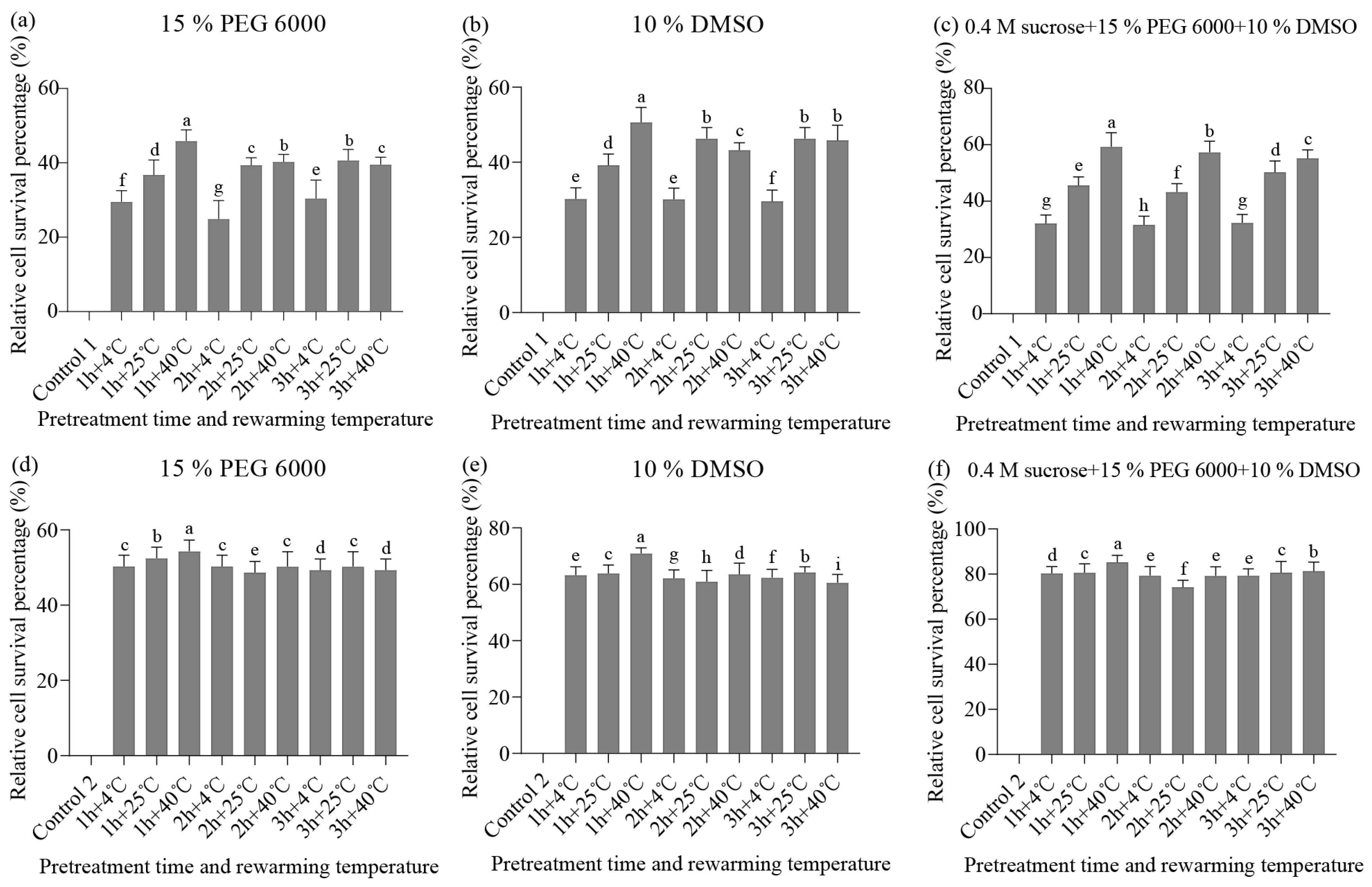
To determine the optimal cryopreservation procedure, we evaluated the relative recovery rates of embryogenic tissues subjected to different treatments during the cryopreservation step. These treatments included using separate or different combinations of cryoprotectants after solid and liquid pre-culture conditions containing 0.4 M sorbitol. We also tested different durations of treatment (1–3 h) as well as different rewarming temperatures (4 °C, 25 °C, and 40 °C). After 14 d of culturing, the embryogenic tissues that survived rewarming started to multiply. The visible proliferation was seen after 4 weeks in culture when measuring recovery. Differences in recovery rates were observed among samples according to different treatments. When pre-cultured with the liquid medium, the relative survival of pretreatment with 10% DMSO alone or 15% PEG6000 alone is 50.6% and 45.9% (Figure 2a,b). The highest relative survival rate of embryogenic tissues (59.3%) was achieved after the pretreatment with 10% DMSO, 0.4 M sucrose, and 15% PEG6000 for 1 h under the rewarming temperature of 40 °C (Figure 2c). In a comparison of the relative survival percentages of embryogenic tissues in three pretreatments, the highest relative survival rate of embryogenic tissues was obtained with pretreatment using 10% DMSO, 0.4 M sucrose, and 15% PEG6000. Further, the relative survival percentage of pre-cultured embryogenic tissues in the solid medium in pretreatment with 10% DMSO, 0.4 M sucrose, and 15% PEG6000 was higher (85.3%) when the pretreatment time and the rewarming temperature were 1 h and 40 °C, respectively (Figure 2d–f). In Experiment 1, a comparison between liquid and solid pre-culture conditions revealed that the relative survival percentage of embryogenic tissues in solid pre-culture was more stable.
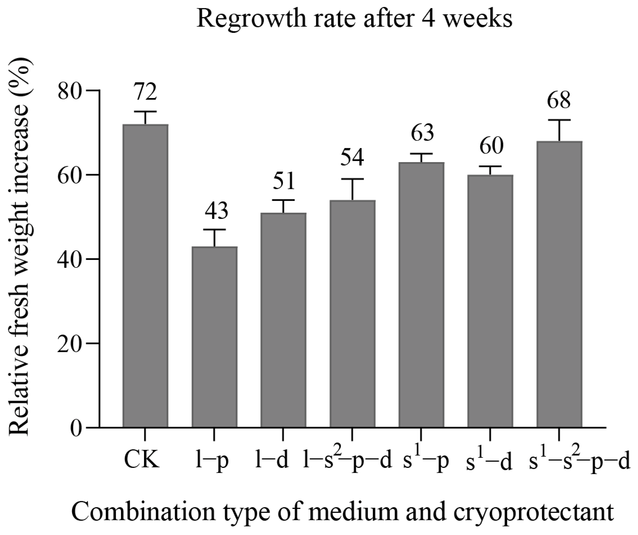
To compare the regrowth rate of embryogenic tissues treated with different cryoprotectants, we exposed them to cryoprotectant for 1 h at a temperature of 4 °C and then rewarmed at 40 °C once the embryogenic tissues returned to normal growth. We found that among the three pretreatments in the liquid medium, using 15% PEG6000 (liquid: 43%, solid: 63%) resulted in the lowest regrowth rate, followed by 10% DMSO (liquid: 51%, solid: 60%). In Experiment 2, using 10% DMSO, 0.4 M sucrose, and 15% PEG6000 yielded the highest embryogenic tissue regrowth rate (liquid: 54%, solid: 68%).
Furthermore, we observed that the embryogenic tissue regrowth rate was lower in the pre-cultured liquid medium than in the pre-culture solid medium with the same pretreatment. Specifically, the embryogenic tissue regrowth rate pre-cultured in the solid medium using 10% DMSO, 0.4 M sucrose, and 15% PEG6000 showed the highest growth rate (Figure 2). In the combination processing of Experiment 1 and Experiment 2, we obtained the best treatments in Experiment 3 and Experiment 4 at 1 h and 40 °C.
Our results suggest that the best cryopreservation procedure involves pre-culture in a solid medium, 1 h pretreatment time in cryoprotectant on ice, and rewarming at 40 °C. After 4 weeks of culture, we found that the regrowth rate of cryopreserved embryogenic tissues in 10% DMSO, 0.4 M sucrose, and 15% PEG6000 was closest to the regrowth rate of non-cryopreserved embryogenic tissues (Figure 3).
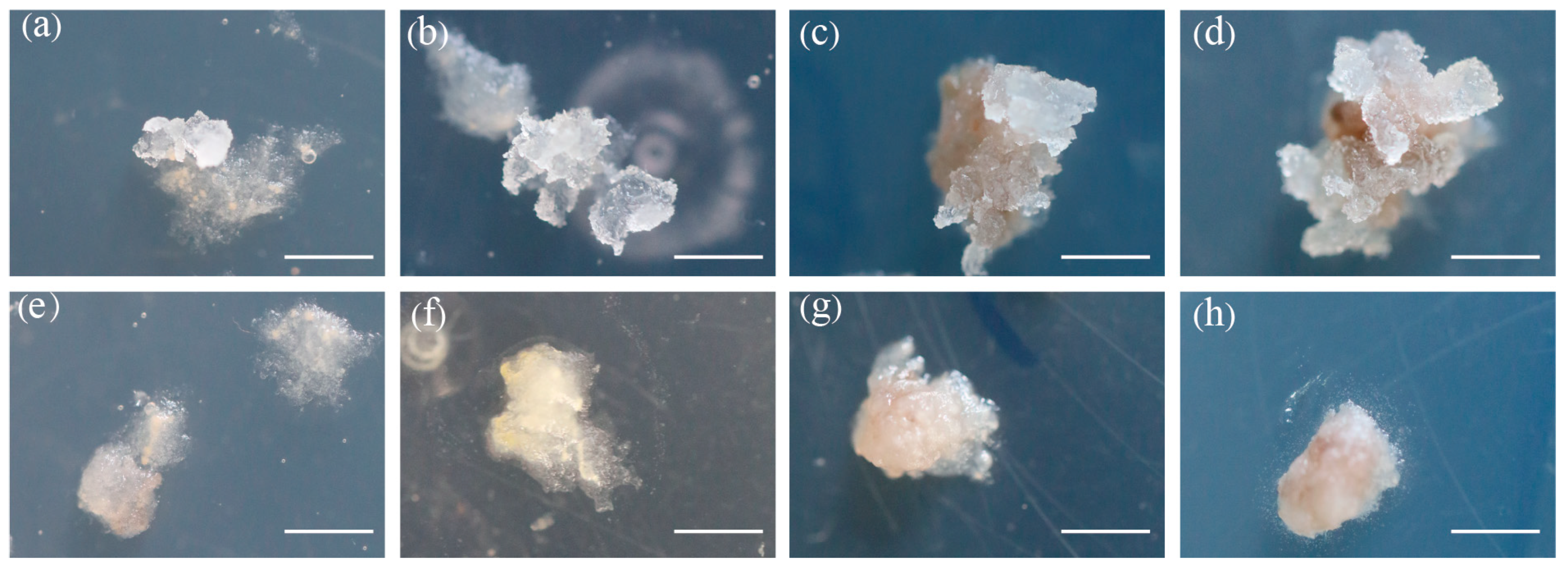
However, the embryogenic tissue pre-cultured in either liquid or solid medium and pretreated only with 0.4 M sucrose did not proliferate after thawing for three weeks (Figure 4e–h).

3.2. Morphological Characterization

To assess the damage caused by cryopreservation on embryogenic tissues, we compared tissue samples that were cryopreserved with those that were not. After rewarming, we observed that the embryogenic tissues pre-cultured in liquid medium had smaller tissue clusters than those pre-cultured in solid medium. We then performed 0.01% FDA staining on the cryo- and non-cryopreserved samples and found that each cryoprotective pretreatment drastically damaged a number of living cells in comparison to non-cryopreserved tissues during cryopreservation (Figure 5). Our results revealed that in the non-cryopreserved embryogenic tissue samples, there were strong green fluorescence signals present in long suspensor cells after staining. However, the fluorescent signal was localized in meristematic cell clumps in cryopreserved tissue, indicating that only the meristematic cells survived cryopreservation while the long vacuolated suspensor cells were disrupted (Figure 5b–d).
To investigate the regrowth of embryogenic tissues following cryopreservation, we stained tissue samples with 1% (w/v) TTC and examined them under a microscope at weekly intervals. We discovered that bipolar structures were present in the regrown tissues after cryopreservation, indicating that the embryogenic tissues could re-store their original structural organization during post-thaw growth (Figure 6). Two weeks after thawing, we observed the presence of embryogenic tissues in the PEM I stage, which is composed of only a tiny piece of the meristematic tissues linked to a little elongated cell (Figure 6b). Three weeks after thawing, the majority of embryogenic tissues belonged to type II (PEM II stage), where similar aggregates as PEM I stage but with more vacuolated cells were observed (Figure 6c). Four weeks after thawing, the embryogenic tissues were mostly in the PEM III stage; the masse have a huge meristematic complex and are encircled by a lot of elongated cells (Figure 6d). Additionally, there are no obvious color or texture distinctions between non-cryopreserved and cryopreserved embryogenic tissue samples (Figure 6a,d). Notably, elongated cells in suspension cells showed non-embryogenic characteristics.

3.3. Maturation Ability and Plantlet Regeneration

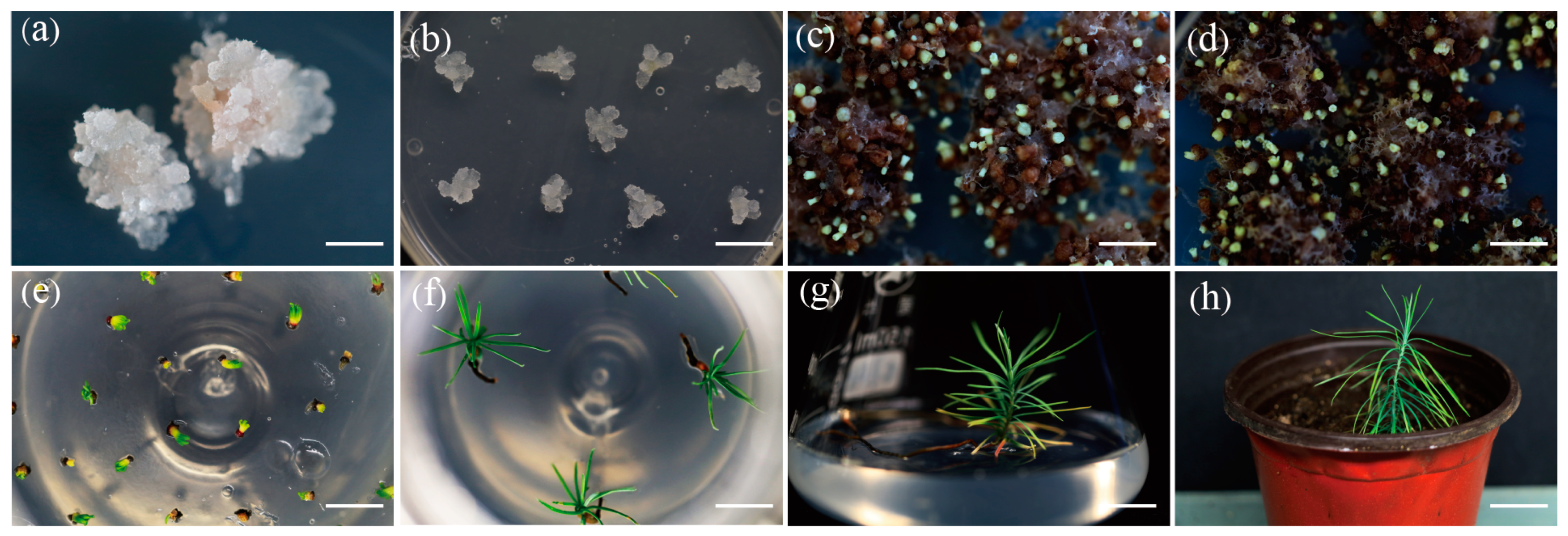
To investigate the effect of cryopreservation on the ability of somatic embryo regeneration from embryogenic tissues, we tested the maturation of embryogenic tissues to assess their differentiation after cryopreservation. We observed that after rewarming and thawing for four weeks, the embryogenic tissues showed higher proliferation (Figure 7a). Subsequently, we transferred 10 g of the embryogenic tissues onto a growth regulator-free proliferation medium for a week before transferring them directly to the maturation medium. A comparison of the maturation ability of cryopreserved and non-cryopreserved tissues revealed that there was no significant difference between them (Figure 7c,d). We calculated the number of somatic embryos generated per gram of embryogenic tissues in cryo- and non-cryopreserved tissues. The number of somatic embryos obtained from matured cryo- and non-cryopreserved tissues were both approximately 30.
After maturing, the cryopreserved embryogenic-tissue-derived somatic embryos were exposed to a 16 h photoperiod at 22 °C for four weeks (Figure 7e). The normally developed cotyledonary embryogenic tissue was then collected and placed in fresh rooting media for three months, during which they gradually developed cotyledons, elongated hypocotyls, and roots (Figure 6f). These plantlets were transferred to fresh rooting media every three weeks for two months to develop into a single plant (Figure 7g). After three weeks, the plantlets were moved to a traditional greenhouse where plantlets were grown at 25 °C and under a 16 h photoperiod (Figure 7h). The survival rate of plants regenerated from matured cryo- and non-cryopreserved tissues were both approaches to 80%.
Overall, our experiments showed that cryopreservation had no effect on the regeneration ability of embryogenic tissues. Our findings suggest that cryopreservation can be used as an effective method for preserving embryogenic tissues and maintaining their reproductive potential.

4. Discussion

Cryopreservation is a valuable technique for the long-term preservation of plant genetic resources while maintaining their genetic stability. Cryopreservation methods have been established for a large number of plant species, and in the late 1980s, the first reports on cryopreservation of conifer tissues were published, including studies on Pinus taeda [26] and P. glauca [2]. Since then, several other conifer species, like P. patula [11], P. mariana [27], P. radiate [14], P. sitchensis [28], P. omorika [29], P. nigra [24], and Torreya taxifolia [30] have also been explored using different methods of freezing preservation. Larch is an important coniferous species with fast growth, high-quality wood, and wide ecological adaptability. In this work, we focused on cryopreserving hybrid larch embryogenic tissue and examined the effect of cryopreservation on the maturation of embryogenic tissue by evaluating the efficiency of somatic embryogenesis. We discussed the steps involved in cryopreserving hybrid larch embryogenic tissues from various aspects, providing insights into the development of effective cryopreservation procedures for larch and further promoting the application of larch cryo-biotechnology.
The quality and type of plant materials (i.e., water content), pretreatment and pre-culture conditions, cryopreservation method, and warming conditions are all critical factors in cryopreservation technologies [3]. For successful cryopreservation, it is important to reduce the intracellular ice crystals in the cell and protect against damage to the membrane and wall of the cell, which ensures that the cells can survive the freezing and thawing processes. Therefore, the most important thing is treating the embryogenic tissues when the cells possess lower water content. As the water content significantly increases in embryogenic tissues, it makes the cells more susceptible to freeze damage [31,32]. The free water content needs to be removed using osmotic agents solely or in combination with a cryoprotectant [33]. The pretreatment and pre-culture conditions are critical in minimizing the formation of intracellular ice crystals during cryopreservation. One key requirement for a successful cryopreservation process is to maintain a lower level of the cell water to prevent the ice crystals from formatting and ensure cell recovery after thawing [34]. In our work, we utilized both solid and liquid media for pre-culturing the embryogenic tissues to condition the osmotic potential of the tissue cells by adding sorbitol. Specifically, we evaluated the effect of using a solid–liquid medium as a pre-culture condition on cryopreservation. Although most current conifer cryopreservation schemes adopt liquid cultures for pretreatment [14,16,23,28,35], some studies have shown that using only liquid cultures can abrupt the homeostasis of embryogenic potential, often resulting in loss of embryogenic potential. To date, the suspension culture is known to offer significant advantages over semi-solid media for the long-term storage of embryogenic tissues [33]. However, based on our study, it was found that a solid medium is more suitable for the pre-culture of larch embryogenic tissues during cryopreservation. Osmotic regulators such as antioxidants [36,37,38], polyvinyl pyrrolidone [39], sorbitol and sugars, and/or elicitors of defense-related proteins in plants [38,40] can be used in the pre-culture step. In conifers, the most commonly used osmotic regulators are sucrose [41,42] and sorbitol [14,15,43]. The most common osmotic regulator for pretreatment of conifer embryogenic tissue was found as sorbitol at 0.4 M concentration [33]. Our work also used 0.4 M sorbitol for the pretreatment of larch embryogenic tissues and observed the improved effects in cryopreservation, confirming the significance of pretreatment for successful cryopreservation.
After the pretreatment of the embryogenic tissues, the freezing method plays a crucial role in successful cryopreservation. Currently, vitrification, two-step cooling methods, encapsulation-dehydration, and V- and D-cryoplatesn are the most commonly used cryopreservation procedures for vegetatively propagated crops [3,44,45,46]. Among them, vitrification and encapsulation-dehydration methods have been continuously improved and are thus the most frequently employed for cryopreservation of clonally propagated plants. In vitrification methods, a concentrated cellular solution is necessary to achieve vitrification. During the osmotic process, when the cell is placed in “vitrification” solutions, the water is removed from the cells [47]. Plant tissue is typically treated with a combination of “vitrification” solutions, such as dimethyl sulfoxide (DMSO), glycerol, or ethylene glycol. In conifers, the widely used cryoprotectant is DMSO [23,24,37], as it can rapidly penetrate the cell wall and plasma membrane. Often, semipermeable or non-permeable cryoprotective substances, such as sugars, sugar alcohols, and high molecular weight additives, like polyethylene glycol (PEG), are mixed with DMSO [28,29]. In these cases, no changes in embryogenic potential were observed after storing the tissue in liquid nitrogen. Therefore, in our work, we compared the effects of three types of cryoprotectants (10% DMSO, 0.4 M sucrose, and 15% PEG6000), whether used alone or combined. The results showed that the highest regrowth efficiency of embryogenic callus was observed in the pretreatment with combined cryoprotectants. The recovery of the pretreated samples was significantly different between pretreatments with DMSO and those without DMSO, demonstrating the significance of this substance. The role of DMSO is to prevent the formation of electrolytes during freezing, which eventually prevents mechanical damage caused by the production of ice crystals. Because of its small molecular weight, at the beginning of freezing, DMSO acts quickly by enhancing the permeability of membranes and promoting cell protection during dehydration [31]. This finding supports the efficacy of our cryopreservation method for hybrid larch embryogenic tissue. Physical dehydration is usually used for encapsulation-dehydration-based cryopreservation techniques to obtain the plant material with optimum water content, and pretreatments are often essential for successful encapsulation-dehydration-based cryo-preservation [48]. Thus, a dehydration-pretreatment step was solely conducted in our work to investigate the effect of dehydration before attempting cryopreservation.
Conifer tissue cryopreservation methods have primarily been based on slow-cooling technology. This may be because conifers typically use embryogenic tissue and cell suspension cultures for cryopreservation, with a small portion being somatic embryos [49]. The embryogenic tissue and cell suspension cultures are often cryo-preserved using the classical slow-cooling protocol. The solutions in the cell are considered to be concentrated enough to vitrify in liquid nitrogen, plunging when the temperature reaches −40 °C. This method is the slow-cooling process that is often used for cryopreservation of cell suspensions and callus tissue [50]. However, related research suggests that the slow-cooling process would damage the cell [34]. Short periods of less than 1 h and extremely long periods of more than 24 h have been observed to be detrimental to the recovery of cryopreserved embryogenic tissues [34]. In our work, we found that the ideal pretreatment time of embryogenic tissues in cryovials packed with ice is 1 h before placing those in a freezing container in a −80 °C freezer. The container freezes the samples at the speed of approximately −1 °C/min in a −80 °C freezer [18,19], which enables slow-cooling of the embryogenic tissues to protect the cells before placing them into LN. Upon long-term cryopreservation, warming is a critical factor for successful regrowth. For conifer embryogenic tissues, cryovials have usually been thawed by transferring those into a water bath at 37–40 °C for several minutes [24,28,29], while in other plants, room temperature may suffice [38,51,52]. In our work, we tested three different warming temperatures (4 °C, 25 °C, and 40 °C) and found that the embryogenic tissues could be recovered under all three warming conditions, with the highest efficiency of callus regrowth observed during rapid thawing at 40 °C. In addition to the thawing temperature, the genotype effect has also received considerable attention [38], but no significant effects on the quality of the regenerated plants were observed due to cryopreservation or the interaction between genotype and cryopreservation [53].
In our work, we also evaluated the effect of cryopreservation on the embryogenesis of callus by measuring the efficiency of somatic embryogenesis of frozen callus. Either the number of somatic embryos or the survival rate of regenerated plants from cryo- and non-cryopreserved tissues is no significant difference. According to the results, the somatic embryogenesis ability of frozen callus remains unaffected. Salaj et al. [50] have also observed that there is no relevance between post-thawing tissue growth (cryo-tolerance) and the maturation ability of embryogenic tissues. While hybrid larch trees’ cryopreserved lines have been extensively utilized in experiments for many years, there is a lack of research on the cryopreservation techniques used to preserve embryogenic lines of most larch species for long periods. However, the successful experience of utilizing cryopreservation methods for other conifer genera gives rise to promising prospects for the broader application of this technology to preserve embryogenic tissues of larch species [7]. Nevertheless, it is essential to note that although cryopreservation has obvious advantages, the process of freezing and thawing can potentially cause damage to the structure and functions of the cell, leading to loss of totipotency and thereby reducing the tissues’ viability and their potential for recovery.

5. Conclusions

This study demonstrates the feasibility of cryopreserving embryogenic tissue of hybrid larch. The protocol involves pre-culturing embryogenic tissue on the medium containing 0.4 M sorbitol for two days, followed by treatment with 10% DMSO, 0.4 M sucrose, and 15% PEG6000 for one hour in ice, and subsequent immersion of samples in LN. Rapid thawing at 40 °C was used to warm samples for recovery. With this approach, we achieved high survival rates of embryogenic tissues (80%) without compromising the somatic embryogenesis ability. Thus, this cryopreservation protocol is an appropriate method of the larch. Despite the success of the cryopreservation for hybrid larch embryogenic tissues, several obstacles remain to be overcome; it needs to further optimize the preparation phase of tissues for dehydration and the vitrification solution treatment for each tissue and species.

Author Contributions

Conceptualization, M.M., J.Y. and C.L.; experimental design: C.L.; material collection and performing the experiments: M.M., X.W., C.Z. and H.W. data analysis: M.M.; software, M.M.; writing—original draft: M.M.; writing—review and editing: M.M., S.P. and C.L. All authors approved the final draft. All authors have read and agreed to the published version of the manuscript.

Funding

This work was supported by the Major Projects of Agricultural Biological Breeding of China (Grant No. 2022ZD0401602).

Data Availability Statement

The data is available on request from the corresponding author.

Conflicts of Interest

The authors declare no conflict of interest.

References

  1. Normah, M.N.; Sulong, N.; Reed, B.M. Cryopreservation of shoot tips of recalcitrant and tropical species: Advances and strategies. Cryobiology 2019, 87, 1–14. [Google Scholar] [CrossRef]
  2. Kartha, K.K.; Fowke, L.C.; Leung, N.L.; Caswell, K.L.; Hakman, I. Induction of somatic embryos and plantlets from cryopreserved cell cultures of white spruce (Picea glauca). J. Plant Physiol. 1988, 132, 529–539. [Google Scholar] [CrossRef]
  3. Bettoni, J.C.; Bonnart, R.; Volk, G.M. Challenges in implementing plant shoot tip cryopreservation technologies. Plant Cell Tissue Org. 2021, 144, 21–34. [Google Scholar] [CrossRef]
  4. Wang, M.R.; Lambardi, M.; Engelmann, F.; Pathirana, R.; Panis, B.; Volk, G.M.; Wang, Q.C. Advances in cryopreservation of in vitro-derived propagules: Technologies and explant sources. Plant Cell Tissue Org. Cult. 2021, 144, 7–20. [Google Scholar] [CrossRef]
  5. Ren, L.; Wang, M.R.; Wang, Q.C. ROS-induced oxidative stress in plant cryopreservation: Occurrence and alleviation. Planta 2021, 254, 124. [Google Scholar] [CrossRef] [PubMed]
  6. Ozudogru, E.A.; Lambardi, M. Cryotechniques for the long-term conservation of embryogenic cultures from woody plants. Methods Mol. Biol. 2016, 1359, 537–550. [Google Scholar]
  7. Shmakov, V.N.; Konstantinov, Y.M. Somatic embryogenesis in Larix: The state of art and perspectives. Vavilovskii Z. Genet. Sel. 2020, 24, 575–588. [Google Scholar] [CrossRef]
  8. Li, B.; Wyckoff, G.W. Breeding strategies for Larix decidua, L. leptolepis and their hybrids in the United States. For. Genet. 1994, 1, 65–72. [Google Scholar]
  9. Larkin, P.J.; Scowcroft, W.R. Somaclonal variation-a novel source of variability from cell cultures for plant improvement. Theor. Appl. Genet. 1981, 60, 197–214. [Google Scholar] [CrossRef]
  10. Klimaszewska, K.; Ward, C.; Cheliak, W.M. Cryopreservation and plant regeneration from embryogenic cultures of larch (Larix×eurolepis) and black spruce (Picea mariana). J. Exp. Bot. 1992, 43, 73–79. [Google Scholar] [CrossRef]
  11. Yan, X.; Wang, K.; Zheng, K.; Zhang, L.; Ye, Y.; Qi, L.; Zhu, M. Efficient organogenesis and taxifolin production system from mature zygotic embryos and needles in larch. For. Res. 2023, 3, 4. [Google Scholar] [CrossRef]
  12. Zhu, T.; Wang, J.; Hu, J.; Ling, J. Application of the somatic embryogenesis technique in conifer species. For. Res. 2022, 2, 18. [Google Scholar]
  13. Ford, C.S.; Jones, N.B.; Staden, J.V. Cryopreservation and plant regeneration from somatic embryos of Pinus patula. Plant Cell Rep. 2000, 19, 610–615. [Google Scholar] [CrossRef] [PubMed]
  14. Hargreaves, C.; Grace, L.; Holden, D. Nurse culture for efficient recovery of cryopreserved Pinus radiata D. Don embryogenic cell lines. Plant Cell Rep. 2002, 21, 40–45. [Google Scholar]
  15. Mathur, G.; Alkutkar, V.A.; Nadgauda, R.S. Cryopreservation of embryogenic culture of Pinus roxburghii. Bio. Plant. 2003, 46, 205–210. [Google Scholar] [CrossRef]
  16. Carneros, E.; Hernández, I.; Díaz-Sala, C.; Toribio, M.; Celestino, C. Effect of different cryoprotectant procedures on the recovery and maturation ability of cryopreserved Pinus pinea embryogenic lines of different ages. Vitro Cell. Dev. Biol. Plant 2017, 53, 469–477. [Google Scholar] [CrossRef]
  17. Gupta, P.K.; Durzan, D.J. Biotechnology of somatic polyembryogenesis and plantlet regeneration in loblolly pine. Nat. Biotechnol. 1987, 5, 147–151. [Google Scholar] [CrossRef]
  18. Touchell, D.; Chiang, V.; Tsai, C.J. Cryopreservation of embryogenic cultures of Picea mariana (black spruce) using vitrification. Plant Cell Rep. 2002, 21, 118–124. [Google Scholar]
  19. Gale, S.; John, A.; Harding, K.; Benson, E.E. Developing cryopreservation for Picea sitchensis (sitka spruce) somatic embryos: A comparison of vitrification protocols. CryoLetters 2008, 29, 135–144. [Google Scholar]
  20. Hazubska-Przybył, T.; Chmielarz, P.; Michalak, M.; Bojarczuk, K. Cryopreservation of embryogenic tissues of Picea omorika (Serbian spruce). Plant Cell Tissue Org Cult. 2010, 102, 35–44. [Google Scholar] [CrossRef]
  21. Dutra, N.T.; Silveira, V.; de Azevedo, I.G.; Gomes-Neto, L.R.; Façanha, A.R.; Steiner, N.; Guerra, M.P.; Floh, E.I.; Santa-Catarina, C. Polyamines affect the cellular growth and structure of pro-embryogenic masses in Araucaria angustifolia embryogenic cultures through the modulation of proton pump activities and endogenous levels of polyamines. Physiol. Plant. 2013, 148, 121–132. [Google Scholar] [CrossRef] [PubMed]
  22. Salaj, T.; Matušíková, I.; Fráterová, L.; Piršelová, B.; Salaj, J. Regrowth of embryogenic tissues of Pinus nigra following cryopreservation. Plant Cell Tissue Org. Cult. 2011, 106, 55–61. [Google Scholar] [CrossRef]
  23. Ma, X.; Bucalo, K.; Determann, R.O.; Cruse-Sanders, J.M.; Pullman, G.S. Somatic embryogenesis, plant regeneration, and cryopreservation for Torreya taxifolia, a highly endangered coniferous species. Vitro Cell. Dev. Biol. Plant 2012, 48, 324–334. [Google Scholar] [CrossRef]
  24. McLellan, M.R.; Day, J.G. Cryopreservation and freeze-drying protocols. Methods Mol. Biol. 1995, 38, 1–5. [Google Scholar]
  25. Varis, S.; Ahola, S.; Jaakola, L.; Aronen, T. Reliable and practical methods for cryopreservation of embryogenic cultures and cold storage of somatic embryos of norway spruce. Cryobiology 2017, 76, 8–17. [Google Scholar] [CrossRef]
  26. Zamecnik, J.; Faltus, M.; Bilavcik, A. Vitrification solutions for plant cryopreservation: Modification and properties. Plants 2021, 10, 2623. [Google Scholar] [CrossRef]
  27. Lineros, Y.; Balocchi, C.; Muñoz, X.; Sánchez, M.; Ríos, D. Cryopreservation of Pinus radiata embryogenic tissue: Effects of cryoprotective pretreatments on maturation ability. Plant Cell Tissue Org. Cult. 2018, 135, 357–366. [Google Scholar] [CrossRef]
  28. Peng, C.; Gao, F.; Wang, H.; Shen, H.; Yang, L. Optimization of maturation process for somatic embryo production and cryopreservation of embryogenic tissue in Pinus koraiensis. Plant Cell Tissue Org. Cult. 2021, 144, 185–194. [Google Scholar] [CrossRef]
  29. Marum, L.; Estêvão, C.; Oliveira, M.M.; Amâncio, S.; Rodrigues, L.; Miguel, C. Recovery of cryopreserved embryogenic cultures of maritime pine-effect of cryoprotectant and suspension density. CryoLetters 2004, 25, 363–374. [Google Scholar] [PubMed]
  30. Uchendu, E.E.; Muminova, M.; Gupta, S.; Reed, B.M. Antioxidant and anti-stress compounds improve regrowth of cryopreserved rubus shoot tips. Vitro Cell. Dev. Biol. Plant 2010, 46, 386–393. [Google Scholar] [CrossRef]
  31. Mathew, L.; McLachlan, A.; Jibran, R.; Burritt, D.J.; Pathirana, R. Cold, antioxidant and osmotic pre-treatments maintain the structural integrity of meristematic cells and improve plant regeneration in cryopreserved kiwifruit shoot tips. Protoplasma 2018, 255, 1065–1077. [Google Scholar] [CrossRef]
  32. Volk, G.M.; Shepherd, A.N.; Bonnart, R. Successful cryopreservation of vitis shoot tips: Novel pre-treatment combinations applied to nine species. CryoLetters 2018, 39, 322–330. [Google Scholar]
  33. Wang, Q.; Laamanen, J.; Uosukainen, M.; Valkonen, J.P. Cryopreservation of in vitro-grown shoot tips of raspberry (Rubus idaeus L.) by encapsulation–vitrification and encapsulation–dehydration. Plant Cell Rep. 2005, 24, 280–288. [Google Scholar] [CrossRef]
  34. Pathirana, R.; McLachlan, A.; Hedderley, D.; Panis, B.; Carimi, F. Pre-treatment with salicylic acid improves plant regeneration after cryopreservation of grapevine (Vitis spp.) by droplet vitrification. Acta. Physiol. Plant. 2016, 38, 12. [Google Scholar] [CrossRef]
  35. Varis, S.A.; Virta, S.; Montalbán, I.A.; Aronen, T. Reducing pre- and post-treatments in cryopreservation protocol and testing storage at −80 °C for norway spruce embryogenic cultures. Int. J. Mol. Sci. 2022, 23, 15516. [Google Scholar] [CrossRef] [PubMed]
  36. Laine, E.; Bade, P.; David, A. Recovery of plants from cryopreserved embryogenic cell suspensions of Pinus caribaea. Plant Cell Rep. 1992, 11, 295–298. [Google Scholar] [CrossRef]
  37. Vondráková, Z.; Cvikrová, M.; Eliásová, K.; Martincová, O.; Vágner, M. Cryotolerance in norway spruce and its association with growth rates, anatomical features and polyamines of embryogenic cultures. Tree Physiol. 2010, 30, 1335–1348. [Google Scholar] [CrossRef] [PubMed] [Green Version]
  38. Kaviani, B.; Kulus, D. Cryopreservation of endangered ornamental plants and fruit crops from tropical and subtropical regions. Biology 2022, 11, 847. [Google Scholar] [CrossRef] [PubMed]
  39. Dinato, N.B.; Santos, I.R.I.; Vigna, B.B.Z.; Ferreira de Paula, A.; Favero, A.P. Perspective: Pollen cryopreservation for plant breeding and genetic resources conservation. CryoLetters 2020, 41, 115–127. [Google Scholar]
  40. Wang, M.R.; Chen, L.; Teixeira da Silva, J.A.; Volk, G.M.; Wang, Q.C. Cryobiotechnology of apple (Malus spp.): Development, progress and future prospects. Plant Cell Rep. 2018, 37, 689–709. [Google Scholar] [CrossRef]
  41. Serrano-Martinez, F.; Casas, J.L. Cryopreservation of Tetraclinis articulata (vahl.) masters. CryoLetters 2011, 32, 248–255. [Google Scholar]
  42. Bi, W.L.; Pan, C.; Hao, X.Y.; Cui, Z.H.; Kher, M.M.; Marković, Z.; Teixeira da Silva, J.A. Cryopreservation of grapevine (Vitis spp.)—A review. Vitro Cell. Dev. Biol.-Plant 2017, 53, 449–460. [Google Scholar] [CrossRef]
  43. Castander-Olarieta, A.; Moncaleán, P.; Montalbán, I.A. Somatic embryogenesis in pines. Methods Mol. Biol. 2022, 2527, 41–56. [Google Scholar] [PubMed]
  44. Volk, G.M. Application of functional genomics and proteomics to plant cryopreservation. Curr. Genom. 2010, 11, 24–29. [Google Scholar] [CrossRef] [Green Version]
  45. Popova, E.; Kulichenko, I.; Kim, H.H. Critical role of regrowth conditions in post-cryopreservation of in vitro plant germplasm. Biology 2023, 12, 542. [Google Scholar] [CrossRef]
  46. Schumacher, H.M.; Westphal, M.; Heine-Dobbernack, E. Cryopreservation of plant cell lines using alginate encapsulation. Methods. Mol. Biol. 2021, 2180, 639–645. [Google Scholar]
  47. Niino, T.; Yamamoto, S.I.; Fukui, K.; Martínez, C.R.C.; Arizaga, M.V.; Matsumoto, T.; Engelmann, F. Dehydration improves cryopreservation of mat rush (Juncus decipiens Nakai) basal stem buds on cryo-plates. CryoLetters 2013, 34, 549–560. [Google Scholar] [PubMed]
  48. Yamamoto, S.I.; Rafique, T.; Priyantha, W.S.; Fukui, K.; Matsumoto, T.; Niino, T. Development of a cryopreservation procedure using aluminium cryo-plates. CryoLetters 2011, 32, 256–265. [Google Scholar]
  49. Bradai, F.; Sanchez-Romero, C. Effect of cryopreservation on the ex vitro establishment of olive plants regenerated via somatic embryogenesis. Plants 2021, 10, 396. [Google Scholar] [CrossRef]
  50. Salaj, T.; Klubicová, K.; Matusova, R.; Salaj, J. Somatic embryogenesis in selected conifer trees Pinus nigra Arn. and Abies Hybrids. Front. Plant Sci. 2019, 10, 13. [Google Scholar] [CrossRef] [Green Version]
  51. Li, X.Y.; Huang, F.H.; Gbur, E.E., Jr. Effect of basal medium, growth regulators and phytagel concentration on initiation of embryogenic cultures from immature zygotic embryos of loblolly pine (Pinus taeda L.). Plant Cell Rep. 1998, 17, 298–301. [Google Scholar] [CrossRef] [PubMed]
  52. Filonova, L.H.; Bozhkov, P.V.; Arnold, S.V. Developmental pathway of somatic embryogenesis in Picea abies as revealed by time-lapse tracking. J. Exp. Bot. 2000, 51, 249–264. [Google Scholar] [CrossRef] [PubMed] [Green Version]
  53. Miguel, C.; Gonçalves, S.; Tereso, S.; Marum, L.; Maroco, J.; Margarida Oliveira, M. Somatic embryogenesis from 20 open-pollinated families of portuguese plus trees of maritime pine. Plant Cell Tissue Org. Cult. 2004, 76, 121–130. [Google Scholar] [CrossRef]
Figure 1. Flowchart depicts the major steps of the cryopreservation for embryogenic tissues. Major steps of cryopreservation procedure include pre-culture, cryoprotectant addition, freezing, and thawing. Step 1 is the pretreatment. Step 2–5 is the part of freezing, and step 6–10 is the part of thawing.
Forests 14 01621 g001
Figure 2. Effects of pretreatment time and rewarming temp on the survival rates. (ac) The pre-culture medium is the liquid; (df) the pre-culture medium is the solid. The control is the embryogenic tissues have not been treated with cryoprotectant (control 1: The embryogenic tissues pre-cultured in liquid medium; control 2: The embryogenic tissues pre-cultured in solid medium). 1 h + 4 °C: The embryogenic tissues on the ice for pretreatment with 1 h and rewarming in 4 °C; 1 h + 25 °C: The embryogenic tissues on the ice for pretreatment with 1 h and rewarming in 25 °C; 1 h + 40 °C: The embryogenic tissues on the ice for pretreatment with 1 h and rewarming in 40 °C; 2 h + 4 °C: The embryogenic tissues on the ice for pretreatment with 2 h and rewarming in 4 °C; 2 h + 25 °C: The embryogenic tissues on the ice for pretreatment with 2 h and rewarming in 25 °C; 2 h + 40 °C: The embryogenic tissues on the ice for pretreatment with 2 h and rewarming in 40 °C; 3 h + 4 °C: The embryogenic tissues on the ice for pretreatment with 3 h and rewarming in 4 °C; 3 h + 25 °C: The embryogenic tissues on the ice for pretreatment with 3 h and rewarming in 25 °C; 3 h + 40 °C: The embryogenic tissues on the ice for pretreatment with 3 h and rewarming in 40 °C; Data are presented as the mean over at least three replicates. Different letters (a–i) above the column chart indicate significant differences by the ANOVA test. The significance level a–i ranges from low to high (ANOVA test; p = 0.05).
Forests 14 01621 g002
Figure 3. The regrowth rate of embryogenic tissues treated with different cryoprotectants at the best pretreatment time on the ice and the best rewarming temperature. (l: liquid medium; s1: solid medium; s2: 0.4 M sucrose; p: 15% PEG6000; d: 10% DMSO). The relative fresh weight increase was used to calculate the regrowth rate. The CK is the growth rate of non-cryopreserved embryogenic tissues (control). Data are presented as the mean over at least three replicates. The mean values are shown on the top of each column.
Forests 14 01621 g003
Figure 4. The growth state of cryopreserved embryogenic tissues grew after 2 weeks of rewarming. (a,b) The embryogenic tissues pre-cultured in liquid medium and the cryoprotectants are 15% PEG6000 and 10% DMSO, respectively; (c,d) The embryogenic tissues pre-cultured in solid medium and the cryoprotectants are 15% PEG 6000 and 10% DMSO, respectively, bar = 0.5 cm; (e) The embryogenic tissues pre-cultured in liquid medium and no cryoprotectants, bar = 0.5 cm; (f) The embryogenic tissues pre-cultured in liquid medium and the cryoprotectants is 0.4 M sucrose, bar = 0.5 cm; (g) The embryogenic tissues pre-cultured in solid medium and no cryoprotectants, bar = 0.5 cm; (h) The embryogenic tissues pre-cultured in solid medium and the cryoprotectants is 0.4 M sucrose, bar = 0.5 cm.
Forests 14 01621 g004
Figure 5. The FDA staining of thawed cryopreserved embryogenic tissues. (a) The non-cryopreserved embryogenic tissues as a contrast, bar = 50 μm; (b) The embryogenic tissues pre-cultured in solid medium and the cryoprotectants is 0.4 M sucrose, 15% PEG6000 and 10% DMSO, bar = 50 μm; (c) The embryogenic tissues pre-cultured in solid medium and the cryoprotectants is 10% DMSO, bar = 50 μm; (d) The embryogenic tissues pre-cultured in solid medium and the cryoprotectants is 0.4 M sucrose, 15% PEG6000 and 10% DMSO, bar = 50 μm. MC: meristematic cells, S: suspensor cells.
Forests 14 01621 g005
Figure 6. The TTC staining of thawed cryopreserved tissues in the sub-cultured period. (a) The non-cryopreserved embryogenic tissues as a control, bar = 200 μm; (b) pro-embryogenic masses (PEMs) I, the masse only have a small portion of meristematic tissue attached to a little elongated cell, bar = 100 μm; (c) PEMs II, the masse have more elongated cells link to meristematic tissue, bar = 200 μm; (d) PEMs III, the masse have a large size meristematic complex and are surrounded by a high number of elongated cells, bar = 200 μm. EC: embryogenic cells, S: suspensor cells.
Forests 14 01621 g006
Figure 7. Somatic embryogenesis and plant regeneration after rewarming cryopreserved tissues of Larix. (a) The cryopreserved embryogenic tissues proliferation after 4 weeks of rewarming, bar = 0.5 cm; (b) Regulator-free culture of cryopreserved embryogenic tissues after rewarming, bar = 0.5 cm; (c) Somatic embryogenesis of cryopreserved embryogenic tissues cultured in the maturation medium for 4 weeks, bar = 1.0 cm; (d) Somatic embryogenesis of non-cryopreserved embryogenic tissues cultured in the maturation medium for 4 weeks, bar = 1 cm; (e) Somatic embryos (SE) were exposed to a 16 h photoperiod at 22 °C for 4 weeks, bar = 0.5 cm; (f) The normally developed cotyledonary SE were transferred into fresh rooting media for 3 months and gradually developed cotyledons, elongated hypocotyls, and roots, bar = 1 cm; (g) Transferred the plantlets to 1/2 MS media for 2 months and gradually developed into a single plant, bar = 1.0 cm; (h) The normally grown plantlet acclimatized in soil, bar = 3 cm.
Forests 14 01621 g007
Table 1. The type of cultured media.
Medium TypeFunctionPGRs
BM1Basal proliferation medium0.2 µM 6-BA, 0.4 µM 2,4-D
BM2Recover proliferation medium2.0 µM 6-BA, 4.0 µM 2,4-D
BM3Regulator-free proliferation mediumnone
BM4Maturated medium30 mg/L ABA
1/2MSRooting mediumnone
Disclaimer/Publisher’s Note: The statements, opinions and data contained in all publications are solely those of the individual author(s) and contributor(s) and not of MDPI and/or the editor(s). MDPI and/or the editor(s) disclaim responsibility for any injury to people or property resulting from any ideas, methods, instructions or products referred to in the content.