Article

14-3-3 Proteins Participate in Regulation of Natural Rubber Biosynthesis in Hevea brasiliensis

1
Key Laboratory of Biology and Genetic Resources of Tropical Crops, Ministry of Agriculture and Rural Affairs, Institute of Tropical Bioscience and Biotechnology, Chinese Academy of Tropical Agricultural Sciences, Haikou 571101, China
2
College of Forestry, Hebei Agricultural University, Baoding 071000, China
3
Tropical Crop Genetic Improvement Key Laboratory of Zhanjiang, South Subtropical Crops Research Institute, Chinese Academy of Tropical Agricultural Sciences, Zhanjiang 524091, China
*
Author to whom correspondence should be addressed.
These authors contributed equally to this work.
Forests 2023, 14(5), 911; https://doi.org/10.3390/f14050911
Submission received: 22 March 2023 / Revised: 24 April 2023 / Accepted: 26 April 2023 / Published: 28 April 2023
(This article belongs to the Special Issue Stress Resistance of Rubber Trees: From Genetics to Ecosystem)

Abstract

:
Plant 14-3-3 proteins mediate a wide range of functionally diverse proteins through protein–protein interactions that are typically phosphorylation-dependent. However, the interactions between 14-3-3 proteins and the major regulators of nature rubber (NR) biosynthesis in H. brasiliensis have not been fully elucidated. In this study, we obtained 81 essential client proteins that interacted with H. brasiliensis 14-3-3 proteins (HbGF14s) through yeast two-hybrid (Y2H) screening. These interaction partners were involved in plant signal transduction, metabolism, development, and NR biosynthesis including small rubber particle protein (SRPP), rubber elongation factor (REF), and MYC2, etc. Furthermore, the interaction of HbGF14c and HbSRPP of H. brasiliensis was confirmed in plants through bimolecular fluorescence complementation (BiFC) assays and in vitro with Pull-down assays. Specifically, the RVSSYLP motif was found to mediate the interaction between HbSRPP and HbGF14c. The findings of this study provide a theoretical basis for the elucidation of the molecular regulation mechanism of the 14-3-3 proteins involved in NR biosynthesis, which could be used to enhance the production of rubber trees through genetic improvement.

1. Introduction

14-3-3 proteins are a type of acidic regulatory protein with a strong regulatory effect in eukaryotes [1]. Numerous 14-3-3 proteins have been identified in plants [2]. The 14-3-3 protein family is a small protein family. For example, 15 such protein genes were identified in Arabidopsis thaliana [3,4]. Twelve genes encoding 14-3-3 proteins exist in tomatoes [5,6]. In rice, the 14-3-3 family contains eight members [4,7]. In tobacco, the 14-3-3 family consists of 17 members [8,9]. In cotton, there are 25 genes encoding 14-3-3 proteins [10]. Ten 14-3-3 proteins were identified, named HbGF14a to HbGF14j in Hevea brasiliensis [11].
In plants, 14-3-3 proteins play a crucial role in regulating various physiological and biochemical processes through interactions with target proteins such as metabolism, growth, development, abiotic stress, and signaling pathways [12,13,14,15,16]. So far, over 300 target proteins of 14-3-3 proteins have been proven to be key components of the cellular metabolic network [17]. The 14-3-3 protein includes homodimers and heterodimers that can bind to the domain of one target protein or two target proteins simultaneously [18]. The majority of the target proteins binding to 14-3-3 proteins contain conserved phosphorylated serine (Ser) or threonine (Thr) sequence motifs [19]. The interactions between 14-3-3 proteins and client proteins led to alterations in protein characteristics such as structure, activity, stability, and subcellular localization [4,20,21,22,23].
H. brasiliensis has the advantages of a large production volume, long production time, and easy collection and has become the major source of natural rubber (NR) [24]. NR contains cis-1,4-polyisoprene that is synthesized from isopentenyl pyrophosphate (IPP) via the mevalonic acid pathway, with the synthesis occurring on the surface of rubber particles in the latex of rubber trees [25]. Many proteins and enzymes participate in NR biosynthesis, such as 3-hydroxy-3-methyl glutaryl coenzyme A reductase (HMGR), farnesyl pyrophosphate synthase (FPS), small rubber particle protein (SRPP), rubber elongation factor (REF), and rubber transferase (a cis-prenyltransferase that catalyzes pyrophosphate into rubber-molecule HRT) [25,26,27,28,29,30,31]. We reported that the 14-3-3 protein (HbGF14a) binds to a RING zinc finger protein (HbRZFP1) to mediate the expression of HRT2 in H. brasiliensis, which is the first time that 14-3-3 proteins have taken part in NR biosynthesis [32]. However, the interactions between other HbGF14s and the major regulators of NR biosynthesis in H. brasiliensis remain unclear.
In this study, we obtained 81 target proteins that bind to 14-3-3 proteins through a yeast two-hybrid (Y2H) screening. HbGF14c protein was applied as the bait protein to obtain a H. brasiliensis small rubber particle protein (HbSRPP). The interactions between the HbGF14c protein and HbSRPP were further proven through bimolecular fluorescence complementation (BiFC), Y2H, and Pull-down assays, providing a theoretical foundation for further studies of the molecular regulation mechanism of 14-3-3 proteins participating in NR biosynthesis.

2. Materials and Methods

2.1. Plant Materials

Hevea brasiliensis Müll. Arg. cultivar CATAS7-33-97 was grown at the National Rubber Tree Varieties Resource Garden of the Chinese Academy of Tropical Agricultural Sciences (Danzhou, Hainan, China). Latex was collected directly into liquid nitrogen and stored in an ice kettle for RNA extraction. At the same time, 30 seeds of tobacco (Nicotiana benthamiana) were planted in pots under 16 h light/8 h dark at 26/21 °C with 70%–80% relative humidity.

2.2. The Construction of a Y2H Library

The total RNA of latex was extracted using the method described previously [33]. The mRNA was purified using a poly A+ RNA kit referring to the manufacturer’s instructions (Clontech/Takara Bio, Kusatsu, Japan). The SMART oligo-dT technique was used to synthesize the first-strand cDNAs, and an LD-PCR was used to synthesize double-strand cDNAs. The Y2H library was constructed according to the Make Your Own “Mate & Plate™” Library System User Manual (Clontech). The construction of the Y2H library was performed as follows. A cDNA library was constructed that linearized pGADT7-Rec, and purified double-strand cDNAs were co-transformed to Y187 yeast and placed at the synthetically defined (SD) medium without leucine (SD/-Leu) at 30 °C for 3–4 days. The library solution was divided into multiple 1 mL aliquots for short-term use and several 50 mL aliquots for long-term storage, which were then kept at a temperature of −80 °C. The cDNA library was diluted 10, 100, and 10,000 times and grown on 100 μL SD/-Leu with three biological replicates. The number of colonies was then recorded. The cell density in the Y2H library was measured using a hemacytometer (Thermo Fisher Scientific, Waltham, MA, USA), and the density of a high-quality Y2H library should reach 2 × 10⁷ cfu/mL. The titer and transformation efficiency were calculated using the following formula:
titer   =   Numbers   of   colonies plating   volume mL ×   dilution   factor
transformation   efficiency   = cfu   x   Suspension   Volume   mL Vol .   plated   mL   x   amount   of   DNA   µ g
Finally, the colonies were selected for PCR identification using the pGADT7-F/R primers. The recombination rate was calculated using the following formula:
  recombination   rate   =   numbers   of   positive   colonies   numbers   of   selected   colonies
The primers for the bait vector of HbGF14s are given in Supplementary Table S1.
The full-length cDNA of HbGF14s was introduced to pGBKT7 as the bait vector, which was introduced into the yeast Y2HGold strain and placed at the SD/-Trp screening medium. Then, 4–5 mL of bait vector concentrate was mixed with 1 mL of Y2H library solution in 50 mL of 2 × YPDA + Kan liquid medium. After the appearance of clover-shaped cells, the yeast solution was collected and grown on the screening culture medium. The independent clones were selected for plasmid sequencing, and the results were analyzed using the basic local alignment search tool (BLAST) on the National Center for Biotechnology Information (NCBI) website (www.ncbi.nlm.nih.gov, accessed on 21 March 2023).

2.3. Targeted Y2H Analysis

To clarify the binding of 14-3-3 protein (HbGF14c) to HbSRPP, Y2H assays were carried out according to MatchmakerTM Gold Yeast Two-Hybrid Systems (Clontech). Full-length HbGF14c cDNA and HbSRPP cDNA were introduced to pGBKT7 and pGADT7 as the prey and bait vectors, respectively (primers are shown in Supplementary Table S1). Then, the prey and bait vectors were transformed together into the yeast Y2HGold strain. Transformed colonies were placed at double-drop-out (DDO) medium (SD/-Trp/-Leu) and quadruple-dropout (QDO) medium (SD/-Trp/-Leu/-His/-Ade) and incubated at 28 °C for 3–5 days.
The cDNA encoding the site-directed mutated HbSRPP (S185A) was amplified with PCR (the primers are shown in Supplementary Table S1). The plasmid containing cDNA coding HbSRPP and S185A was introduced into the pGADT7 vector, and that containing cDNA coding HbGF14c was introduced into the pGBKT7 vector. Then, the vectors were transformed together into yeast and developed on DDO and QDO medium incubated at 28 °C for 3–5 days. The pGBKT7-53 and pGADT7-T vectors were transformed together into yeast as positive controls. The pGBKT7-lam and pGADT7-T vectors were transformed together into yeast as negative controls.

2.4. BIFC Assays

Full length HbGF14c cDNA and HbSRPP cDNA (not their stop codons) were introduced to pGN1 (split GFP N-terminal fragment expression) and p2GC (split GFP C-terminal fragment expression) vectors, as described previously [34] (primers are shown in Supplementary Table S1), and introduced into the Agrobacterium tumefaciens GV3101 strain. Next, GV3101 strains that contained GC-HbGF14c and GN-HbSRPP along with strains that contained GC-HbSRPP and GN-HbGF14c, were injected into the epidermal cells of tobacco leaves that were 6 weeks old at an optical density (OD600) of 0.8. Then, GV3101 strains containing GN-HbGF14c and GC; GN and GC-HbSRPP, GN-HbSRPP and GC; and GN and GC-HbGF14c were used as negative controls. The infected cells were grown on Murashige and Skoog (MS) medium at 25 °C under dark conditions [35]. After 2–3 days, the expression of GFP was observed with an OLYMPUS FLUOVIEW FV1000 confocal laser scanning microscope.

2.5. In Vitro Pull-Down Analysis

The cDNAs encoding HbGF14c and HbSRPP were introduced into pGEX-6P-1 (containing a glutathione S-transferase (GST) tag) and pET28a (containing a histidine (HIS) tag) vectors to transform into Escherichia coli BL21 (DE3) expressing strains (for the primers see Supplementary Table S1). The recombinant proteins, HbGF14c-GST and HbSRPP-His, were purified using a GST-binding column (Merck Millipore, Billerica, MA, USA) and HiTrap affinity column (GE Healthcare) according to the instructions of manufacturers. In vitro binding tests were performed as described previously [32]. An immunoblot analysis including anti-GST or anti-His antibodies was performed to detect the interaction between HbGF14c and HbSRPP. Polyhistidine-tagged HbGF14c was introduced into E. coli BL21 (DE3) strains and used as a control.

3. Results

3.1. Construction of cDNA Library

Total RNA was extracted from latex of H. brasiliensis (Figure 1A). The double-strand (ds) cDNAs were synthesized, with the majority ranging in size from 500 to 4000 bp (Figure 1B). To ensure the production of a high-quality cDNA library, the ds cDNA underwent purification to eliminate small fragments between 200–500 bp (Figure 1C).

3.2. Construction of a Y2H Library and Screening of Proteins Interacting with HbGF14s

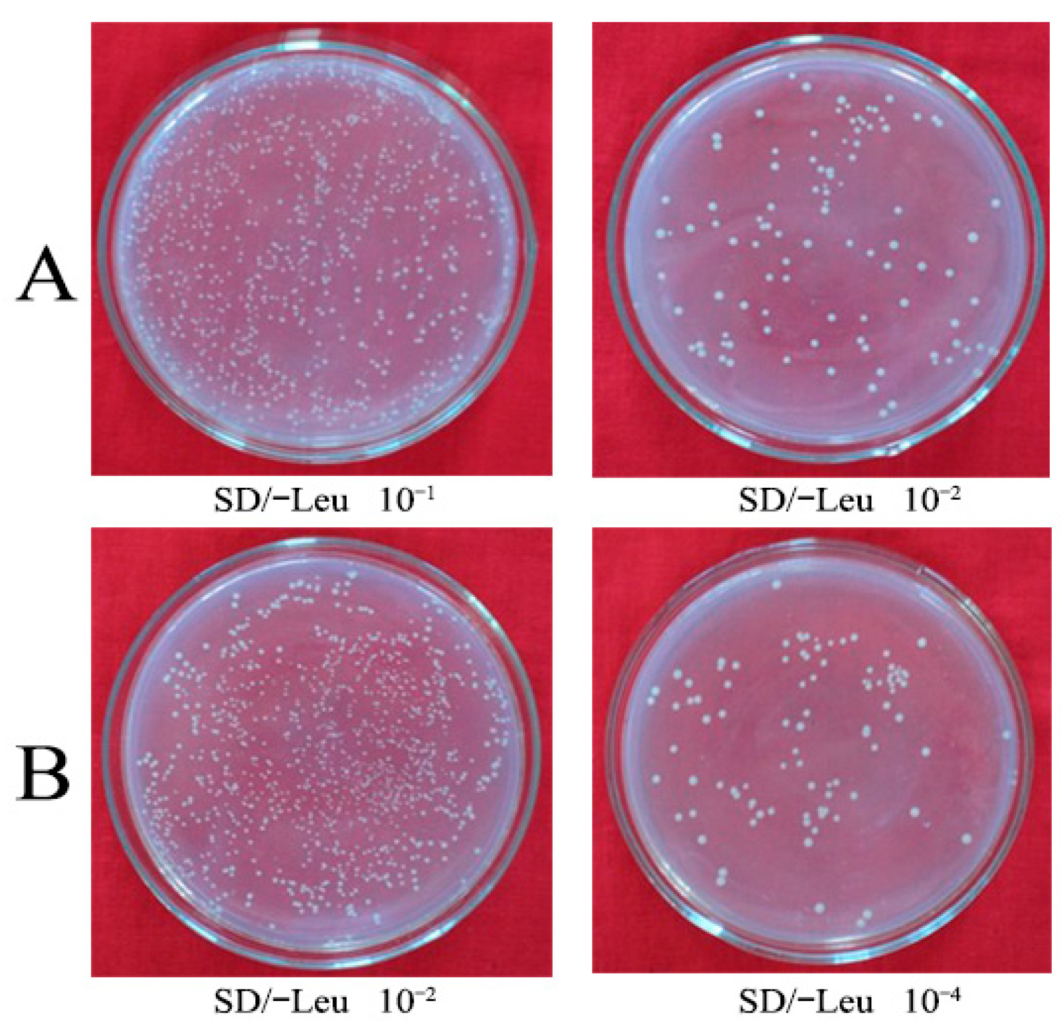
To identify the interaction proteins of HbGF14s, a Y2H assay was performed wherein HbGF14s were used to screen the Y2H library. The linearized pGADT7-Rec plasmid and purified ds cDNA were co-transformed into yeast Y187. In total, 401 colonies were grown on an SD/-Leu plate coated with 10−2 dilution (Figure 2A). The number of independent clones in the library was 2.8 × 10⁶ cfu/mL. On the SD/-Leu plate coated with 10−4 dilution, 246 colonies were grown (Figure 2B). The titer was 4.92 × 10⁷ cfu/mL.
Twenty-four colonies were identified through PCR. The results showed that the inserted fragments were mainly distributed in 500–2000 bp, and some yeast cells contained multiple recombinant library plasmids (Figure 3). The library recombination rate was 96%.
Screening obtained 81 cDNAs encoding proteins interacting with HbGF14s. Based on the BLAST analysis, these cDNA coding proteins were divided into 11 categories according to their functions: stress-related proteins (1%), heat shock proteins (2%), oxidative stress proteins (5%), rubber biosynthesis-related proteins (12%), transcription associated proteins (11%), protein/DNA/RNA-binding proteins (9%), ATPase (1%), signal proteins (20%), protein metabolism-related proteins (5%), carbohydrate metabolism-related proteins (7%), and unknown proteins (27%) (Figure 4). Among these, 17 proteins interacting with HbGF14s were high homologous with H. brasiliensis, including SRPP, REF, MYC2, etc. (Table 1). These proteins participate in many physiological and biochemical processes of laticifer cells, including NR biosynthesis, stress-dependent responses, defense, protein turnover, and transcription. Although SRPP and REF are involved in NR biosynthesis and play a role in increasing molecular weight, quality and yield of NR, the regulatory mechanisms governing their interactions with other proteins have been insufficiently investigated.

3.3. Analysis of the Interaction between HbGF14c and HbSRPP

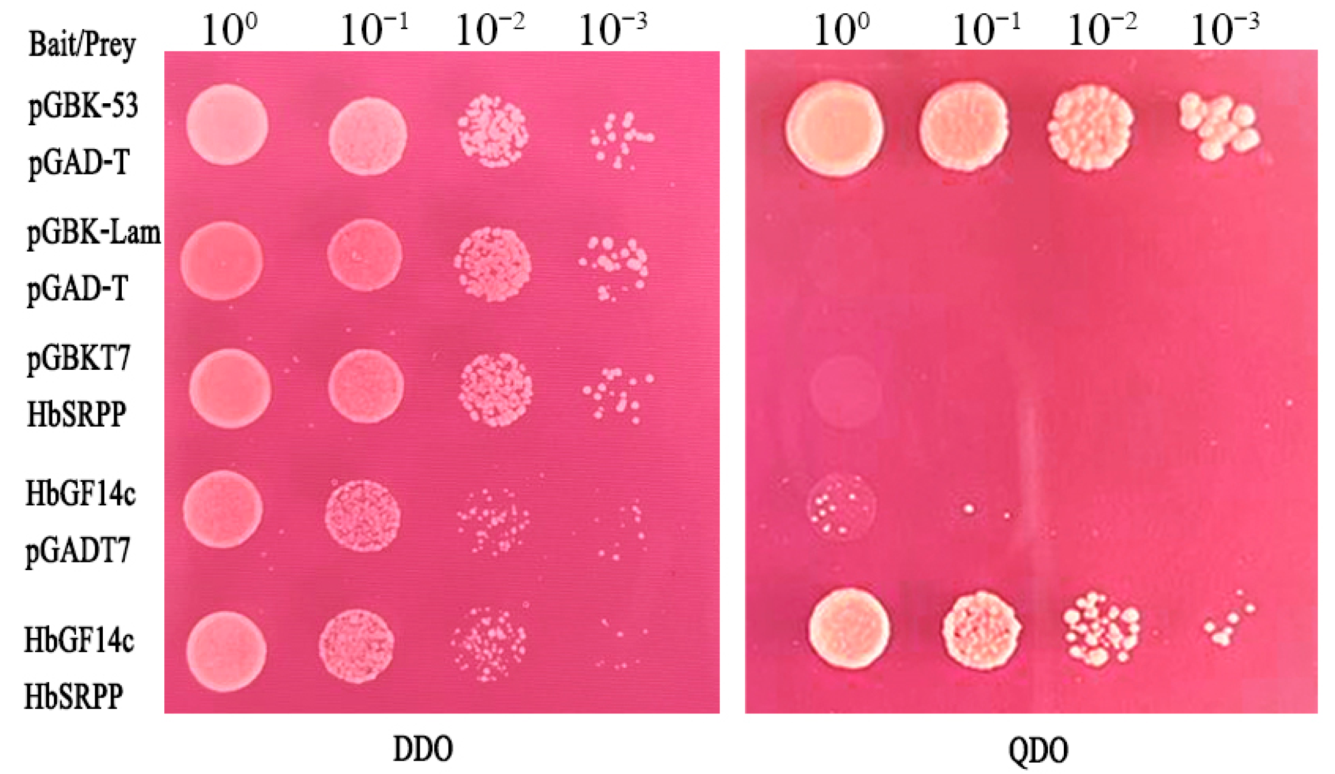
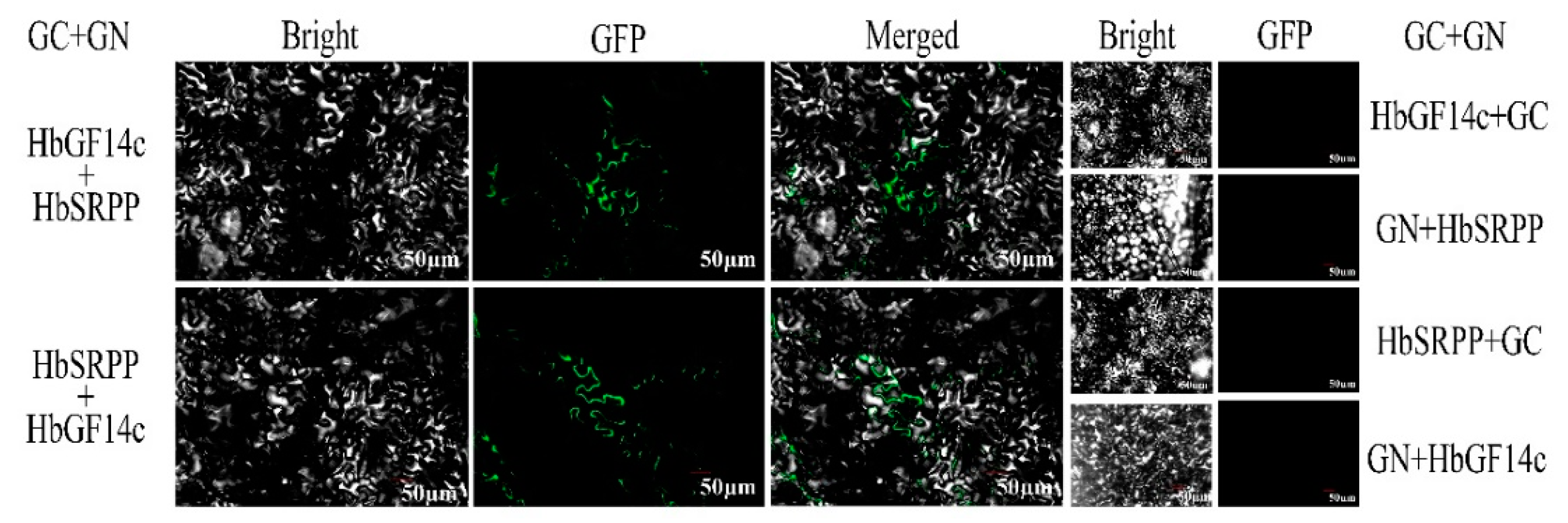
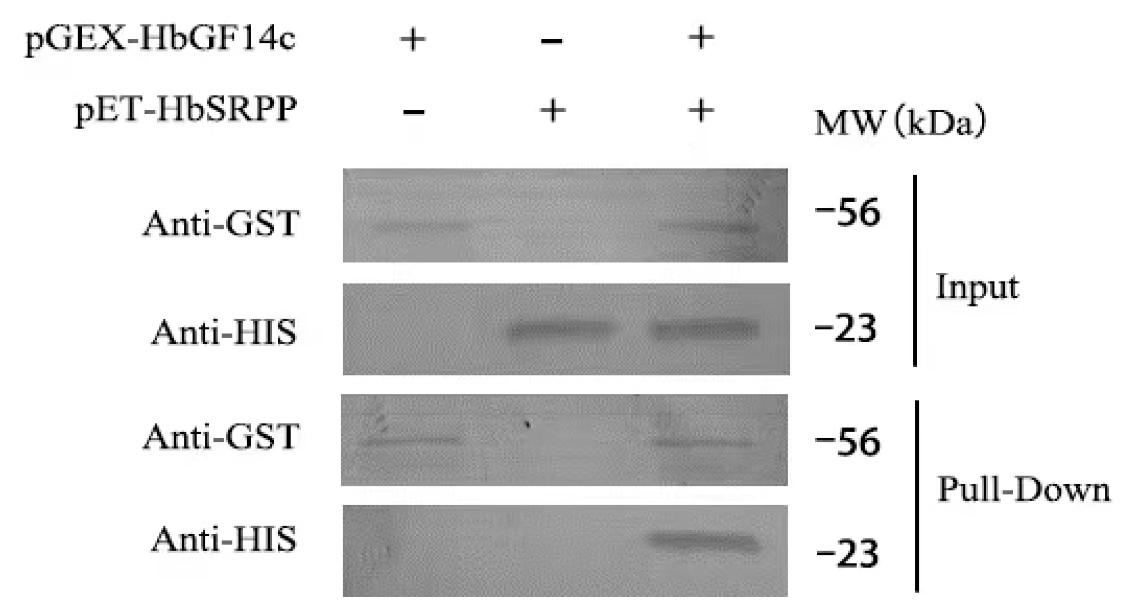
To exclude the false positives of the Y2H screening interacting proteins, we performed a Y2H assay and in-plant BIFC assays to certify the interaction between HbSRPP and HbGF14c proteins. The Y2H assay showed that pGBKT7-HbSRPP and pGADT7-HbGF14c grew on DDO and QDO screening plates together with the positive control (Figure 5). The BIFC assay showed green fluorescence in the tobacco cells co-infected with GC-HbGF14c and GN-HbSRPP as well as GC-HbSRPP and GN-HbGF14c that was mainly concentrated in the cytoplasm, while the control group GN-HbGF14c and GC, GN and GC-HbSRPP, GN-HbSRPP and GC, and GN and GC-HbGF14c had no fluorescence (Figure 6). The interaction between HbSRPP with the HbGF14c protein was further confirmed through an in vitro Pull-down assay (Figure 7). These results implied that HbGF14c specifically interacted with HbSRPP.

3.4. HbSRPP Binds to HbGF14c through the RVSSYLP Motif

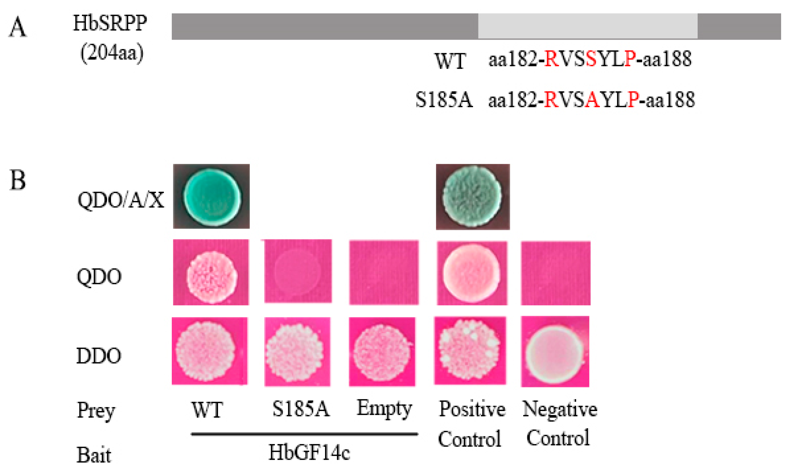
The interaction between the 14-3-3 protein and the target protein involved the combination of phosphoserine/phosphothreonine residues. RXX (pS/pT) XP is a typical 14-3-3 protein-combining motif [36]. The RVSSYLP motif at 182–188 in the amino acid sequence of HbSRPP includes the amino acid serine at position 185, which can be phosphorylated. To determine the binding site of HbGF14c with HbSRPP, we changed S185 to alanine (S185A) to create a mutant of HbSRPP. Notably, S185A eliminated the binding to HbGF14c in the Y2H assay (Figure 8). Therefore, the HbSRPP–HbGF14c interaction was dependent on the combining site of the phosphorylated S185 residue in the RVSSYLP motif.

4. Discussion

14-3-3 proteins participate in almost all plant physiological activities. The interaction between 14-3-3 proteins and many target proteins modulates the activity of the interacting proteins, which regulate a series of growth and development in plants. Many interacting proteins of the 14-3-3 protein family have been identified [37,38]. The molecular mechanisms of 14-3-3 proteins binding to target proteins have also been studied [39,40,41]. In H. brasiliensis, the 14-3-3 protein HbGF14a interacts with HbRZFP1 to regulate the expression of HRT2 [32]. However, the regulatory metabolism of the other 14-3-3 proteins and their interacting proteins in NR biosynthesis remains little understood. In this study, we applied the Y2H system and the latex cDNA library from H. brasiliensis to construct a high-quality Y2H library. HbGF14s, which were highly expressed in laticifer cells, were used as the bait protein, and 81 proteins that bind to 14-3-3 proteins were screened. SRPP, REF, and MYC2 were obtained by applying HbGF14c and HbGF14f as the bait proteins, respectively (Table 1). SRPP and REF were the most abundant proteins above rubber particles, and both play important roles in NR biosynthesis [30,31,42,43]. The 14-3-3 proteins can change the structures of the target proteins. For example, the conformational change of Arabidopsis N-acetyltransferase (AANAT) was modulated by interacting with 14-3-3 protein [20]. It is possible that HbGF14c interacts with SRPP or REF to alter their structure and regulate NR biosynthesis. Jasmonate signaling was proven to positively regulate NR biosynthesis [44,45]. MYC2 is an important transcription factor that activates many jasmonate-responsive genes [46,47,48]. Additionally, HbMYC2 activates expression of HbFPS1 and HbSRPP1 to participate in NR biosynthesis [48]. The binding of 14-3-3 proteins to a client protein impacts the dephosphorylation of the client protein. It was demonstrated that in Arabidopsis, 14-3-3 proteins regulate the nuclear localization and dephosphorylation of PIF7 by preventing its binding to 14-3-3 proteins [49]. The interaction of 14-3-3 proteins with transcription factors can alter transcriptional activity. In Nicotiana, the binding of 14-3-3 proteins enhanced the protein stability and transcriptional activity of AtWRI1 [23]. 14-3-3 proteins can hind or expose the input or output signals of client proteins to regulate their subcellular localization and nucleocytoplasmic shuttling. The interaction between GmMYB133 and 14-3-3 proteins was shown to regulate the subcellular localization of GmMYB133, with the former acting as a positive regulator of the soybean isoflavone synthesis pathway [14]. Based on this, we speculate that the HbGF14f protein may bind to the HbMYC2 protein and potentially affect its transcriptional activity or subcellular localization, thereby regulating the expression of HbFPS1 or HbSRPP1 and participating in NR biosynthesis. Further analysis is required to fully elucidate the interaction mechanisms of HbGF14s with the identified proteins.
The involvement of HblMYB19, HblMYB44, and HbMYC2b in NR biosynthesis has been confirmed through their positive regulation of the HbSRPP gene expression, whereas HbWRKY1, HbWRKY14, and HbMADS4 have been shown to inhibit the expression of the HbSRPP gene [50,51,52,53,54]. Our previous studies have primarily focused on the transcriptional regulation mechanism of the HbSRPP gene, with little attention given to the interaction mechanism between the HbSRPP protein and other proteins. In this study, we used Y2H, BiFC, and Pull-down assays to confirm the interaction of HbGF14c with HbSRPP. Additionally, two conserved phosphorylation motifs in the target protein were bound to the 14-3-3 protein, i.e., the model Ⅰ:RXX (pS/pT) XP and the model Ⅱ: LX (R/K) SX (pS/pT) XP. The 14-3-3 binding site is a match with the RXX (pS/pT) XP motif [36]. The S185A mutation in HbSRPP abolished its binding to HbGF14c in yeast, demonstrating that HbSRPP interacts with HbGF14c through the RVSSYLP motif. The results show that HbGF14c may regulate NR biosynthesis by interacting with the target protein HbSRPP.

5. Conclusions

The findings from this study demonstrate the involvement of 14-3-3 proteins in the process of NR biosynthesis through interactions with client proteins. These results provide a theoretical foundation for the use of biotechnology to modify the expression of key regulators of NR biosynthesis, which may lead to improvements in NR production.

Supplementary Materials

The following supporting information can be downloaded at https://www.mdpi.com/article/10.3390/f14050911/s1: Table S1: Primers used in this study.

Author Contributions

The study was conceived and directed by Y.W. and S.P. and Y.W. conducted the analysis and wrote the manuscript. M.Z., Z.Y., Y.W., H.L., D.G. and J.Z. performed experiments and analyzed the data. All authors have read and agreed to the published version of the manuscript.

Funding

This study was supported by Hainan Provincial Natural Science Foundation of China (2019RC304, 323RC539) and Central Public-interest Scientific Institution Basal Research Fund for Chinese Academy of Tropical Agricultural Sciences (1630052022009).

Data Availability Statement

Not available.

Acknowledgments

We thank Xiuchun Zhang and Mengbin Ruan (Chinese Academy of Tropical Agricultural Sciences) for sharing the BiFC vector.

Conflicts of Interest

The authors declare no conflict of interest.

References

  1. Gökirmak, T.; Paul, A.L.; Ferl, R.J. Plant phosphopeptide-binding proteins as signalling mediators. Curr. Opin. Plant Biol. 2010, 13, 527–532. [Google Scholar] [CrossRef] [PubMed]
  2. Yashvardhini, N.; Bhattacharya, S.; Chaudhuri, S.; Sengupta, D.N. Molecular characterization of the 14-3-3 gene family in rice and its expression studies under abiotic stress. Planta 2018, 247, 229–253. [Google Scholar] [CrossRef] [PubMed]
  3. Cao, A.Q.; Jain, A.; Baldwin, J.C.; Raghothama, K.G. Phosphate differentially regulates 14-3-3 family members and GRF9 plays a role in Pi-starvation induced responses. Planta 2007, 226, 1219–1230. [Google Scholar] [CrossRef] [PubMed]
  4. Chevalier, D.; Morris, E.R.; Walker, J.C. 14-3-3 and FHA domains mediate phosphoprotein interactions. Annu. Rev. Plant Biol. 2009, 60, 67–91. [Google Scholar] [CrossRef] [PubMed]
  5. Xu, W.F.; Jia, L.G.; Shi, W.M.; Baluska, F.; Kronzucker, H.J.; Liang, J.S.; Zhang, J.H. The Tomato 14-3-3 protein TFT4 modulates H+ efflux, basipetal auxin transport, and the PKS5-J3 pathway in the root growth response to alkaline stress. Plant Physiol. 2013, 163, 1817–1828. [Google Scholar] [CrossRef]
  6. Xu, W.F.; Shi, W.M. Expression profiling of the 14-3-3 gene family in response to salt stress and potassium and iron deficiencies in young tomato (Solanum lycopersicum) roots: Analysis by realtime RT-PCR. Ann. Bot. 2016, 98, 965–974. [Google Scholar] [CrossRef]
  7. Chen, F.; Li, Q.; Sun, L.X.; He, Z.H. The rice 14-3-3 gene family and its involvement in responses to biotic and abiotic stress. DNA Res. 2006, 13, 53–63. [Google Scholar] [CrossRef]
  8. Konagaya, K.; Matsushita, Y.; Kasahara, M.; Nyunoya, H. Members of 14-3-3 protein isoforms interacting with the resistance gene product N and the elicitor of Tobacco mosaic virus. J. Gen. Plant Pahtology 2004, 70, 221–231. [Google Scholar] [CrossRef]
  9. Lee, J.H.; Kwak, G.; Lim, Y.P.; Oh, M.H. 14-3-3 proteins contribute to leaf and root development via brassinosteroid insensitive 1 in Arabidopsis thaliana. Genes Genom. 2020, 42, 347–354. [Google Scholar] [CrossRef]
  10. Sun, G.L.; Xie, F.L.; Zhang, B.H. Transcriptome-wide identification and stress properties of the 14-3-3 gene family in cotton (Gossypium hirsutum L.). Funct. Integr. Genom. 2011, 11, 627–636. [Google Scholar] [CrossRef]
  11. Yang, Z.P.; Li, H.L.; Guo, D.; Tang, X.; Peng, S.Q. Identification and characterization of the 14-3-3 gene family in Hevea brasiliensis. Plant Physiol. Biochem. 2014, 80, 121–127. [Google Scholar] [CrossRef]
  12. Denison, F.C.; Paul, A.L.; Zupanska, A.K.; Ferl, R.J. 14-3-3 proteins in plant physiology. Semin. Cell Dev. Biol. 2011, 22, 720–727. [Google Scholar] [CrossRef] [PubMed]
  13. Liu, Z.Y.; Jia, Y.X.; Ding, Y.L.; Shi, Y.T.; Li, Z.; Guo, Y.; Gong, Z.Z.; Yang, S.H. Plasma membrane CRPK1-mediated phosphorylation of 14-3-3 proteins induces their nuclear import to fine-tune CBF signaling during cold response. Mol. Cell 2017, 66, 117–128. [Google Scholar] [CrossRef]
  14. Bian, S.M.; Li, R.H.; Xia, S.Q.; Liu, Y.J.; Jin, D.H.; Xie, X.; Dhaubhadel, S.; Zhai, L.L.; Wang, J.Y.; Li, X.Y. Soybean CCA1-like MYB transcription factor GmMYB133 modulates iso-flavonoid biosynthesis. Biochem. Biophys. Res. Commun. 2018, 507, 324–329. [Google Scholar] [CrossRef]
  15. Chen, Y.S.; Ho, T.H.D.; Liu, L.H.; Lee, D.H.; Lee, C.H.; Chen, Y.R.; Lin, S.Y.; Lu, C.A.; Yu, S.M. Sugar starvation-regulated MYBS2 and 14-3-3 protein interactions enhance plant growth, stress tolerance, and grain weight in rice. Proc. Natl. Acad. Sci. USA 2019, 116, 21925–21935. [Google Scholar] [CrossRef] [PubMed]
  16. Sang, N.; Liu, H.; Ma, B.; Huang, X.Z.; Zhuo, L.; Sun, Y.Q. Roles of the 14-3-3 gene family in cotton flowering. BMC Plant Biol. 2021, 21, 162. [Google Scholar] [CrossRef]
  17. de Boer, A.H.; van Kleeff, P.J.M.; Gao, J. Plant 14-3-3 proteins as spiders in a web of phosphorylation. Protoplasma 2013, 250, 425–440. [Google Scholar] [CrossRef]
  18. Paul, A.L.; Denison, F.C.; Schultz, E.R.; Zupanska, A.K.; Ferl, R.J. 14-3-3 phosphoprotein interaction networks—Does isoform diversity present functional interaction specification? Front. Plant Sci. 2012, 3, 190. [Google Scholar] [CrossRef] [PubMed]
  19. Aitken, A. Functional specificity in 14-3-3 isoform interactions through dimer formation and phosphorylation. Chromosome location of mammalian isoforms and variants. Plant Mol. Biol. 2002, 50, 993–1010. [Google Scholar] [CrossRef]
  20. Obsil, T.; Obsilova, V. Structural basis of 14-3-3 protein functions. Semin. Cell Dev. Biol. 2011, 22, 663–672. [Google Scholar] [CrossRef]
  21. Shin, R.; Jez, J.M.; Basra, A.; Zhang, B.; Schachtman, D.P. 14-3-3 proteins fine-tune plant nutrient metabolism. FEBS Lett. 2011, 585, 143–147. [Google Scholar] [CrossRef] [PubMed]
  22. Xu, L.; Li, T.; Wu, Z.; Feng, H.; Yu, M.; Zhang, X.; Chen, B. Arbuscular mycorrhiza enhances drought tolerance of tomato plants by regulating the 14-3-3 genes in the ABA signaling pathway. Appl. Soil Ecol. 2018, 125, 213–221. [Google Scholar] [CrossRef]
  23. Kong, Q.; Ma, W. WRINKLED1 as a novel 14-3-3 client: Function of 14-3-3 proteins in plant lipid metabolism. Plant Signal. Behav. 2018, 13, e1482176. [Google Scholar] [CrossRef] [PubMed]
  24. Backhaus, R.A. Rubber formation in plants—A mini-review. Isr. J. Bot. 1985, 34, 283–293. [Google Scholar]
  25. Chow, K.S.; Wan, K.L.; Isa, M.N.; Bahari, A.; Tan, S.H.; Harikrishna, K.; Yeang, H.Y. Insights into rubber biosynthesis from transcriptome analysis of Hevea brasiliensis latex. J. Exp. Bot. 2007, 58, 2429–2440. [Google Scholar] [CrossRef] [PubMed]
  26. Dennis, M.S.; Light, D.R. Rubber elongation factor from Hevea brasiliensis. Identification, characterization and role in rubber biosynthesis. J. Biol. Chem. 1989, 264, 18608–18617. [Google Scholar] [CrossRef] [PubMed]
  27. Chye, M.L.; Tan, C.T.; Chuan, H. Three genes encode 3-hydroxy-3-methy lglutaryl-coenzyme A reductase in Hevea brasiliensis: hmg1 and hmg3 are differentially expressed. Plant Mol. Biol. 1992, 19, 19473–19484. [Google Scholar] [CrossRef]
  28. Adiwilaga, K.; Kush, A. Cloning and characterization of cDNA encoding farnesyl diphosphate synthase from rubber tree (Hevea brasiliensis). Plant Mol. Biol. 1996, 30, 935–946. [Google Scholar] [CrossRef]
  29. Oh, S.K.; Kang, H.; Shin, D.H.; Yang, J.; Chow, K.S.; Yeang, H.Y.; Wagner, B.; Breiteneder, H.; Han, K.H. Isolation, characterization, and functional analysis of a novel cDNA clone encoding a small rubber particle protein from Hevea brasiliensis. J. Biol. Chem. 1999, 274, 17132–17138. [Google Scholar] [CrossRef]
  30. Yamashita, S.; Yamaguchi, H.; Waki, T.; Aoki, Y.; Mizuno, M.; Yanbe, F.; Ishii, T.; Funaki, A.; Tozawa, Y.; Miyagi-Inoue, Y.; et al. Identification and reconstitution of the rubber biosynthetic machinery on rubber particles from Hevea brasiliensis. Elife 2016, 5, e19022. [Google Scholar] [CrossRef]
  31. Brown, D.; Feeney, M.; Ahmadi, M.; Lonoce, C.; Sajari, R.; Di-Cola, A.; Frigerio, L. Subcellular localization and interactions among rubber particle proteins from Hevea brasiliensis. J. Exp. Bot. 2017, 68, 5045–5055. [Google Scholar] [CrossRef] [PubMed]
  32. Guo, D.; Yang, Z.P.; Li, H.L.; Wang, Y.; Zhu, J.H.; Peng, S.Q. The 14-3-3 protein HbGF14a interacts with a RING zinc finger protein to regulate expression of the rubber transferase gene in Hevea brasiliensis. J. Exp. Bot. 2018, 69, 1903–1912. [Google Scholar] [CrossRef] [PubMed]
  33. Tang, C.R.; Qi, J.Y.; Li, H.P.; Zhang, C.L.; Wang, Y.K. A convenient and efficient protocol for isolating high-quality RNA from latex of Hevea brasiliensis (para rubber tree). J. Biochem. Biophys. Methods 2007, 70, 749–754. [Google Scholar] [CrossRef] [PubMed]
  34. Liu, L.Y.; Wang, H.Y.; Fu, Y.; Tang, W.; Zhao, P.J.; Liu, Z.X.; Wu, K.X.; Zhang, X.C. Turnip crinkle virus-encoded suppressor of RNA silencing interacts with Arabidopsis SGS3 to enhance virus infection. Mol. Plant Pathol. 2023, 24, 154–166. [Google Scholar] [CrossRef] [PubMed]
  35. Sparkes, I.A.; Runions, J.; Kearns, A.; Hawes, C. Rapid, transient expression of fluorescent fusion proteins in tobacco plants and generation of stably transformed plants. Nat. Protoc. 2006, 1, 2019–2025. [Google Scholar] [CrossRef]
  36. Johnson, C.; Crowther, S.; Stafford, M.J.; Campbell, D.G.; Toth, R.; MacKintosh, C. Bioinformatic and experimental survey of 14-3-3-binding sites. Biochem. J. 2010, 427, 69–78. [Google Scholar] [CrossRef]
  37. Ma, W.; Kong, Q.; Mantyla, J.J.; Yang, Y.; Ohlrogge, J.B.; Benning, C. 14-3-3 protein mediates plant seed oil biosynthesis through interaction with AtWRI1. Plant J. 2016, 88, 228–235. [Google Scholar] [CrossRef]
  38. Fukayama, H.; Miyagawa, F.; Shibatani, N.; Koudou, A.; Sasayama, D.; Hatanaka, T.; Azuma, T.; Yamauchi, Y.; Matsuoka, D.; Morita, R. CO2-responsive CCT protein interacts with 14-3-3 proteins and controls the expression of starch synthesis-related genes. Plant Cell Environ. 2021, 44, 2480–2493. [Google Scholar] [CrossRef]
  39. Aitken, A. 14-3-3 proteins: A historic overview. Semin. Cancer Biol. 2006, 16, 162–172. [Google Scholar] [CrossRef]
  40. Kumar, K.; Muthamilarasan, M.; Bonthala, V.S.; Roy, R.; Prasad, M. Unraveling 14-3-3 proteins in C4 panicoids with emphasis on model plant Setaria italica reveals phosphorylation dependent subcellular localization of RS splicing factor. PLoS ONE 2015, 10, e0123236. [Google Scholar] [CrossRef]
  41. Camoni, L.; Visconti, S.; Aducci, P.; Marra, M. 14-3-3 proteins in plant hormone signaling: Doing several things at once. Front. Plant Sci. 2018, 9, 297. [Google Scholar] [CrossRef]
  42. Berthelot, K.; Lecomte, S.; Estevez, Y.; Peruch, F. Hevea brasiliensis REF (Hevb 1) and SRPP (Hevb 3): An overview on rubber particle proteins. Biochimie 2014, 106, 1–9. [Google Scholar] [CrossRef] [PubMed]
  43. Chow, K.S.; Mat-Isa, M.N.; Bahari, A.; Ghazali, A.K.; Alias, H.; Mohd-Zainuddin, Z.; Hoh, C.C.; Wan, K.L. Metabolic routes affecting rubber biosynthesis in Hevea brasiliensis latex. J. Exp. Bot. 2012, 63, 1863–1871. [Google Scholar] [CrossRef] [PubMed]
  44. Peng, S.Q.; Xu, J.; Li, H.L.; Tian, W.M. Cloning and molecular characterization of HbCOI1 from Hevea brasiliensis. Biosci. Biotechnol. Biochem. 2009, 73, 665–670. [Google Scholar] [CrossRef] [PubMed]
  45. Tian, W.M.; Huang, W.F.; Zhao, Y. Cloning and characterization of HbJAZ1 from the laticifer cells in rubber tree (Hevea brasiliensis Müll. Arg.). Trees 2010, 24, 771–779. [Google Scholar] [CrossRef]
  46. Dombrecht, B.; Xue, G.P.; Sprague, S.J.; Kirkegaard, J.A.; Ross, J.J.; Reid, J.B.; Fitt, G.P.; Sewelam, N.; Schenk, P.M.; Manner, J.M.; et al. MYC2 differentially modulates diverse jasmonate-dependent functions in Arabidopsis. Plant Cell 2007, 19, 2225–2245. [Google Scholar] [CrossRef]
  47. Zhao, Y.; Zhou, L.M.; Chen, Y.Y.; Yang, S.G.; Tian, W.M. MYC genes with differential responses to tapping, mechanical wounding, ethrel and methyl jasmonate in laticifers of rubber tree (Hevea brasiliensis Müll. Arg.). J. Plant Physiol. 2011, 168, 1649–1658. [Google Scholar] [CrossRef]
  48. Deng, X.M.; Guo, D.; Yang, S.G.; Shi, M.J.; Chao, J.Q.; Li, H.L.; Peng, S.Q.; Tian, W.M. Jasmonate signalling in the regulation of rubber biosynthesis in laticifer cells of rubber tree, Hevea brasiliensis. J. Exp. Bot. 2018, 69, 3559–3571. [Google Scholar] [CrossRef]
  49. Xu, H.; Qian, Z.; Yupei, J.; Chuanwei, Y.; Qianyue, W.; Lin, L. Shade-induced nuclear localization of PIF7 is regulated by phosphorylation and 14-3-3 proteins in Arabidopsis. Elife 2018, 7, e31636. [Google Scholar]
  50. Li, H.L.; Qu, L.; Guo, D.; Wang, Y.; Zhu, J.H.; Peng, S.Q. Histone deacetylase interacts with a WRKY transcription factor to regulate the expression of the small rubber particle protein gene from Hevea brasiliensis. Ind. Crops Prod. 2020, 145, 111989. [Google Scholar] [CrossRef]
  51. Guo, D.; Li, H.L.; Wang, Y.; Zhu, J.H.; Peng, S.Q. A myelocytomatosis transcription factor from Hevea brasiliensis positively regulates the expression of the small rubber particle protein gene. Ind. Crops Prod. 2019, 133, 90–97. [Google Scholar] [CrossRef]
  52. Wang, Y.; Zhan, D.F.; Li, H.L.; Guo, D.; Zhu, J.H.; Peng, S.Q. Transcriptome-wide identification and characterization of MYB transcription factor genes in the laticifer cells of Hevea brasiliensis. Front. Plant Sci. 2017, 8, 1974. [Google Scholar] [CrossRef] [PubMed]
  53. Li, H.L.; Wei, L.R.; Guo, D.; Wang, Y.; Zhu, J.H.; Chen, X.T.; Peng, S.Q. HbMADS4, a MADS-box transcription factor from Hevea brasiliensis, negatively regulates HbSRPP. Front. Plant Sci. 2016, 7, 1709. [Google Scholar] [CrossRef] [PubMed]
  54. Wang, Y.; Guo, D.; Li, H.L.; Peng, S.Q. Characterization of HbWRKY1, a WRKY transcription factor from Hevea brasiliensis that negatively regulates HbSRPP. Plant Physiol. Biochem. 2013, 71, 283–289. [Google Scholar] [CrossRef]
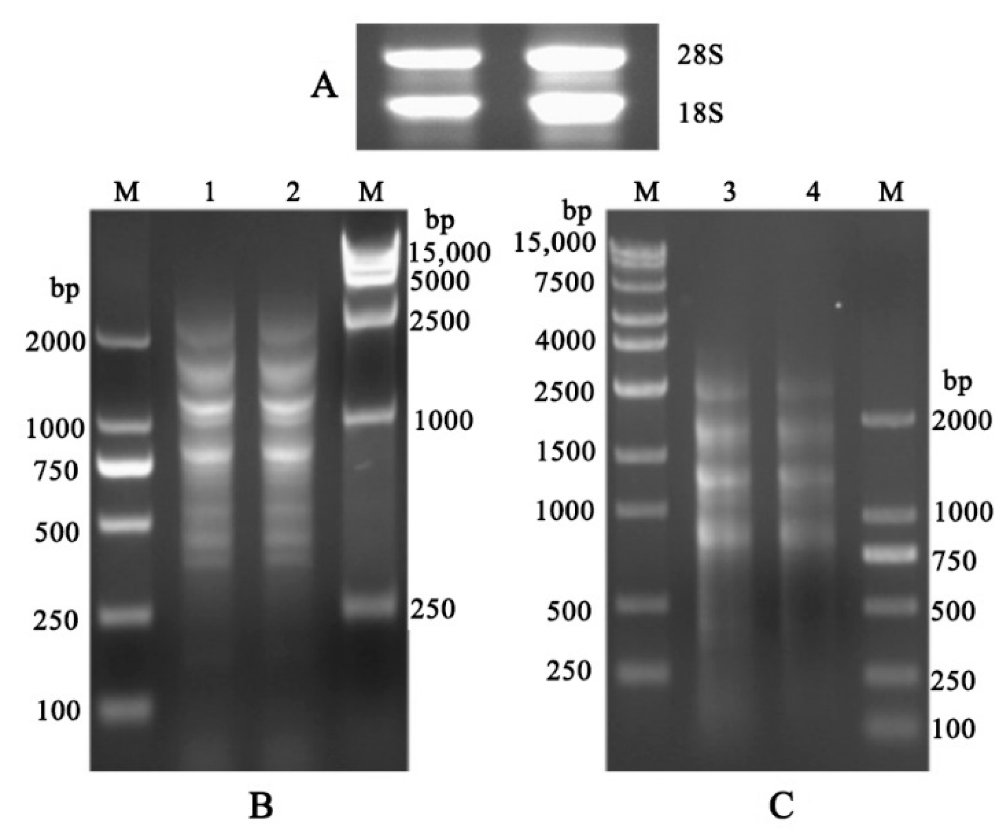
Figure 1. Construction of cDNA library. (A) extraction of RNA. (B) Synthesis of the cDNA library. (C) Purification of the cDNA library. Lane M, DNA 2000, 15,000 marker, bp base pairs. Lane 1 and 2, Synthesis of ds cDNA. Lane 3 and 4, purification of ds cDNA.
Forests 14 00911 g001
Figure 2. Analysis of the quality of the Y2H library. (A) The growth of independent clones. Three biological replicates were used. (B) Determination of the library titer.
Forests 14 00911 g002
Figure 3. Determination of the recombinant rate of the library. Lanes 1–24, the colonies selected were amplified with PCR with the pGADT7-F/R primers. Lane M, DNA 2000 marker, bp base pairs.
Forests 14 00911 g003
Figure 4. Classification of the novel putative 14-3-3 interactors. The 14-3-3 interactors were analyzed using BLAST on the NCBI website (www.ncbi.nlm.nih.gov, accessed on 21 March 2023).
Forests 14 00911 g004
Figure 5. The Y2H assay results for the interaction of HbGF14c with HbSRPP Yeast strains expressing the indicated constructs were grown on DDO medium (left panel) and QDO medium (right panel). The dilution is displayed at the bottom of the figure.
Forests 14 00911 g005
Figure 6. The BiFC assay results for the interaction between HbGF14c and HbSRPP. The expression of GFP was observed with an OLYMPUS FLUOVIEW FV1000 confocal laser scanning microscope. The negative controls were the GV3101 strain containing GN-HbGF14c and GC, GN and GC-HbSRPP, GN-HbSRPP and GC, and GN and GC-HbGF14c.
Forests 14 00911 g006
Figure 7. In vitro Pull-down assay results for the interactions of HbGF14c with HbSRPP. Immunoblot assays including anti-GST and anti-His antibodies were performed to detect interactions.
Forests 14 00911 g007
Figure 8. Identification of the typical motif in HbSRPP for HbGF14c–HbSRPP interactions. (A) Schematic diagram of the HbSRPP mutant construct. (B) Y2H analysis of the interactions of HbSRPP (WT) or its mutants (S185A) with HbGF14c. The positive controls were yeast co-transformants with pGBKT753 plus pGADT7-T, and the negative controls were pGBKT7-lam plus pGADT7-T.
Forests 14 00911 g008
Table 1. Identified cDNA clones from the HbGF14s yeast two hybrid screen.
IDBaitHomologous ProteinsFunctionScoreE-ValueHomology
1HbGF14aHEV2.1NRB1406.00 × 10−53Id. 97%; Po. 97%
2HbGF14aHEV2.2NRB1406.00 × 10−53Id. 97%; Po. 97%
3HbGF14bHEV2.2NRB4073.00 × 10−162Id. 95%; Po. 99%
4HbGF14cRubber elongation factor(REF)NRB5084.00 × 10−141Id. 99%; Po. 99%
5HbGF14cSmall rubber particle protein (SRPP)NRB6748.00 × 10−69Id. 94%; Po. 94%
6HbGF14eglutaredoxinNRB2176.00 × 10−54Id. 96%; Po. 98%
7HbGF14frubber elongation factor NRB4693.00 × 10−129Id. 99%; Po. 99%
8HbGF14cHevein (HEV1) precursorNRB, S7743.00 × 10−80Id. 97%; Po. 98%
9HbGF14cLatex abundant protein 1NRB, S3879.00 × 10−36Id. 99%; Po. 99%
10HbGF14aC3HC4-type RING zinc finger proteinS4442.00 × 10−121Id. 97%; Po. 97%
11HbGF14aelicitor-responsive protein S3771.00 × 10−101Id. 97%; Po. 98%
12HbGF14fMYC2S2823.00 × 10−130Id. 98%; Po. 99%
13HbGF14bDnaJ proteinD3793.00 × 10−102Id. 100%; Po. 100%
14HbGF14atranslation elongation factor 1-alphaPT5360Id. 97%; Po. 98%
15HbGF14atranslation elongation factor 1B gammaPT3293.00 × 10−87Id. 92%; Po. 96%
16HbGF14cChaperone protein dnaJPT3887.00 × 10−36Id. 77%; Po. 80%
17HbGF14g18S ribosomalT3788.00 × 10−102Id.100%; Po.100%
Note: Refer to the known laticifer process for classification as far as possible: NRB: Nature rubber biosynthesis, S: stress-related response, D: defense, PT: protein turnover, T: transcription. These cDNA coding homologous proteins were analyzed using BLAST on the NCBI website (www.ncbi.nlm.nih.gov, accessed on 21 March 2023).
Disclaimer/Publisher’s Note: The statements, opinions and data contained in all publications are solely those of the individual author(s) and contributor(s) and not of MDPI and/or the editor(s). MDPI and/or the editor(s) disclaim responsibility for any injury to people or property resulting from any ideas, methods, instructions or products referred to in the content.