Article

Comparison of Chemosensory Receptor Genes in the Antennae Transcriptomes of Sirex noctilio and Sirex nitobei (Hymenoptera: Siricidae)

1
The Key Laboratory for Silviculture and Conservation of the Ministry of Education, School of Forestry, Beijing Forestry University, Beijing 100083, China
2
Anhui Forestry Pest Control and Quarantine Bureau, Hefei 230001, China
3
Institute of Medicinal Plant Development, Chinese Academy of Medical Sciences and Peking Union Medical College, Beijing 100193, China
*
Authors to whom correspondence should be addressed.
These authors contributed equally to this work.
Forests 2022, 13(9), 1495; https://doi.org/10.3390/f13091495
Submission received: 10 August 2022 / Revised: 5 September 2022 / Accepted: 12 September 2022 / Published: 15 September 2022

Abstract

:
The woodwasp Sirex noctilio Fabricius is a worldwide quarantine pest for forestry that mainly harms conifers, especially Pinus species. Sirex nitobei Matsumura, a native species from China and closely related to S. noctilio. Olfaction and gustation play a vital role in the life movements of woodwasps, but the molecular mechanisms of chemoperception in these species remain unclear. We performed a comparative analysis of 41 odorant receptors (ORs), 13 ionotropic receptors (IRs), and 8 gustatory receptors (GRs) of S. noctilio and 43 ORs, 16 IRs, and 10 GRs of S. nitobei. Phylogenetic analysis showed that two species-specific OR subfamilies were identified in each species. In addition to conserved “antennal IRs”, “divergent IRs”, including 7 SnocIRs and 7 SnitIRs, were found. Moreover, a sugar receptor subfamily (SnocGR5 and SnitGR11), a carbon dioxide receptor subfamily (SnocGR2/GR3 and SnitGR3), and a fructose receptor subfamily (SnitGR9) emerged, but bitter receptors were not identified. The tissue-specific expression profiles showed 36 ORs were enriched in the antennae of S. noctilio. Among them, 19 ORs were female-biased, whereas 4 ORs (SnocOR6/15/18/30) were male-biased. In addition, 34 ORs were highly expressed in S. nitobei antennae, of which 22 ORs were female-biased, whereas SnitOR2/18/30 were male-biased. Seven IRs were enriched in the antennae of both species, of which SnocIR4, 6, 10, 11, and 12 were significantly male-biased, while SnitIR4, 6, 10, 11, and 12 were significantly female-biased. Three GRs were highly expressed in the antennae of both species. SnocGR2 and SnocGR6 were also highly expressed in the head and leg, respectively. In the present study, a total 62 and 69 chemosensory receptor genes were identified in the antennal transcriptomes of S. noctilio and S. nitobei, respectively. Although most receptor genes are homologous, there are also some specific receptor genes, suggesting similarities and differences in molecular mechanisms between the two closely related species. OR genes may be involved in different physiological functions by whether they are expressed in olfactory organs, or obvious gender bias. Our results provide a foundation for further investigating the molecular mechanisms of chemoreception in these two closely related woodwasp species, and establishes a starting point for further research on molecular mechanisms of the olfactory system in symphyta woodwasps.

1. Introduction

Sirex noctilio Fabricius (Hymenoptera: Siricidae), also known as the “European woodwasp” [1], is a major quarantine pest of forestry worldwide. S. noctilio is native to Eurasia and North Africa and primarily harms conifers, especially Pinus species [2,3]. In its native range, S. noctilio is not considered an important forest pest, but in countries of the southern hemisphere it is considered to be a major invasive pest, where it causes high mortality and significant economic losses in exotic pine plantations [4,5]. Due to trade and tourism, the rate of biological invasion by S. noctilio has greatly increased [6], and S. noctilio has been introduced into nine countries and attracted worldwide attention. It first invaded New Zealand in 1900, then Australia in 1952, South America in 1980, and South Africa in 1994 [7,8]. In 2004, S. noctilio was first trapped in North America [9]. It was first discovered in China in Daqing, Heilongjiang Province in 2013 and continued to spread to northeastern China and Inner Mongolia [10]. S. nitobei Matsumura is closely related to S. noctilio and is native to China, Japan, and Korea. It is widely distributed in China and can attack P. armandii Franch, P. tabuliformis Carrière, and Larix spp. In China, both species commonly attack P. sylvestris var. mongolica Litv [11].
S. noctilio and S. nitobei are both xylophagous insects. Female adults inject eggs, phytotoxic mucus, and a symbiotic white-rot fungus Amylostereum areolatum (Fr.) Boidininto into their host trees [12,13]. The mucus inhibits the transport of photosynthates and water, resulting in a weakened tree more susceptible to symbiotic fungi [12]. The mucus also reduces the moisture of the host to a level suitable for larvae and pupae survival [14]. Larvae feed on fungal hyphae and decomposed wood by fungi, while adults do not need be fed, and only live for about a week. The combined actions of the insects, mucus, and fungi ultimately lead to the death of the host tree [15,16,17]. Because the larvae live in the trunk and the adults live only a short time, traditional control methods are not effective. Parasites and parasitoids are currently used as a biological control for S. noctilio and S. nitobei infestation [18,19,20]. Trap trees are also used for control, but this method is destructive to trees and not economical [21]. In the United States, pheromone preparations are being explored as approaches for woodwasp control. These include a 70/30 blend of α/β pinene as an aggregation pheromone, and a 100:1:1 ratio of (Z)-3-decenol, (Z)-4-decenol, and (E, E)-2, 4-decadienal as a species-specific attractant [22,23]. S. nitobei, as a native species in China, was reported to attack only dead trees, and combined with their low abundance and little economic value, attracted no serious attention. To date, the molecular mechanisms underlying chemoperception in the two closely related, economically important species of woodwasps are still unknown.
Insects are able to respond quickly to their environment due to the sensitivity and selectivity of the peripheral olfactory and gustatory system for intra- or inter-species chemical communication as well as for communication between insects and plants. Molecular substances in the environment are detected via chemosensory organs to regulate behavior and improve survival. Therefore, chemoreception plays a critical role in insects by functioning in identification of food resources, mating, and oviposition sites, as well as predator avoidance [24]. Proteins in these systems include odorant binding proteins (OBPs), chemosensory proteins (CSPs), odorant receptors (ORs), ionotropic receptors (IRs), gustatory receptors (GRs), sensory neuron membrane proteins (SNMPs), and odorant-degrading enzymes (ODEs). Of these, the chemosensory receptors (ORs, IRs, and GRs) are membrane proteins [24,25,26]. ORs and IRs are mainly located on the dendrites of olfactory sensory neurons (OSNs) of sensilla basiconica in antennae. ORs are ligand-gated ion channels involved in odorant recognition and signal transduction, and possess seven transmembrane domains. In insect species, the C-terminus of ORs is extracellular, while the N-terminal is intracellular, a configuration opposite to that found in mammals [27,28,29]. There are two types of odorant receptors. The first is the conventional odorant receptor (ORx), which is highly variable and shows low homology between different insect species. According to their functions, ORx are further classified into general ORs and sex pheromone receptors (PRs); the former perceives non-pheromonal compounds in the environment with higher specificity, and PRs recognize sex pheromones that activate specific neural pathways that function in mate searching. Notably, PRs are conserved in Lepidoptera [30]. ORs can be activated by either a combination of OBP and an odorant molecule or solely by an odorant molecule; upon activation, the ion channel stimulates nerve impulses, converting chemical signals into electrical signals for transmission [26]. The identical ORs are expressed by OSNs and join into a single glomerulus of the antennal lobe and are then transported by another group of fibers to higher brain centers to guide insect behavior [31,32]. The other type of ORs is the olfactory receptor co-receptor OR83b, which was renamed ORco [33]. Compared with ORx, ORco is widely expressed and highly conserved among different insects. Furthermore, it cannot recognize odorant molecules alone and functions in a heteromeric complex with ORs (ORx/ORco) [29]. In addition, ORco can facilitate correct localization of ORs on the dendritic membrane and enhance OR sensitivity to odorants [34,35,36]. Since the discovery of the first insect olfactory and taste gene in the Drosophila melanogaster Meigen genome, these genes have been found in many insects, such as Lepidoptera, Hymenoptera, and Coleoptera [37,38]. With the application of genomic and transcriptomic sequencing methods, ORs have been identified in many Hymenopterans, including 174 in Apis mellifera Linnaeus, 72 in Cephus cinctus Norton, 76 in Meteorus pulchricornis Wesmael, 54 in Microplitis mediator Haliday, and 301 in Nasonia vitripennis Walker. AmOR11 was identified as a receptor of the queen substance 9-oxo-2-decenoic acid (9-ODA) in honey bees [39].
IRs are a subfamily of ionotropic glutamate receptors (iGluRs) and are more conserved and ancient than ORs. They are found in nematodes, mollusks, annelids, and arthropods. IRs perceive amines and acids, while ORs mainly sense alcohols, ketones, and esters [40,41]. IRs are located on the dendritic membrane of OSNs in ceoloconic sensilla and are divided into two subfamilies, “antennal IRs” and “divergent IRs”. The former is specifically expressed in antennae and are relatively more conserved than “divergent IRs” [42,43]. Notably, IR8a and IR25a are co-receptors that have functions similar to ORco and are co-expressed with other IRs [44,45]. The latter are species-specific, exhibit low amino acid sequence similarity, and have no obvious orthologous relationship among insect species. Interestingly, in D. melanogaster, IR76b is a Na+ channel and salt receptor and widely expressed in larval gustatory receptor neurons (GRNs), which are involved in the taste perception of amino acids. This finding suggests that IRs not only participate in the olfactory function, but also taste [46].
Insect GRs play important roles in host identification and feeding preferences. All GRs have three conserved transmembrane domains (TM6, ICL3, and TM7). GRs are primarily expressed in GRNs and are also found in OSNs [47,48]. They can recognize soluble compounds or pheromones, such as sugars, salts, amino acids, and bitter substances [49]. Furthermore, some GRs expressed in the antennae are also involved in the recognition of carbon dioxide [50]. Sugar receptors, such as DmelGR5a, DmelGR61a, and DmelGR64f in D. melanogaster, stimulate feeding [51,52]. Bitter receptors, such as DmelGR66a, are involved in identifying toxic secondary metabolites produced by plants [53,54]. Carbon dioxide receptors, such as DmelGR21a and DmelGR63a, are used to detect healthy trees as well as find food and oviposition sites [55]. A gustatory sensillum contains multiple GRNs and a mechanosensory neuron (MSN), implying that GRs can also participate in mechanoreception [56,57].
From the perspective of ecological control strategy, olfactory and gustatory proteins were recently identified as new targets for integrated pest control, as they represent a more environmentally friendly approach than other methods [58]. In this study, the transcriptomes of S. noctilio and S. nitobei were analyzed and the expression patterns of S. noctilio 62 and S. nitobei 69 chemosensory genes in different tissues and both females and males were obtained by using reverse transcription PCR (RT-PCR) and quantitative real-time PCR (RT-qPCR). We performed a phylogenetic analysis of Hymenopteran chemosensory receptors and analyzed their abundance of expression. In order to explore the differences between invasive and native species at the olfactory level, we conducted a thorough, comparative study of chemosensory receptor genes between S. noctilio and S. nitobei to provide a foundation with which to study the molecular mechanisms of olfaction in these two closely related species. Our results will facilitate further exploration of receptor function in woodwasps, as well as new ecological approaches for pest control.

2. Materials and Methods

2.1. Insects

S. noctilio and S. nitobei were collected from Duerbert Mongolian Autonomous County, Heilongjiang Province and Yushu, Jilin Province, China. In April–May 2018, the woodwasp-damaged P. sylvestris var. mongolica were cut down, divided into 1-m sections, and sent to the Phytosanitary Laboratory of Beijing Forestry University. The wood sections were sealed at both ends with paraffin and placed in net cages (3 × 3 × 3 m) at 24 °C and 65% relative humidity for eclosion. Upon emergence, the male and females were placed in separate plastic boxes marked with their species, emergence date, and sex. We used virgin adults for all experiments. Tissues were excised and put into Trizol reagent or frozen quickly in liquid nitrogen and then stored at −80 °C until RNA extraction.

2.2. RNA Extraction and cDNA Synthesis

20 antennas, 10 heads (without antennae), 15 pairs of legs (including forefoot, middle foot, and hind foot) and 10 external genitals were dissected from both female and male S. noctilio and S. nitobei and stored in Trizol reagent (Invitrogen, Carlsbad, CA, USA). Total RNA from female antennae (FA), male antennae (MA), female heads (FH), male heads (MH), female legs (FL), male legs (ML), female external genitals (FG), and male external genitals (MG) was extracted using the RNeasy Plus Mini Kit (Qiagen, Hilden, Germany) according to the manufacturer’s instructions. The quality and integrity of RNA were determined using a NanoDrop 8000 (Thermo, Waltham, MA, USA) and 1% agarose gel electrophoresis, respectively. High-quality RNA (1 µg total RNA from each sample) was used to synthesize first-strand cDNA using the PrimeScriptTM RT Reagent Kit with gDNA Eraser Kit (TaKaRa, Shiga, Japan). The first reaction contained 2 µL of 5 × gDNA Eraser Buffer and 1 µL of gDNA Eraser to remove genomic DNA at 42 °C for 2 min. The second reaction contained 10 µL of the reaction solution obtained above, 4 µL of RNase-free water, 4 µL of 5×PrimeScript Buffer 2, 1 µL of RT Primer Mix, and 1 µL of PrimeScript RT Enzyme Mix I. The reactions were incubated at 37 °C for 15 min, 85 °C for 5 s, and then held at 4 °C. All operations were performed on ice, and the resulting cDNA was stored at −20 °C.

2.3. Sequence and Phylogenetic Analysis of Chemosensory Receptor Genes

The amino acid sequence identities of 131 chemosensory receptors of S. noctilio and S. nitobei from our previous transcriptome analyses [58], were analyzed by GeneDoc software. All amino acid sequences of candidate chemosensory receptors from S. noctilio, S. nitobei and other Hymenopteran species underwent multiple sequence alignment using ClustalX 2.0 software (Dublin, Ireland) [59]. A total of 262 Hymenopteran OR protein sequences were used to construct phylogenetic trees. These included 33 ORs from S. noctilio, 35 ORs from S. nitobei, 72 ORs from C. cinctus, 73 ORs from M. pulchricornis, 18 ORs from A. mellifera, and 31 ORs from N. vitripennis (Supplementary File S4, Table S8). An IR phylogenetic tree was constructed based on 135 protein sequences from S. noctilio (11), S. nitobei (11), and 5 other Hymenopteran species, M. pulchricornis (11), M. mediator (6), N. vitripennis (61), A. mellifera (9), and C. cinctus (26) (Supplementary File S4, Table S8). In addition, 116 GR amino acid sequences from Hymenopteran species were used to construct phylogenetic trees. These included sequences from S. noctilio (4), S. nitobei (4), C. cinctus (37), A. mellifera (12), N. vitripennis (57), and M. mediator (2) (Supplementary File S4, Table S8). The phylogenetic trees were constructed by MEGA 7.0, and the settings used were the neighbor-joining method with default parameters and 1000 bootstrap replicates [60,61].

2.4. Tissue-and Sex-Specific Expression Profiles of Chemosensory Receptor Genes

In order to compare the expression levels of chemosensory receptor genes, we used FPKM (Fragments Per Kilobase of exon model per Million mapped reads) values to estimate gene expression in our previously published antennal transcriptomes [62]. Heatmaps of chemosensory receptor gene expression in S. noctilio and S. nitobei were made using R version 3.4.2 (R Development Core Team, The R Foundation for Statistical Computing, Vienna, Austria) [63]. We identified 41 and 43 ORs, 13 and 16 IRs, 8 and 10 GRs in S. noctilio and S. nitobei, respectively, and collected four tissues (antennae, legs, external genitals, and heads without antennae) from S. noctilio and S. nitobei, and performed RT-PCR and RT-qPCR to measure the chemosensory receptor expression profiles in both sexes and different tissues. Apart from the antennae samples, the cDNAs of other tissues were mixed (female: male = 1:1) prior to analysis the OR expression profiles. β-Tubulin was used as a reference gene, and each experiment was run in triplicate [58]. The reactions consisted of 12.5 µL of 2× Master Mix (RuibioBioTechCo, Beijing, China), 10.5 µL of ddH2O, 1 µL of template, and 0.5 µL of both forward and reverse primer (10 µM). PCR reactions were cycled on Bio-Rad T100 Thermal Cycler PCR machine (Bio-Rad, Hercules, CA, USA), which used the following parameters: 94 °C for 3 min, 34 cycles at 94 °C for 30 s, 55 °C for 30 s, 72 °C for 30 s, and 72 °C for 5 min. PCR products were verified by 1% agarose gel electrophoresis and viewed with a DOC gel imaging system.
The expression profiles of all IRs and GRs in both female and male tissues were determined using RT-qPCR on a Bio-Rad CFX Connect PCR system (Bio-Rad, Hercules, CA, USA). The qPCR reactions consisted of 12.5 µL of SYBR Premix Ex Taq II (TaKaRa, Shiga, Japan), 1 µL of both forward and reverse primer, 2 µL of sample cDNA, and 8.5 µL of sterilized H2O. The qPCR reactions were cycled using the following parameters: 95 °C for 30 s followed by 40 cycles at 95 °C for 5 s and 60 °C for 30 s. Fluorescence signal intensity was measured from 60 °C to 95 °C, and the resulting melt curve was used to check the specificity of primers and for the existence of primer dimers. All experimental operations were performed on ice, and gene-specific primers were designed by Primer-BLAST (http://www.ncbi.nlm.nih.gov/tools/primer-blast/, accessed on 20 December 2018) (Supplementary File S5, Tables S9 and S10). β-Tubulin was used as reference gene [58]. Each experiment was run in triplicate (technical replicates) and included three biological replicates. The relative expression levels of chemosensory receptor genes were relative to male external genitals (MG) and calculated using the 2−∆∆Ct method. The GraphPad Prism 7.0 (GraphPad Software, La Jolla, CA, USA) was used for analysis and plotting. [64,65]. Differences in the relative expression levels of chemosensory receptor genes were analyzed by SPSS 21.0 (SPSS Inc., Chicago, IL, USA) by a one-way nested analysis of variance (ANOVA) followed by Duncan’s new multiple range test (α = 0.05).

3. Results

3.1. Analysis of Gene Expression Abundance from Antennal Transcriptome

The gene expression abundance in the S. noctilio and S. nitobei antennae were 41 and 43 ORs, 13 and 16 IRs, 8 and 10 GRs, respectively [58], and were evaluated using FPKM values (Figure 1). As chemosensory receptors are membrane proteins, their expression levels are relatively low. We found that the FPKM values of most receptors were less than 10, which is considered a weak expression. Of the ORs, 41.7% of the genes had an FPKM value greater than 10, while for IRs and GRs, only 17.2% and 16.7% of the receptors had FPKM values greater than 10, respectively.
The three biological replicates (FA and MA) clustered together on the heatmap dendrogram, indicating that the sequencing results were reproducible. Cluster analysis of 41 S. noctilio OR genes revealed that ORs can be divided into three groups. The first group consists of 16 ORs (Cluster 1), and displayed no significant differences in expression between male and female antennae. The second group include the 20 OR genes (Cluster 2) that display similar expression patterns and are highly expressed in female antennae. In contrast, 5 ORs (Cluster 3) were relatively highly expressed in male antennae (Figure 1A).
Similarly, 43 OR genes were classified into three groups in S. nitobei (Figure 1B). Clusters 2 and 3 contained 7 and 26 ORs that were relatively highly expressed in male and female antennae, respectively. The remaining 10 ORs (Cluster 1) displayed no significant differences in expression between male and female antennae.
IR and GR genes also clustered into three groups in both S. noctilio and S. nitobei. Of these, the three biological replicates of IRs were not clustered together, so the data are not available for further analysis. 4 IRs (Cluster 3) were relatively highly expressed in S. nitobei female antennae, while 10 (Cluster 1) were highly expressed male antennae (Supplementary File S1, Figure S1).
In both species, three GRs were highly expressed in female antennae (Cluster 1), while 3 S. noctilio GRs and 5 S. nitobei GRs (Cluster 2 and 3, respectively) displayed higher expression in male antennae (Supplementary File S1, Figure S2). Generally, most chemosensory receptor genes displayed relatively high mRNA abundance in female antennae. OR genes exhibited higher expression than IRs and GRs.

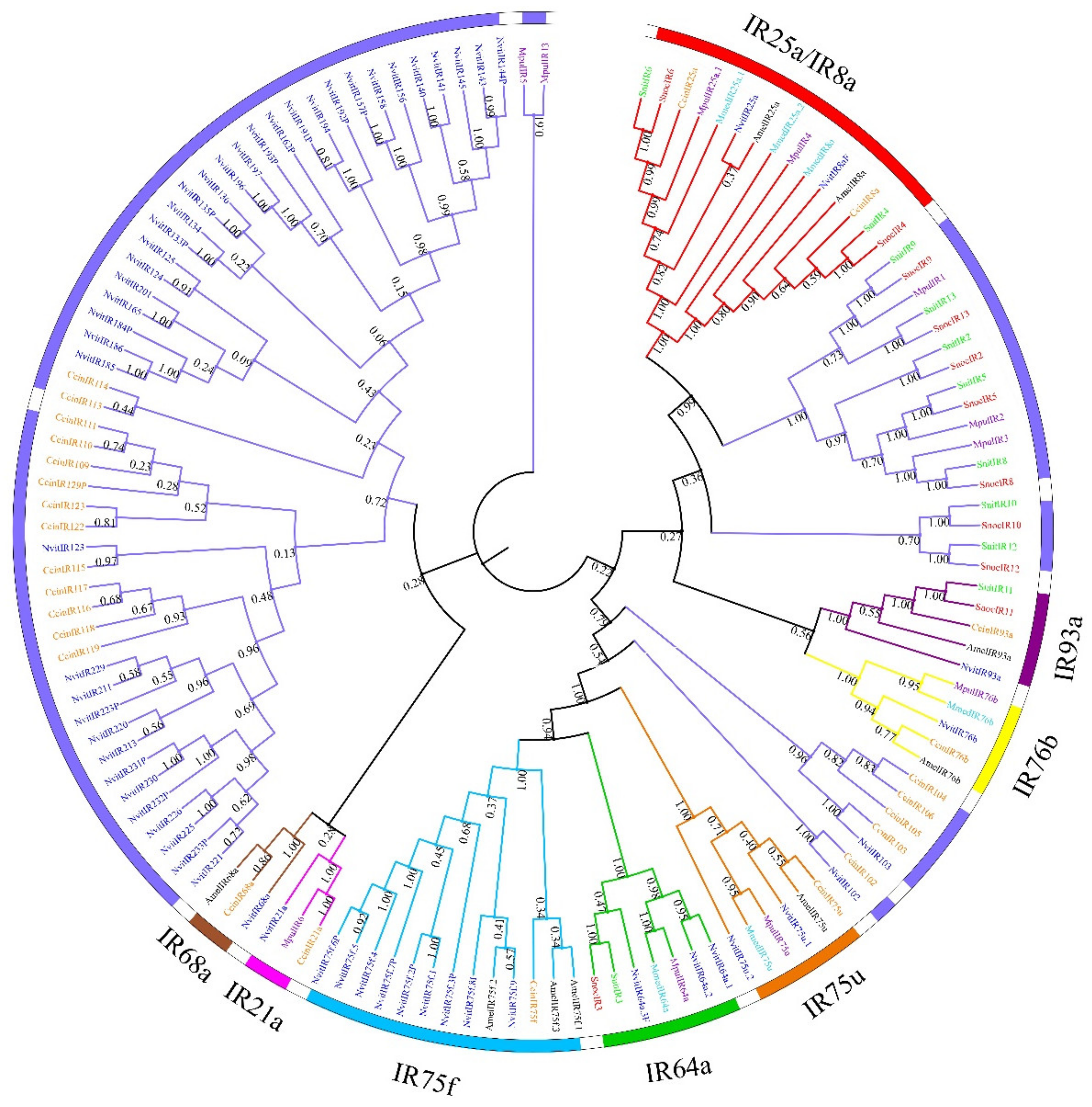
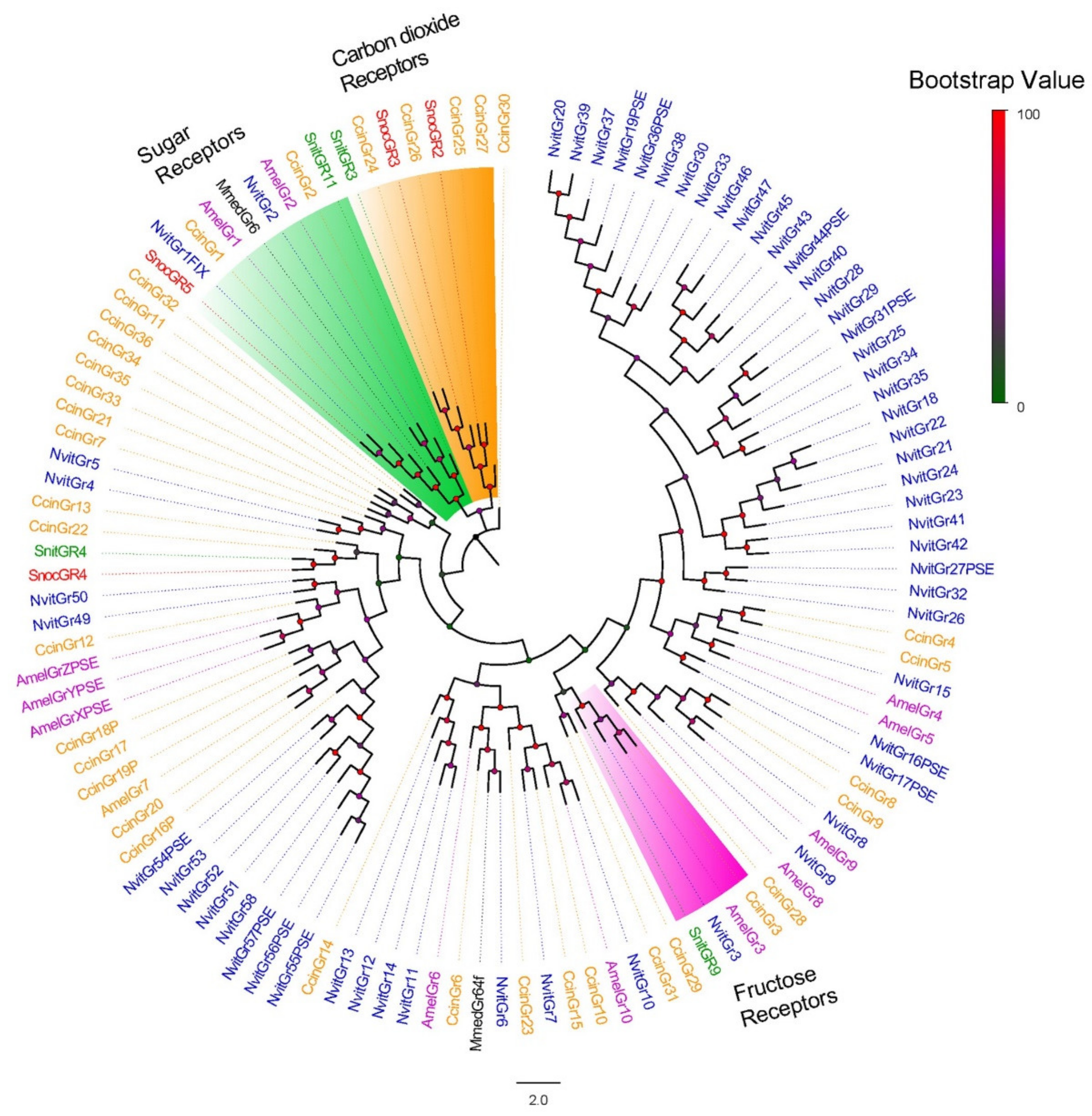
3.2. Phylogenetic Analysis of Chemosensory Receptors of S. noctilio and S. nitobei among Hymenoptera

A total of 262 OR protein sequences from S. noctilio, S. nitobei and four other Hymenopteran species (N. vitripennis, A. mellifera, C. cinctus, and M. pulchricornis) were used to construct a phylogenetic tree (Figure 2). SnocORco and SnitORco were clustered together with ORco from other species, consistent with the finding that ORco represent a highly conserved co-receptor gene subfamily. Most of the SnocORs were orthologous to SnitORs and clustered together with high bootstrap values. Our phylogenetic analysis showed two species-specific branches in S. noctilio and S. nitobei that exhibited low homology with other Hymenopteran ORs. The first branch included 6 SnocORs (SnocOR20, 12, 9, 22a, 17, and 23) and 7 SnitORs (SnitOR20a/b, 12, 9, 22a, 17, and 1b), and the second branch included 7 SnocORs (SnocOR21, 24, 25, 26, 28a, 28b, and 27) and 7 SnitORs (SnitOR21a, 21b, 24, 25, 28a, 28b, and 27). The remaining SnocORs and SnitORs were dispersed amongst other branches and clustered with ORs from N. vitripennis, A. mellifera, C. cinctus, and M. pulchricornis. However, no ORs clustered with AmelOR11, which encodes a pheromone receptor (PR) in A. mellifera.
Our analysis also revealed >90% amino acid similarity for 28 pairs of orthologous ORs between S. noctilio and S. nitobei. ORco, OR35, OR19, and OR15 displayed similarities as high as 99%, likely because they are closely related proteins specific to Sirex spp. However, the homology between ORs in the same species was relatively low (between 0%–86% and 0%–89% in S. noctilio and S. nitobei, respectively), except for two ORs expressed on the same transcript. Among ORs, only 5.2% and 5.1% displayed intra-species amino acid similarities greater than 40% (Supplementary File S2, Tables S1, S2 and S7).
The phylogenetic tree of IRs was constructed using 11 SnocIRs, 11 SnitIRs, and IRs from 5 other Hymenopteran species (M. pulchricornis, M. mediator, N. vitripennis, A. mellifera, and C. cinctus) (Figure 3). IR93a, 64a, 21a, 75u, 75f, 76b, and 68a are “antennal IRs”, a group of conserved IRs specifically expressed in antennae [43,44]. SnocIR11 and SnitIR11 were part of the IR93a subgroup, and SnocIR3 and SnitIR3 clustered within the IR64a subgroup. These IRs may be “antennal IRs”, specifically expressed in antennae and related to olfactory sensation. However, the 21a, 75u, 75f, 76b, and 68a subgroups were absent from S. noctilio and S. nitobei. We also identified a species-specific branch of “divergent IRs,” which included 7 SnocIRs (SnocIR9, 13, 2, 5, 8, 10, and 12) and 7 SnitIRs (SnitIR9, 13, 2, 5, 8, 10, and 12). These IRs exhibit low amino acid sequence identity with other Hymenopteran species. In addition, 2 SnocIRs (SnocIR6 and SnocIR4) and 2 SnitIRs (SnitIR6 and SnitIR4) were classified into the IR25a and IR8a co-receptor subfamily groups [44].
Amino acid sequences of GRs from S. noctilio and S. nitobei and 4 additional Hymenopteran species were used to construct a GR phylogenetic tree (Figure 4). SnocGR5 and SnitGR11 clustered with sugar receptors from other Hymenopteran insects, such as NvitGR1/GR2, AmelGR1/GR2, and CcinGR1/GR2; thus, we speculate that SnocGR5 and SnitGR11 likely encode sugar receptors. SnocGR2/3 and SnitGR3 displayed high homology with known carbon dioxide receptors, suggesting that they may be involved in CO2 detection. SnitGR9 clustered with other known fructose receptors, such as NvitGR3, AmelGR3, and CcinGR3. The amino acid similarities of three pairs of orthologous GRs were all greater than 90%, but overall, the similarity between GRs in the same species is less than that of ORs and IRs. We observed intra-species amino acid similarities between 0%–27% for GRs (Supplementary File S2, Tables S5–S7).

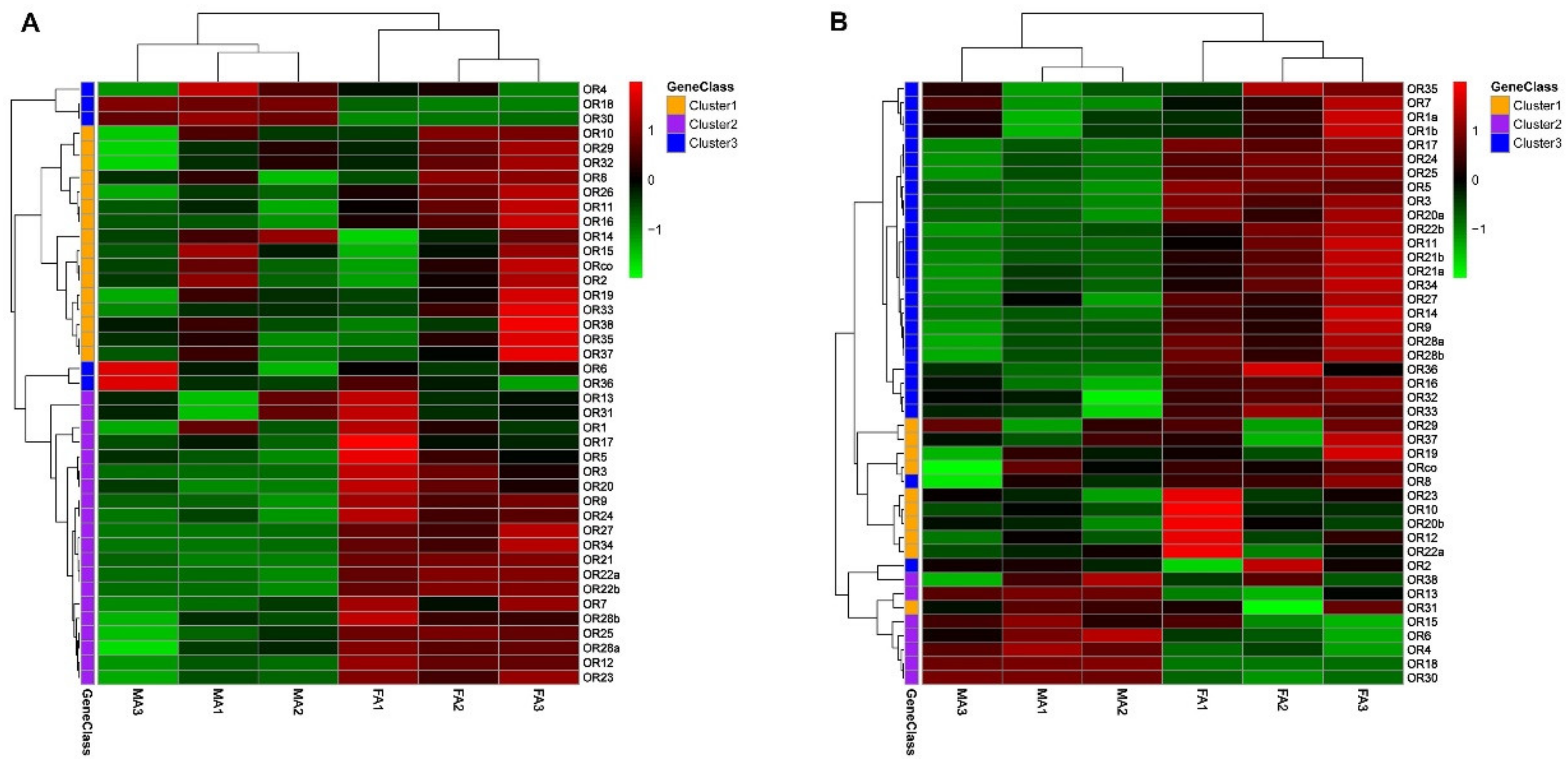
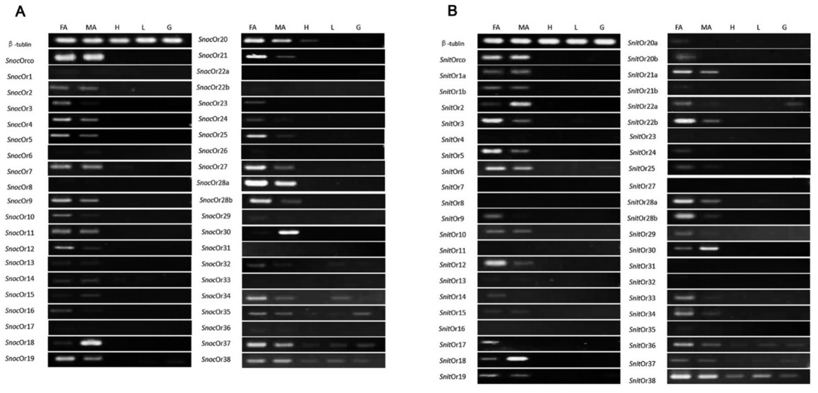
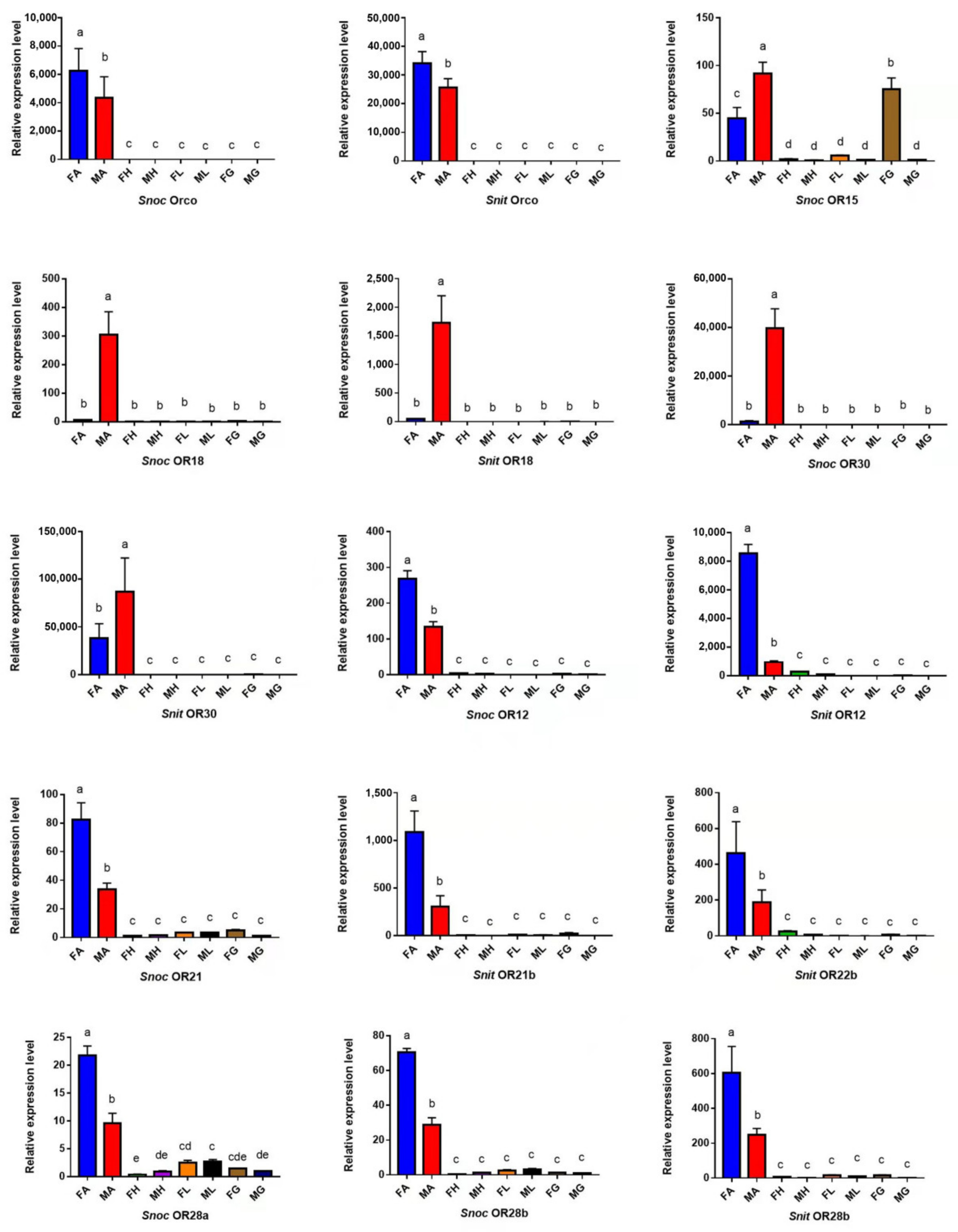
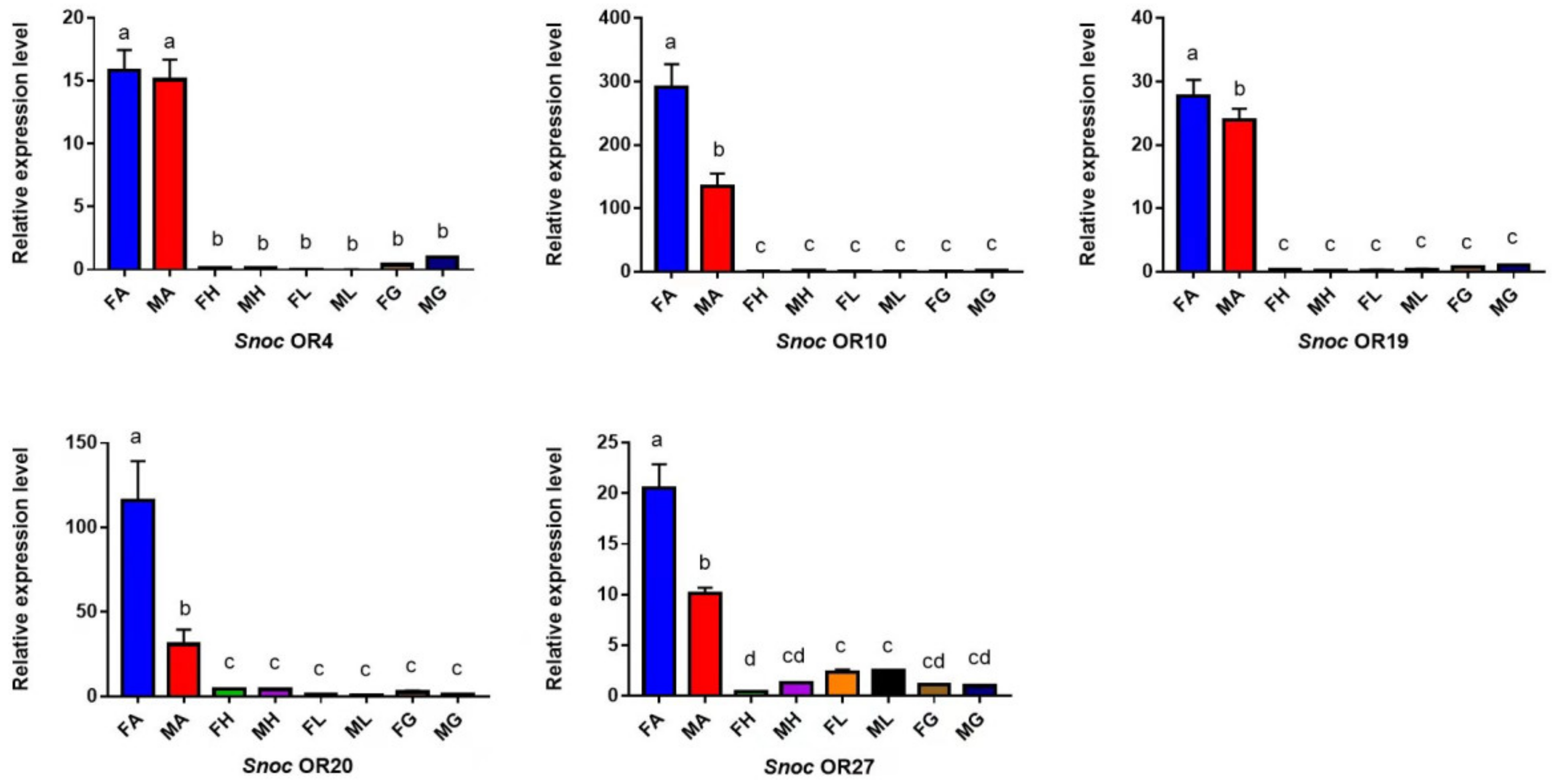
3.3. The Specific Expression Profiles of S. noctilio and S. nitobei OR Genes

The results showed that 41 and 43 OR genes in S. noctilio and S. nitobei were examined in different tissues (antennae, leg, external genitals, and head without antennae) using RT-qPCR with gene-specific primers (Figure 5, Supplementary File S3). In S. noctilio, we detected 36 ORs (SnocORco, SnocOR2-7, SnocOR9-21, SnocOR23-30, SnocOR32, and SnocOR34-38) that were highly expressed in antennae compared with others. Besides, 19 OR genes (SnocOR3-5, SnocOR9-10, SnocOR12, SnocOR16, SnocOR19- 21, SnocOR23-25, SnocOR27-28b, SnocOR32, SnocOR34, and SnocOR37) were found to be significantly female-biased, while 4 OR genes (SnocOR6, SnocOR15, SnocOR18, and SnocOR30) were found to be significantly male-biased. The remaining 13 OR genes (SnocORco, SnocOR2, SnocOR7, SnocOR11, SnocOR13-14, SnocOR17, SnocOR22b, SnocOR26, SnocOR29, SnocOR35-36, and SnocOR38) displayed no significant differences in expression between male and female antennae. SnocOR7 and SnocOR20 were also expressed in the head, SnocOR32 and SnocOR34 were also expressed in the leg, and SnocOR19 was also expressed in external genitals. SnocOR35, SnocOR37, and SnocOR38 were expressed in all tissues. In S. nitobei, 34 OR genes were highly expressed in antennae than other tissues. Of these, 23 OR genes (SnitOR3, SnitOR5, SnitOR6, SnitOR9, SnitOR12, SnitOR14, SnitOR17, SnitOR19, SnitOR20a and 20b, SnitOR21a and 21b, SnitOR22a and 22b, SnitOR24-25, SnitOR28a-29, and SnitOR33-36) were female-biased. SnitOR2, SnitOR18, and SnitOR30 were significantly male-biased. The remaining 9 OR genes (SnitORco, SnitOR1a and 1b, SnitOR6, SnitOR10, SnitOR13, SnitOR15, and SnitOR37-38) displayed similar expression levels in both male and female antennae. However, SnitOR6 and SnitOR22a were expressed not only in antennae but also in external genitals, and SnitOR36, SnitOR37, and SnitOR38 were expressed in all tissues. Together, our results suggest that OR genes are highly expressed in antennae, and approximately 50% and 10% of OR genes are female- and male-biased, respectively.
Based on the results of RT-qPCR, 20 OR genes were selected for qPCR. The two were consistent. SnocORco and SnitORco were highly expressed in male and female antennae, and females were higher than males. SnocOR15, SnocOR18, SnocOR30, SnitOR18 and SnitOR30 were significantly more highly expressed in male antennae. In addition, SnocOR15 was also highly expressed in female external genitalia, and the remaining 13 OR genes were female biased (Figure 6).

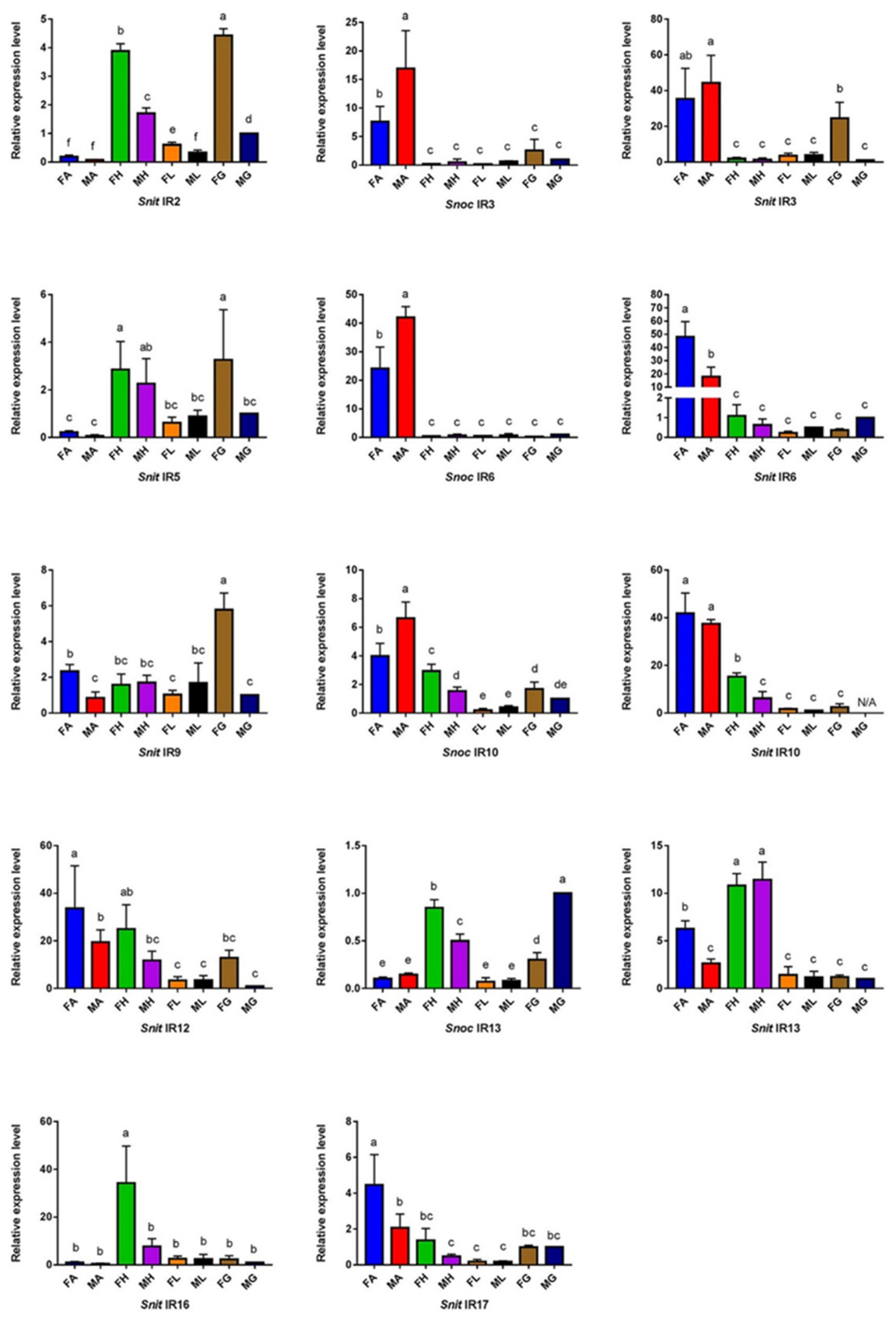
3.4. The Specific Expression Profiles of S. noctilio and S. nitobei IR Genes

We also determined the expression patterns of 13 and 16 IR genes in different tissues (FA, MA, FH, MH, FL, ML, FG, and MG) in S. noctilio and S. nitobei, using qPCR with specific primers (Figure 7). We identified 13 IR genes that displayed significantly higher expression in antennae (SnocIR3/SnitIR3, SnocIR4/SnitIR4, SnocIR6/SnitIR6, SnocIR10/SnitIR10, SnocIR11/SnitIR11, SnocIR12/SnitIR12, and SnitIR17). Most homologous genes between two species displayed similar expression patterns, but, surprisingly, a few homologous genes displayed opposite expression profiles in the antennae. For example, SnocIR4, 6, 10, 11, and 12 were significantly male-biased, while SnitIR4, 6, 10, 11, and 12 were significantly female-biased. In addition, the relative expression levels of nine IR genes (SnocIR1/SnitIR1, SnocIR2/SnitIR2, SnocIR5/SnitIR5, SnocIR8/SnitIR8, and SnocIR7) were found to be significantly higher in the head and female external genitals than other tissues. Four IRs (SnitIR13, 14, 15, and 16) were highly expressed in the head, SnocIR13 was highly expressed in the male external genitals and female head. Only SnocIR9 and SnitIR9 were specifically expressed in the female external genitals.

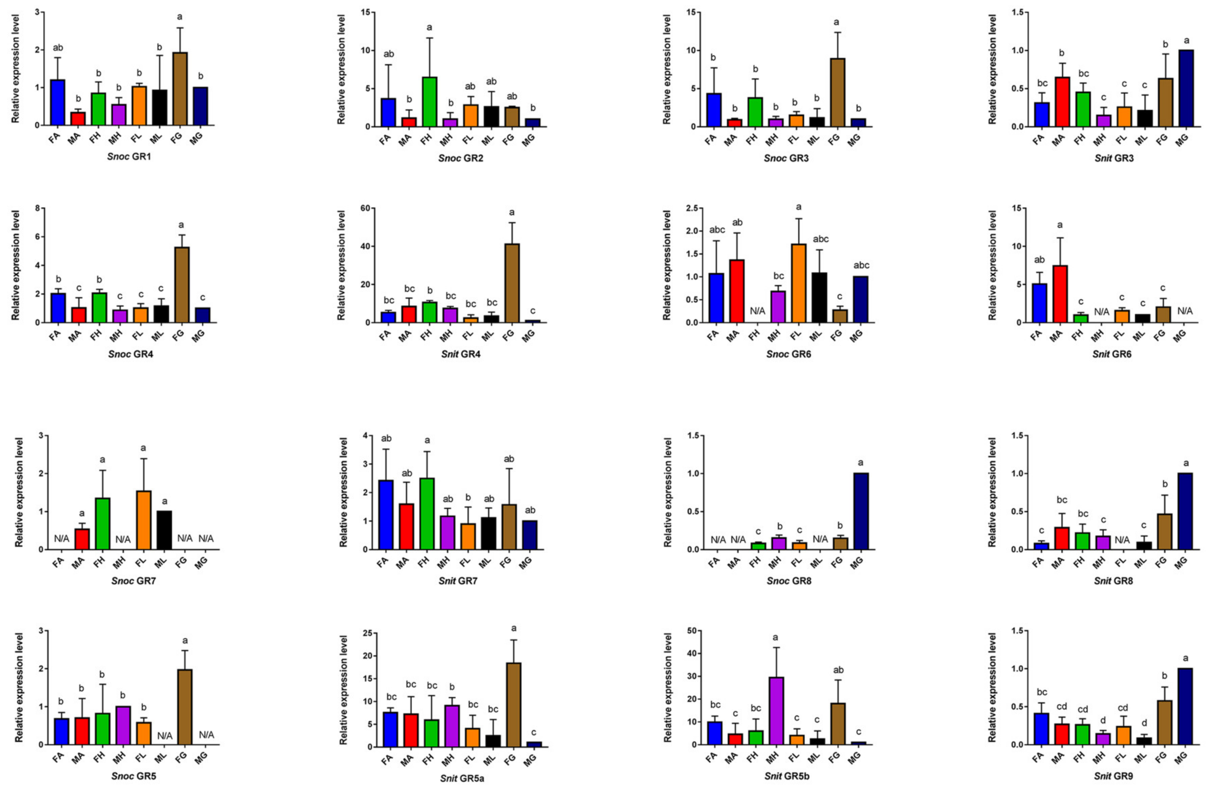
3.5. The Specific Expression Profiles of S. noctilio and S. nitobei GR Genes

We identified a total of 18 GR genes, including 8 and 10 GRs in S. noctilio and S. nitobei, respectively. Twelve of the 18 GR genes were found to be significantly more highly expressed in the external genitals (Figure 8). Eight GR genes (SnocGR1, SnocGR3, SnocGR5, SnitGR5a and 5b, SnitGR10, SnocGR4, and SnitGR4) displayed higher expression in the female external genitals than other tissues, while the remaining 4 GRs (SnitGR3, SnitGR9, SnocGR8 and SnitGR8) were highly expressed in the male external genitals. Expression of 6 GR genes (SnocGR1, SnocGR2, SnocGR6 and SnitGR6, SnitGR10, and SnitGR11) was higher in the antennae, and SnocGR2 and SnitGR5b were highly expressed in the head. Only SnocGR6 was mainly expressed in the leg, and the expression of SnitGR7 exhibited no significant tissue-specific expression. In general, the S. noctilio and S. nitobei more than 50% of GR genes were relatively highly expressed in the external genitalia, while one-third were highly expressed in the antennae. A few genes were highly expressed in the head or leg.

4. Discussion

Compared with Symphyta species, studies on chemosensation have focused on the Apocrita, including A. mellifera, Campoletis chlorideae, N. vitripennis, M. pulchricornis, M. mediator, Macrocentrus cingulum Brischke [66,67,68,69,70,71]. In the Hymenoptera, there were some species with more ORs than siricids (41 and 43 in S. noctilio S. nitobei, respectively), such as 60 in M. mediator [69], 72 in C. cinctus [71], 80 in Chouioia cunea Yang [72], 99 in M. pulchricornis [66], 301 in N. vitripennis [73], 174 in A. mellifera [49], 211 in C. chlorideae [70] and 109 in M. cingulum [74].
S. noctilio and S. nitobei are both xylophagous insects and live in pure or mixed coniferous forests, which is a relatively more simple chemical environment and biological habitat than those of social insects and parasitoids [49]. Similarly, in Coleoptera, there are some species with less ORs than the siricids, such as 2 in Agrilus planipennis Fairmaire, 9 in Monochamus alternatus Hope and 37 in Anoplophora glabripennis Motschulsky [75,76,77] (Figure 9), all of which belong to boring pests. This could also be the cause of why the number of ORs in the siricids is low. In addition, sequencing depth and sampling status also contributed to the differences in OR number [35,69]. The phylogenetic tree result showed that both SnocORco and SnitORco were clustered to the honey bee and other Hymenoptera ORco [73,78], which indicates that the ORcos of the wasp could act together with ORs as in other insects.
In the current study, 36 S. noctilio ORs and 34 S. nitobei ORs (70 of the 84 ORs) were highly expressed in the antennae, consistent with previous studies on the functions of insect ORs in insect olfaction [79]. Some ORs were expressed not only in the antennae but also in non-olfactory organs [72]. For example, SnocOR7 and SnocOR20 were expressed in the head, SnocOR19, SnitOR6, and SnitOR22a were expressed in the external genitalia, and SnocOR19 and SnocOR34 were expressed in the foot. Six ORs were found to be expressed in all tissues, indicating these six ORs may participate in the non-olfactory organs, a hypothesis that will require further validation [69].
Among the antennae-enriched ORs, 19 S. noctilio ORs and 22 S. nitobei ORs were mainly expressed in females, suggesting that these receptor genes may be involved in female behaviors. Furthermore, four S. noctilio ORs and 3 S. nitobei ORs were mainly expressed in males, possibly related to the identify of pheromones. Cooperband et al. (2012) [23] reported that an aggregated pheromone of S. noctilio is released by males and attracts both males and females. However, most OR genes are female-biased except for OR18 and OR30, which may be involved in the detection of pheromones and are highly expressed in the male antennae of the wood wasp, which should be further investigated. ORs are membrane proteins with relatively low expression levels compared with OBPs and CSPs; there are only 15 ORs in S. noctilio and 20 ORs in S. nitobei whose FPKM values are greater than 10. Therefore, subtle changes in the samples may alter the expression pattern of ORs.
The genes of the two closely related woodwasps have a high sequence similarity, which is due to the recognition of the same host plant. In contrast, within the same species, chemosensory receptors exhibited a very low similarity. Specifically, GR genes displayed intra-species similarity of only 20%–27%. This may be due to rapid gene duplication and the differentiation of gene functions during evolution to adapt to environmental changes [80]. Interestingly, we identified two species-specific OR clades which may be related to the recognition and/or adaptation of species-specific host plant volatiles or interspecies communication [39]. More interestingly, some closely related OR genes, such as OR4/16/23/27/32, were highly expressed in female antennae in S. noctilio but were barely expressed in S. nitobei. Similarly, SnitOR17/22a/22b/33/36 displayed a female-biased expression but almost no expression in S. noctilio. The expression of OR6 was significantly female-biased in S. nitobei, but in S. noctilio, the gene was expressed only in male antennae. Similarly, OR2 was highly expressed in male antennae in S. nitobei but displayed no significant sex-biased expression in S. noctilio.
IRs are a relatively ancestral and conserved receptor family. Total 13 and 16 IR transcripts were identified in S. noctilio and S. nitobei, respectively. There are species with more IRs than the siricids, such as 66 in D. melanogaster [44,45], while there are also species with similar ORs to the siricids, such as 13 in M. cingulum [74] and 10 in C. cunea [72].
Two “antennae IRs” IR11 and IR3 were identified in the woodwasp species, which belong to the IR93a and IR64a branches, respectively, and are expressed in sacculus neurons [81] (Shanbhag et al., 1995). IR64a (together with IR8a) has an olfactory function and detects acid substances, but the function of IR93a (and IR25a) is still unknown [43,82]. In addition to identifying 4 “antennal IRs”, the “divergent IRs” branch includes seven SnitIRs and seven SnocIRs. This finding is consistent with the seven “divergent IRs” found in M. mediator [83]. These “divergent IRs” may exhibit gustatory functions or participate in other physiological activities [84].
The four “antennal IRs” were significantly highly expressed in antennae, consistent with our phylogenetic analysis. Among the “divergent IRs”, IR10 and IR12 are highly expressed in the antennae in both species, while others are highly expressed in non-olfactory tissues. For example, SnitIR14 and 15 are highly expressed in the head, and SnocIR9 and SnitIR9 are highly expressed in the female external genitalia. This finding is similar to those from previous studies found that IRs not only participate in the olfactory recognition process but also have a gustatory function [85,86].
Two IRco genes were identified in M. mediator, whereas in the woodwasp species C. cunea and M. pulchricornis studies, only one IR25a homolog was found [69,72]. In our study, IR6 and IR4 are speculated as co-receptors of the woodwasps based on the two genes (SnocIR6 and SnitIR6) and another two genes (SnocIR4 and SnitIR4) were clustered into a highly conserved IR25a or IR8a subfamily, respectively.
A total of 18 GRs (8 in S. noctilio and 10 in S. nitobei) were identified in the study. The number of GRs in the honey bee is similar to those of the siricids, though there are species with more GRs than the siricids, such as 35 in C. cinctus, 58 in N. vitripennis (58), and 68 in D. melanogaster [48,71] (Figure 9).
Interestingly, 12 of 18 GRs were highly expressed in the external genitalia, and only a few GRs, such as SnocGR6, SnitGR6, and SnitGR11, were expressed in the antennae, head, and foot. This finding is inconsistent with the high expression reported in taste organs, such as the antennae and mouthparts. In honey bees, gustatory sensilla are mainly distributed in the antennae, mouthparts, and forefeet. In D. melanogaster, the labellum, pharynx, wing margins, legs, and ovipositor are important gustatory organs [56,87]. A possible reason for this difference is that woodwasp adults do not feed and only live for a week to complete mating and oviposition activities, so gustatory receptors are mainly expressed in the external genitalia for selecting the most suitable location for oviposition [57,88].
In S. noctilio and S. nitobei, GRs exhibit very low identity (0%–27%) as previously reported [89]. Despite this, we identified two sugar receptors, three carbon dioxide receptors, and one fructose receptor in the two closely related woodwasp species. Notably, we did not identify any bitter receptors, as there are in honey bees [87,90].

5. Conclusions

In summary, we described the tissue- and sex-specific expression of 62 S. noctilio and 69 S. nitobei chemosensory receptors. Combined with antennal transcriptome analysis, our analyses revealed that most receptor genes are prominently expressed in antennae. Of these, OR18 and OR30 were highly expressed in male antennae, suggesting that they may be associated with pheromone perception. Our data will provide a foundation for the further exploration of the functions of olfactory receptors, and the molecular mechanisms of chemoreception in Hymenoptera, especially for the molecular studies of chemoreception in the two closely related woodwasp species.

Supplementary Materials

The following supporting information can be downloaded at: https://www.mdpi.com/article/10.3390/f13091495/s1, Supplementary File S1 (Figure S1 Expression profiles of IR gens in antennae.; Figure S2 Expression profiles of GR gens in antennae.); Supplementary File S2 (Table S1 Amino acid sequence similarity of OR gens in S. noctilo.; Table S2 Amino acid sequence similarity of OR gens in S. nitobei.; Table S3 Amino acid sequence similarity of IR gens in S. noctilo.; Table S4 Amino acid sequence similarity of IR gens in S. nitobei.; Table S5 Amino acid sequence similarity of GR gens in S. noctilo.; Table S6 Amino acid sequence similarity of GR gens in S. nitobe.; Table S7 Comparison of homologous chemosensory gens in S. noctilo and S. nitobei.); Supplementary File S3 (Tissue-specific expression of candidate OR genes in S. noctilo and S.nitobei were evaluated by RT-PCR); Supplementary File S4 (Table S8 Gene names and GenBank accession numbers of other Hymenoptera ORs, IRs and GRs used in phylogenetic trees. Supplementary File S5 (Table S9 Primes used for RT-PCR.; Table S10 Primes used for quantitative real time-PCR.)

Author Contributions

W.W. carried out the majority of the bioinformatics studies and participated in performing the experiments; E.H. performed the RT-qPCR and wrote part of the manuscript; B.G., M.M. and H.Y. collected insects; J.Z. participated in the design of the study and revised the draft of the manuscript; P.L. and H.Q. proposed the project, designed the whole study, wrote and revised the manuscript. All authors have read and agreed to the published version of the manuscript.

Funding

This research was funded by the National Natural Science Foundation of China (Grant No. 31570643, 81774015), National Key R&D Program of China (2021YFD1400900). The funders had no role in the design of the study, data collection and analysis, decision to publish, or preparation of the manuscript.

Institutional Review Board Statement

Not applicable.

Informed Consent Statement

Not applicable.

Data Availability Statement

Not applicable.

Conflicts of Interest

The authors declare no conflict of interest.

References

  1. Ciesla, W.M. European Woodwasp: A potential threat to North America’s conifer forests. J. For. 2003, 101, 18–23. [Google Scholar]
  2. Spradbery, J.P.; Kirk, A.A. Aspects of the ecology of siricid woodwasps (Hymenoptera: Siricidae) in Europe, North Africa and Turkey with special reference to the biological control of Sirex noctilio F. in Australia. Bull. Entomol. Res. 1978, 68, 341–359. [Google Scholar] [CrossRef]
  3. Carnegie, A.J. Predicting the potential distribution of Sirex noctilio (Hymenoptera: Siricidae), a significant exotic pest of Pinus plantations. Ann. For. Sci. 2006, 63, 119–128. [Google Scholar] [CrossRef]
  4. Haugen, D.; Underdown, M. Release of parasitoids for Sirex noctilio control by transporting infested logs. Austral. For. 1990, 53, 266–270. [Google Scholar] [CrossRef]
  5. Slippers, B.; Hurley, B.P.; Wingfield, M.J. Sirex Woodwasp: A Model for Evolving Management Paradigms of Invasive Forest Pests. Annu. Rev. Èntomol. 2015, 60, 601–619. [Google Scholar] [CrossRef] [PubMed]
  6. Haack, R.A. Intercepted Scolytidae (Coleoptera) at U.S. ports of Entry: 1985–2000. Integr. Pest Manag. Rev. 2001, 6, 253–282. [Google Scholar] [CrossRef]
  7. Miller, D.; Clark, A.F. Sirex noctilio (Hym.) and its Parasite in New Zealand. Bull. Èntomol. Res. 1935, 26, 149–154. [Google Scholar] [CrossRef]
  8. Gilbert, J.M.; Miller, L.W. An Outbreak of Sirex noctilio F. In Tasmania. Aust. For. 1952, 16, 63–69. [Google Scholar] [CrossRef]
  9. Hoebeke, E.R.; Haugen, D.A.; Haack, R.A. Sirex noctilo: Discovery of a palearctic siricid woodwasp in New York (revised on 7 July 2005). Newsl. Mich. Entomol. Soc. 2005, 50, 24–25. [Google Scholar]
  10. Li, D.; Shi, J.; Lu, M.; Ren, L.; Zhen, C.; Luo, Y. Detection and Identification of the Invasive Sirex noctilio (Hymenoptera: Siricidae) Fungal Symbiont, Amylostereum areolatum (Russulales: Amylostereacea), in China and the Stimulating Effect of Insect Venom on Laccase Production by A. areolatum YQL03. J. Econ. Èntomol. 2015, 108, 1136–1147. [Google Scholar] [CrossRef]
  11. Wang, L.; Ren, L.; Li, C.; Gao, C.; Liu, X.; Wang, M.; Luo, Y. Effects of endophytic fungi diversity in different coniferous species on the colonization of Sirex noctilio (Hymenoptera: Siricidae). Sci. Rep. 2019, 9, 5077. [Google Scholar] [CrossRef]
  12. Coutts, M. The Mechanism of Pathogenicity of Sirex noctilio on Pinus Radiata II. Effects of S. Noctilio Mucus. Aust. J. Biol. Sci. 1969, 22, 1153–1162. [Google Scholar] [CrossRef]
  13. Taylor, K.L.; Kitching, R.L.; Jones, R.E. The Sirex woodwasp: Ecology and control of an introduced forest insect. In The Ecology of Pests: Some Australian Case Histories; CSIRO: Canberra, Australia, 1981; pp. 231–248. [Google Scholar]
  14. Talbot, P.H.B. The Sirex-Amylostereum-Pinus Association. Annu. Rev. Phytopathol. 1977, 15, 41–54. [Google Scholar] [CrossRef]
  15. Parkin, E.A. Symbiosis and Siricid Woodwasps. Ann. Appl. Biol. 1942, 29, 268–274. [Google Scholar] [CrossRef]
  16. Thompson, B.M.; Grebenok, R.J.; Behmer, S.T.; Gruner, D.S. Microbial Symbionts Shape the Sterol Profile of the Xylem-Feeding Woodwasp, Sirex noctilio. J. Chem. Ecol. 2013, 39, 129–139. [Google Scholar] [CrossRef]
  17. Madden, J.; Coutts, M. The role of fungi in the biology and ecology of woodwasps (Hymenoptera: Siricidae). In Insect-Fungus Symbiosis: Nutrition, Mutualism, and Commensalism; Allanheld Osmun: London, UK, 1979; pp. 165–174. [Google Scholar]
  18. Zondag, R. A nematode infection of Sirex noctilio (F.) in New Zealand. N. Z. J. For. Sci. 1969, 12, 732–747. [Google Scholar]
  19. Ryan, K.; De Groot, P.; Smith, S.M. Evidence of interaction between Sirex noctilio and other species inhabiting the bole of Pinus. Agric. For. Èntomol. 2012, 14, 187–195. [Google Scholar] [CrossRef]
  20. Hurley, B.P.; Slippers, B.; Wingfield, M.J. A comparison of control results for the alien invasive woodwasp, Sirex noctilio, in the southern hemisphere. Agric. For. Èntomol. 2007, 9, 159–171. [Google Scholar] [CrossRef]
  21. Zylstra, K.E.; Dodds, K.J.; Francese, J.A.; Mastro, V. Sirex noctilio in North America: The effect of stem-injection timing on the at-tractiveness and suitability of trap trees. Agric. For. Entomol. 2010, 12, 243–250. [Google Scholar]
  22. Bashford, R. The development of static trapping systems to monitor for wood-boring insects in forestry plantations. Aust. For. 2008, 71, 236–241. [Google Scholar] [CrossRef]
  23. Cooperband, M.F.; Böröczky, K.; Hartness, A.; Jones, T.H.; Zylstra, K.E.; Tumlinson, J.H.; Mastro, V.C. Male-Produced Pheromone in the European Woodwasp, Sirex noctilio. J. Chem. Ecol. 2012, 38, 52–62. [Google Scholar] [CrossRef] [PubMed]
  24. Leal, W.S. Odorant Reception in Insects: Roles of Receptors, Binding Proteins, and Degrading Enzymes. Annu. Rev. Èntomol. 2013, 58, 373–391. [Google Scholar] [CrossRef] [PubMed]
  25. Vogt, R.G.; Riddiford, L.M. Pheromone binding and inactivation by moth antennae. Nature 1981, 293, 161–163. [Google Scholar] [CrossRef] [PubMed]
  26. Pelosi, P.; Iovinella, I.; Zhu, J.; Wang, G.; Dani, F.R. Beyond chemoreception: Diverse tasks of soluble olfactory proteins in insects. Biol. Rev. 2018, 93, 184–200. [Google Scholar] [CrossRef] [Green Version]
  27. Tsitoura, P.; Andronopoulou, E.; Tsikou, D.; Agalou, A.; Papakonstantinou, M.P.; Kotzia, G.A.; Labropoulou, V.; Swevers, L.; Georgoussi, Z.; Iatrou, K. Expression and Membrane Topology of Anopheles gambiae Odorant Receptors in Lepidopteran Insect Cells. PLoS ONE 2010, 5, e15428. [Google Scholar] [CrossRef]
  28. Katritch, V.; Cherezov, V.; Stevens, R.C. Structure-Function of the G Protein–Coupled Receptor Superfamily. Annu. Rev. Pharmacol. Toxicol. 2013, 53, 531–556. [Google Scholar] [CrossRef]
  29. Neuhaus, E.M.; Gisselmann, G.; Zhang, W.; Dooley, R.; Störtkuhl, K.; Hatt, H. Odorant receptor heterodimerization in the olfactory system of Drosophila melanogaster. Nat. Neurosci. 2005, 8, 15–17. [Google Scholar] [CrossRef]
  30. Nakagawa, T.; Sakurai, T.; Nishioka, T.; Touhara, K. Insect Sex-Pheromone Signals Mediated by Specific Combinations of Olfactory Receptors. Science 2005, 307, 1638–1642. [Google Scholar] [CrossRef]
  31. Di, C.; Ning, C.; Huang, L.Q.; Wang, C.Z. Design of larval chemical attractants based on odorant response spectra of odorant re-ceptors in the cotton bollworm. Insect. Biochem. Mol. Biol. 2017, 84, 48–62. [Google Scholar] [CrossRef]
  32. Yang, K.; Huang, L.-Q.; Ning, C.; Wang, C.-Z. Two single-point mutations shift the ligand selectivity of a pheromone receptor between two closely related moth species. eLife 2017, 6, e29100. [Google Scholar] [CrossRef]
  33. Vosshall, L.B.; Hansson, B.S. A Unified Nomenclature System for the Insect Olfactory Coreceptor. Chem. Sens. 2011, 36, 497–498. [Google Scholar] [CrossRef]
  34. Jones, W.; Nguyen, T.-A.; Kloss, B.; Lee, K.; Vosshall, L. Functional conservation of an insect odorant receptor gene across 250 million years of evolution. Curr. Biol. 2005, 15, R119–R121. [Google Scholar] [CrossRef]
  35. Gu, S.-H.; Zhou, J.-J.; Gao, S.; Wang, D.-H.; Li, X.-C.; Guo, Y.-Y.; Zhang, Y.-J. Identification and comparative expression analysis of odorant binding protein genes in the tobacco cutworm Spodoptera litura. Sci. Rep. 2015, 5, 13800. [Google Scholar] [CrossRef]
  36. Cattaneo, A.M.; Gonzalez, F.; Bengtsson, J.M.; Corey, E.A.; Jacquin-Joly, E.; Montagné, N.; Salvagnin, U.; Walker, W.B.; Witzgall, P.; Anfora, G. Candidate pheromone receptors of codling moth Cydia pomonella respond to pheromones and kairomones. Sci. Rep. 2017, 7, 41105. [Google Scholar] [CrossRef] [Green Version]
  37. Clyne, P.J.; Warr, C.G.; Carlson, J.R. Candidate Taste Receptors in Drosophila. Science 2000, 287, 1830–1834. [Google Scholar] [CrossRef]
  38. Vosshall, L.B.; Amrein, H.; Morozov, P.S.; Rzhetsky, A.; Axel, R. A Spatial Map of Olfactory Receptor Expression in the Drosophila Antenna. Cell 1999, 96, 725–736. [Google Scholar] [CrossRef]
  39. Wanner, K.W.; Nichols, A.S.; Walden, K.K.O.; Brockmann, A.; Luetje, C.W.; Robertson, H.M. A honey bee odorant receptor for the queen substance 9-oxo-2-decenoic acid. Proc. Natl. Acad. Sci. USA 2007, 104, 14383–14388. [Google Scholar] [CrossRef]
  40. Hallem, E.A.; Dahanukar, A.; Carlson, J.R. Insect odor and taste receptors. Annu. Rev. Èntomol. 2006, 51, 113–135. [Google Scholar] [CrossRef]
  41. Silbering, A.F.; Rytz, R.; Grosjean, Y.; Abuin, L.; Ramdya, P.; Jefferis, G.; Benton, R. Complementary Function and Integrated Wiring of the Evolutionarily Distinct Drosophila Olfactory Subsystems. J. Neurosci. 2011, 31, 13357–13375. [Google Scholar] [CrossRef]
  42. Guo, M.; Krieger, J.; Große-Wilde, E.; Mißbach, C.; Zhang, L.; Breer, H. Variant Ionotropic Receptors Are Expressed in Olfactory Sensory Neurons of Coeloconic Sensilla on the Antenna of the Desert Locust (Schistocerca gregaria). Int. J. Biol. Sci. 2014, 10, 1–14. [Google Scholar] [CrossRef]
  43. Rytz, R.; Croset, V.; Benton, R. Ionotropic Receptors (IRs): Chemosensory ionotropic glutamate receptors in Drosophila and beyond. Insect Biochem. Mol. Biol. 2013, 43, 888–897. [Google Scholar] [CrossRef] [PubMed]
  44. Croset, V.; Rytz, R.; Cummins, S.F.; Budd, A.; Brawand, D.; Kaessmann, H.; Gibson, T.J.; Benton, R. Ancient Protostome Origin of Chemosensory Ionotropic Glutamate Receptors and the Evolution of Insect Taste and Olfaction. PLoS Genet. 2010, 6, e1001064. [Google Scholar] [CrossRef] [PubMed]
  45. Benton, R.; Vannice, K.S.; Gomez-Diaz, C.; Vosshall, L.B. Variant Ionotropic Glutamate Receptors as Chemosensory Receptors in Drosophila. Cell 2009, 136, 149–162. [Google Scholar] [CrossRef] [PubMed]
  46. Zhang, Y.V.; Ni, J.; Montell, C. The Molecular Basis for Attractive Salt-Taste Coding in Drosophila. Science 2013, 340, 1334–1338. [Google Scholar] [CrossRef] [Green Version]
  47. Crava, C.M.; Ramasamy, S.; Ometto, L.; Anfora, G.; Rota-Stabelli, O. Evolutionary Insights into Taste Perception of the Invasive Pest Drosophila suzukii. G3 Genes Genomes Genet. 2016, 6, 4185–4196. [Google Scholar] [CrossRef]
  48. Scott, K.; Brady, R.; Cravchik, A.; Morozov, P.; Rzhetsky, A.; Zuker, C.; Axel, R. A chemosensory gene family encoding candidate gustatory and olfactory receptor in Drosophila. Cell 2001, 104, 661–673. [Google Scholar] [CrossRef]
  49. Robertson, H.M.; Wanner, K.W. The chemoreceptor superfamily in the honey bee, Apis mellifera: Expansion of the odorant, but not gustatory, receptor family. Genome Res. 2006, 16, 1395–1403. [Google Scholar] [CrossRef]
  50. Chyb, S.; Dahanukar, A.; Wickens, A.; Carlson, J.R. Drosophila Gr5a encodes a taste receptor tuned to trehalose. Proc. Natl. Acad. Sci. USA 2003, 100, 14526–14530. [Google Scholar] [CrossRef]
  51. Dahanukar, A.; Lei, Y.-T.; Kwon, J.Y.; Carlson, J.R. Two Gr Genes Underlie Sugar Reception in Drosophila. Neuron 2007, 56, 503–516. [Google Scholar] [CrossRef]
  52. Jiao, Y.; Moon, S.J.; Wang, X.; Ren, Q.; Montell, C. Gr64f Is Required in Combination with Other Gustatory Receptors for Sugar Detection in Drosophila. Curr. Biol. 2008, 18, 1797–1801. [Google Scholar] [CrossRef]
  53. Thorne, N.; Chromey, C.; Bray, S.; Amrein, H. Taste Perception and Coding in Drosophila. Curr. Biol. 2004, 14, 1065–1079. [Google Scholar] [CrossRef] [PubMed]
  54. Wang, Z.; Singhvi, A.; Kong, P.; Scott, K. Taste Representations in the Drosophila Brain. Cell 2004, 117, 981–991. [Google Scholar] [CrossRef]
  55. Jones, W.; Cayirlioglu, P.; Kadow, I.G.; Vosshall, L. Two chemosensory receptors together mediate carbon dioxide detection in Drosophila. Nature 2007, 445, 86–90. [Google Scholar] [CrossRef]
  56. Montell, C. A taste of the Drosophila gustatory receptors. Curr. Opin. Neurobiol. 2009, 19, 345–353. [Google Scholar] [CrossRef] [Green Version]
  57. Stocker, R.F. The organization of the chemosensory system in Drosophila melanogaster: A rewiew. Cell Tissue Res. 1994, 275, 3–26. [Google Scholar] [CrossRef] [PubMed]
  58. Guo, B.; Hao, E.H.; Qiao, H.L.; Wang, J.Z.; Wu, W.W.; Zhou, J.J.; Lu, P.F. Antennal transcriptome analysis of olfactory genes and char-acterizations of odorant binding proteins in two woodwasps, Sirex noctilio and Sirex nitobei (Hymenoptera: Siricidae). BMC Genom. 2021, 22, 172. [Google Scholar] [CrossRef] [PubMed]
  59. Larkin, M.A.; Blackshields, G.; Brown, N.P.; Chenna, R.; McGettigan, P.A.; McWilliam, H.; Valentin, F.; Wallace, I.M.; Wilm, A.; Lopez, R.; et al. Clustal W and Clustal X version 2.0. Bioinformatics 2007, 23, 2947–2948. [Google Scholar] [CrossRef]
  60. Saitou, N.; Nei, M. The neighbor-joining method: A new method for reconstructing phylogenetic trees. Mol. Biol. Evol. 1987, 4, 406–425. [Google Scholar] [CrossRef]
  61. Kumar, S.; Stecher, G.; Tamura, K. Mega7: Molecular evolutionary genetics analysis version 7.0 for bigger datasets. Mol. Biol. Evol. 2016, 33, 1870–1874. [Google Scholar] [CrossRef]
  62. Haas, B.J.; Papanicolaou, A.; Yassour, M.; Grabherr, M.; Blood, P.D.; Bowden, J.; Couger, M.B.; Eccles, D.; Li, B.; Lieber, M.; et al. De novo transcript sequence reconstruction from RNA-seq using the Trinity platform for reference generation and analysis. Nat. Protoc. 2013, 8, 1494–1512. [Google Scholar] [CrossRef]
  63. Wilkinson, L.; Friendly, M. The History of the Cluster Heat Map. Am. Stat. 2009, 63, 179–184. [Google Scholar] [CrossRef]
  64. Ginzinger, D.G. Gene quantification using real-time quantitative PCR: An emerging technology hits the mainstream. Exp. Hematol. 2002, 30, 503–512. [Google Scholar] [CrossRef]
  65. Livak, K.J.; Schmittgen, T.D. Analysis of relative gene expression data using real-time quantitative PCR and the 2−∆∆ct method. Methods 2001, 25, 402–408. [Google Scholar] [CrossRef]
  66. Sheng, S.; Liao, C.-W.; Zheng, Y.; Zhou, Y.; Xu, Y.; Song, W.-M.; He, P.; Zhang, J.; Wu, F.-A. Candidate chemosensory genes identified in the endoparasitoid Meteorus pulchricornis (Hymenoptera: Braconidae) by antennal transcriptome analysis. Comp. Biochem. Physiol. Part D Genom. Proteom. 2017, 22, 20–31. [Google Scholar] [CrossRef]
  67. Werren, J.H.; Richards, S.; Desjardins, C.A.; Niehuis, O.; Gadau, J.; Colbourne, J.K.; The Nasonia Genome Working Group. Functional and Evolutionary Insights from the Genomes of Three Parasitoid Nasonia Species. Science 2010, 327, 343–348. [Google Scholar] [CrossRef]
  68. Ahmed, T.; Zhang, T.-T.; Wang, Z.-Y.; He, K.-L.; Bai, S.-X. Identification and expression pattern analysis of chemosensory receptor genes in the Macrocentrus cingulum (Hymenoptera: Braconidae) antennae. Eur. J. Èntomol. 2016, 113, 76–83. [Google Scholar] [CrossRef]
  69. Wang, S.-N.; Peng, Y.; Lu, Z.-Y.; Dhiloo, K.H.; Gu, S.-H.; Li, R.-J.; Zhou, J.-J.; Zhang, Y.-J.; Guo, Y.-Y. Identification and Expression Analysis of Putative Chemosensory Receptor Genes in Microplitis mediator by Antennal Transcriptome Screening. Int. J. Biol. Sci. 2015, 11, 737–751. [Google Scholar] [CrossRef]
  70. Sun, Y.L.; Dong, J.F.; Ning, C.; Ding, P.P.; Huang, L.Q.; Sun, J.G.; Wang, C.Z. An odorant receptor mediates the attractiveness of cis-jasmone to Campoletis chlorideae, the endoparasitoid of Helicoverpa armigera. Insect Mol. Biol. 2019, 28, 23–34. [Google Scholar] [CrossRef]
  71. Robertson, H.M.; Waterhouse, R.M.; Walden, K.K.O.; Ruzzante, L.; Reijnders, M.J.M.F.; Coates, B.S.; Legeai, F.; Gress, J.C.; Biyiklioglu, S.; Weaver, D.K.; et al. Genome sequence of the wheat stem sawfly, Cephus cinctus, representing an early-branching lineage of the Hymenoptera, illuminates evolution of hymenopteran chemoreceptors. Genome Biol. Evol. 2018, 10, 2997–3011. [Google Scholar] [CrossRef]
  72. Zhao, Y.; Wang, F.; Zhang, X.; Zhang, S.; Guo, S.; Zhu, G.; Qiang, L.; Min, L. Transcriptome and expression patterns of chemosensory genes in antennae of the parasitoid wasp Chouioia cunea. PLoS ONE 2016, 11, e0148159. [Google Scholar] [CrossRef]
  73. Robertson, H.M.; Gadau, J.; Wanner, K.W. The insect chemoreceptor superfamily of the parasitoid jewel wasp Nasonia vitripennis. Insect Mol. Biol. 2010, 19, 121–136. [Google Scholar] [CrossRef] [PubMed]
  74. Ahmed, T.; Zhang, T.; Wang, Z.; He, K.; Bai, S. Gene set of chemosensory receptors in the polyembryonic endoparasitoid Macro-centrus cingulum. Sci. Rep. 2016, 6, 24078. [Google Scholar] [CrossRef] [PubMed]
  75. Hu, P.; Wang, J.; Cui, M.; Tao, J.; Luo, Y. Antennal transcriptome analysis of the Asian longhorned beetle Anoplophora glabripennis. Sci. Rep. 2016, 6, 26652. [Google Scholar] [CrossRef]
  76. Wang, J.; Li, D.-Z.; Min, S.-F.; Mi, F.; Zhou, S.-S.; Wang, M.-Q. Analysis of chemosensory gene families in the beetle Monochamus alternatus and its parasitoid Dastarcus helophoroides. Comp. Biochem. Physiol. Part D Genom. Proteom. 2014, 11, 1–8. [Google Scholar] [CrossRef] [PubMed]
  77. Mamidala, P.; Wijeratne, A.J.; Wijeratne, S.; Poland, T.; Qazi, S.S.; Doucet, D.; Cusson, M.; Beliveau, C.; Mittapalli, O. Identification of Odor-Processing Genes in the Emerald Ash Borer, Agrilus planipennis. PLoS ONE 2013, 8, e56555. [Google Scholar] [CrossRef] [PubMed]
  78. Goldman-Huertas, B.; Mitchell, R.F.; Lapoint, R.T.; Faucher, C.P.; Hildebrand, J.G.; Whiteman, N.K. Evolution of herbivory in Drosophilidae linked to loss of behaviors, antennal responses, odorant receptors, and ancestral diet. Proc. Natl. Acad. Sci. USA 2015, 112, 3026–3031. [Google Scholar] [CrossRef] [PubMed]
  79. He, P.; Zhang, J.; Liu, N.-Y.; Zhang, Y.-N.; Yang, K.; Dong, S.-L. Distinct Expression Profiles and Different Functions of Odorant Binding Proteins in Nilaparvata lugens Stål. PLoS ONE 2011, 6, e28921. [Google Scholar] [CrossRef] [PubMed]
  80. Zhou, J.-J.; Vieira, F.G.; He, X.-L.; Smadja, C.; Liu, R.; Rozas, J.; Field, L.M. Genome annotation and comparative analyses of the odorant-binding proteins and chemosensory proteins in the pea aphid Acyrthosiphon pisum. Insect Mol. Biol. 2010, 19, 113–122. [Google Scholar] [CrossRef]
  81. Shanbhag, S.R.; Singh, K.; Singh, R.N. Fine structure and primary sensory projections of sensilla located in the sacculus of the antenna of Drosophila melanogaster. Cell Tissue Res. 2010, 282, 237–249. [Google Scholar] [CrossRef]
  82. Ai, M.; Min, S.; Grosjean, Y.; Leblanc, C.; Bell, R.; Benton, R.; Suh, G.S.B. Acid sensing by the Drosophila olfactory system. Nature 2010, 468, 691–695. [Google Scholar] [CrossRef]
  83. Wang, S.-N.; Peng, Y.; Lu, Z.-Y.; Dhiloo, K.H.; Zheng, Y.; Shan, S.; Li, R.-J.; Zhang, Y.-J.; Guo, Y.-Y. Cloning and expression profile of ionotropic receptors in the parasitoid wasp Microplitis mediator (Hymenoptera: Braconidae). J. Insect Physiol. 2016, 90, 27–35. [Google Scholar] [CrossRef]
  84. Gallio, M.; Ofstad, T.A.; Macpherson, L.J.; Wang, J.W.; Zuker, C.S. The Coding of Temperature in the Drosophila Brain. Cell 2011, 144, 614–624. [Google Scholar] [CrossRef]
  85. Chauhan, P.; Hansson, B.; Kraaijeveld, K.; De Knijff, P.; Svensson, E.I.; Wellenreuther, M. De novo transcriptome of Ischnura elegans provides insights into sensory biology, colour and vision genes. BMC Genome 2014, 15, 808. [Google Scholar] [CrossRef] [PubMed]
  86. Barbarossa, I.T.; Muroni, P.; Dardani, M.; Casula, P.; Angioy, A.M. New insight into the antennal chemosensory function of Opius concolor (Hymenoptera: Braconidae). It. J. Zool. 1998, 65, 367–370. [Google Scholar] [CrossRef]
  87. De Brito Sanchez, M.G. Taste perception in honey bees. Chem. Sens. 2011, 36, 675–692. [Google Scholar] [CrossRef]
  88. Yang, C.-H.; Belawat, P.; Hafen, E.; Jan, L.Y.; Jan, Y.-N. Drosophila Egg-Laying Site Selection as a System to Study Simple Decision-Making Processes. Science 2008, 319, 1679–1683. [Google Scholar] [CrossRef] [PubMed]
  89. Robertson, H.M.; Warr, C.G.; Carlson, J.R. Molecular evolution of the insect chemoreceptor gene superfamily in Drosophila mela-nogaster. Proc. Natl. Acad. Sci. USA 2003, 100, 14537–14542. [Google Scholar] [CrossRef] [PubMed]
  90. De Brito Sanchez, M.G.; Giurfa, M.; de Paula Mota, T.R.; Gauthier, M. Electrophysiological and behavioural characterization of gustatory responses to antennal ‘bitter’ taste in honeybees. Eur. J. Neurosci. 2005, 22, 3161–3170. [Google Scholar] [CrossRef] [PubMed]
Figure 1. Expression profiles of OR genes in antennae. (A) S. noctilio, (B) S. nitobei. FA: female antennae, MA: male antennae, the FPKM-values were used for calculating transcript abundance.
Forests 13 01495 g001
Figure 2. Neighbor-joining tree of 262 ORs from Hymenoptera species. The S. noctilio genes are displayed in red and S. nitobei genes are displayed in green. Mpul: M. pulchricornis; Ccin: C. cinctus; Nv: N. vitripennis; Am: A. mellifera. The Amino acid sequences used for the phylogenetic tree were listed in Supplementary File S4.
Forests 13 01495 g002
Figure 3. Phylogenetic tree of ionotropic receptor (IRs). The S. noctilio genes are displayed in red and S. nitobei genes are displayed in green. Mpul: M. pulchricornis; Mmed: M. mediator; Ccin: C. cinctus; Nvit: N. vitripennis; Amel: A. mellifera. The Amino acid sequences used for the phylogenetic tree are listed in Additional File S4. Four pairs of orthologous IRs exhibited more than 90% amino acid similarity, but compared to ORs, the homology between IRs in the same species was extremely low (0%–44%). Only one pair of IRs displayed an amino acid similarity greater than 40% between the two closely related woodwasp species (Supplementary File S2, Tables S3, S4 and S7).
Forests 13 01495 g003
Figure 4. Phylogenetic tree of gustatory receptor (GRs). The S. noctilio genes are displayed in red and S. nitobei genes are displayed in green. Mmed: M. mediator; Ccin: C. cinctus; Nvit: N. vitripennis; Amel: A. mellifera. The Amino acid sequences used for the phylogenetic tree are listed in Supplementary File S4.
Forests 13 01495 g004
Figure 5. Tissue-specific expression of candidate OR genes were evaluated by RT-PCR. (A) S. noctilio, (B) S. nitobei. FA: female antennae; MA: male antennae; H: head; L: legs; G: genitals. β-Tubulin used as a reference gene for each cDNA template.
Forests 13 01495 g005
Figure 6. Relative expression of S. noctilio and S. nitobei ORs in different parts of female and male adults by qPCR. FA: female antennae; MA: male antennae; FH: female head; MH: male head; FL: female legs; ML: male legs; FG: female genitals; MG: male genitals. The standard error was represented by the bar and the significant differences (p < 0.05) were indicated by different small letters (a–e) above each bar. N/A indicates that the transcript level is too low to measure.
Forests 13 01495 g006aForests 13 01495 g006b
Figure 7. Relative expression levels of S. noctilio and S. nitobei IRs in different tissues of female and male adults by qPCR. FA: female antennae; MA: male antennae; FH: female head; MH: male head; FL: female legs; ML: male legs; FG: female genitals; MG: male genitals. The standard error was represented by the bar and the significant differences (p < 0.05) were indicated by different small letters (a–f) above each bar. N/A indicates that the transcript level is too low to measure.
Forests 13 01495 g007
Figure 8. Relative expression levels of S. noctilio and S. nitobei GRs in different tissues of female and male adults by qPCR. FA: female antennae; MA: male antennae; FH: female head; MH: male head; FL: female legs; ML: male legs; FG: female genitals; MG: male genitals. The bar represents standard error and the different small letters (a–e) above each bar indicate significant differences (p < 0.05). N/A indicates that the transcript level is too low to measure.
Forests 13 01495 g008
Figure 9. The number of ORs and GRs in 17 different Hymenoptera insects. The number near the bar column represents the number of genes in different insects, and the red and green dashed lines represent the average number of OR and GR genes in 17 species of Hymenoptera, respectively.
Forests 13 01495 g009
Publisher’s Note: MDPI stays neutral with regard to jurisdictional claims in published maps and institutional affiliations.

