Article

Influence of Physico-Chemical Factors on the Efficiency and Metabolite Profile of Adult Pinus radiata D. Don Bud Organogenesis

by
Alejandra Rojas-Vargas
1,2,
Ander Castander-Olarieta
2,
Itziar A. Montalbán
2 and
Paloma Moncaleán
2,*
1
Instituto de Investigación y Servicios Forestales, Universidad Nacional, Heredia 86-3000, Costa Rica
2
NEIKER-BRTA, Department of Forestry Sciences, 01192 Arkaute, Spain
*
Author to whom correspondence should be addressed.
Forests 2022, 13(9), 1455; https://doi.org/10.3390/f13091455
Submission received: 21 July 2022 / Revised: 2 September 2022 / Accepted: 8 September 2022 / Published: 10 September 2022
(This article belongs to the Special Issue Application of Biotechnology Techniques on Tree Species—Series II)

Abstract

:
Genetic improvement programs for conifer forest species face the challenge of propagating elite individuals with superior characteristics in the present landscape of climate change; the problem is focused on the fact that when these individuals have shown the desirable traits, they have changed phase and therefore have lost the ability to be propagated by traditional methods. Based on our previous works on Pinus spp. regeneration of adult trees through organogenesis and trying to improve the protocol in Pinus radiata, our objective was to analyze the influence of collection dates and different 6-benzyladenine (BA) concentrations in the first phase of shoot induction, as well as the effect of different light types on the success of root induction. Moreover, we were interested in studying the effect of the abovementioned physico-chemical factors on the amino acid and carbohydrate content in the shoots developed in vitro. Reinvigorated shoots were obtained in both BA concentrations (22 or 44 μM), although the highest BA concentration showed the best results in terms of shoot induction (explants forming shoots (46%) and number of shoots per explant (1.95 ± 0.52)) when using initial explants collected in the first week of February. The percentage of explants forming shoots (EFS) was genotype-dependent. Explants from genotype A induced with the highest BA concentration showed the highest EFS (91%). With respect to the light treatment applied, significant differences in root induction (20%) and in the number of roots per explant (4.62 ± 0.65) were observed in shoots cultured under white FL. Finally, significant differences in different phases of the rooting process were detected in the amounts of fructose, glucose and sucrose and in the content of threonine and tyrosine.

1. Introduction

Conifers are an important gymnosperm group, distributed worldwide, and they form extensive, circumboreal forests across North America and Eurasia [1]. Conifers are particularly important since they supply over 50% of the world’s timber harvest and present technical and economic advantages for wood-based industries. In forestry plantations, conifers are preferred over angiosperms as they produce a much faster economic yield with more predictable shapes and sizes of timber [2].
Among all conifers, the family Pinaceae far outweighs others in economic importance and the genera Pinus ranks first. Radiata pine (Pinus radiata D. Don), native to the Central Coast of California and Mexico, is the most planted pine species, and its plantations have been extended throughout Australia, Chile, New Zealand, South Africa and Northern Spain [3,4]. However, in recent years, many countries have reported problems regarding the commercial viability of plantations due to effects provoked by fungus and other stresses derived from climate change [4,5]. Brown spot needle blight caused by Lecanosticta acicola and Dothistroma needle blight caused by the fungi Dothistroma septosporum and D. pini are forest diseases that affect this species [6,7]. For this reason, great efforts are being carried out to develop efficient methods to propagate selected trees with tolerance to several biotic and abiotic stresses. In this sense, in vitro methods provide tools that can be used for the clonal propagation of Pinus species, as well as for the creation of backup collections as an alternative to the in situ conservation of species [8,9,10,11].
In in vitro culture, the physico-chemical environment is very important and sometimes determines the success of the morphogenic process. The type and concentration of plant growth regulators, vitamins, basal media, solidifying agents, pH, etc., are crucial to obtain a highly efficient in vitro regeneration procedure [12,13]. In this sense, cytokinins added to the culture medium directly affect organogenesis, having a direct response to the induction of axillary shoot buds due to their effect on the endogenous phytohormone balance [12,14].
Light also affects plant morphogenesis, and it may not only induce plant development but also induce photo-inhibition when leaves are exposed to more light than they can utilize [15]. Usually, fluorescent tubes (FL) are used for plant micropropagation in growth chambers, with irradiances between 25 and 150 mmol m−2 s−1 for a 16 h photoperiod [16]. However, this illumination method has some drawbacks, such as its short lifespan (10,000 h) and the fact that it produces heat, which leads to the need for an extensive cooling system and high maintenance costs [15,17]. Light-emitting diodes (LEDs) are being used currently as an alternative light source for controlled-environment horticulture technology [18]. The high efficiency in the energy conversion of LEDs reduces the heat emissions and thereby saves energy. LEDs are also environmentally safer than FL tubes and are made of recyclable material [19].
When optimizing a tissue culture protocol, the source of explant, the explant type and its developmental stage are also key factors [20]. In the specific case of cells from adult explants, they are less prone to initiating dedifferentiation and reprogramming processes than juvenile explant cells [21]. Moreover, the effect of the initial explant collection date on the morphogenic and embryogenic success has been demonstrated [22].
The development of a successful tissue culture protocol to achieve in vitro shoots from Pinus adult trees requires not only the optimization of adventitious shoot initiation and elongation but also the improvement of the rooting process. There are many chemical factors involved in the success of the process; the carbohydrate source has been described as one of them, involved in the growth of adventitious roots in shoots [23]. Traditionally, sucrose is commonly used in tissue culture because it is readily assimilated by plants, but, in some cases, high concentrations of this compound could have negative effects in the root elongation phase [20,23].
In the case of Pinus, the Neiker-BRTA research group established successful protocols for adult pine organogenesis in Pinus pinaster Ait., Pinus sylvestris L., Pinus pinea L., Pinus radiata and Pinus halepensis Mill. [9,10,11,24,25]. These kinds of procedures showed low success and were sometimes extremely dependent on the genotype. For this reason, and trying to increase the yield of this in vitro organogenic tool, our main goal was to study the effects of different physico-chemical factors on the organogenesis process of Pinus radiata buds from adult trees. Moreover, we focused our study on the involvement of the assayed environmental conditions in the content of amino acids and carbohydrates during the rooting phase.

2. Materials and Methods

2.1. Plant Material

Vegetative shoot buds (1–7 cm length) (Figure 1a) were collected every 15 days from 13 January to the last week of January 2020, and every 8 days from the first to the fourth week of February 2020, comprising a total of 6 collection dates. Eight 13-year-old adult trees (A, B, C, D, E, F, G and J), located in Neiker, Arkaute (Spain; 42°51′08.5″ N, 2°37′37.1″ W), and one 9-year-old juvenile somatic tree (N) (Spain; 42°51′5.51″ N, 2°37′19.43″ W) were chosen to carry out the experiment. After cutting and collecting buds from the trees, they were wrapped in moist paper to prevent dehydration and stored in polyethylene bags at 4 °C for a maximum of 3 days. The vegetative buds were disinfected as follows: all explants were washed with commercial detergent, rinsed under running water for 10 min, immersed in ethanol 96% for 2 min and then washed three times with sterile distilled water. After this, explants were disinfected in 1 mL L−1 silver nanoparticle solution (Argovit®, Vector Vita LLC, Novosibirsk, Russia) (1.2% (w/w) of metallic silver stabilized with 18.8% (w/w) of polyvinylpyrrolidone (PVP) suspended in water (80% w/w)) for 15 min and then rinsed three times with sterile distilled water [26]. Finally, the bud scales were removed, and the explants were cut transversely with a surgical scalpel blade into 0.5–1.0-cm-thick slices (Figure 1b) under sterile conditions in a laminar-flow unit. The slices were directly used to study the effect of 6-benzyladenine (BA, Duchefa Biochemie, Haarlem, Netherlands) on the induction medium.

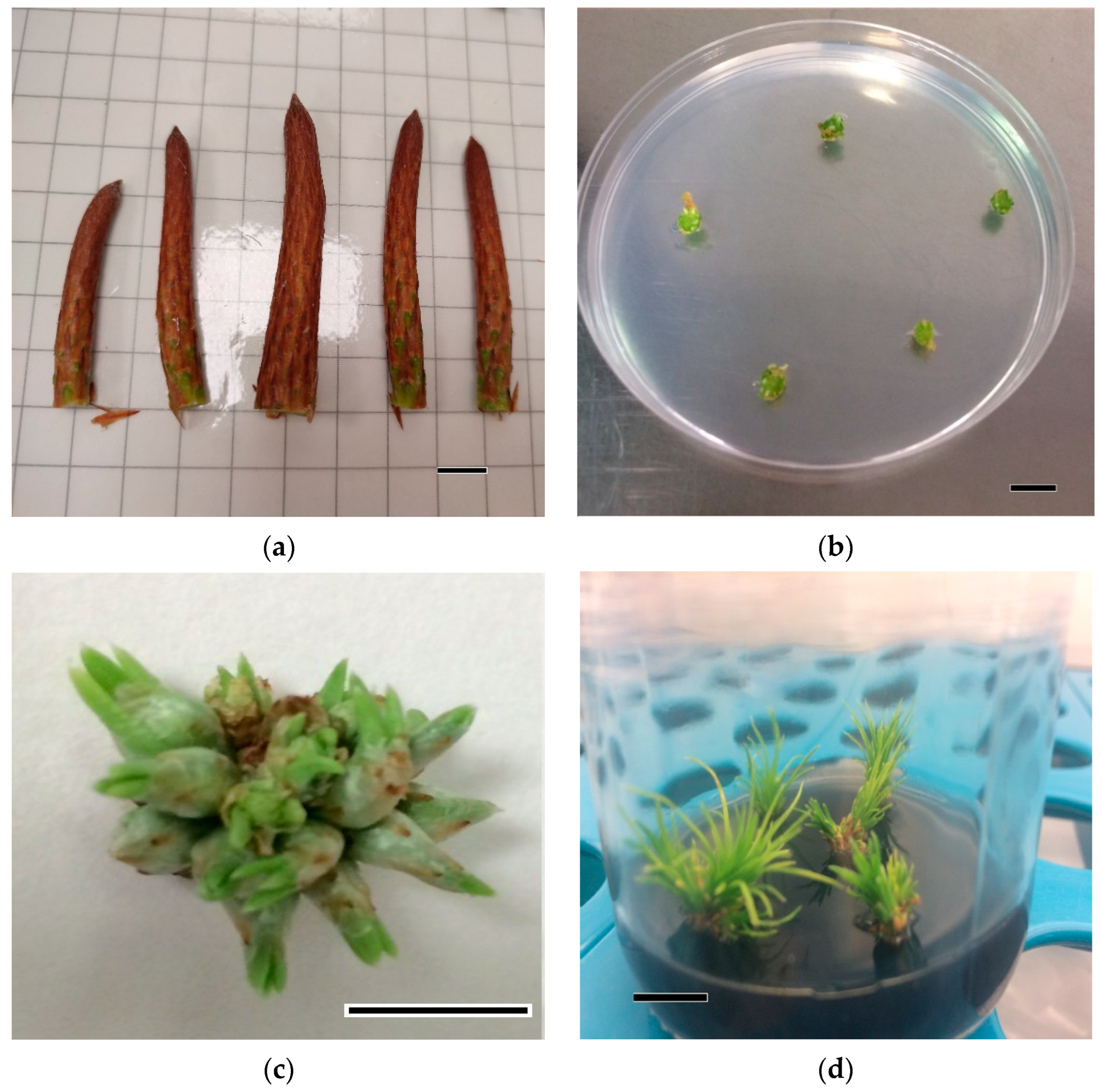
2.2. Organogenic Process

Four to five slices were cultivated vertically on Petri dishes (90 × 15 mm) containing 20 mL of bud induction medium (IM) (Figure 1b). IM consisted of Quoirin and Lepoivre (LP) medium [27], as modified by Aitken-Christie et al. [28], supplemented with 3% (w/v) sucrose and solidified with 8 gL−1 Difco Agar®. The effect of two concentrations of BA was evaluated (22 and 44 μM). The pH of the medium was adjusted to 5.8 before autoclaving (121 °C, 20 min). Four to five Petri dishes per BA concentration and genotype were laid on the growth chamber at a temperature of 21 ± 1 °C, under a 16 h photoperiod, with 120 µmol m−2 s−1 of light intensity provided by cool white fluorescent tubes (TLD 58 W/33; Philips, France).

2.2.1. Shoot Growth and Elongation

As soon as bud induction was observed (between 7 and 9 weeks in IM) (Figure 1c), explants were transferred to Petri dishes with elongation medium (EM). EM consisted of hormone-free LP medium supplemented with 2 gL−1 activated charcoal and 3% (w/v) sucrose and solidified with 8.5 gL−1 Difco Agar®. The pH of the medium was adjusted to 5.8. After 35–50 days in culture (when elongating needle fascicles were evident), explants were subcultured into baby food jars (150 mL) containing 25 mL of EM (Figure 1d). The shoots were subcultivated every six weeks to the same medium. The light intensity, photoperiod and temperature of the growth chamber were the same as described above.
When shoots were 1.0–1.5 cm in length in the original bud slice, they were separated and cultivated individually in fresh EM (Figure 1d). The explants that showed new axillary shoot formation were cultivated into fresh EM, comprising a total of 6 subcultures, and the remaining explants showing secondary needles were separated and cultivated again with IM treatment to promote axillary bud development (re-induction).

2.2.2. Root Induction and Acclimatization of Rooted Plants

After the elongation phase, shoots of at least 2.0–2.5 cm length were used for root induction. The explants were cultivated in Ecoboxes (Eco2box/green filter: a polypropylene vessel with a “breathing” hermetic cover, Duchefa®) containing 100 mL of root induction medium (RIM), which consisted of LP medium supplemented with a mixture of 5 µM 1-Naphthaleneacetic acid (NAA) and 10 µM indole-3-butyric acid (IBA), 8 gL−1 Difco Agar® and (A) 3% (w/v) sucrose or (B) 1.5% (w/v) sucrose. Eight shoots per genotype and BA concentration were cultivated for each sucrose concentration. The pH of the culture media was adjusted to 5.8 before autoclaving. The shoots were placed in dim light at 21 ± 1 °C for 8 days, followed by four weeks under a 16 h photoperiod. Two different light treatments were tested: (A) white light, 120 µmol m−2 s−1 of light intensity provided by cool white fluorescent tubes (TLD 58 W/33; Philips, France); and (B) red light, 67 µmol m−2 s−1 of light intensity provided by adjustable LEDs (RB4K Grow LEDs, GureLED-Meetthings, Vitoria-Gasteiz, Spain).
After four weeks of culture in RIM, explants were cultured in Ecoboxes containing 100 mL of root expression medium (REM), which consisted of LP medium supplemented with 2 gL−1 activated charcoal, 3% (w/v) sucrose and 8.5 gL−1 Difco Agar®, for six weeks. Then, explants with visible roots were transferred to wet peat moss (Pindstrup, Aarhus, Denmark): vermiculite (8:2, v/v) and acclimatized in the greenhouse under controlled conditions at 21 ± 1 °C, decreasing the humidity progressively over one month from 95 to 80%.

2.3. Metabolite Extraction and Soluble Sugar and Amino Acid Quantification

Metabolite extractions from explants collected after four weeks of culture in RIM treatments and after six weeks cultured in REM medium were performed following the protocol described by Valledor et al. [29]. Five to eight explants were taken from each condition (previous BA treatment, light type and sucrose content in the medium); no particular selection of the material was made. Each explant was considered as a biological replicate. Explants were ground in liquid nitrogen and 100 mg of the resulting fine powder was transferred to 2 mL microcentrifuge tubes containing 800 µL cold metabolite extraction buffer (methanol:chloroform:water, 2.5:1:0.5, v:v:v). Then, tubes were centrifuged at 20,000× g 6 min at 4 °C and the supernatants were transferred to new tubes containing 800 µL of phase separation mix (chloroform:water, 1:1, v:v). After centrifugation at 10,000× g for 5 min at room temperature, the upper aqueous phases containing polar metabolites were saved in new 1.5 mL tubes. Aliquots of 200 mL were then obtained from these tubes and completely dried on a Speedvac to remove the remaining methanol.
Finally, the samples were resuspended in 100 µL ultra-pure water and soluble sugars and amino acids were quantified by High-Performance Liquid Chromatography (HPLC) (Agilent 1260 Infinity II, Agilent Technologies, Santa Clara, CA, USA). Soluble sugars, including sugar alcohol (fructose, galactose glucose, mannitol and sucrose), were separated using a Hi-Plex Ca column (7.7 mm × 300 mm, 8 µm) and detected using a refractive index detector at a flow rate of 0.15 mL min−1 pure water at 80 °C for 30 min, as described by Castander-Olarieta et al. [30]. In the case of amino acids, a Poroshell 120 column (4.6 mm × 100 mm, 2.7 µm, Agilent Technologies, Santa Clara, CA, USA) was used, coupled to a fluorescence detector. The samples were injected into the column at a flow rate of 1.5 mL min−1 at 40 °C for 18 min with a discontinuous gradient. Solvent A was a mixture of 10 mM Na2HPO4 (Scharlau Chemie, Barcelona, Spain) and 10 mM Na2B4O7 (pH 8.2) (Scharlau Chemie, Barcelona, Spain) and solvent B was acetonitrile: methanol:water (45:45:10, v:v:v) (Scharlau Chemie, Barcelona, Spain, HPLC-grade). The gradient program was the following: min 0–13.40, solvent A 98% and solvent B 2%, min 13.40–13.50, solvent A 43% and solvent B 57%, min 13.50–15.80, solvent B 100%, and min 15.80–18, solvent A 98% and solvent B 2%. Amino acids were estimated after pre-column derivatization by mixing 1 µL sample with 2.5 µL borate buffer (Agilent Technologies, Santa Clara, CA, USA), 32 µL diluent (100 mL solvent A and 0.4 mL concentrated H3PO4) (PanReac AppliChem, Barcelona, Spain, pure pharma-grade) and 0.5 µL o-phthaldialdehyde (OPA) (Agilent Technologies, Santa Clara, CA, USA). In the specific case of amino acids hydroxyproline and proline, samples were mixed with 0.5 µL of 9-fluorenyl-methylchloroformate protecting group (FMOC) (Agilent Technologies, Santa Clara, CA, USA) instead of OPA. Detection was performed by analyzing fluorescence with excitation at 260 nm and emission at 450 nm for OPA derivatives and emission at 325 nm for FMOC derivatives.
In the case of amino acids, concentrations were determined from internal calibration curves constructed with the corresponding commercial standards (Agilent Technologies, Santa Clara, CA, USA). Carbohydrates such as fructose, galactose, mannitol and sucrose (Merck, Darmstadt, Germany) and glucose (Duchefa Biochemie, Haarlem, The Netherlands) were determined from internal calibration curves constructed with the corresponding commercial standards. Results were conveniently adjusted considering the concentration step after methanol removal (2 times), and the results were expressed as μmol g FW–1. In the case of certain amino acids, samples were re-injected after 1/32 dilution to avoid signal saturation, and the resulting concentrations were conveniently adjusted.

2.4. Data Collection and Statistical Analysis

Two to four Petri dishes per genotype (nine genotypes) and four to five bud slices per Petri dish were cultured for each BA concentration and collection date. Contamination, survival and the percentage of explants forming shoots (EFS) (%) at each collection date were measured after two months of culture. When the axillary shoots were isolated and cultured individually in elongation medium, the EFS (%) and the mean number of shoots per explant (NS/E) were calculated with respect to the non-contaminated explants. A logistic regression model was used to analyze the collection date, genotype and BA concentration’s effects on the EFS (%). Tukey’s post hoc test (α = 0.05) was used for multiple comparisons. Data on the NS/E were analyzed by analysis of variance (ANOVA). When necessary, multiple comparisons were made using Duncan’s post hoc test (α = 0.05).
The root induction percentage (RI) (%), the mean number of roots per explant (NR/E) and the length of the longest root (LLR) (cm) were recorded after six weeks of culture in REM medium. A completely randomized design was carried out using eight stems per BA and sucrose concentrations and light treatments.
To assess the effect of the cytokinin and sucrose concentration and light treatments’ effects on the RI (%), a logistic regression was performed. Data on NR/E were x2 transformed to meet homoscedasticity and were analyzed by ANOVA. Data on LLR (cm) were subjected to ANOVA. When necessary, multiple comparisons were made using Duncan’s post hoc test (α = 0.05). The acclimatization percentage was calculated after four weeks under ex vitro conditions.
Data on free amino acid and carbohydrate content after four weeks of culture in RIM medium and after six weeks cultured in REM medium were analyzed. The confirmation of the homogeneity of variances and normality of the data was performed, and, when necessary, they were log(x) and x transformed. Data on free amino acids and carbohydrates were subjected to ANOVA, and, when necessary, multiple comparisons were made using Duncan’s post hoc test (α = 0.05). The data were analyzed using R Core Team software® (version 4.2.1, Vienna, Austria).

3. Results

3.1. Organogenic Process

Genotype A showed the highest survival of the explants, independently of the collection date (72%). On the contrary, the lowest survival was observed in genotype J (43%). The highest survival values of the explants were observed on the first collection date (second week of January). In the same way, the highest contamination rates were recorded in samples collected in the third and fourth weeks of February (44% and 43%, respectively), and the lowest were found from January to the second week of February (between 17 and 34%).
The collection date had a significant effect on the EFS (%) (Figure 2, Supplementary Table S1). A significantly higher EFS (%) was obtained during the first week of February than during the third and fourth weeks of February (4%). Shoots induced in the second and third weeks of January and the second week of February displayed intermediate values (29 to 34%).
Reinvigorated shoots were obtained after culture media were supplemented with both BA concentrations (22 and 44 μM). Regarding EFS (%), BA concentrations alone did not provoke any statistically significant differences in the EFS (%) (Supplementary Table S2), but the genotype and the interaction between genotype and BA concentration showed significant differences (Figure 3). A significantly higher EFS (%) was obtained in buds of genotype A induced with 44 μM BA than in the rest of the genotypes and BA concentrations tested. Buds of genotype N cultured with 44 μM BA showed a significantly lower EFS (%) than buds from genotypes A, F, G and J and genotypes E and J cultured at 22 and 44 μM BA, respectively (between 33 and 45%, Figure 3).
No statistically significant differences were found in NS/E when the effect of the BA concentration in the culture media during the induction phase was analyzed (Supplementary Table S3). The NS/E ranged from 1.66 ± 0.40 to 1.95 ± 0.52 in explants induced with 22 and 44 µM BA, respectively.

Rooting Induction and Acclimatization of Rooted Plants

The different BA concentrations (22 and 44 µM) used in the shoot induction phase did not have a statistically significant effect on RI (18% and 14%), on NR/E (4.50 and 3.56) or on LLR (1.83 cm and 1.60 cm), respectively (Supplementary Tables S4 and S5).
A significantly higher RI (%) was observed in shoots cultured under white FL when compared with shoots growing under red LEDs (Figure 4a). In the same way, NR/E was significantly higher in shoots cultured under white FL than in those under red LEDs (Figure 4b). Light treatments did not provoke any statistically significant differences in the LLR. Shoots growing under red LEDs showed the longest length (2.22 cm) (Supplementary Tables S4 and S6).
For the LLR, a significant effect of the interaction between BA and light treatment (Figure 5a) and between BA and sucrose concentration (Figure 5b) was observed. Explants induced with 44 μM BA and exposed to red LEDs displayed higher values compared with explants induced at the lowest BA concentration and exposed to the same light conditions (Figure 5a). On the contrary, explants cultured in the presence of 22 μM BA and exposed to white FL displayed significantly higher values on LLR compared with explants induced at the highest BA concentration and exposed to the same light conditions (Figure 5a). Additionally, explants induced in the presence of 44 μM BA and cultured in medium supplemented with 1.5% sucrose during root induction showed significantly higher values than shoots induced at the same BA concentration followed by a medium supplemented with 3.0% sucrose (Figure 5b). When a culture medium containing 22 μM BA was used for shoot induction, regardless of the sucrose concentration evaluated, intermediate values of LLR were obtained (Figure 5b).
Regarding the sucrose concentrations, shoots cultured in medium with 3% sucrose showed a significantly higher RI value (22%) than those cultured in 1.5% sucrose (9%). No statistically significant differences in NR/E (3.73 and 4.10) and LLR values (2.01 and 1.55 cm) were observed in shoots cultured in the presence of 1.5 and 3% sucrose, respectively (Supplementary Tables S5 and S6).
Additionally, the interaction between BA concentration and light treatment and between BA and sucrose concentrations assayed throughout shoot induction did not show significant differences in RI (%) and NR/E (Supplementary Tables S4 and S5). In the same way, no statistically significant differences were observed in the interaction between light treatment and sucrose concentration evaluated during the shoot induction phase in RI (%), NR/E and LLR (Supplementary Table S6).
Shoots induced in the presence of 22 μM BA, rooted in medium supplemented with 3% sucrose and exposed to white FL from genotypes A, E and K developed in vitro roots (Figure 1e). In all genotypes, in the bases of shoots, a large and profuse callus was observed (Figure 1e). Acclimatized shoots were successfully obtained only from genotype K (33.33%) (Figure 1f).

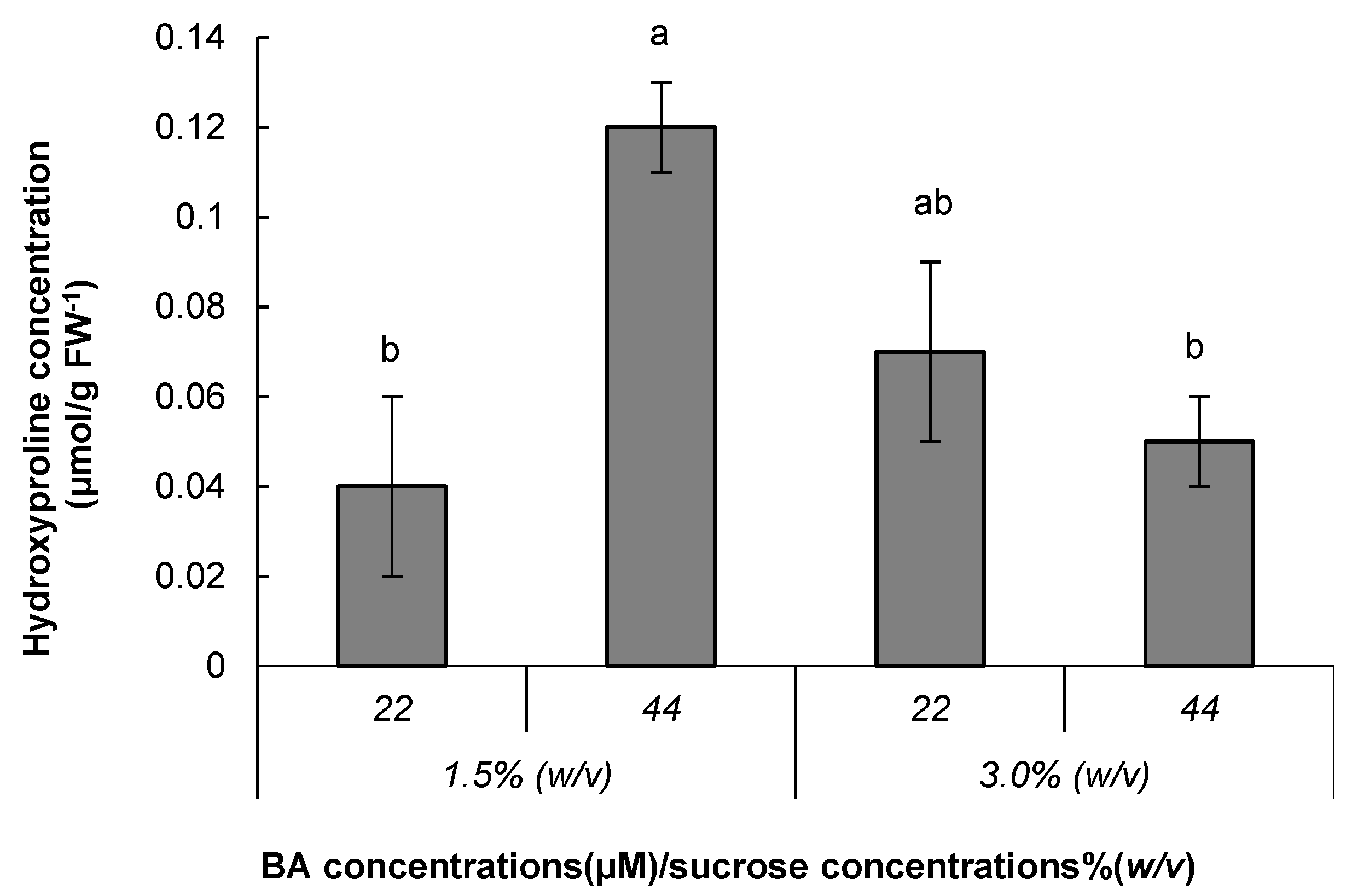
3.2. Metabolite Analysis

BA concentrations and light treatments, as well as the interaction between both variables, did not show significant differences for the concentration of carbohydrates in shoots analyzed after four weeks under RIM treatment (Supplementary Tables S7–S9). However, explants cultured in media with the highest BA concentration presented a lower sucrose concentration than explants cultured in media with 22 μM BA (Table 1). Additionally, the levels of all carbohydrates analyzed were lower in explants cultured under red LEDs, except for the galactose concentration (Table 1).
Regarding the effect of the sucrose concentration on the carbohydrate levels of shoots analyzed after root induction treatment, no significant differences were found in galactose and mannitol concentrations (Supplementary Tables S8 and S9). Galactose displayed values from 2.41 to 4.85 μmol g FW−1 and mannitol from 6.69 to 7.87 μmol g FW−1 in media supplemented with 1.5 and 3% sucrose, respectively. In the case of fructose, glucose and sucrose, significantly higher concentrations were obtained in shoots cultured in the presence of the highest sucrose concentration when compared with the results at the lowest sucrose concentration (Figure 6a–c). The interaction between BA and light treatment, the interaction between BA and sucrose concentration and the interaction between light treatment and sucrose concentration did not show statistically significant differences (Supplementary Tables S7–S9).
At the end of the rooting phase, no statistically significant differences were found for any of the carbohydrates analyzed in shoots during the root expression phase in relation to BA, light treatments, sucrose concentrations or the interactions among them (Supplementary Tables S10–S12). The highest glucose content (26.44 μmol g FW−1) was found when shoots were cultured in the presence of the lowest BA concentration during the induction phase (Table 2). Additionally, shoots cultured under white FL exhibited an increase in fructose, glucose and sucrose levels compared with those cultured under red LEDs (Table 2). The fructose, glucose and sucrose levels decreased in shoots cultured during the induction phase in medium supplemented with the highest sucrose concentration. The galactose concentration in shoots after the rooting phase was the lowest of all the sugars analyzed, independently of the chemical characteristics of the culture media.
Amino acid concentrations in shoots after root induction were not significantly different in relation to BA, light treatments, sucrose concentrations or the interactions between them (Table 3 and Supplementary Tables S13–S15). Alanine, arginine, asparagine and glutamine levels were the highest in shoots after the rooting phase, regardless of the in vitro treatment applied during the induction phase (Table 3).
Shoots cultured with the highest sucrose concentration showed higher levels of alanine, arginine and glutamic acid than those cultured at the lowest sucrose concentration (Table 3). In contrast, the asparagine content was higher in rooted shoots coming from explants cultured in induction media supplemented with 1.5% sucrose than in those cultured with 3.0% sucrose.
After six weeks of culture in REM medium, no significant differences were found for the amino acid concentrations in shoots coming from explants induced in the presence of different concentrations of BA, light treatment and the interaction between both variables (Supplementary Tables S16–S18). However, the concentrations of alanine, arginine, glutamine, glycine, and lysine exhibited a decrease when shoots had been cultured with the highest BA concentration (Table 4). Pinus radiata shoots contained concentrations below 1.00 μmol g FW−1 of aspartic acid, glycine, hydroxyproline, histidine, isoleucine, leucine, lysine, methionine, phenylalanine, threonine, tryptophan, tyrosine and valine, regardless of the BA concentration used in the induction media (Table 4).
Arginine, glutamine and lysine concentrations were the highest in shoots growing under white FL. In contrast, concentrations of alanine, arginine, asparagine, aspartic acid, cysteine, glutamic acid, glycine, hydroxyproline, isoleucine, leucine, methionine, phenylalanine, proline, serine, tryptophan, tyrosine and valine increased in shoots exposed to red LEDs (Table 4).
Threonine and tyrosine concentrations in rooted shoots cultured in induction media with the lowest sucrose concentration were significantly higher than in shoots cultured in media with the highest sucrose concentration (Table 4). Although not significant, it is worth noting that alanine, arginine, asparagine, aspartic acid, cysteine, glutamic acid, glutamine, glycine, hydroxyproline, histidine, isoleucine, leucine, proline, serine and valine levels at the end of the rooting phase were also lower in shoots cultured in medium with the highest sucrose concentration during root induction (Table 4).
Significantly higher values of hydroxyproline were found in shoots developed in a culture medium with 44 μM BA and 1.5% sucrose than in those from 44 μM BA and 3.0% sucrose. Additionally, the hydroxyproline levels were significantly higher in shoots cultured in a medium with 44 μM BA and 1.5% sucrose than in those from 22 μM BA and 1.5% sucrose (Figure 7). The lowest hydroxyproline level was obtained in shoots induced under the lowest BA concentration and cultured in medium supplemented with 1.5% sucrose during the root induction phase (Figure 7).
After the rooting expression phase, the interaction between light treatment and sucrose concentration showed statically significant differences for the aspartic acid, isoleucine, phenylalanine, threonine and tyrosine levels (Figure 8a–e). Significantly higher concentrations of the abovementioned amino acids were detected in shoots from medium with the lowest sucrose concentration and exposed to red LEDs (Figure 8a–e).
Additionally, shoots coming from root induction at 3.0% sucrose and exposed to red LEDs presented the lowest aspartic acid, isoleucine, threonine and tyrosine levels (Figure 8a–e).

4. Discussion

In this study, the genotype affected the survival of the explants independently of the collection date. This result could be partially explained since the genotype is an endogenous factor that has a significant role in the regenerative potential, overall repeatability and reliability of the tissue culture protocol [31]. As was reviewed in [32,33], the response of explants to particular tissue culture conditions is highly dependent on the genetic and physiological determination. In this sense, Pinus radiata, P. taeda L. and P. halepensis showed a differential organogenic response in vitro depending on the genotype in previous studies [25,34,35].
The highest survival and the lowest contamination rates were found during the first collection date. Several protocols mention that factors such as the genotype, explant source and plant growth regulators influence the in vitro regeneration of plants via organogenesis or somatic embryogenesis [36,37,38]. In our case, the developmental stage of the explant can be related to the contamination rate. On the first collection date, explants consisted of apical shoot buds with closed scales; this source of explant could be better for the effective elimination of contamination. Analogous results were obtained in Pseudotsuga menziesii Mirb. Franco [39] organogenesis, where spring buds showed higher contamination rates than winter buds.
Silver nanoparticles have various applications in plant biotechnology, mainly to eradicate microbial contamination and promote in vitro development [40]. In this sense, the use of a silver nanoparticle solution in our study favored the disinfection efficiency of the explants, in agreement with the results for Fragaria × ananassa, Vanilla planifolia Andrews and Psidium friedrichsthalianum (O. Berg) Nied, where the contamination rates of 23, 28 and 40%, respectively [37,40,41], were reported. In contrast, in P. halepensis, a negative effect of the silver nanoparticle solution for the sterilization of apical shoot buds was found with a contamination rate of 89% [25].
The timing of explant collection and the explant developmental stage influence the response to organogenesis or somatic embryogenesis [42,43]. When the EFS was evaluated, apical buds collected in the first week of February showed higher percentages than those collected during the third and fourth weeks of February. A similar pattern has been reported in other species, such as Quercus alba L. and Allamanda cathartica L., showing that the explant developmental stage or the season of collection of explants played an important role in the in vitro response [43,44]. During our study, a morphogenic peak from dormancy to bud initiation was observed during the January and February collection dates, wherein tissues became very active in terms of morphogenesis by increasing the shoot formation. These results are in accordance with those obtained in shoot explants of Larix decidua Mill. [45,46], where two short morphogenic peaks were observed, the first before bud break and the second in late summer.
When we focused on the study of the effect of the BA concentration on EFS, no statistically significant differences were obtained. In plant tissue culture, BA is the most commonly used cytokinin [47]; in addition, BA, used alone or in combination with other cytokinins, has been shown to efficiently promote in vitro bud induction [9,48]. In our experiments, BA at the concentrations tested (22 and 44 μM) provided a satisfactory organogenic response, and, although not significant, a slightly higher response in explants cultured in media supplemented with 44 μM BA was observed. An analogous result was obtained in P. halepensis using the same BA concentration [25]. Likewise, in P. pinea, a higher number of buds formed per explant was observed during the first 16 days of culture at the highest concentration of BA tested (44.4 μM) [49]. This behavior may be attributed to the maturity of the explant used, as, most likely, the aged buds are not as physiologically active as the young ones. For the above, higher concentrations of exogenously applied growth regulators may be needed in mature explants to obtain similar morphogenic responses, as reported for Tetraclinis articulata (Vahl) Masters [50]. In this regard, Wendling et al. [51] mentioned that, through the continuous in vitro subculture of shoots in media supplemented with cytokinins, reinvigoration can be obtained. In our study, all genotypes evaluated were able to develop reinvigorated shoots in media with both BA concentrations tested. These results agreed with those observed in P. pinea, P. pinaster, P. silvestris and P. halepensis, where reinvigorated axillary shoots were obtained through organogenesis, using buds as initial explants [9,10,11,25].
In order to obtain a root induction response in the explants, the culture medium was supplemented with NAA combined with IBA, a mixture traditionally used to induce root differentiation in Pinus species [11,24,34,52]. However, in this work, the rooting percentage was low, as observed in P. pinea when NAA alone was applied [11]. Montalbán et al. [24], studying P. radiata, reported that IBA alone was more efficient in the adventitious root induction of shoots coming from explants of juvenile origin. In Pinus elliottii Engelm. var. elliottii, Nunes et al. [53] observed that different concentrations and combinations of IBA (4.9 and 9.8 μM) and NAA (1.1, 2.1 and 2.7 μM) led to low root frequency using shoots developed from juvenile material. Likewise, in P. halepensis, long exposure to IBA was not effective in inducing roots in shoots of mature origin [25]. It is widely recognized that the most common problem encountered in the micropropagation of mature conifers is the adventitious root formation in shoots [11].
As reviewed by Bairu et al. [54], the development of basal callus is one of the main physiological disorders that affects the rooting competence of microplants; this basal callus could interfere with physiological processes by trapping essential growth constituents such as plant growth regulators and creating a physical barrier. In our work, in the bases of shoots, a large and profuse callus was observed, and this fact could have affected the development of adventitious roots. In contrast, in slash pine, when inducing adventitious roots using IBA combined with NAA, no callus formation was observed in shoots developed from juvenile explants [53]. Similarly, in Pinus. roxburghii Sarg., using shoots developed from juvenile explants, the root induction medium supplemented with NAA and solidified with 0.6% agar produced shoots with low or no callusing at the base of the shoots [55]. In P. taeda [35,56], a combination of auxin (2.68 μM NAA) with cytokinin (0.44 μM BA) was evaluated and showed root induction of 55.6 to 47.5%, respectively. More recently, a pulse with IBA 500 mg L−1 for 5 min was used as an ex vitro rooting preconditioning treatment in Mitragyna parvifolia (Roxb.) Korth., obtaining approximately 90% micropropagated shoots rooted ex vitro [57]. Soumare et al. [58] reported that the use of plant-growth-promoting microbes, specifically Streptomyces griseorubens and Norcardiopsis alba, increased the rooting and root hair in maize. In this sense, these recent strategies, or physical factors such as the photoperiod, temperature and substrates for rooting, could be considered to improve P. radiata rooting.
Carbohydrates in plants have several essential functions: they are basic elements of macromolecules, constitute substrates for respiration, play an important role in the synthesis pathway of many compounds and are indispensable for many other processes related to plant development or gene expression [59,60]. In our work, the sucrose treatment had a significant effect on the RI percentage, and the highest result was observed in 3% (w/v) sucrose. Similar results were observed when comparing sucrose concentrations (1–9%) for the root induction of Apple Rootstock MM 10, concluding that sucrose had a direct effect on rooting [60]. Likewise, in banana (Musa sp) and Metroxylon sagu Rottb., the highest values for rooting were found in a medium supplemented with 3% (w/v) sucrose [61,62]. In Eucalyptus globulus Labill. and P. pinea, glucose had a positive effect during root induction [63,64]. Additionally, in E. globulus [63], it was reported that the carbohydrates are an energetic requirement for root development and sucrose, which is commonly used in tissue culture because it is the main sugar translocated in the phloem of several plants [20,23].
The light type and wavelength specificity have an influence on morphogenetic responses such as adventitious root formation [23]. In several studies, LED lights have been used as an alternative to conventional lighting sources for plant tissue culture [65]. Supporting this, the spectral properties of LEDs have been found to regulate the morphological, anatomical and physiological responses of in vitro plants [65]. In this work, it was stated that the effectiveness of red LEDs on rooting depends on the genotype and the concentration of growth regulators applied in the root induction medium [65]. LED treatments (red or blue) showed a higher response in the rooting of plantlets in Tripterospermum japonicum Maxim, Gossypium hirsutum L. and Doritaenopsis [66,67,68]. In Anthurium, root formation was progressively induced under a higher portion of red LEDs in a mixed circuit of blue and red [69]. However, in this work, shoots grown under white FL showed a significantly higher RI and NR/E. A similar pattern has been reported in Handroanthus ochraceus (Cham.) Mattos, Achilea millefolium L. and Alocasia amazonica, where higher values of root induction were obtained in plants growing under low FL irradiances [16,70,71].
When studying the effect of the BA concentration and light treatment on LLR, explants induced with 44 μM BA and exposed to red LEDs showed the highest values. A similar pattern has been reported in Vitis ficifolia Bunge var. ganebu, where longer roots were obtained in plants growing under red LEDs [72]. In contrast, higher values of root length were obtained in plants of H. ochraceus exposed to low FL irradiances [16].
Metabolite analysis showed significant differences for several carbohydrate and amino acid concentrations in the different treatments tested. Carbohydrates in in vitro culture media have functions such as the maintenance of osmotic potential and to serve as a carbon source for developmental processes including root induction [73,74]. When studying the effect of the sucrose concentration supplemented in RIM, higher fructose, glucose and sucrose content was found in shoots growing in culture medium supplemented with the highest sucrose concentration. In A. amazonica, media supplemented with 3 or 6.0% sucrose showed higher glucose, fructose and sucrose content in in vitro leaves compared with ex vitro leaves [75]. Moreover, Mingozzi et al. [76], studying Cydonia oblonga Mill., concluded that the sucrose content in leaves was associated with its content in the culture media, and shoots cultured with 30 gL−1 sucrose showed higher content of sucrose, glucose and fructose. Additionally, [15] explained that in vitro plants release invertase enzymes to the culture medium that act in the hydrolysis of sucrose, giving rise to glucose and fructose. In the present work, the levels of the carbohydrates were unaffected by lighting conditions. Contrasting our results, the content of sucrose, starch and soluble sugars was higher in plantlets of Doritaenopsis and G. hirsutum exposed to red plus blue and red LEDs, respectively [67,68].
Amino acid metabolism plays an essential role in plant protein biosynthesis, represents a building block for other biosynthesis pathways and is essential during signaling processes [77]. Apart from the abovementioned functions, amino acids may also be involved in the plant stress response through intracellular pH regulation and the detoxification of reactive oxygen species [78,79]. Tyrosine is the precursor of secondary metabolites such as betacyanin in Alternanthera brasiliana (L.) Kuntze and certain enzymes such as tyrosine decarboxylase, which is responsible for redirecting essential primary metabolites into secondary metabolic pathways [80,81]. In addition, in P. radiata embryonal masses initiated under stressful conditions, Castander et al. [4] discussed that tyrosine could be involved in antioxidative processes and osmotic adjustment. Likewise, the tyrosine metabolism pathway serves as a starting point for the production of tyrosine-derived metabolites essential to plant survival, such as tocopherols, plastoquinone and ubiquinone [82]. With regard to threonine, it is an essential amino acid, and it is a substrate for isoleucine synthesis [83]. Moreover, threonine metabolites have an important role in plant growth and development, cell division and responses to abiotic stresses [83,84]. However, in our study, the shoots with higher content of these two amino acids showed worse in vitro rooting (those coming from root induction medium with 1.5% (w/v) sucrose, at the end of the rooting phase).
Krasenski et al., Lugan et al. and Joshi et al. [83,85,86] observed amino acid accumulation in plants exposed to abiotic stress, such as drought, extreme temperature, salinity and osmotic stress. In this sense, tobacco plants showed high levels of aromatic amino acids, and this fact was related to the improvement of salt stress tolerance [87]. In this study, our results suggest that low sucrose concentrations could be related to abiotic stress similar to that observed in nutrient deficiencies or carbohydrate starvation, such as the results observed in Camellia sinensis (L.) Kuntze leaves, where increased threonine levels were observed under phosphate starvation [88]. However, the amino acid pool differs and changes depending on the developmental and physiological stage of the plant [77].

5. Conclusions

The regeneration of P. radiata from the vegetative shoot buds of adult trees via organogenesis was achieved, and it depended on environmental physico-chemical factors. The collection date and genotype had a strong effect on the efficiency of the process. Reinvigorated shoots from adult trees were obtained at both of the BA concentrations tested. The optimal result in terms of shoot induction was obtained at the highest BA concentration. Our results suggest that the use of white FL and a 3% sucrose concentration was better for root induction. The low RI percentage suggests that further research should be encouraged to improve this phase, which is still a bottleneck in the micropropagation of mature conifers.
Moreover, shoots showing better in vitro rooting had higher fructose, glucose and sucrose content, whereas those from the worse root treatment showed higher threonine and tyrosine levels.

Supplementary Materials

The following supporting information can be downloaded at: https://www.mdpi.com/article/10.3390/f13091455/s1. Table S1: Statistical analysis for explants forming shoots [EFS (%)] in different collection dates from Pinus radiata D. Don cultured in LP medium [27] modified by Aitken-Christie et al. [28]; Table S2: Statistical analysis for explants forming shoots [EFS (%)] from nine different genotypes of Pinus radiata D. Don cultured in LP medium [27] modified by Aitken-Christie et al. [28] supplemented with 6-benzyladenine (BA) (22 and 44 μM); Table S3: ANOVA for number of shoots formed per explant (NS/E) from Pinus radiata D. Don cultured in LP medium [27] modified by Aitken-Christie et al. [28] supplemented with 6-benzyladenine (BA) (22 and 44 μM); Table S4: Statistical analysis for root induction (%), number roots per explant, and length of longest root from Pinus radiata D. Don cultured in LP medium [27] modified by Aitken-Christie et al. [28] according to 6-benzyladenine (BA) concentrations and light treatments applied in the root induction phase; Table S5: Statistical analysis for root induction (%), number roots per explant, and length of longest root from Pinus radiata D. Don cultured in LP medium [27] modified by Aitken-Christie et al. [28] according to and 6-benzyladenine (BA) and sucrose concentrations applied in the root induction phase; Table S6: Statistical analysis for root induction (%), number roots per explant, and length of longest root from Pinus radiata D. Don cultured in LP medium [27] modified by Aitken-Christie et al. [28] according to light treatments and sucrose concentrations applied in the root induction phase; Table S7: ANOVA for carbohydrates content of Pinus radiata D. Don cultured in LP medium [27] modified by Aitken-Christie et al. [28] supplemented with a mixture of 5 µM 1-Naphthaleneacetic acid (NAA) and 10 µM indole-3-butyric acid (IBA) according to 6-benzyladenine (BA) concentrations and light treatments; Table S8: ANOVA for carbohydrates content of Pinus radiata D. Don cultured in LP medium [27] modified by Aitken-Christie et al. [28] supplemented with a mixture of 5 µM 1-Naphthaleneacetic acid (NAA) and 10 µM indole-3-butyric acid (IBA) according to 6-benzyladenine (BA) and sucrose concentrations; Table S9: ANOVA for carbohydrates content of Pinus radiata D. Don cultured in LP medium [27] modified by Aitken-Christie et al. [28] supplemented with a mixture of 5 µM 1-Naphthaleneacetic acid (NAA) and 10 µM indole-3-butyric acid (IBA) according to light treatments and sucrose concentrations; Table S10: ANOVA for carbohydrates content of Pinus radiata D. Don cultured in LP medium [27] modified by Aitken-Christie et al. [28] supplemented with 2 gL−1 activated charcoal, according to 6-benzyladenine (BA) concentrations and light treatments; Table S11: ANOVA for carbohydrates content of Pinus radiata D. Don cultured in LP medium [27] modified by Aitken-Christie et al. [28] supplemented with 2 gL−1 activated charcoal, according to 6-benzyladenine (BA) concentrations and sucrose; Table S12: ANOVA for carbohydrates content of Pinus radiata D. Don cultured in LP medium [27] modified by Aitken-Christie et al. [28] supplemented with 2 gL−1 activated charcoal, according to light treatments and sucrose concentrations; Table S13: ANOVA for amino acid content of Pinus radiata D. Don cultured in LP medium [27] modified by Aitken-Christie et al. [28] supplemented with a mixture of 5 µM 1-Naphthaleneacetic acid (NAA) and 10 µM indole-3-butyric acid (IBA) according to 6-benzyladenine (BA) concentrations and light treatments; Table S14: ANOVA for amino acid content of Pinus radiata D. Don cultured in LP medium [27] modified by Aitken-Christie et al. [28] supplemented with a mixture of 5 µM 1-Naphthaleneacetic acid (NAA) and 10 µM indole-3-butyric acid (IBA) according to 6-benzyladenine (BA) and sucrose concentrations; Table S15: ANOVA for amino acid content of Pinus radiata D. Don cultured in LP medium [27] modified by Aitken-Christie et al. [28] supplemented with a mixture of 5 µM 1-Naphthaleneacetic acid (NAA) and 10 µM indole-3-butyric acid (IBA) according to light treatments and sucrose concentrations;Table S16: ANOVA amino acid content of Pinus radiata D. Don cultured in LP medium [27] modified by Aitken-Christie et al. [28] supplemented with 2 gL−1 activated charcoal according to 6-benzyladenine (BA) concentrations and light treatments; Table S17: ANOVA for amino acid content of Pinus radiata D. Don cultured in LP medium [27] modified by Aitken-Christie et al. [28] supplemented with 2 gL−1 activated charcoal according to 6-benzyladenine (BA) and sucrose concentrations; Table S18: ANOVA for amino acid content of Pinus radiata D. Don cultured in LP medium [27] modified by Aitken-Christie et al. [28] supplemented with 2 gL−1 activated charcoal according to light treatments and sucrose concentrations.

Author Contributions

Conceptualization, P.M. and I.A.M.; methodology, P.M. and I.A.M.; formal analysis, A.R.-V.; investigation, A.R.-V. and A.C.-O.; data curation, A.R.-V.; writing—original draft preparation, A.R.-V.; writing—review and editing, A.R.-V., P.M., I.A.M. and A.C.-O.; supervision, P.M. and I.A.M. All authors have read and agreed to the published version of the manuscript.

Funding

This research was funded by MINECO project (AGL2016-76143-C4-3R), MICINN (PID2020-112627RB-C32), CYTED (P117RT0522), DECO (Basque government) and MULTIFOREVER project, supported under the umbrella of ERA-NET Cofund ForestValue by ANR (FR), FNR (DE), MINCyT (AR), MINECO-AEI (ES), MMM (FI), VINNOVA (SE) [ForestValue has received funding from the European Union’s Horizon 2020 Research and Innovation Programme under grant agreement no. 773324], Universidad Nacional de Costa Rica and Instituto de Investigación y Servicios Forestales (INISEFOR).

Acknowledgments

The authors are grateful to Sonia Suárez Álvarez M. Sc. (NEIKER-BRTA, Spain) for kindly providing the facilities for HPLC analysis.

Conflicts of Interest

The authors declare no conflict of interest. The funders had no role in the design of the study; in the collection, analyses, or interpretation of data; in the writing of the manuscript; or in the decision to publish the results.

Abbreviations

ANOVA—Analysis of variance, BA—6-Benzyladenine, EFS—Explants forming shoots, EM—Elongation medium, FL—Fluorescent light, IBA—Indole-3-butyric acid, IM—Induction medium, LEDs—Light-emitting diodes, LLR—Length of the longest root, NAA—1-Naphthaleneacetic acid, NR/E—Number of roots per explant, NS/E—number of shoots per explant, PVP—Polyvinylpyrrolidone, REM—Root expression medium, RI—Root induction, RIM—Root induction medium.

References

  1. Neale, D.B.; Wheeler, N.C. The Conifers. In The Conifers: Genomes, Variation and Evolution; Springer Nature: Cham, Switzerland, 2019; pp. 1–21. [Google Scholar] [CrossRef]
  2. Farjon, A. The Kew review: Conifers of the world. Kew Bull. 2018, 73, 8. [Google Scholar] [CrossRef]
  3. Escandón, M.; Valledor, L.; Pascual, J.; Pinto, G.; Cañal, M.J.; Meijón, M. System-wide analysis of short-term response to high temperature in Pinus radiata. J. Exp. Bot. 2017, 68, 3629–3641. [Google Scholar] [CrossRef]
  4. Castander-Olarieta, A.; Montalbán, I.A.; De Medeiros Oliveira, E.; Dell’Aversana, E.; D’Amelia, L.; Carillo, P.; Steiner, N.; Fraga, H.P.F.; Guerra, M.P.; Goicoa, T.; et al. Effect of thermal stress on tissue ultrastructure and metabolite profiles during initiation of radiata pine somatic embryogenesis. Front. Plant Sci. 2019, 9, 2004. [Google Scholar] [CrossRef]
  5. Allen, C.D.; Macalady, A.K.; Chenchouni, H.; Bachelet, D.; McDowell, N.; Vennetier, M.; Kitzberger, T.; Rigling, A.; Breshears, D.D.; Hogg (Ted), E.H.; et al. A global overview of drought and heat-induced tree mortality reveals emerging climate change risks for forests. For. Ecol. Manag. 2010, 259, 660–684. [Google Scholar] [CrossRef]
  6. Jankovský, L.; Palovčíková, D.; Dvořák, M.; Tomšovský, M. Records of Brown spot needle blight related to Lecanosticta acicola in the Czech Republic. Plant Prot. Sci. 2009, 45, 16–18. [Google Scholar] [CrossRef]
  7. Woods, A.J.; Martín-García, J.; Bulman, L.; Vasconcelos, M.W.; Boberg, J.; La Porta, N.; Peredo, H.; Vergara, G.; Ahumada, R.; Brown, A.; et al. Dothistroma needle blight, weather and possible climatic triggers for the disease’s recent emergence. For. Pathol. 2016, 46, 443–452. [Google Scholar] [CrossRef]
  8. Reed, B.M.; Sarasan, V.; Kane, M.; Bunn, E.; Pence, V.C. Biodiversity conservation and conservation biotechnology tools. In Vitro Cell. Dev. Biol. Plant. 2011, 47, 1–4. [Google Scholar] [CrossRef]
  9. De Diego, N.; Montalbán, I.A.; Fernandez de Larrinoa, E.; Moncaleán, P. In vitro regeneration of Pinus pinaster adult trees. Can. J. For. Res. 2008, 38, 2607–2615. [Google Scholar] [CrossRef]
  10. De Diego, N.; Montalbán, I.A.; Moncaleán, P. In vitro regeneration of adult Pinus sylvestris L. Trees. S. Afr. J. Bot. 2010, 76, 158–162. [Google Scholar] [CrossRef] [Green Version]
  11. Cortizo, M.; de Diego, N.; Moncaleán, P.; Ordás, R.J. Micropropagation of adult stone pine (Pinus pinea L.). Trees Struct. Funct. 2009, 23, 835–842. [Google Scholar] [CrossRef]
  12. Phillips, G.C. In vitro morphogenesis in plants—Recent advances. In Vitro Cell. Dev. Biol. Plant. 2004, 40, 342–345. [Google Scholar] [CrossRef]
  13. Kulus, D.; Woźny, A. Influence of light conditions on the morphogenetic and biochemical response of selected ornamental plant species under in vitro conditions: A mini-review. BioTechnologia 2020, 101, 75–83. [Google Scholar] [CrossRef]
  14. De Almeida, M.; de Almeida, C.V.; Graner, E.M.; Brondani, G.E.; de Abreu-Tarazi, M.F. Pre-procambial cells are niches for pluripotent and totipotent stem-like cells for organogenesis and somatic embryogenesis in the peach palm: A histological study. Plant Cell Rep. 2012, 31, 1495–1515. [Google Scholar] [CrossRef] [PubMed]
  15. Thorpe, T.; Stasolla, C.; Yeung, E.C.; de Klerk, G.J.; Roberts, A.; George, E.F. The components of plant tissue culture media II: Organic additions, osmotic and pH effects, and support systems. In Plant Propagation by Tissue Culture, 3rd ed.; George, E.F., Hall, M.A., de Klerk, G.J., Eds.; Springer: Dordrecht, The Netherlands, 2008; pp. 115–173. [Google Scholar]
  16. Larraburu, E.E.; Correa, G.S.; Llorente, B.E. In vitro development of yellow lapacho (Bignoniaceae) using high-power light emitting diode. Rev. Arvore 2018, 42, e420508. [Google Scholar] [CrossRef]
  17. Jao, R.C.; Fang, W. An adjustable light source for photo-phyto related research and young plant production. Appl. Eng. Agric. 2003, 19, 601–608. [Google Scholar] [CrossRef]
  18. Riikonen, J.; Kettunen, N.; Gritsevich, M.; Hakala, T.; Särkkä, L.; Tahvonen, R. Growth and development of Norway spruce and Scots pine seedlings under different light spectra. Environ. Exp. Bot. 2016, 121, 112–120. [Google Scholar] [CrossRef]
  19. Astolfi, S.; Marianello, C.; Grego, S.; Bellarosa, R. Preliminary investigation of LED lighting as growth light for seedlings from different tree species in growth chambers. Not. Bot. Horti. Agrobot. Cluj-Napoca 2012, 40, 31–38. [Google Scholar] [CrossRef]
  20. Kumar, N.; Reddy, M.P. In vitro plant propagation: A review. J. For. Environ. Sci. 2011, 27, 61–72. [Google Scholar] [CrossRef]
  21. Narváez, I.; Martín, C.; Jiménez-Díaz, R.M.; Mercado, J.A.; Pliego-Alfaro, F. Plant regeneration via somatic embryogenesis in mature wild olive genotypes resistant to the defoliating pathotype of Verticillium dahliae. Front. Plant Sci. 2019, 10, 1471. [Google Scholar] [CrossRef]
  22. Montalbán, I.A.; De Diego, N.; Igartua, E.A.; Setién, A.; Moncaleán, P. A combined pathway of somatic embryogenesis and organogenesis to regenerate radiata pine plants. Plant Biotechnol. Rep. 2011, 5, 177–186. [Google Scholar] [CrossRef]
  23. Ragonezi, C.; Klimaszewska, K.; Castro, M.R.; Lima, M.; de Oliveira, P.; Zavattieri, M.A. Adventitious rooting of conifers: Influence of physical and chemical factors. Trees Struct. Funct. 2010, 24, 975–992. [Google Scholar] [CrossRef]
  24. Montalbán, I.A.; De Diego, N.; Moncaleán, P. Testing novel cytokinins for improved in vitro adventitious shoots formation and subsequent ex vitro performance in Pinus radiata. Forestry 2011, 84, 363–373. [Google Scholar] [CrossRef]
  25. Pereira, C.; Montalbán, I.A.; Pedrosa, A.; Tavares, J.; Pestryakov, A.; Bogdanchikova, N.; Canhoto, J.; Moncaleán, P. Regeneration of Pinus halepensis (Mill.) through organogenesis from apical shoot buds. Forests 2021, 12, 363. [Google Scholar] [CrossRef]
  26. Stephano-Hornedo, J.L.; Torres-Gutiérrez, O.; Toledano-Magaña, Y.; Gradilla-Martínez, I.; Pestryakov, A.; Sánchez-González, A.; García-Ramos, J.C.; Bogdanchikova, N. ArgovitTM silver nanoparticles to fight Huanglongbing disease in Mexican limes (Citrus aurantifolia Swingle). RSC Adv. 2020, 10, 6146–6155. [Google Scholar] [CrossRef] [PubMed]
  27. Quoirin, M.; Lepoivre, P. Études des milieux adaptés aux cultures in vitro de Prunus. Acta Hortic. 1977, 78, 437–442. [Google Scholar] [CrossRef]
  28. Aitken-Christie, J.; Singh, A.P.; Davis, H. Multiplication of meristematic tissue: A new tissue culture system for radiata pine. In Genetic Manipulation of Woody Plants; Hanover, J.W., Keathley, D.E., Eds.; Plenum Publishing Corp: New York, NY, USA, 1988; pp. 413–432. [Google Scholar] [CrossRef]
  29. Valledor, L.; Escandón, M.; Meijón, M.; Nukarinen, E.; Cañal, M.J.; Weckwerth, W. A universal protocol for the combined isolation of metabolites, DNA, long RNAs, small RNAs, and proteins from plants and microorganisms. Plant J. 2014, 79, 173–180. [Google Scholar] [CrossRef] [PubMed]
  30. Castander-Olarieta, A.; Pereira, C.; Montalbán, I.A.; Mendes, V.M.; Correia, S.; Suárez-Álvarez, S.; Manadas, B.; Canhoto, J.; Moncaleán, P. Proteome-wide analysis of heat-stress in Pinus radiata somatic embryos reveals a combined response of sugar metabolism and translational regulation mechanisms. Front. Plant Sci. 2021, 12, 631239. [Google Scholar] [CrossRef]
  31. Coleman, G.D.; Ernst, S.G. In vitro shoot regeneration of Populus deltoides: Effect of cytokinin and genotype. Plant Cell Rep. 1989, 8, 459–462. [Google Scholar] [CrossRef]
  32. Jiménez, V.M. Involvement of plant hormones and plant growth regulators on in vitro somatic embryogenesis. Plant Growth Regul. 2005, 47, 91–110. [Google Scholar] [CrossRef]
  33. Fehér, A. Why somatic plant cells start to form embryos? In Somatic Embryogenesis; Mujib, A., Šamaj, J., Eds.; Plant Cell Monographs; Springer: Berlin/Heidelberg, Germany, 2005; Volume 2, pp. 85–101. [Google Scholar] [CrossRef]
  34. Montalbán, I.A.; Novák, O.; Rolčik, J.; Strnad, M.; Moncaleán, P. Endogenous cytokinin and auxin profiles during in vitro organogenesis from vegetative buds of Pinus radiata Adult Trees. Physiol. Plant. 2013, 148, 214–231. [Google Scholar] [CrossRef]
  35. Cézar, T.M.; Higa, A.R.; Koehler, H.S.; Ribas, L.L.F. Influence of culture medium, explant length and genotype on micropropagation of Pinus taeda L. Ciênc. Florest. 2015, 25, 13–22. [Google Scholar] [CrossRef]
  36. Parzymies, M. Nano-silver particles reduce contaminations in tissue culture but decrease regeneration rate and slows down growth and development of Aldrovanda vesiculosa explants. Appl. Sci. 2021, 11, 3653. [Google Scholar] [CrossRef]
  37. Tung, H.T.; Thuong, T.T.; Cuong, D.M.; Luan, V.Q.; Hien, V.T.; Hieu, T.; Nam, N.B.; Phuong, H.T.N.; Van The Vinh, B.; Khai, H.D.; et al. Silver nanoparticles improved explant disinfection, in vitro growth, runner formation and limited ethylene accumulation during micropropagation of strawberry (Fragaria × Ananassa). Plant Cell. Tissue Organ Cult. 2021, 145, 393–403. [Google Scholar] [CrossRef]
  38. Rojas-Vargas, A.; Castander-Olarieta, A.; Moncaleán, P.; Montalbán, I.A. Optimization of the micropropagation of elite adult trees of Sequoia sempervirens: Forest species of interest in the Basque Country, Spain. Rev. Bionatura. 2021, 6, 1511–1519. [Google Scholar] [CrossRef]
  39. Traore, A.; Xing, Z.; Bonser, A.; Carlson, J. Optimizing a protocol for sterilization and in vitro establishment of vegetative buds from mature douglas fir trees. J. Am. Soc. Hortic. Sci. 2005, 40, 1464–1468. [Google Scholar] [CrossRef]
  40. Pastelín-Solano, M.C.; Ramírez-Mosqueda, M.A.; Bogdanchikova, N.; Castro-González, C.G.; Bello-Bello, J.J. Silver nanoparticles affect the micropropagation of vanilla (Vanilla planifolia Jacks. Ex Andrews). Agrociencia 2020, 54, 1–13. [Google Scholar]
  41. Andújar, I.; González, N.; García-Ramos, J.C.; Bogdanchikova, N.; Pestryakov, A.; Escalona, M.; Concepción, O. ArgovitTM Silver nanoparticles reduce contamination levels and improve morphological growth in the in vitro culture of Psidium friedrichsthalianum (O. Berg) Nied. SN Appl. Sci. 2020, 2, 2110. [Google Scholar] [CrossRef]
  42. Andersone, U.; Ievinsh, G. Changes of morphogenic competence in mature Pinus sylvestris L. buds in vitro. Ann. Bot. 2002, 90, 293–298. [Google Scholar] [CrossRef] [Green Version]
  43. Corredoira, E.; San-José, M.C.; Vieitez, A.M. Induction of somatic embryogenesis from different explants of shoot cultures derived from young Quercus alba Trees. Trees Struct. Funct. 2012, 26, 881–891. [Google Scholar] [CrossRef]
  44. Khanam, M.N.; Anis, M. Organogenesis and efficient in vitro plantlet regeneration from nodal segments of Allamanda cathartica L. using TDZ and ultrasound assisted extraction of quercetin. Plant Cell. Tissue Organ Cult. 2018, 134, 241–250. [Google Scholar] [CrossRef]
  45. Bonga, J.M. The effect of various culture media on the formation of embryo-like structures in cultures derived from explants taken from mature Larix decidua. Plant Cell. Tissue Organ Cult. 2004, 77, 43–48. [Google Scholar] [CrossRef]
  46. Bonga, J.M. Can explant choice help resolve recalcitrance problems in in vitro propagation, a problem still acute especially for adult conifers? Trees Struct. Funct. 2017, 31, 781–789. [Google Scholar] [CrossRef]
  47. Phillips, G.C.; Garda, M. Plant tissue culture media and practices: An overview. In Vitro Cell. Dev. Biol. Plant. 2019, 55, 242–257. [Google Scholar] [CrossRef]
  48. Valverde-Cerdas, L.; Rojas-Vargas, A.; Hine-Gómez, A. In vitro propagation of Albizia guachapele, Cedrela odorata, Platymiscium pinnatum and Guaiacum sanctum. Plant Tissue Cult. Biotechnol. 2008, 18, 151–156. [Google Scholar] [CrossRef]
  49. Moncaleán, P.; Alonso, P.; Centeno, M.L.; Cortizo, M.; Rodríguez, A.; Fernández, B.; Ordás, R.J. Organogenic responses of Pinus pinea cotyledons to hormonal treatments: BA metabolism and cytokinin content. Tree Physiol. 2005, 25, 1–9. [Google Scholar] [CrossRef]
  50. Juan-Vicedo, J.; Serrano-Martinez, F.; Cano-Castillo, M.; Casas, J.L. In vitro propagation, genetic assessment, and, medium-term conservation of coastal endangered species Tetraclinis articulata (Vahl) Masters (Cupressaceae) from adult trees. Plants 2022, 11, 187. [Google Scholar] [CrossRef]
  51. Wendling, I.; Trueman, S.J.; Xavier, A. Maturation and related aspects in clonal forestry-part II: Reinvigoration, rejuvenation and juvenility maintenance. New For. 2014, 45, 473–486. [Google Scholar] [CrossRef]
  52. Álvarez, J.M.; Majada, J.; Ordás, R.J. An improved micropropagation protocol for maritime pine (Pinus pinaster Ait.) isolated cotyledons. Forestry 2009, 82, 175–184. [Google Scholar] [CrossRef] [Green Version]
  53. Nunes, S.; Sousa, D.; Pereira, V.T.; Correia, S.; Marum, L.; Santos, C.; Dias, M.C. Efficient protocol for in vitro mass micropropagation of slash pine. In Vitro Cell. Dev. Biol. Plant. 2018, 54, 175–183. [Google Scholar] [CrossRef]
  54. Bairu, M.W.; Kane, M.E. Physiological and developmental problems encountered by in vitro cultured plants. Plant Growth Regul. 2011, 63, 101–103. [Google Scholar] [CrossRef]
  55. Kalia, R.K.; Arya, S.; Kalia, S.; Arya, I.D. Plantlet regeneration from fascicular buds of seedling shoot apices of Pinus roxburghii Sarg. Biol. Plant. 2007, 51, 653–659. [Google Scholar] [CrossRef]
  56. De Oliveira, L.F.; Ribas, L.L.F.; Quoirin, M.; Koehler, H.S.; Amano, E.; Higa, A.R. Micropropagation of Pinus taeda L. from juvenile material. Tree For. Sci. Biotechnol. 2012, 6, 96–101. [Google Scholar]
  57. Patel, A.K.; Lodha, D.; Shekhawat, N.S. An improved micropropagation protocol for the ex situ conservation of Mitragyna parvifolia (Roxb.) Korth (Rubiaceae): An endangered tree of pharmaceutical importance. In Vitro Cell. Dev. Biol. Plant. 2020, 56, 817–826. [Google Scholar] [CrossRef]
  58. Soumare, A.; Boubekri, K.; Lyamlouli, K.; Hafidi, M.; Ouhdouch, Y.; Kouisni, L. Efficacy of phosphate solubilizing actinobacteria to improve rock phosphate agronomic effectiveness and plant growth promotion. Rhizosphere 2021, 17, 100284. [Google Scholar] [CrossRef]
  59. Gibson, S.I. Plant sugar-response pathways. part of a complex regulatory web. Plant Physiol. 2000, 124, 1532–1539. [Google Scholar] [CrossRef]
  60. Calamar, A.; De Klerk, G.J. Effect of sucrose on adventitious root regeneration in apple. Plant Cell. Tissue Organ Cult. 2002, 70, 207–212. [Google Scholar] [CrossRef]
  61. Ahmed, S.; Sharma, A.; Bhushan, B.; Singh, A.K.; Wali, V.K. Effect of carbohydrate source, pH and supporting media on in vitro rooting of banana (Musa Spp.) cv. Grand naine plantlets. Afr. J. Agric. Res. 2014, 9, 1135–1140. [Google Scholar] [CrossRef]
  62. Sumaryono, W.M.; Ratnadewi, D. Effect of carbohydrate source on growth and performance of in vitro sago palm (Metroxylon sagu Rottb.) plantlets. HAYATI J. Biosci. 2012, 19, 88–92. [Google Scholar] [CrossRef]
  63. Corrêa, L.D.R.; Paim, D.C.; Schwambach, J.; Fett-Neto, A.G. Carbohydrates as regulatory factors on the rooting of Eucalyptus saligna Smith and Eucalyptus globulus Labill. Plant Growth Regul. 2005, 45, 63–73. [Google Scholar] [CrossRef]
  64. Zavattieri, A.; Lima, M.; Sobral, V.; Oliveira, P.; Costa, A. Effects of carbon source, carbon concentration and culture conditions on in vitro rooting of Pinus pinea L. microshoots. Acta Hortic. 2009, 812, 173–180. [Google Scholar] [CrossRef]
  65. Gupta, S.; Jatothu, B. Fundamentals and applications of light-emitting diodes (LEDs) in in vitro plant growth and morphogenesis. Plant Biotechnol. Rep. 2013, 7, 211–220. [Google Scholar] [CrossRef]
  66. Moon, H.K.; Park, S.Y.; Kim, Y.W.; Kim, C.S. Growth of Tsuru-Rindo (Tripterospermum japonicum) cultured in vitro under various sources of light-emitting diode (LED) irradiation. J. Plant Biol. 2006, 49, 174–179. [Google Scholar] [CrossRef]
  67. Li, H.; Xu, Z.; Tang, C. Effect of light-emitting diodes on growth and morphogenesis of upland cotton (Gossypium hirsutum L.) plantlets in vitro. Plant Cell. Tissue Organ Cult. 2010, 103, 155–163. [Google Scholar] [CrossRef]
  68. Shin, K.S.; Murthy, H.N.; Heo, J.W.; Hahn, E.J.; Paek, K.Y. The effect of light quality on the growth and development of in vitro cultured Doritaenopsis plants. Acta Physiol. Plant. 2008, 30, 339–343. [Google Scholar] [CrossRef]
  69. Budiarto, K. Spectral quality affects morphogenesis on Anthurium plantlet during in vitro culture. Agrivita 2010, 32, 234–240. [Google Scholar] [CrossRef]
  70. Alvarenga, I.C.A.; Pacheco, F.V.; Silva, S.T.; Bertolucci, S.K.V.; Pinto, J.E.B.P. In vitro culture of Achillea millefolium L.: Quality and intensity of light on growth and production of volatiles. Plant Cell. Tissue Organ Cult. 2015, 122, 299–308. [Google Scholar] [CrossRef]
  71. Jo, E.A.; Tewari, R.K.; Hahn, E.J.; Paek, K.Y. Effect of photoperiod and light intensity on in vitro propagation of Alocasia amazonica. Plant Biotechnol. Rep. 2008, 2, 207–212. [Google Scholar] [CrossRef]
  72. Poudel, P.R.; Kataoka, I.; Mochioka, R. Effect of red- and blue-light-emitting diodes on growth and morphogenesis of grapes. Plant Cell. Tissue Organ Cult. 2008, 92, 147–153. [Google Scholar] [CrossRef]
  73. Yaseen, M.; Ahmad, T.; Sablok, G.; Standardi, A.; Hafiz, I.A. Review: Role of carbon sources for in vitro plant growth and development. Mol. Biol. Rep. 2013, 40, 2837–2849. [Google Scholar] [CrossRef]
  74. Moradi, P.; Ford-Lloyd, B.; Pritchard, J. Metabolomic approach reveals the biochemical mechanisms underlying drought stress tolerance in thyme. Anal. Biochem. 2017, 527, 49–62. [Google Scholar] [CrossRef]
  75. Jo, E.A.; Tewari, R.K.; Hahn, E.J.; Paek, K.Y. In vitro sucrose concentration affects growth and acclimatization of Alocasia amazonica plantlets. Plant Cell. Tissue Organ Cult. 2009, 96, 307–315. [Google Scholar] [CrossRef]
  76. Mingozzi, M.; Morini, S.; Lucchesini, M.; Mensuali-Sodi, A. Effects of leaf soluble sugars content and net photosynthetic rate of quince donor shoots on subsequent morphogenesis in leaf explants. Biol. Plant. 2011, 55, 237–242. [Google Scholar] [CrossRef]
  77. Hildebrandt, T.M.; Nunes Nesi, A.; Araújo, W.L.; Braun, H.P. Amino acid catabolism in plants. Mol. Plant. 2015, 8, 1563–1579. [Google Scholar] [CrossRef]
  78. Campalans, A.; Messeguer, R.; Goday, A.; Pagès, M. Plant responses to drought, from ABA signal transduction events to the action of the induced proteins. Plant Physiol. Biochem. 1999, 37, 327–340. [Google Scholar] [CrossRef]
  79. Nuccio, M.L.; Rhodes, D.; McNeil, S.D.; Hanson, A.D. Metabolic engineering of plants for osmotic stress resistance. Curr. Opin. Plant Biol. 1999, 2, 128–134. [Google Scholar] [CrossRef]
  80. Silva, N.C.B.; Macedo, A.F.; Lage, C.L.S.; Esquibel, M.A.; Sato, A. Developmental effects of additional ultraviolet a radiation, growth regulators and tyrosine in Alternanthera brasiliana (L.) Kuntze cultured in vitro. Braz. Arch. Biol. Technol. 2005, 48, 779–786. [Google Scholar] [CrossRef]
  81. Zhang, J.X.; Ma, L.Q.; Yu, H.S.; Zhang, H.; Wang, H.T.; Qin, Y.F.; Shi, G.L.; Wang, Y.N. A tyrosine decarboxylase catalyzes the initial reaction of the salidroside biosynthesis pathway in Rhodiola sachalinensis. Plant Cell Rep. 2011, 30, 1443–1453. [Google Scholar] [CrossRef]
  82. Xu, J.J.; Fang, X.; Li, C.Y.; Yang, L.; Chen, X.Y. General and Specialized Tyrosine Metabolism Pathways in Plants. Abiotech 2020, 1, 97–105. [Google Scholar] [CrossRef]
  83. Joshi, V.; Joung, J.G.; Fei, Z.; Jander, G. Interdependence of threonine, methionine and isoleucine metabolism in plants: Accumulation and transcriptional regulation under abiotic stress. Amino Acids 2010, 39, 933–947. [Google Scholar] [CrossRef]
  84. Muthuramalingam, P.; Krishnan, S.R.; Pandian, S.; Mareeswaran, N.; Aruni, W.; Pandian, S.K.; Ramesh, M. Global analysis of threonine metabolism genes unravel key players in rice to improve the abiotic stress tolerance. Sci. Rep. 2018, 8, 9270. [Google Scholar] [CrossRef]
  85. Krasensky, J.; Jonak, C. Drought, salt, and temperature stress-induced metabolic rearrangements and regulatory networks. J. Exp. Bot. 2012, 63, 1593–1608. [Google Scholar] [CrossRef]
  86. Lugan, R.; Niogret, M.F.; Leport, L.; Guégan, J.P.; Larher, F.R.; Savouré, A.; Kopka, J.; Bouchereau, A. Metabolome and water homeostasis analysis of Thellungiella salsuginea suggests that dehydration tolerance is a key response to osmotic stress in this halophyte. Plant J. 2010, 64, 215–229. [Google Scholar] [CrossRef]
  87. Oliva, M.; Guy, A.; Galili, G.; Dor, E.; Schweitzer, R.; Amir, R.; Hacham, Y. Enhanced production of aromatic amino acids in tobacco plants leads to increased phenylpropanoid metabolites and tolerance to stresses. Front. Plant Sci. 2021, 11, 604349. [Google Scholar] [CrossRef]
  88. KC, S.; Liu, M.; Zhang, Q.; Fan, K.; Shi, Y.; Ruan, J. Metabolic Changes of Amino Acids and Flavonoids in Tea Plants in Response to Inorganic Phosphate Limitation. Int. J. Mol. Sci. 2018, 19, 3683. [Google Scholar] [CrossRef] [Green Version]
Figure 1. Organogenic process in apical shoot buds from Pinus radiata D. Don adult trees: (a) apical shoot buds, bar = 1 cm; (b) shoot bud slices in culture, bar = 1 cm; (c) shoot bud slice after induction with 22 μM 6-benzyladenine (BA), bar = 1 cm; (d) shoots in elongation medium, bar = 1 cm; (e) rooted shoots, bar = 1 cm; (f) acclimatized shoots, bar = 3 cm.
Forests 13 01455 g001aForests 13 01455 g001b
Figure 2. Explants forming shoots (%) shown in buds of Pinus radiata D. Don adult trees collected on different dates and cultured in Quoirin and Lepoivre (LP) medium [27], modified according to Aitken-Christie et al. [28]. Data are presented as mean values ± S.E. Different letters indicate significant differences by Tukey’s post hoc test (p < 0.05).
Forests 13 01455 g002
Figure 3. Explants forming shoots (%) shown in buds of different genotypes of Pinus radiata D. Don cultured in Quoirin and Lepoivre (LP) medium [27], modified according to Aitken-Christie et al. [28], supplemented with 6-benzyladenine (BA at 22 and 44 μM). Data are presented as mean values ± S.E. Different letters indicate significant differences by Tukey’s post hoc test (p < 0.05).
Forests 13 01455 g003
Figure 4. Effect of light treatments (white fluorescent (F) and red LEDs (R)) on root induction (%) (a) and number of roots per explant (b) of Pinus radiata D. Don shoots cultured in Quoirin and Lepoivre (LP) medium [27], modified according to Aitken-Christie et al. [28]. Data are presented as mean values ± S.E. Different letters indicate significant differences by Tukey’s post hoc test (a) and by Duncan’s post hoc test (b) (p < 0.05).
Forests 13 01455 g004
Figure 5. Effect of 6-benzyladenine (BA) concentration (22 and 44 μM), light treatment (white fluorescent (F) and red LEDs (R)) (a) and BA concentration (22 and 44 μM) and sucrose concentration (1.5% and 3.0% (w/v) (b) on length of the longest root (cm) of Pinus radiata D. Don shoots cultured in Quoirin and Lepoivre (LP) medium [27] modified according to Aitken-Christie et al. [28]. Data are presented as mean values ± S.E. Different letters indicate significant differences by Duncan’s post hoc test (p < 0.05).
Forests 13 01455 g005
Figure 6. Effect of sucrose concentration (1.5% and 3.0% (w/v)) on fructose (a), glucose (b) and sucrose (c) concentration of Pinus radiata D. Don shoots cultured in rooting medium (Quoirin and Lepoivre (LP) medium [27], modified according to Aitken-Christie et al. [28], supplemented with a mixture of 5 µM 1−Naphthaleneacetic acid (NAA) and 10 µM indole−3−butyric acid (IBA)). Data are presented as mean values ± S.E. Different letters indicate significant differences by Duncan’s post hoc test (p < 0.05).
Forests 13 01455 g006aForests 13 01455 g006b
Figure 7. Effect of 6-Benzyladenine (BA) (22 and 44 μM) and sucrose concentration (1.5 and 3.0% (w/v)) on hydroxyproline amino acid level of Pinus radiata D. Don shoots cultured in Quoirin and Lepoivre (LP) medium [27], modified according to Aitken-Christie et al. [28], supplemented with 2 gL−1 activated charcoal. Data are presented as mean values ± S.E. Different letters indicate significant differences by Duncan’s post hoc test (p < 0.05).
Forests 13 01455 g007
Figure 8. Effect of light treatment (white fluorescent (F) or red LEDs (R)) and sucrose concentration (1.5 and 3.0% (w/v)) on aspartic acid (a), isoleucine (b), phenylalanine (c), threonine (d) and tyrosine (e) amino acid levels of Pinus radiata D. Don shoots cultured in Quoirin and Lepoivre (LP) medium [27], modified according to Aitken-Christie et al. [28], supplemented with 2 gL−1 activated charcoal. Data are presented as mean values ± S.E. Different letters indicate significant differences by Duncan’s post hoc test (p < 0.05).
Forests 13 01455 g008aForests 13 01455 g008b
Table 1. Effect of 6-benzyladenine (BA) concentration (22 and 44 μM) and light treatment (white fluorescent or red LEDs) on carbohydrate content of Pinus radiata D. Don shoots cultured in rooting medium (Quoirin and Lepoivre (LP) medium [27], modified according to Aitken-Christie et al. [28], supplemented with a mixture of 5 µM 1-Naphthaleneacetic acid (NAA) and 10 µM indole-3-butyric acid (IBA)).
Carbohydrates
(μmol g FW−1)
Retention Time (min)BA Concentration (μM)Light Treatment
2244White FluorescentRed LEDs
Fructose18.07621.36 ± 6.2822.71 ± 2.9024.91 ± 5.2119.20 ± 2.59
Galactose16.0964.82 ± 2.182.75 ± 0.282.96 ± 0.374.25 ± 1.82
Glucose14.65313.66 ± 4.3715.50 ± 2.1716.70 ± 3.5712.66 ± 2.19
Mannitol22.9117.59 ± 0.457.04 ± 0.447.36 ± 0.367.15 ± 0.55
Sucrose12.64012.29 ± 3.825.67 ± 0.8310.73 ± 3.305.70 ± 0.99
Data are presented as mean values ± S.E.
Table 2. Effect of 6-Benzyladenine (BA) (22 and 44 μM) and sucrose concentration (1.5 and 3.0% (w/v)) and light treatment (white fluorescent or red LEDs) on carbohydrate content of Pinus radiata D. Don shoots cultured in Quoirin and Lepoivre (LP) medium [27], modified according to Aitken-Christie et al. [28], supplemented with 2 gL−1 activated charcoal after the rooting phase.
Carbohydrates
(μmol g FW−1)
Retention Time
(min)
BA Concentration (μM)Light TreatmentSucrose Concentration (w/v)
2244White FluorescentRed LEDs1.5%3.0%
Fructose18.07625.74 ± 7.1822.87 ± 1.9725.98 ± 4.7921.57 ± 2.3627.17 ± 4.4419.58 ± 1.95
Galactose16.0963.12 ± 0.593.87 ± 0.594.00 ± 0.473.19 ± 0.753.42 ± 0.663.86 ± 0.53
Glucose14.65326.44 ±15.0114.41 ± 1.5524.22 ± 9.8812.46 ± 1.3722.50 9.1113.51±2.28
Mannitol22.9115.38 ± 0.755.85 ± 0.365.66 ± 0.565.71 ± 0.415.83 ± 0.565.49 ± 0.33
Sucrose12.64010.10 ± 4.245.29 ± 0.798.63 ± 2.885.15 ± 0.948.38 ± 2.715.12 ± 0.79
Data are presented as mean values ± S.E.
Table 3. Effect of 6-Benzyladenine (BA) (22 and 44 μM) and sucrose concentration (1.5 and 3.0% (w/v)) and light treatment (white fluorescent or red LEDs) on amino acid content (μmol g FW−1) of Pinus radiata D. Don shoots cultured in rooting medium (Quoirin and Lepoivre (LP) medium [27], modified according to Aitken-Christie et al. [28], supplemented with mixture of 5 µM 1-Naphthaleneacetic acid (NAA) and 10 µM indole-3-butyric acid (IBA)).
Free Amino Acids
(μmol g FW−1)
Retention Time
(min)
BA Concentration (μM)Light TreatmentSucrose Concentration (w/v)
2244White FluorescentRed LEDs1.5%3.0%
Alanine5.32319.14 ± 9.7216.08 ±4.8215.40 ± 6.7119.43 ± 6.6411.98 ± 4.5422.54 ± 8.08
Arginine5.03529.98 ± 10.3928.22 ±7.3434.10 ± 8.7722.8 ± 7.7222.45 ± 6.7435.34 ± 9.64
Asparagine3.11513.72 ± 8.2724.78 ±8.1816.09 ± 7.6725.70 ± 9.3930.53 ± 9.6810.52 ± 6.05
Aspartic acid1.2040.57 ± 0.120.61 ± 0.070.68 ± 0.100.50 ± 0.050.60 ± 0.080.59 ± 0.10
Cysteine7.1403.47 ± 0.824.36 ± 0.554.61 ± 0.673.32 ± 0.603.95 ± 0.794.07 ± 0.53
Glutamic acid1.8499.31 ± 8.075.55 ± 2.6411.78 ± 6.171.42 ± 0.103.60 ± 2.0410.39 ± 6.54
Glutamine3.82752.78 ± 16.6670.16 ±8.5865.37 ± 12.3861.26 ± 11.1963.54 ± 12.5763.41 ± 11.29
Glycine4.2310.11 ± 0.020.12 ± 0.010.13 ± 0.020.10 ± 0.010.10 ± 0.020.13 ± 0.01
Hydroxyproline10.3300.02 ± 0.010.03 ± 0.010.02 ± 0.010.03 ± 0.010.03 ± 0.010.03 ± 0.01
Histidine4.0160.15 ± 0.040.18 ± 0.020.18 ± 0.030.16 ± 0.020.18 ± 0.030.17 ± 0.02
Isoleucine9.0790.08 ± 0.010.10 ± 0.010.09 ± 0.010.09 ± 0.010.10 ± 0.010.09 ± 0.01
Leucine9.5920.09 ± 0.020.11 ± 0.010.11 ± 0.010.10 ± 0.010.10 ± 0.010.11 ± 0.01
Lysine10.0180.14 ± 0.030.17 ± 0.020.16 ± 0.030.16 ± 0.010.17 ± 0.020.14 ± 0.02
Methionine7.9050.02 ± 0.010.03 ± 0.010.02 ± 0.010.03 ± 0.010.02 ± 0.010.03 ± 0.01
Phenylalanine8.9270.05 ± 0.010.06 ± 4.9−30.06 ± 0.010.06 ± 0.010.05 ± 0.010.06 ± 4.3−3
Proline12.5430.27 ± 0.070.30 ± 0.080.33 ± 0.090.23 ± 0.060.25 ± 0.070.31 ± 0.08
Serine3.3353.82 ± 3.161.01 ± 0.113.18 ± 2.240.83 ± 0.120.92 ± 0.143.27 ± 2.42
Threonine4.3860.12 ± 0.020.15 ± 0.010.14 ± 0.020.12 ± 0.010.13 ± 0.020.14 ± 0.02
Tryptophan8.6530.05 ± 0.010.06 ± 0.010.05 ± 0.010.06 ± 5.0−30.05 ± 0.010.06 ± 4.6−3
Tyrosine6.3340.06 ± 0.010.09 ± 0.010.08 ± 0.010.08 ± 0.010.08 ± 0.010.08 ± 0.01
Valine7.7600.29 ± 0.070.40 ± 0.040.38 ± 0.050.33 ± 0.040.32 ± 0.050.39 ± 0.05
Data are presented as mean values ± S.E.
Table 4. Effect of 6-Benzyladenine (BA) (22 and 44 μM) and sucrose concentration (1.5 and 3.0% (w/v)) and light treatment (white fluorescent or red LEDs) on amino acid levels (μmol g FW−1) of Pinus radiata D. Don shoots cultured in Quoirin and Lepoivre (LP) medium [27], modified according to Aitken-Christie et al. [28], supplemented with 2 gL−1 activated charcoal after the rooting phase.
Free Amino Acids
(μmol g FW−1)
Retention Time
(min)
BA Concentration (μM)Light TreatmentSucrose Concentration (w/v)
2244White FluorescentRed LEDs1.5%3.0%
Alanine5.32327.12 ± 9.1215.29 ± 5.1718.89 ± 7.5519.96 ± 5.6224.53 ± 6.8712.74 ± 5.63
Arginine5.03567.60 ± 24.4463.97 ± 15.279.68 ± 19.5549.47 ± 16.8068.70 ± 15.7260.71 ± 23.06
Asparagine3.1150.76 ± 0.093.98 ± 3.300.73 ± 0.075.17 ± 4.504.56 ± 3.800.64 ± 0.06
Aspartic acid1.2040.81 ± 0.080.87 ± 0.080.82 ± 0.040.89 ± 0.120.90 ± 0.090.78 ± 0.06
Cystine7.1407.08 ± 0.747.39 ± 0.537.17 ± 0.607.41 ± 0.627.74 ± 0.666.69 ± 0.42
Glutamic acid1.84919.35 ± 10.9927.51 ± 6.8520.38 ± 8.1829.34 ± 8.3531.59 ± 9.1915.67 ± 5.12
Glutamine3.82752.86 ± 12.0739.56 ± 6.6649.22 ± 10.5138.70 ± 5.3846.58 ± 5.0641.08 ± 12.57
Glycine4.2310.20 ± 0.020.18 ± 0.020.18 ± 0.020.19 ± 0.030.20 ± 0.030.17 ± 0.01
Hydroxyproline 10.3300.05 ± 0.010.09 ± 0.010.07 ± 0.010.08 ± 0.010.09 ± 0.020.06 ± 0.0047
Histidine4.0160.24 ± 0.030.23 ± 0.020.23 ± 0.030.23 ± 0.030.26 ± 0.030.20 ± 0.02
Isoleucine9.0790.17 ± 0.010.17 ± 0.020.16 ± 0.010.18 ± 0.020.19 ± 0.020.15 ± 0.01
Leucine9.5920.22 ± 0.020.21 ± 0.020.20 ± 0.010.22 ± 0.030.22 ± 0.020.19 ± 0.01
Lysine10.0180.41 ± 0.160.25 ± 0.040.36 ± 0.110.25 ± 0.040.30 ± 0.040.32 ± 0.13
Methionine7.9050.02 ± 0.010.04 ± 0.010.03 ± 0.010.04 ± 0.010.03 ± 0.010.03 ± 0.01
Phenylalanine8.9270.11 ± 0.010.10 ± 0.010.09 ± 0.010.11 ± 0.010.10 ± 0.010.10 ± 0.01
Proline12.5431.00 ± 0.181.02 ± 0.260.91 ± 0.171.12 ± 0.321.28 ± 0.280.67 ± 0.13
Serine3.3351.44 ± 0.141.41 ± 0.151.31 ± 0.111.53 ± 0.191.60 ± 0.161.19 ± 0.09
Threonine4.3860.23 ± 0.020.24 ± 0.020.24 ± 0.010.24 ± 0.030.27 ± 0.02 a0.21 ± 0.01 b
Tryptophan8.6530.15 ± 0.020.12 ± 0.010.14 ± 0.010.12 ± 0.020.13 ± 0.010.13 ± 0.01
Tyrosine6.3340.14 ± 0.010.13 ± 0.010.13 ± 4.0−30.14 ± 0.010.15 ± 0.01 a0.12 ± 0.01 b
Valine7.7600.63 ± 0.090.56 ± 0.060.56 ± 0.050.61 ± 0.090.63 ± 0.070.52 ± 0.06
Data are presented as mean values ± S.E. Different letters indicate significant differences by Duncan’s post hoc test (p < 0.05).
Publisher’s Note: MDPI stays neutral with regard to jurisdictional claims in published maps and institutional affiliations.

