Article

Arthrinium arundinis, a Novel Causal Agent of Moso Bamboo (Phyllostachys edulis) Culm Rhomboid Rot and Its Sensitivity to Fungicides

Forest Protection Institute in Forestry College, Fujian Agriculture and Forestry University, Fuzhou 350002, China
*
Author to whom correspondence should be addressed.
Forests 2022, 13(10), 1616; https://doi.org/10.3390/f13101616
Submission received: 27 August 2022 / Revised: 27 September 2022 / Accepted: 29 September 2022 / Published: 2 October 2022
(This article belongs to the Special Issue Detection and Analysis of Forest Pathogens)

Abstract

:
Culm rhomboid rot is a new disease of Moso bamboo (Phyllostachys edulis) and mainly occurs on Moso bamboo grown at an altitude above 800 m. The typical symptoms start with black spots and irregular shapes, which expand vertically into an elongated, fusiform, or rhomboid shape up to 15 cm in length. Eleven fungal isolates were isolated from infected tissue, and the isolate BBB1 was confirmed as the pathogen following Koch’s postulates in vitro and in vivo. Regarding morphology, BBB1 was identified as Arthrinium sp.–conidia single-celled, brown to dark, smooth, globose to subglobose, with an equatorial slit, 5.0–9.3 × 3.1–7.3 μm in size. The conidiophores were reduced to conidiogenous cells, pale brown to dark, smooth, and ampulliform. Phylogenetic analysis based on ITS-TEF1-α-TUB2 concatenate sequences identified BBB1 as A. arundinis. Furthermore, the sensitivity of the pathogen against six common fungicides was investigated with mycelial growth assays. Prochloraz exhibited the most potent inhibition with an EC50 value of 0.019 mg/L; another effective fungicide was difenoconazole, with an EC50 value of 0.195 mg/L. This is the first report of A. arundinis causing culm rhomboid rot of Moso bamboo in China; the results provide a basis for managing Moso bamboo culm rhomboid rot.

1. Introduction

Moso bamboo (Phyllostachys edulis), an economically important species of the subfamily Bambusoideae of the family Poaceae, is a temperate species of giant timber bamboo native to China that is now widely distributed in East and Southeast Asia, Africa, and the USA [1,2]. A grown-up Moso bamboo culm can reach a height of up to 28 m and a diameter of up to 20 cm, with very high mechanical strength, and its physical properties boast an average breaking tenacity more than three times that of cotton, wool, rayon, or polyester. These extraordinary properties of culms qualify Moso bamboo as industrial bamboo [2]. In addition, Moso bamboo plays an important role in controlling soil erosion and other climate regulation because of its rapid growth [3]. As an important part of forest resources in southern China, Moso bamboo covers more than 4 million ha2 in subtropical areas [4]. However, intensive production makes Moso bamboo susceptible to infectious diseases. Several Moso bamboo diseases, such as dieback blight (Ceratosphaeria phyllostachydis), foot rot (Arthrinium phaeospermum), leaf spot (Alternaria alternata), sooty blotch (Meliola phyllostachydis), root rot (Fusarium acuminatum), black spot (Coccostroma arundinariae), and leaf rust (Puccinia phyllostachydis), have been found in China and have caused great losses [5].
In 2015, we found a new disease of Moso bamboo in Guangze County (117°20′29.75″ E, 27°32′48.45″ N), Nanping City, Fujian Province, China, named culm rhomboid rot. The typical symptoms commonly appear on aged (five-year-old and above) Moso bamboo with black spots on the surface of the culm, and with the interior of these spots turning black. A few years later, in 2018, the same disease was discovered in Huajiashan Moso Bamboo Farm (117°50′19.52″ E, 27°11′58.12″ N), Jianyang District, and then in Wuyishan County (117°39′24.11″ E, 27°42′1.37″ N), Nanping City in 2020. According to our survey, the disease infected approximately 30% of the Moso bamboo in Huajiashan Moso Bamboo Farm in 2018, and the incidence reached 36% in 2020 (unpublished survey data). To our knowledge, there has been no report of such a disease in Moso bamboo (P. edulis) in southeast China. A similar symptom record has been reported for fishscale bamboo (P. heteroclada) in Sichuan Province (103°3′0.41″ E, 30°1′5.85″ N), China, and three fungal species, Parakarstenia phyllostachydis, Neostagonosporella sichuanensis, and Podonectria sichuanensis, were collected and identified from the lesion tissues; nevertheless, none of them were tested in terms of their pathogenicity toward fishscale bamboo [6,7,8]. In addition, Fusarium oxysporum was considered the pathogen of rhomboid rot in Moso bamboo in Jiangxi Province (114°7′4.48″ E, 26°39′18.29″ N), China [9]. However, the F. oxysporum we isolated from the diseased Moso bamboo culms could not reproduce rhomboid rot symptoms. Thus, there is confusion in the research concerning the pathogen of the culm rhomboid rot disease of Moso bamboo.
As a pervasive genus, Arthrinium has been isolated from soils, plants, animals, and even the ocean [10,11,12,13,14,15,16]. Arthrinium currently comprises ca. 120 species (http://www.indexfungorum.org, accessed on 1 October 2022) and most of them have been recorded as plant pathogens or endophytes, especially on bamboo [5,10,17,18,19,20,21,22]. Arthrinium species share basauxic conidiophores with terminal and intercalary polyblastic conidiogenous cells and brown, unicellular conidia with an equatorial slit, which serves as a reference for morphological taxonomy, but it is difficult to distinguish among Arthrinium, Cordella, and Pteroconium based on these morphological characteristics [23,24]. In 2013, Crous et al. regarded morphological characteristics in distinguishing these genera as phylogenetically insignificant and considered Cordella and Pteroconium as generic synonyms of Arthrinium based on molecular phylogenetic data [11]. At present, the taxonomy of Arthrinium fungi is mainly based on a combination of morphology and molecular phylogenetic data. On A. arundinis, published by Dyko and Sutton in 1979, its conidiophores reduce to conidiogenous cells; it is pale brown, smooth, ampulliform, conidia brown, smooth, globose in the surface view, (5–)6–7 μm, lenticular in the side view, 3–4 μm diameter, and it has a pale equatorial slit [11].
A. arundinis can be found in various plant hosts worldwide, several of which are plant pathogens. A. arundinis causes leaf edge spots on peaches and leaf blight in tea plants in China [25,26]; it also causes kernel blight on barley in the USA and the leaf spots on rosemary in Iran [27,28]. Reports on Arthrinium-related bamboo diseases have begun emerging quite recently, and some new species and/or new bamboo hosts have been reported [5,17,18,20,21]. A. arundinis causes brown culm streaks of early spring bamboo shoots (P. praecox), with small yellow or sandy beige cycle round spots (2 mm) appearing initially, then developing into brown streaks (10 to 15 × 1 to 2 mm) [17].
The methods of managing bamboo diseases mainly include removing the infected culms and spraying fungicides. However, there are few studies on fungicide susceptibility screening for the Arthrinium pathogens of bamboo. The objectives of this study were: (i) To isolate and confirm the pathogen of Moso bamboo culm rhomboid rot following Koch’s postulates; (ii) to identify the species of the pathogen of Moso bamboo culm rhomboid rot through morphological characteristics and gene phylogenetic analysis; and (iii) to determine the sensitivity of this pathogen against six common fungicides. The results of this study will provide a basis for the identification and management of this disease.

2. Materials and Methods

2.1. Field survey and Fungal Isolation

The incidence of culm rhomboid rot in Moso bamboo was surveyed in Huajiashan Moso Bamboo Farm (117°50′19.52″ E, 27°11′58.12″ N), Jianyang District and Dazhulan Bamboo Forest (117°39′24.11″ E, 27°42′1.37″ N), Wuyishan County, Nanping City, Fujian Province, China. The symptoms were photographed using a Canon EOS M6 digital camera, and the size of the spots (50 spots) was measured. Specimens of culm rhomboid rot in Moso bamboo were collected from Huajiashan Moso Bamboo Farm in April 2021 (average precipitation of 211 mm, average temperature of 20 °C, and altitude of 840 m) and kept in the Forest Protection Institute of Fujian Agriculture and Forestry University.
For pathogen isolation, the specimens were cut into small pieces (5 × 5 mm) from lesion margins, and the small pieces were surface-disinfected by submerging them into 2% sodium hypochlorite solution for 2 min and then in 75% ethanol for 1 min, followed by washing thrice with sterile water, consecutively. The sterilized pieces were wiped dry with sterilized filter paper and then placed into Petri dishes containing PDA (five pieces per dish) amended with 50 μg/mL of ampicillin. The Petri dishes were incubated at 25 °C with a 12 h light/dark cycle. After two days, hyphal tips from the leading edge fungal colonies that grew from tissues were transferred to fresh PDA to obtain pure cultures, and the pure cultures were maintained at 25 °C.

2.2. Pathogenicity Assay

2.2.1. Inoculation under Laboratory Conditions

The pathogenicity of all of the isolates was assayed on culm chips of Moso bamboo in vitro. The culms of the Moso bamboo were cut and split into pieces (5 × 10 cm). Then, the pieces were sterilized by spraying them with 75% ethanol and wounded with sterile drills. Mycelial plugs (5 mm diameter) from the edge of each isolate colony were placed on the artificial wounds of the Moso bamboo pieces. The control Moso bamboo pieces were inoculated using PDA plugs without fungus. All of the treatments were placed in moisture chambers containing wet paper towels to retain their moisture and were kept at 25 °C with a 12 h light/dark cycle. The symptoms were recorded over 10 days. Three replicates were repeated for each treatment, and the experiment was conducted twice. Isolates causing symptoms similar to those in the wild were selected for pathogenicity tests under field conditions.

2.2.2. Inoculation under Field Conditions

The isolate BBB1, respective to the suspected causal agent, was used to confirm the pathogenicity under field conditions. One-year-old and five-year-old healthy Moso bamboos without disease symptoms in nature in the bamboo garden of Fujian Agriculture and Forestry University were used for artificial inoculation. The culm surfaces were sterilized with 75% ethanol and wounded using sterilized drills. The mycelial plugs of BBB1 were obtained and inoculated on the culms following the method described above. After this, sterilized absorbent cotton moistened with sterile water was used to cover the wounds, and then the absorbent cotton was sealed with plastic wrap. Control culms were treated with pure PDA plugs following the same procedure. Developing rot culms were collected for the re-isolation of the fungus for pathogen verification and the completion of Koch’s postulates [29]. Each treatment set three replicates and the experiment was conducted twice from May to August 2021 (temperature: 22–29 °C, average precipitation: 212 mm).

2.3. Morphological Characterization

For morphological identification, the purified isolate BBB1 was incubated on PDA for 4 days at 25 °C. The characteristics of the colony and the diameter, color, and texture were measured and recorded daily, and the growth rate of the mycelia was calculated using the following formula: Growth rate (cm/day) = 1/2 (Dia2 − Dia1), where Dia2 is the colony diameter after culturing for 24 h in Dia1. The asexual reproduction characters were observed based on cultures on malt extract agar (MEA; malt extract 20 g and 15 g agar) and according to the synoptic keys proposed for Arthrinium species identification [11]. The shape and size of the microscopic structures were observed and measured using a Nikon microscope (ECLIPSE NI-U, Nikon, Tokyo, Japan) connected to a Nikon DS-Ri2 camera and software. At least 50 conidiogenous cells and conidia were measured to calculate the mean size.

2.4. Molecular and Phylogenetic Analysis

The genomic DNA of the isolate BBB1 was extracted using an SP Fungal DNA Kit (D5542, Omega Bio-tek, Norcross, GA, USA). The ITS was amplified using the primer ITS1/ITS4 [30]. The partial sequence of TEF1-α and TUB2 were amplified following the method of Carbone and Kohn and Glass and Donaldson [31,32], respectively. PCR reactions were performed in a C1000 Touch Thermal Cycler (Bio-Rad, USA) with a total volume of 50 μL. Amplicons were sequenced by Sangon Biotech Company (Shanghai, China), and DNA sequences were aligned and edited using MEGA 7.0 software [33]. Twenty-four strains involving 10 species of Arthinium were used for the phylogenetic analyses, and their ITS, TEF1-α, and TUB2 sequences were downloaded from GenBank and are listed in Table 1. Phylogenetic analyses were conducted in the MEGA 7.0 software using the maximum likelihood (ML) method based on the concatenated ITS-TEF1-α-TUB2 sequences [34]. For ML analysis, the branch stability was determined by 1000 bootstrap replicates, and Nigrospora bambusa was used as the outgroup.

2.5. Sensitivity of Isolate BBB1 to Fungicides

Six common fungicides—difenoconazole (37% a.i.), hymexazol (99% a.i.), iprodione (50% a.i.), mancozeb (80% a.i.), prochloraz (45% a.i.), and propiconazole (50% a.i.) —were used to determine the sensitivity of the isolate BBB1 in vitro. Stock solutions of the active ingredient at 103 mg/L were created by dissolving the fungicides in sterile distilled water, and adding it into PDA to obtain a range of final concentrations, as follows: difenoconazole: 0.09375, 0.1875, 0.375, 0.75, and 1.5 mg/L; hymexazol: 50, 100, 150, 200, and 250 mg/L; iprodione: 4, 5, 6, 7, and 8 mg/L; mancozeb: 100, 200, 300, 400, and 500 mg/L; prochloraz: 0.00625, 0.0125, 0.025, 0.05, and 0.1 mg/L; propiconazole: 0.625, 1.25, 2.5, 5, and 10 mg/L. PDA without fungicide was used as the negative control. PDA plugs (5 mm in diameter) were removed from the edge of four-day-old cultures and transferred to the center of the PDA plates amended with the fungicides and negative control. Three replicates of each treatment were conducted. The colony diameter was measured in two perpendicular directions after incubation for 4 days at 25 °C. The experiment was conducted twice independently. The inhibition rate of mycelial growth related to the control was calculated for all the fungicide concentrations.
Inhibition   rate   ( % ) = Diameter   of   control   fungal   colony     Diameter   of   treated   fungal   colony Diameter   of   control   fungal   colony   ×   100 %
The median effective concentration (EC50) value was calculated by linear regressions of the inhibition rates of mycelial growth (dependent variable, Y) versus the log10 transformation of the fungicide concentrations (independent variable, X).

3. Results

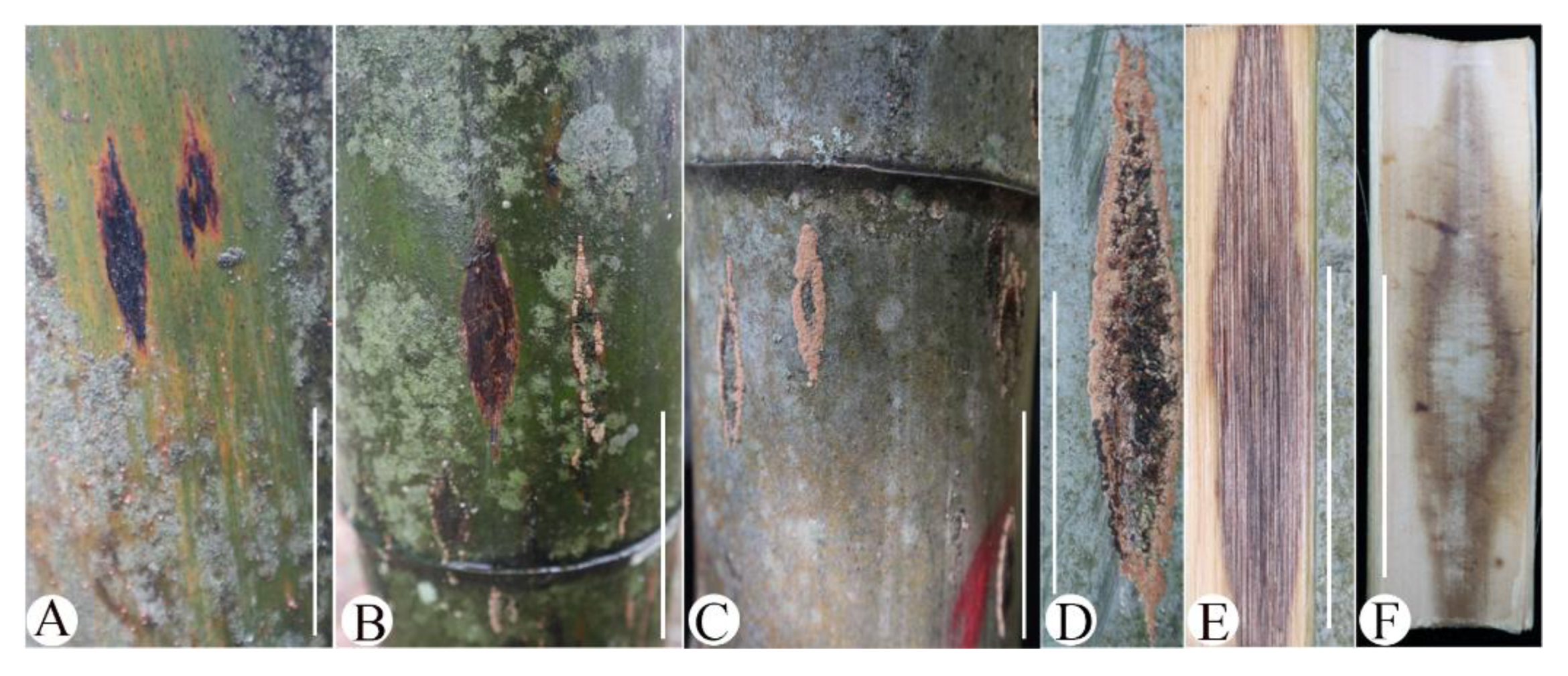
3.1. Symptoms Description, Fungal Isolation, and Pathogenicity

From the field survey, rhomboid rot culms were widespread in the Moso bamboo forest cultured at an altitude above 800 m. Symptoms developed on the culm of the Moso bamboo, especially on the middle and lower culms of aged (five-year-old and above) Moso bamboo. The typical symptoms started with black spots and irregular shapes that expanded vertically into an elongate, fusiform, or rhomboid shape, with diseased spots reaching up to 15 cm in length (Figure 1A,B,D). Necrosis did not only occur on the surface, but also invaded the culm wall and entered the inner cavity of the culm, and the infected area of the bamboo cavity turned black and became necrotic (Figure 1E,F). In addition, macroscopic pink fungal ascomata appeared, surrounding the spots (Figure 1B–D). Diseased spots can spread over the whole Moso bamboo culm when the disease becomes severe, causing the entire Moso bamboo to wilt.
Eleven fungal isolates were isolated from the infected tissue specimens, identified as six different species (Figure S1) according to the colony characteristics. The fungus with pink ascomata could not grow on PDA and was identified as Podonectria according to Figure S2 and Yang et al. [6]. All of the isolates and Podonectria ascospores were inoculated on culm pieces in the laboratory, and only five isolates represented by BBB1 caused black spots around the inoculation sites after 10 days post-incubation (dpi). Moreover, the symptoms were similar to the disease symptoms observed in the wild. The spots turned black both outside and inside the bamboo culm pieces (Figure 2A), and no symptoms developed on the control (Figure 2B). The same fungus was recovered from the experimental inoculation. The isolates were deposited in the Forest Protection Institute of Fujian Agriculture and Forestry University, Fuzhou City, Fujian Province, China.
In the field, isolate BBB1 could infect the one-year-old Moso bamboo culms and cause culm necrosis after 14 dpi (Figure 2E). The spots on the five-year-old culms extended to a rhomboid shape of approximately 2.8 cm (length) × 1.0 cm (width) in size after four months post-inoculation (Figure 2D). The same fungus was reisolated from the transitional area between the diseased and healthy tissues based on morphological and molecular characterizations.

3.2. Morphological Characteristics of Pathogenic Fungus

On PDA, colonies of BBB1 grew and reached 7.36–7.80 (average of 7.56) cm in diameter after four days of incubation (1.89 cm per day). The colonies were dirty white with aerial mycelia (Figure 3A). On MEA, colonies of BBB1 were white with aerial mycelia (Figure 3B). Both hyphae were smooth, hyaline, branched, and septate on PDA and MEA (Figure 3A,B). The conidiophores were reduced to conidiogenous cells and measured 7.0–12.6 μm × 2.7–4.6 μm (length × width; average of 9.4 × 3.7 μm; n = 50), were pale brown to dark, smooth, and ampulliform (Figure 3C). The conidia were 5.0–9.3 × 3.1–7.3 μm (average of 6.4 × 5.4 μm; n = 100), single-celled, brown to dark, smooth, anomalous in shape (globose to subglobose), and sometimes with an equatorial slit (Figure 3C,D). This fungus was identified as a member of the genus Arthrinium.

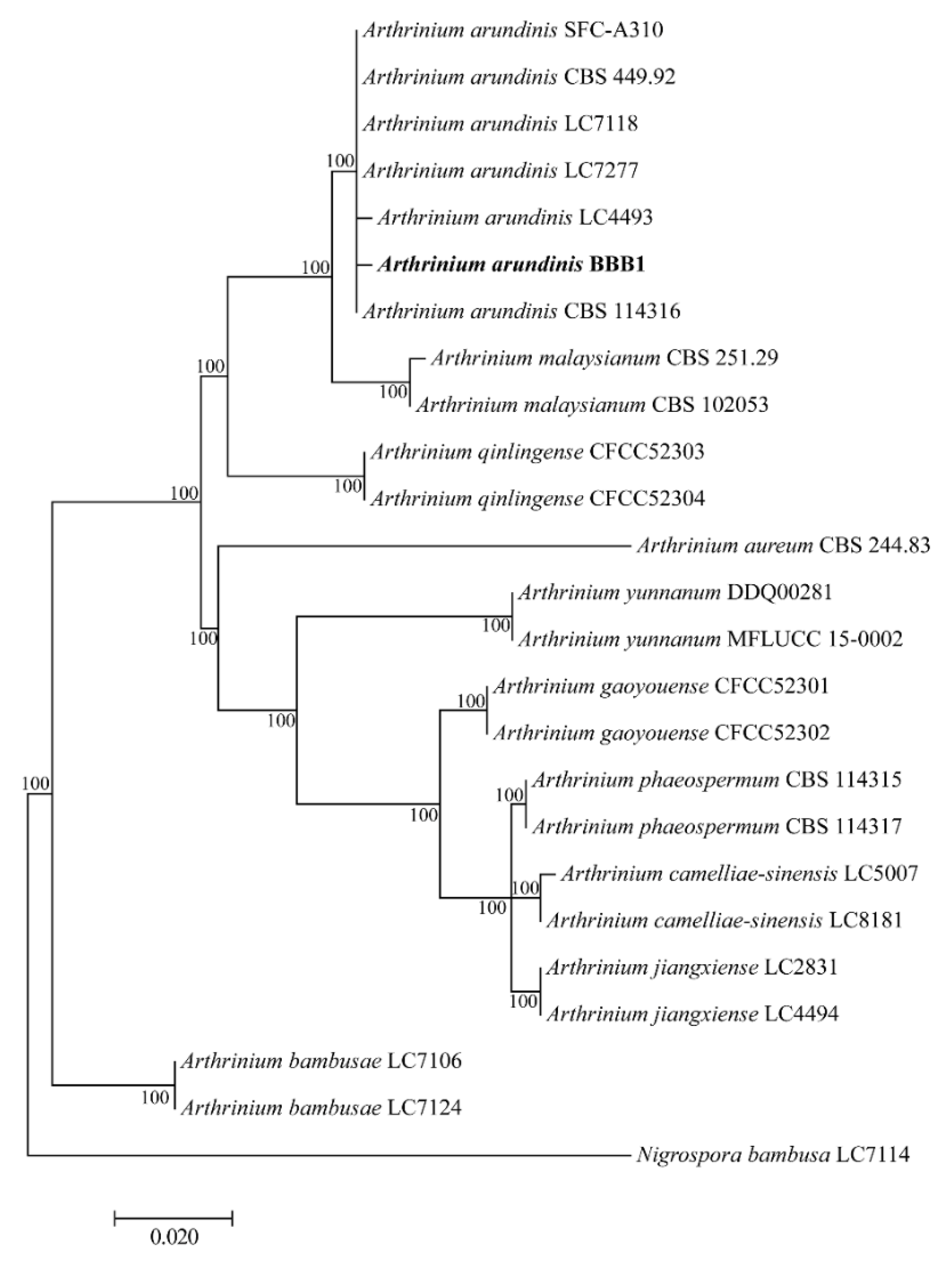
3.3. Phylogenetic Analysis of the Pathogenic Fungus

The partial sequences ITS (547 bp), TEF1-α (323 bp), and TUB2 (509 bp) of the isolate BBB1 were obtained. The sequences were kept in the GenBank database with accession numbers: OK639113, OL944302, and OL944301, respectively. The ITS sequence of BBB1 showed a 99.8% similarity with A. arundinis strains such as DUCC7561 (MT582801) and ZMQRS8 (MR446201). The TEF1-α sequence showed a 99.0% similarity with A. arundinis strains EGG3 (MF627423) and CBS:732.71 (KF145002). The TUB2 sequence showed 98.8% similarity with A. arundinis strains CBS:732.71 (KF144980) and CBS:464.83 (KF144979). The phylogenetic analyses based on the concatenated ITS–TEF1-αTUB2 sequences showed that the isolate BBB1 clustered with A. arundinis type specimens with high support (Figure 4). Collectively, the morphology, molecular characterization, and pathogenicity test confirmed A. arundinis as the causal agent of Moso bamboo culm rhomboid rot.

3.4. Sensitivity of A. arundinis BBB1 to Fungicides

The sensitivity of A. arundinis isolate BBB1 from Moso bamboo against six commonly used fungicides was determined with mycelial growth assays in vitro. The six tested fungicides were difenoconazole, hymexazol, iprodione, mancozeb, prochloraz, and propiconazole, and their EC50 values were 0.195, 170.520, 5.627, 267.514, 0.019, and 4.324 mg/L, respectively (Table 2). Prochloraz exhibited the most potent inhibition against the mycelial growth of the pathogen isolate BBB1, with an EC50 value of 0.019 mg/L. Another effective fungicide was difenoconazole, with an EC50 value of 0.195 mg/L. Meanwhile, hymexazol and mancozeb needed high concentrations to inhibit the growth of the pathogen isolate BBB1.

4. Discussion

Arthrinium is a globally distributed genus inhabiting a wide range of hosts as saprophytes and endophytes on plants. The genus also includes phytopathogenic species such as A. arundinis, which causes kernel blight of barley in the USA, and A. marii, which causes olive tree dieback in Italy [27,35]. Recently, some new species and/or new bamboo hosts have been reported [5,17,18,20,21]. In this study, A. arundinis was isolated from the culms of Moso bamboo (P. edulis) and confirmed as the pathogen of culm rhomboid rot by fulfilling Koch’s postulates. A. arundinis has been reported to cause the brown culm streaks on P. praecox, which the streaks expanding to a maximum of 0.3 cm [17], but we found that A. arundinis infected Moso bamboo causing lesions of up to 15 cm in length and invading the inside of the culms. The significant differences in the disease symptoms may be due to the different hosts or differences in the pathogenicity of the pathogens. In addition, similar behaviors were not only reported for Arthrinium on Moso bamboo but Fusarium oxysporum has been reported to cause culm rhomboid rot in Moso bamboo in Jiangxi Province, China [9]. Unfortunately, none of the Fusarium strains (Figure S1B–E) we isolated exhibited pathogenicity. To the best of our knowledge, this is the first report of A. arundinis causing culm rhomboid rot of Moso bamboo in China or worldwide.
According to our survey, the disease seriously occurred on aged (five-year-old and above) Moso bamboo in the wild; in the artificial inoculation test, A. arundinis could cause disease in new (one-year-old) and aged (five-year-old) Moso bamboo culm, and wounds were required for disease occurrence both in the laboratory and under field conditions (inoculation without wounds could not induce rot; data not shown). Furthermore, we found that swarms of scale insects inhabit aged Moso bamboo culms (Figure S2A,B); do the biting spots caused by scale insects contribute to A. arundinis infection? It has long been assumed that F. verticillioides is an opportunistic fungus, where it takes advantage of the openings left by Diatraea saccharalis caterpillar attack to infect sugarcane. One report presented by Franco et al. in 2021 showed that volatile compounds from F. verticillioides attract D. saccharalis caterpillars. Once they become adults, the fungus is transmitted vertically to their offspring, which continues the cycle by inoculating the fungus in healthy sugarcane [36]. The interaction among bamboo, scale insects, and A. arundinis needs further investigation. Based on this, we infer that the management of scale insects on aged Moso bamboo may be important for controlling culm rhomboid rot disease.
Initially, we supposed that the pink fruitbody fungi around the diseased spots were the pathogen. However, the pure culture could not be obtained, and the ascospores collected from the fruitbody could not induce the rot in Moso bamboo culms. The non-pathogenic pink fruitbody fungi were found around the rot spots on Moso bamboo in Fujian Province and identified as Podonectria based on its morphological characteristics and habitats (Figure S2C–E,H,I). It was difficult to obtain a pure colony of Podonectria, a fungal genus, under cultural conditions and which used to be entomopathogenic fungi of armored scale insects [37,38,39]. Podonectria has also been reported on P. heteroclada from Sichuan Province, China, considered to be a factor that expands necrotic spots [6,40]. We found that Podonectria not only colonized around the necrotic spots, but it was also widely distributed via armored scale insects (Figure S2A,B). The function of Podonectria in this disease is unknown, and the relationship and interactions among A. arundinis, Podonectria, and armored scale insects should be investigated further. During the field survey, we found that the surface of the Moso bamboo culms was easily covered with a complex of dirt, moss, and sooty mold under a highly humid environment, which resulted in the diseased spots hardly being observed, but the pink ascomata of Podonectria around the necrosis were obvious (Figure S2F,G). It is certain that Podonectria acted as a marker for culm rhomboid rot spots in the early stage.
Several species of Arthrinium have been documented as plant pathogens, although information on pathogen biology and disease epidemiology and management is generally lacking [35]. The sensitivity of A. arundinis BBB1 against six commonly used fungicides (difenoconazole, prochloraz, propiconazole, iprodione, hymexazol, and mancozeb) was evaluated. Prochloraz exhibited the highest toxicity to fungal growth. Prochloraz is a DMI (demethylation inhibitor)-imidazole fungicide with a biochemical mode of action based on the inhibition of the ergosterol biosynthesis pathway and has been used as a protectant or eradicate [41,42]. Another effective fungicide is difenoconazole, which is a triazole fungicide. Considering the production of resistance to prochloraz, we recommend that prochloraz and difenoconazole be applied alternately for disease management.
The field survey found that disease occurrence is very complex. Although we determined the pathogen and provided some reasonable fungicides based on a sensitivity test of the pathogen indoors, the plant–pathogen–scale insect interaction also needs to be paid more attention in the field control.

5. Conclusions

In conclusion, we confirmed the causal agent of culm rhomboid rot disease in Moso bamboo and identified the pathogen as A. arundinis according to its morphology and phylogenetic analysis. This is the first report of A. arundinis as the pathogen of Moso bamboo culm rhomboid rot. Wounds on the Moso bamboo culm surface were required for disease occurrence. A. arundinis was more sensitive to the fungicides prochloraz (Ec50 value of 0.019 mg/L) and difenoconazole (EC50 value of 0.195 mg/L). This study could provide a scientific basis for the prevention and control of Moso bamboo culm rhomboid rot disease in Fujian Province, China.

Supplementary Materials

The following supporting information can be downloaded at: https://www.mdpi.com/article/10.3390/f13101616/s1, Figure S1: Six representative strains of 11 isolates obtained from the diseased Moso bamboo culms. Figure S2: Habitats and morphological characteristics of Podonectria. A and B, The fungi and nymph in the wild: pink (the fruiting body of fungi), and white (the nymph of insects); C, Ascomata; D, Longitudinal section of stroma; E, Pseudoparaphyses and Asci; F and G, Diseased spots covered with dirt; H, Asci; I, Ascospore; Scale bar: D–E: 20 μm; H–I: 10 μm.

Author Contributions

Conceptualization, Q.Z., Z.S. and F.Z.; methodology, software, validation, S.Z.; formal analysis, S.Z., H.Z., Y.L. and Q.Z.; investigation, S.Z. and Z.S.; writing—original draft preparation, S.Z.; writing—review and editing, Q.Z.; visualization, H.Z. and Y.L.; supervision, project administration, and funding acquisition, Q.Z. All authors have read and agreed to the published version of the manuscript.

Funding

This research was funded by the Science and Technology Innovation Project of Straits Science and Technology Research Institute (PT2021009) and the National Natural Science Foundation of China (31901312).

Data Availability Statement

Not applicable.

Acknowledgments

Thanks to Zhongyi Wu, Huajiashan Moso Bamboo Farm, for providing disease samples. The authors are grateful to the anonymous reviewers and Academic Editor for their valuable comments on the revision of this article.

Conflicts of Interest

The authors declare no conflict of interest.

References

  1. Kellogg, E.A. Flowering Plants. Monocots; Kubitzki, K., Ed.; Springer: Hamburg, Germany, 2015; pp. 87–91. [Google Scholar] [CrossRef]
  2. Wang, S.; Chen, T.H.; Liu, E.U.; Liu, C.P. Accessing the nursing behaviour of moso bamboo (Phyllostachys edulis) on carbohydrates dynamics and photosystems. Sci. Rep. 2020, 10, 1015. [Google Scholar] [CrossRef] [Green Version]
  3. Ramakrishnan, M.; Yrjl, K.; Vinod, K.K.; Sharma, A.; Zhou, M. Genetics and genomics of moso bamboo (Phyllostachys edulis): Current status, future challenges, and biotechnological opportunities toward a sustainable bamboo industry. Food Energy Secur. 2020, 9, e229. [Google Scholar] [CrossRef]
  4. Song, X.; Chen, X.; Zhou, G.; Jiang, H.; Peng, C. Observed high and persistent carbon uptake by moso bamboo forests and its response to environmental drivers. Agr. Forest Meteorol. 2017, 247, 467–475. [Google Scholar] [CrossRef]
  5. Xu, M.Q.; Dai, Y.C.; Fan, S.H.; Jin, L.X.; Lu, Q.; Tian, G.Z.; Wang, L.F. Records of bamboo diseases and the taxonomy of their pathogens in China (I). For. Res. 2006, 19, 692–699. (In Chinese) [Google Scholar]
  6. Yang, C.L.; Xu, X.; Liu, Y. Podonectria Sichuanensis, a potentially mycopathogenic fungus from Sichuan Province in China. Phytotaxa 2019, 402, 219–231. [Google Scholar] [CrossRef]
  7. Yang, C.L.; Xu, X.L.; Wanasinghe, D.N.; Jeewon, R.; Phookamsak, R.; Liu, Y.G.; Liu, L.J.; Hyde, K.D. Neostagonosporella sichuanensis gen. et sp. nov. (Phaeosphaeriaceae, Pleosporales) on Phyllostachysheteroclada (Poaceae) from Sichuan Province, China. MycoKeys 2019, 46, 119–150. [Google Scholar] [CrossRef] [PubMed]
  8. Yang, C.L.; Baral, H.O.; Xu, X.; Liu, Y. Parakarstenia phyllostachydis, a new genus and species of non-lichenized Odontotremataceae (Ostropales, Ascomycota). Mycol. Prog. 2019, 18, 833–845. [Google Scholar] [CrossRef]
  9. Wang, H.X.; Xu, X.J.; Peng, J.S.; Liu, Y.X.; Zhang, Z.B. Isolation and identification of pathogen infecting moso bamboo in Jinggangshan. J. Bamboo Res. 2012, 31, 42–46. (In Chinese) [Google Scholar]
  10. Ramos, H.P.; Braun, G.H.; Pupo, M.T.; Said, S. Antimicrobial activity from endophytic fungi Arthrinium state of Apiospora montagnei Sacc. and Papulaspora immersa. Braz. Arch. Biol. Technol. 2010, 53, 629–632. [Google Scholar] [CrossRef]
  11. Crous, P.W.; Groenewald, J.Z. A phylogenetic re-evaluation of Arthrinium. IMA Fungus 2013, 4, 133–154. [Google Scholar] [CrossRef] [Green Version]
  12. Wang, M.; Tan, X.M.; Liu, F.; Cai, L. Eight new Arthrinium species from China. Mycokeys 2018, 34, 1–24. [Google Scholar] [CrossRef]
  13. Pintos, A.; Alvarado, P.; Planas, J.; Jarling, R. Six new species of Arthrinium from Europe and notes about A. caricicola and other species found in Carex spp. hosts. Mycokeys 2019, 49, 15–48. [Google Scholar] [CrossRef] [PubMed] [Green Version]
  14. Kallol, D.; Lee, S.Y.; Choi, H.W.; Eom, A.H.; Cho, Y.J.; Jung, H.Y. Taxonomy of Arthrinium minutisporum sp. nov., Pezicula neosporulosa, and Acrocalymma pterocarpi: New Records from Soil in Korea. Mycobiology 2020, 6, 450–463. [Google Scholar] [CrossRef]
  15. Senanayake, I.C.; Bhat, J.D.; Cheewangkoon, R.; Xie, N. Bambusicolous Arthrinium Species in Guangdong Province, China. Front. Microbiol. 2020, 11, 602773. [Google Scholar] [CrossRef]
  16. Kwon, S.L.; Park, M.S.; Jang, S.; Lee, Y.M.; Kim, J.J. The genus Arthrinium (Ascomycota, Sordariomycetes, Apiosporaceae) from marine habitats from Korea, with eight new species. IMA Fungus 2021, 12, 1–26. [Google Scholar] [CrossRef]
  17. Chen, K.; Wu, X.Q.; Huang, M.X.; Han, Y.Y. First report of brown culm streak of Phyllostachys praecox caused by Arthrinium arundinis in Nanjing, China. Plant Dis. 2014, 98, 1274. [Google Scholar] [CrossRef]
  18. Li, B.J.; Liu, P.Q.; Jiang, Y.; Weng, Q.Y.; Chen, Q.H. First report of culm rot caused by Arthrinium phaeospermum on Phyllostachys viridis in China. Plant Dis. 2016, 100, 1013. [Google Scholar] [CrossRef]
  19. Dai, D.Q.; Phookamsak, R.; Wijayawardene, N.N.; Li, W.J.; Bhat, D.J.; Xu, J.C.; Taylor, J.E.; Hyde, K.D.; Chukeatirote, E. Bambusicolous fungi. Fungal Divers. 2017, 82, 1–105. [Google Scholar] [CrossRef]
  20. Yang, C.L.; Xu, X.; Liu, Y. First report of bamboo blight disease caused by Arthrinium yunnanum on Phyllostachys heteroclada in Sichuan, China. Plant Dis. 2018, 103, 1026. [Google Scholar] [CrossRef]
  21. Yang, C.L.; Xu, X.; Liu, Y. First report of bamboo blight disease caused by Arthrinium pseudoparenchymaticum on Dendrocalamus latiflorus in Sichuan, China. Plant Dis. 2019, 103, 1411. [Google Scholar] [CrossRef]
  22. Feng, Y.; Liu, J.K.J.; Lin, C.G.; Chen, Y.Y.; Liu, Z.Y. Article additions to the genus Arthrinium (Apiosporaceae) from bamboos in China. Front. Microbiol. 2021, 12, 661281. [Google Scholar] [CrossRef] [PubMed]
  23. Ellis, M.B. Dematiaceous Hyphomycetes. Commonwealth Mycological Institute. Kew. 1971, pp. 608. Available online: https://www.scirp.org/(S(lz5mqp453edsnp55rrgjct55))/reference/ReferencesPapers.aspx?ReferenceID=2024742 (accessed on 28 September 2022).
  24. Hyde, K.D.; Fröhlich, J.; Taylor, J.E. Fungi from palms. XXXVI. Reflections on unitunicate Ascomycetes with Apiospores. Sydowia 1998, 50, 21–80. [Google Scholar]
  25. Ji, Z.L.; Zhang, S.W.; Zhu, F.; Wan, B.X.; Liang, R.Z. First report of Arthrinium arundinis causing leaf edge spot of peach in China. Plant Dis. 2020, 104, 11. [Google Scholar] [CrossRef]
  26. Kuberan, T.; Cheng, L.L.; Deng, C.; Deng, W.W.; Zhang, Z. First report of leaf blight caused by Arthrinium arundinis on tea plants in China. Plant Dis. 2019, 103, 12. [Google Scholar] [CrossRef]
  27. Cano, C.M. First report of Arthrinium arundinis causing kernel blight on barley. Plant Dis. 1992, 76, 1077B. [Google Scholar] [CrossRef]
  28. Bagherabadi, S.; Zafari, D.; Anvar, F.G. First report of leaf spot caused by Arthrinium arundinis on rosemary in Iran. J. Plant Pathol. 2014, 96, 126. [Google Scholar] [CrossRef]
  29. Koch, R. Ueber den augenblicklichen Stand der bakteriologischen Choleradiagnose. Z. Für Hyg. Und Infekt. 1893, 14, 319–338. [Google Scholar] [CrossRef] [Green Version]
  30. White, T.J.; Bruns, T.; Lee, S.J.W.T.; Taylor, J.L. Amplification and direct sequencing of fungal ribosomal RNA genes for phylogenetics. In PCR Protocols: A Guide to Methods and Applications; Innis, M.A., Gelfand, D.H., Sninsky, J.J., White, T.J., Eds.; Academic Press: San Diego, CA, USA, 1990; pp. 315–322. [Google Scholar] [CrossRef]
  31. Carbone, I.; Kohn, L.M. A method for designing primer sets for speciation studies in filamentous Ascomycetes. Mycologia 1999, 91, 553–556. [Google Scholar] [CrossRef]
  32. Glass, N.L.; Donaldson, G.C. Development of primer sets designed for use with the PCR to amplify conserved genes from Filamentous Ascomycetes. Appl. Environ. Microb. 1995, 61, 1323–1330. [Google Scholar] [CrossRef] [Green Version]
  33. Huang, L.; Yang, J.Y.; Zhu, Y.N.; Zhu, L.H.; Zhang, Y.F.; Ye, J.R.; Li, D.W. Canker on culm of Bambusa multiplex (Lour.) Raeusch. ex Schult. caused by Fusarium incarnatum (Roberge) Sacc. J. Phytopathol. 2019, 167, 91–97. [Google Scholar] [CrossRef] [Green Version]
  34. Kumar, S.; Stecher, G.; Tamura, K. Mega7: Molecular evolutionary genetics analysis version 7.0 for bigger datasets. Mol. Biol. Evol. 2015, 33, 1870–1874. [Google Scholar] [CrossRef] [PubMed]
  35. Gerin, D.; Nigro, F.; Faretra, F.; Pollastro, S. Identification of Arthrinium marii as causal agent of Olive tree dieback in Apulia (Southern Italy). Plant Dis. 2020, 104, 694–701. [Google Scholar] [CrossRef] [PubMed]
  36. Franco, F.P.; Túler, A.C.; Gallan, D.Z.; Gonçalves, F.G.; Favaris, A.P.; Peñaflor, M.F.G.V.; Leal, W.S.; Moura, D.S.; Bento, J.M.S.; Silva-Filho, M.C. Fungal phytopathogen modulates plant and insect responses to promote its dissemination. ISME J. 2021, 15, 3522–3533. [Google Scholar] [CrossRef] [PubMed]
  37. Rossman, A.Y. Podonectria, a genus in the Pleosporales on scale insects. Mycotaxon 1978, 7, 163–182. [Google Scholar]
  38. Dao, H.T.; Beattie, G.; Rossman, A.Y.; Burgess, L.W.; Holford, P. Four putative entomopathogenic fungi of armoured scale insects on Citrus in Australia. Mycol. Prog. 2016, 15, 47. [Google Scholar] [CrossRef]
  39. Xu, X.L.; Zeng, Q.; Lv, Y.C.; Jeewon, R.; Yang, C.L. Insight into the systematics of novel entomopathogenic fungi associated with armored scale insect, Kuwanaspis howardi (Hemiptera: Diaspididae) in China. J. Fungi. 2021, 7, 628. [Google Scholar] [CrossRef]
  40. Qi, R.; Yang, C.; Li, L.; Liu, C.; Xu, X.; Liu, Y.; Zeng, Q.; Liu, L.; Liu, Y. Pathogenic characteristics, biological characteristics and drug control test of pathogen causing rhombic-spot of Phyllostachys heteroclada. J. Northeast. For. Univ. 2021, 49, 131–135, 141. (In Chinese) [Google Scholar]
  41. Ma, Z.; Michailides, T.J. Advances in understanding molecular mechanisms of fungicide resistance and molecular detection of resistant genotypes in phytopathogenic fungi. Crop Prot. 2005, 24, 853–863. [Google Scholar] [CrossRef]
  42. Mora-Aguilera, J.A.; Ríos-López, E.G.; Yáez-Zúiga, M.; Rebollar-Alviter, A.; Tovar-Pedraza, J.M. Sensitivity to MBC fungicides and prochloraz of Colletotrichum gloeosporioides species complex isolates from mango orchards in Mexico. J. Plant Dis. Protect. 2021, 128, 481–491. [Google Scholar] [CrossRef]
Figure 1. Symptoms of moso bamboo (Phyllostachys edulis) culm rhomboid rot disease in bamboo forests. Scale bar: 5 cm. (A) Black spots and rhomboid shape symptoms on the outer surface; (B) Symptoms covered with a complex of dirt, moss, and sooty mold; (C) Macroscopic pink fungal ascomata appeared, surrounding the spots; (D) Rhomboid shape symptoms with sooty mold and pink fungal ascomata; (E,F) Necrosis occur on the inner cavity of the culm.
Forests 13 01616 g001
Figure 2. Pathogenicity test. (A) Symptoms on Moso bamboo culm inoculated with the isolate BBB1 after 10 days; (B) Control Moso bamboo culm inoculated with PDA plug; (C) Symptoms on 5-year-old Moso bamboo inoculated with the isolate BBB1 after 4 months, the upper three wounds inoculated with the isolate BBB1, and the lower one with PDA plug; (D) Details under the diseased tissues; (E) Symptoms on 1-year-old Moso bamboo inoculated with the isolate BBB1 after 14 days; (F) 1-year-old control Moso bamboo inoculated with PDA plug.
Forests 13 01616 g002
Figure 3. Morphology of the isolate BBB1. (A) Colony morphology of 5-day-old on PDA; (B) Colony morphology of 5-day-old on MEA; (C) Conidiogenous cells; (D) Conidia; Scale bars: 10 μm.
Forests 13 01616 g003
Figure 4. The phylogenetic tree of the isolate BBB1 with allied taxa analyzed using concatenated sequences of ITS, TEF1-α, and TUB2 with the maximum likelihood method. Nigrospora bambusa was used as the outgroup.
Forests 13 01616 g004
Table 1. Details of Arthrinium. spp. used in this study.
SpeciesStrain *SubstratesOriginGeneBank Accession Number
ITSTEFTUB
Arthirinium arundinisBBB1BambooChinaOK639113OL944302OL944301
A. arundinisLC4493Phyllostachys sp.ChinaKY494689KY705088KY806202
A. arundinisLC7277BambooChinaKY494750KY705146KY705218
A. arundinisLC7118BambooChinaKY494723KY705120KY705191
A. arundinisSFC-A310Beach SandKoreaMH498551MH544682MH498509
A. arundinisCBS 449.92BambooCanadaKF144887KF145019KF144977
A. arundinisCBS 114316Hordeum vulgareIranKF144884KF145016KF144974
A. aureumCBS 244.83AirSpainAB220251KF145023KF144981
A. bambusaeLC7106BambooChinaKY494718KY806204KY705186
A. bambusaeLC7124BambooChinaKY494727KY806206KY705195
A. camelliaesinensisLC5007Camellia sinensisChinaKY494704KY705103KY705173
A. camelliaesinensisLC8181Brassica rapaChinaKY494761KY705157KY705229
A. gaoyouenseCFCC 52301Phragmites australisChinaMH197124MH236793MH236789
A. gaoyouenseCFCC 52302Phragmites australisChinaMH197125MH236794MH236790
A. jiangxienseLC2831BambooChinaKY494686KY705085KY806201
A. jiangxienseLC4494Phyllostachys sp.ChinaKY494690KY705089KY705160
A. malaysianumCBS 251.29Cinnamomum camphoraMalaysiaKF144897KF145031KF144989
A. malaysianumCBS 102053Macaranga hullettiiMalaysiaKF144896KF145030KF144988
A. phaeospermumCBS 114315Hordeum vulgareIranKF144905KF145039KF144997
A. phaeospermumCBS 114317Hordeum vulgareIranKF144906KF145040KF144998
A. qinlingenseCFCC 52303Fargesia qinlingensisChinaMH197120MH236795MH236791
A. qinlingenseCFCC 52304Fargesia qinlingensisChinaMH197121MH236796MH236792
A. yunnanumDDQ00281Phyllostachys nigraChinaKU940148N/AN/A
A. yunnanumMFLUCC15-1002Phyllostachys nigraChinaKU940147N/AN/A
Nigrospora BambusaLC7114BambooChinaKY385307KY385313KY385319
* CBS: Westerdijk Fungal Biodiversity Institute, Utrecht, Netherlands; CFCC: China Forestry Culture Collection Center, Beijing, China; DDQ: D.Q. Dai; LC: personal culture collection of Lei Cai, housed in the Institute of Microbiology, Chinese Academy of Sciences, China; MFLUCC: Mae Fah Luang University Culture Collection, Chiang Rai, Thailand; SFC: the Seoul National University Fungus Collection. Sequences in bold were generated in this study.
Table 2. Toxicity of six fungicides against the mycelial growth of Arthrinium arundinis isolate BBB1.
FungicideRegression EquationEC50 * (95% CL)/mg/LCorrelation Coefficient (r)
Difenoconazole y = 1.05 + 1.48 x0.195 (0.130–0.263)0.991
Hymexazol y = −9.09 + 4.07 x170.520 (152.558–193.500)0.988
Iprodione y = −8.3 + 11.06 x5.627 (5.377–5.873)0.992
Mancozeb y = −10.87 + 4.48 x267.514 (240.548–296.180)0.971
Prochloraz y = 2.69 + 1.56 x0.019 (0.014–0.025)0.989
Propiconazole y = −1.27 + 2 x4.324 (3.462–5.659)0.977
* EC50 = median effective concentration; CL = confidence limit.
Publisher’s Note: MDPI stays neutral with regard to jurisdictional claims in published maps and institutional affiliations.

