Article

Effect of an Ectomycorrhizal Fungus on the Growth of Castanea henryi Seedlings and the Seasonal Variation of Root Tips’ Structure and Physiology

1
Key Laboratory of Cultivation and Protection for Non-Wood Forest Trees of Ministry of Education, College of forestry, Central South University of Forestry and Technology, Changsha 410004, China
2
Texas A&M AgriLife Research and Extension Center, 17360 Coit Rd, Dallas, TX 75252, USA
*
Author to whom correspondence should be addressed.
Forests 2021, 12(12), 1643; https://doi.org/10.3390/f12121643
Submission received: 23 August 2021 / Revised: 5 November 2021 / Accepted: 24 November 2021 / Published: 26 November 2021
(This article belongs to the Section Forest Ecophysiology and Biology)

Abstract

:
Castanea henryi is a ubiquitous hardwood chestnut species in southern China and is important both ecologically and economically. It is mainly cultivated for nut production, just like other chestnut species. However, the establishment of C. henryi seedlings in a new orchard has proven to be difficult because few seedlings survive transplanting due to the incompatibility of their coarse root architecture with nutrient-depleted red acid soils in southern China. Root architecture can be profoundly modified and nutrient can be stress alleviated due to the association of roots with ectomycorrhizal (ECM) fungi. Boletus edulis is an ECM fungus with edible and medicinal fruiting bodies. However, its impact on plant growth varies with the plant species it is associated with. In order to elucidate the role of B. edulis in C. henryi afforestation, we evaluated growth parameters and soil enzymatic activities, as well as seasonal variations in physiology and structure of ECM root tips. Growth responses and soil enzymatic activities were measured 6 months after inoculation. The physiological characteristics of root tips were also compared at various seasons throughout the year. B. edulis colonization of C. henryi roots was successful at a 60% colonization rate. Height, base diameter, and biomass (especially the underground part) of inoculated seedlings (JG) were higher than those of uninoculated seedlings (CK). JG had higher root total length, root surface area, root volume, root average diameter, and number of root tips than CK. Additionally, JG exhibited higher total nitrogen and phosphorus content. Abnormal mantle and Harting net were observed in winter. No matter the season, ECM tips had higher antioxidant enzyme activities, root activities, soluble protein content, and lower malondialdehyde compared to non-ECM tips (nE) and those without ECM tips (woE), and there were no differences between nE and woE. It is important to understand the growth of the host plant in response to ECM and that the seasonal variation of ECM root tips is important when growing high-quality C. henryi seedlings, due to the crucial role of B. edulis in improving seedling initial survival rate.

1. Introduction

Chinquapin, Castanea henryi (Skam) Rehder & E.H. Wilson, is important economically for its timber and nuts. Chinquapin is widely distributed in southern China, especially in Fujian, Zhejiang, and Hunan provinces [1,2]. Chinquapin nuts have many desirable characteristics, including a high starch content, a high mineral nutrition content, and eight amino acids important for human health [1,3]. Planting chinquapin for its nut harvest has been the main economic income revenue for many growers in southern China. However, nutrient depletion, common in hilly areas in southern China [4], combined with the frequency of extreme weather has led to a long initial period of seedling establishment or survival stage and a low survival rate of chinquapin seedlings in afforestation [5].
Chinquapin, like other members of the family Fagaceae, forms ectomycorrhizal (ECM) associations with members of the Ascomycota and Basidiomycota fungi [6]. This symbiosis is a ubiquitous mutualism that plays a key role in plant and soil health and in carbon and nutrient cycles [7,8]. Several studies have demonstrated that inoculating with ECM fungi is an effective method to improve seedling resistance to biotic and abiotic stresses, to promote growth, shorten the survival stage, and improve the survival rate in afforestation [9,10,11,12,13,14,15,16,17,18]. In addition, since some ECM fungi have a high economic value as an edible or medicinal product, the use of ECM as a practice in agroforestry has the potential to increase grower income [17,19].
Many studies showed that inoculating with ECM fungi resulted in larger plants, compared to the non-inoculated control [12,13,14]. However, other experiments resulted in smaller plants [16,20,21,22]. The role and effect of ECM on plant growth differed depending on the associated fungal species [17,22,23], the nutritional status of the plant [16,24], and the environment [11,22]. Previous studies have primarily focused on the effects of B. edulis on C. mollissima and Pinus species with inconsistent results [13,22,25]. Therefore, the benefit of B. edulis inoculation in C. henryi is unknown. Such a benefit is of great importance if B. edulis can be used for C. henryi afforestation or as a general practice in agroforestry.
Harley [26] has shown that ECM structures are not static organs but change ontogenetically from a juvenile to a mature state and finally senesce. These structural changes during development are concordant with variations in physiological metabolism of plant cells. Tagu et al. [27] and Acioli-Santos et al. [28] demonstrated that the structural modifications of ECM are influenced by environmental conditions and genetic compatibility of the partner plants. Seasonal variations in temperature, precipitation, and physiology and growth of plants affect the balance of free radical metabolism in plant cells [29], especially in winter (mainly due to chilling or freezing stresses) and summer (mainly due to drought, heat, or flooding stresses). Seasonal variations also result in the accumulation of reactive oxygen species (ROS), which cause lipid peroxidation, enzyme inhibition, and programmed cell death [30,31,32]. Many studies suggest that ECM fungi can effectively improve plant resistance to abiotic stresses [9,11,18], which may be mediated by combinations of improved nutrient and water relations, as well as the modulation of reactive oxygen species’ metabolism.
In this study, we hypothesize that B. edulis promotes the growth of C. henryi seedlings and that the structure and physiology of ECM root tips change seasonally. The aim of this research is to evaluate the feasibility of B. edulis in C. henryi for agroforestry. The major objectives are to (1) study the effects of inoculation with B. edulis on the growth of C. henryi and (2) characterize the seasonal dynamics in the vegetative structures and the physiological characteristics of ECM.

2. Material and Methods

2.1. Study Site

The study was conducted in Changsha city, central south China. The climate in the area is subtropical monsoon with abundant rainfall and heat. Rain is mainly in the summer, and the average annual precipitation is about 1361 mm. Long summers (118–127 days) and winters (117–122 days) and short springs (61–64 days) and autumns (59–69 days) are typical for Changsha. The average minimum temperature in January is 4.4 °C. The study lasted 19 months (January 2018 to July 2019). The total precipitation between January and July was higher in 2019 (1078 mm) than in 2018 (766 mm) (Figure 1).

2.2. Seedlings Production in the Nursery

In January 2018, seeds of C. henryi from Hunan Province were rinsed with tap water for 30 min and then soaked in 2% potassium permanganate solution for 30 s before storage in sand. During the storage period, sand moisture was maintained at 10% (w/w). In March 2018, when roots and stems had emerged and reached at least 5 cm in length, seedlings were transferred to cylindrical plastic pots with a top diameter of 16 cm and height of 18 cm. The pots were filled with a mixture of loess, peat (Klasmann, Niedersachsen, Germany), perlite, and vermiculite 4:1:1:1 (v/v/v/v), with 1710 mg kg−1 total N, 218 mg kg−1 total P, 6958 mg kg−1 total potassium (K), and a pH of 5.6. The substrate mixture was exposed to the sun for 7 days before loading to pots.

2.3. Inoculum Production and Seedling Inoculation

Boletus edulis was provided by China General Microbiological Culture Collection Center. Mycelia of B. edulis was first grown on modified Melin-Norkrans agar (MMN) for 4 weeks at 25 °C. Rounded pieces of mycelia (4 mm diam.) were taken from the colony margin and grown in sterilized MMN liquid culture (25 °C, 180 rev min−1) for 21 days. The liquid was then injected into a sterilized substrate of peat:perlite:sand mixture of 8:1:1 (v/v/v) and was cultured for 45 days at 25 °C. When the seedlings were transferred to the pots 1 month later, the inoculations were made by a manual injection of 10 g of B. edulis substrate per seedling for a total of 150 inoculated seedlings (JG). Another 150 non-inoculated seedlings were used as control plants (CK). They were watered with sterile water and shaded when necessary. No additional fertilizers were applied to the plants during the duration of the trials.

2.4. ECM Colonization, Plant Growth, Root Architecture, and N and P Content

At 6months after inoculation, 10 seedlings were randomly selected from each treatment to characterize ECM colonization, according to the protocol of Lu et al. [22], and to collect data on plant morphology, biomass accumulation, root architecture, and tissue N and P content. Plant morphology was assessed by measuring plant height and diameter at the root collar. Seedlings of JG and CK treatments were carefully harvested by removing all substrate attached to the roots. A root scanner (WinRHIZO PRO 2013, Regent Instruments, Quebec, QC, Canada) was used to analyze the root architecture of JG and CK seedlings. The seedlings were oven dried at 105 °C for 30 min and then at 60 °C for 48 h until a constant weight was reached. The dry weight of the aboveground parts (stem, branch, and leaf) and the underground part (root) was measured. After the tissue of each plant part was crushed, total nitrogen was determined using the Kjeldahl method, and total P was determined using the Automated Discrete Analyzers Model Smart Chem 200 after digestion with H2SO4–H2O2 [33].

2.5. Soil Sample Preparation and Enzymatic Activity Assays

Soil adhering to the roots was collected by vigorous shaking of the seedlings. Approximately 100 g of each soil sample, collected from a pooled sample of 5 seedlings, was then passed through a 0.25 mm sieve, each treatment with 3 soil samples. All samples were stored at 4 °C for no more than 7 days before analysis. Urease activity was determined by indophenol colorimetry, and phosphatase activity was determined by phenyl phosphate disodium colorimetry [22]. These enzymes were chosen for their contribution to nutrient absorption.

2.6. Microscopic Observation

After washing the roots, one part was immediately fixed in 2.5% (w/v) glutaraldehyde in phosphate-buffered saline (PBS; pH 7) overnight at 4 °C. The other part was photographed using a stereoscope (SZX16, Olympus, Tokyo, Japan). Root architecture was measured using a scanning electronic microscope (SEM, JEOL JSM-6380LV, Tokyo, Japan), according to the method described by Baptista et al. [34]. Root scanning of the complete root system was not possible due to its large size and presence of lignified roots, so all primary lateral roots and parts of secondary lateral roots were cut to a length of no more than 10 cm, rearranged on the tray of the scanner to ensure that the roots did not block each other. Using the paraffin sectioning method, cut sections were also observed using an optical microscope (BX-51, Olympus, Tokyo, Japan) [35].

2.7. Root Tip Physiological Characteristics

Roots were sampled in September 2018 (autumn), December 2018 (winter), March 2019 (spring), and July 2019 (summer) to characterize their physiology at various seasons during their growth. Root tips from inoculated seedlings with ECM were noted as wECM. Root tips from inoculated seedlings without ECM were noted as woECM. Root tips from uninoculated seedlings were noted as nECM. Each sample consisted of mixed root tips from at least 5 seedlings, each treatment had 3 root tips samples, and each measurement of root tips physiological characteristics was replicated 3 times.

2.7.1. Root Tip Activity and Antioxidant Enzyme Analysis

Plant root activity was determined by the TTC reduction method [36]. About 0.2 g of C. henryi root tip tissue was placed in a calibration test tube of 15 mL, and tissue was fully immersed in a solution of 0.4% TTC and an equal amount of 0.07 mol L−1 phosphate. Test tubes were then kept at 37 °C for 1.5 h. The reaction was terminated by adding 2 mL of 1 mol L−1 sulfuric acid. Finally, the tissue was ground in a mortar with 10 mL ethyl acetate. After centrifugation, the supernatant was measured using the colorimetric method at a wavelength of 485 nm.
The superoxide dismutase (SOD) activity of roots was determined using the nitro blue tetrazolium chloride (NBT) light reduction method [36]. About 0.2 g of roots and 6 mL phosphate buffer (0.05 mol L−1, pH 7.8) were mixed, ground into a homogenate in an ice bath, transferred to a centrifugal tube, and centrifuged at 4 °C and 15,000 rpm for 10 min. The resultant supernatant was the enzyme solution. One test tube was kept in the dark and used as the reference control, and the other test tubes were reacted under a 4000-lx fluorescent lamp for 30 min. After 30 min, absorbance was recorded at 560 nm. One unit (1 U) of SOD activity was defined as the amount of enzyme required to cause 50% inhibition of NBT reduction under the assay conditions used in this trial.
The peroxidase (POD) activity of roots was determined using the guaiacol oxidation method [36]. About 0.2 g of root tissue was ground into a homogenate in an ice bath with 9 mL of phosphate buffer (0.05 mol L−1, pH 7.0) and centrifuged at 4 °C and 4000 rpm for 10 min A total of 3 mL of a mixture consisting of 0.28 µL of guaiacol and 19 µL of 30% H2O2 in 50 mL of phosphate buffer (0.01 mol L−1, pH 6.0) was added to the reaction mixture, and the mixture was stored in a 4 °C refrigerator. Before colorimetric analysis was conducted, 1 mL of enzyme solution and 1 mL of phosphate buffer (0.05 mol L−1, pH 7.0) were added to the control group. Colorimetric analysis was determined immediately afterwards at 470 nm, and readings were collected once every minute for 4 min. An increase of 0.01 per minute in the reading was defined as one unit of enzyme activity (1 U).
The extraction method of the enzyme catalase (CAT) was similar to the POD described above. Each tube had 1.5 mL of phosphate buffer, 0.2 mL of enzyme solution, and 1.0 mL of distilled water. The enzyme solution added to the blank control tube was similar to the sample tube; however, the enzyme activity was first neutralized in a hot water bath for about 1 h before measurement. All sample tubes and blank controls were then preheated to 25 °C for 3 min, and the reaction was initiated by adding 0.3 mL of H2O2. The reaction was then immediately measured using colorimetry at 240 nm, with one reading per minute for 4 min. A decrease of 0.1 per minute was defined as one unit of enzyme activity (1 U).

2.7.2. Oxidative Damage and Protein Measurement

The content of malondialdehyde (MDA) was determined by the thiobarbituric acid (TBA) heating method [36]. The methodology was as follows: the sample of root tips for each treatment was about 0.2 g. Each sample was ground with 10% trichloroacetic acid (TCA) at 4000 rpm for 10 min. Then, 2 mL of the extraction medium was supplemented with 0.6% TBA and boiled in a hot water bath for 10 min. After rapid cooling, the sample was centrifuged at 4000 rpm for 10 min. The content of MDA was calculated by measuring the supernatant colorimetrically at 532 nm and 600 nm.
The soluble protein content was determined using the Coomassie brilliant blue method [36]. The extraction method was the same as that of SOD. A 0.6 mL sample of the culture fluid was mixed with 5 mL Coomassie brilliant blue G-250 reagent and shaken well. After standing for 2 min, absorbance was recorded at 595 nm.

2.8. Statistical Analysis

One-way analysis of variance (ANOVA) was used in the determination of the effects of inoculation and the least significant difference (LSD) test was used for mean separation. The figures were prepared using Origin Pro 2020 (Origin Laboratory, Northampton, MA, USA).

3. Results

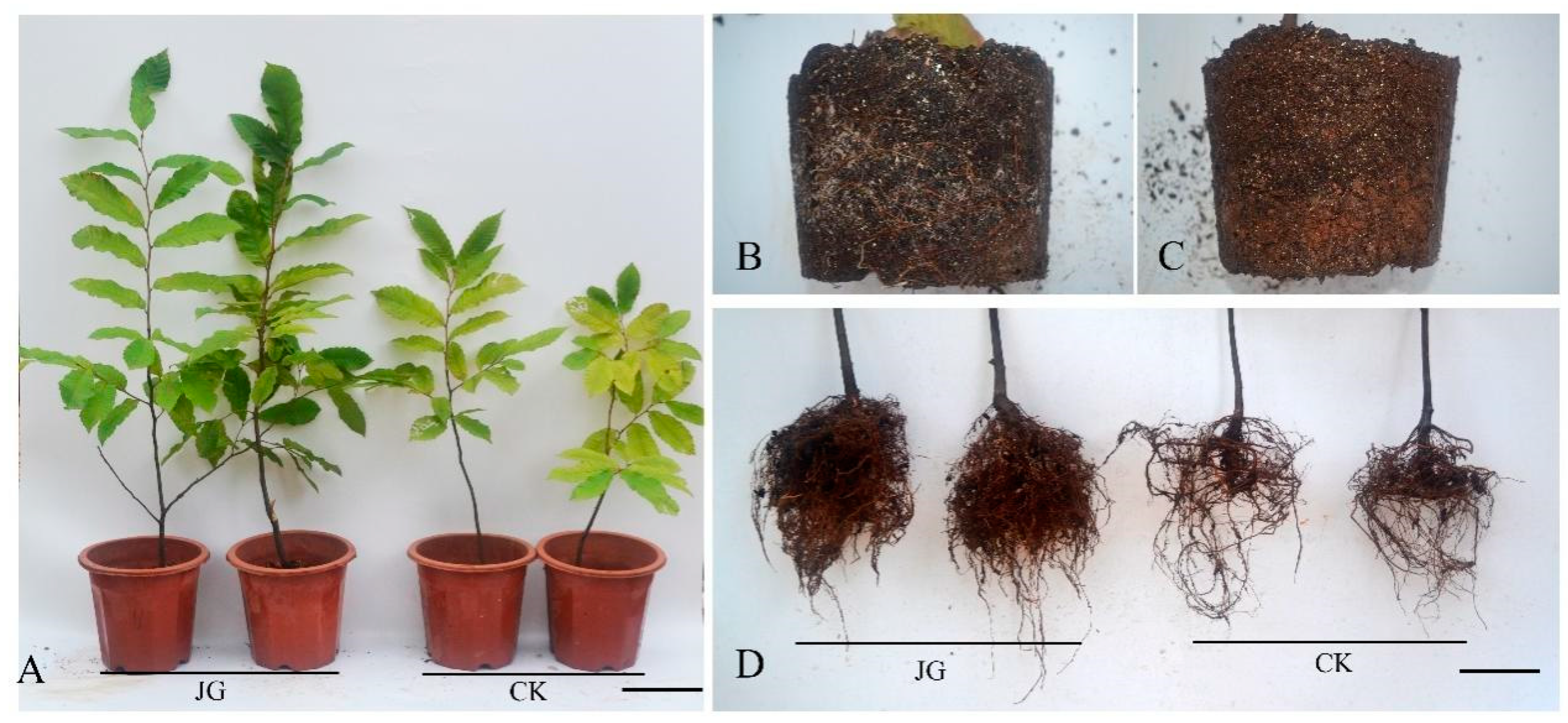
3.1. Root Colonization, Plant Growth, Biomass, and Root Architecture

After a 6-month inoculation period (from April 2018 to October 2018), all JG seedlings were successfully colonized at a rate of 61% (±9.94%) of their root tissue (Figure 2B), while the CK samples showed little colonization of 6.6% (±4.20%) (F = 254; p< 0.001) (Figure 2C). Inoculation with B. edulis significantly improved the growth of C. henryi seedlings and changed the root architecture (p < 0.05) (Figure 2A,D, Figure 3 and Figure 4; Table 1). Seedling height and base diameter of JG plants (46.0 cm and 8.22 mm, respectively) were significantly higher than the CK seedlings (21.1 cm and 5.20 mm, respectively) (Figure 4A). Inoculation with B. edulis significantly increased the biomass of C. henryi seedlings (p < 0.05) (Figure 4B). The biomass of JG (26.7 g seedling−1) was much higher than that of CK (14.8 g seedling−1). More specifically, the aboveground and underground parts of JG were 12.1 g seedling−1 and 14.6 g seedling−1, respectively, with a root:shoot ratio of 1.20. On the other hand, the aboveground and underground parts of CK were 7.83 g seedling−1 and 6.97 g seedling−1, respectively, with a root:shoot ratio of 0.89. The root surface area and root volume of JG were at least 2.5 times that of CK (Table 1).

3.2. Nutrient Content

After a 6-month inoculation period, the total N and P content of JG seedlings was significantly higher than that of CK seedlings (p < 0.05) (Figure 5). The difference in P content between JG and CK seedlings was greater than that of N content (Figure 5). The P content of aboveground (60.8 mg seedling−1) and underground (96.5 mg seedling−1) parts of JG seedlings was 2.7 and 2.8 times that of CK (22.4 mg seedling−1 and 34.4 mg seedling−1), respectively (Figure 5A). The N content of aboveground (168 mg seedling−1) and underground (155 mg seedling−1) parts of JG seedlings was 1.2 and 1.3 times that of CK (136 mg seedling−1 and 118 mg seedling−1), respectively (Figure 5B).

3.3. Enzymatic Activity in the Rhizospheric Soil of Seedlings

Inoculation with B. edulis significantly increased the rhizospheric soil invertase activity and acid phosphatase activity, but decreased the alkaline phosphatase activity, and had no significant effect on urease activity and on neutral phosphatase activity (p < 0.05) (Figure 6). The invertase activity of JG (17.0 glucose mg g−1) was 1.46 times that of CK (11.6 glucose mg g−1) (Figure 6A), and the acid phosphatase activity (pH 5) of JG (2.20 phenol mg g−1) was 17.3 times that of CK (0.13 phenol mg g−1) (Figure 6C). The alkaline phosphatase activity (pH 9) of JG (0.34 glucose mg g−1) was 0.40 times that of CK (0.85 glucose mg g−1) (Figure 6C).

3.4. Root Tips Structure of Each Season

Seasonal changes had significant effects on the structure of ECM of C. henryi (Figure 7) root tips. The hyphae bonded in winter and summer (Figure 7F,G,N,O) and flaked off in winter (Figure 7F), the epidermal cells collapsed (Figure 7G,H), and cortical cells were invaded by the fungus (Figure 7H). No significant changes in the inner mantle or Harting net structure were observed in summer (Figure 7P). ECM had fluffy and energetic epitaxial mycelia (Figure 7C,D,J,K), along with a normal structure of the mantle and Harting net (Figure 7D,L), both in autumn and spring seasons. Rhizomorph was most abundant in spring (Figure 7I,J). Additionally, multiple secondary lateral roots were differentiating in the short roots that formed the ECM in spring (Figure 7L).

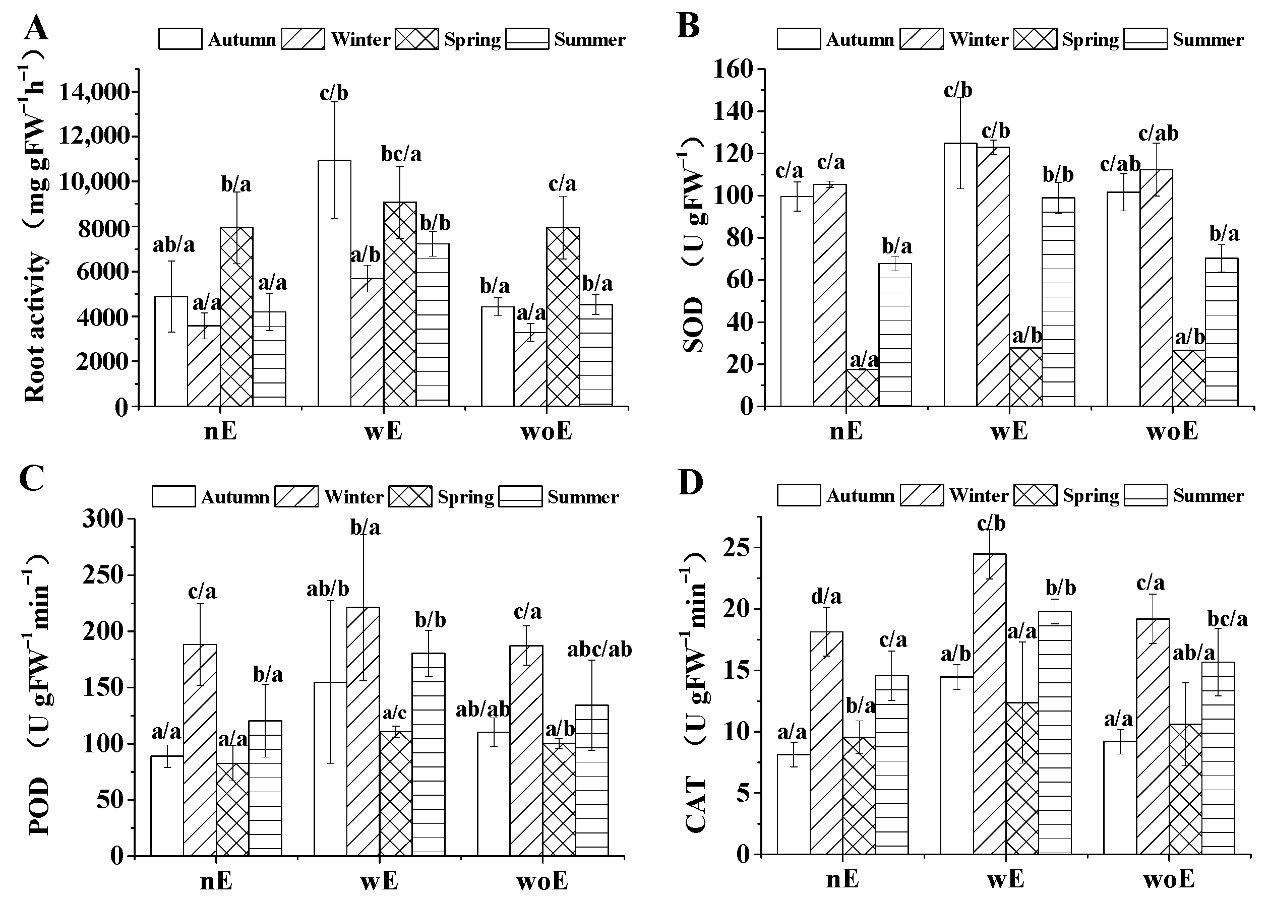
3.5. Root Tips Physiology in Each Season

Seasonal variation had significant effects on root activity, antioxidant enzyme activity, and the level of substances responsible for osmotic adjustment. These significant effects were observed in all treatments, including nE (root tips from uninoculated seedlings), wE (root tips from inoculated seedlings with ECM), and woE (root tips from inoculated seedlings without ECM) (p < 0.05) (Figure 8 and Figure 9). For all treatments, root activity was lowest in winter and highest in summer. Root activity in autumn was the highest both in nE and woE. However, root activities of wE were high in both spring and autumn. No matter the season, root activities of wE were significantly higher than of nE and woE, with no significant difference between nE and woE (p < 0.05) (Figure 8A). For all treatments, SOD activities in autumn and winter were similar and both significantly higher than in spring and summer, with SOD activities in spring being the lowest (p < 0.05). No matter the season, SOD activities of wE were significantly higher than nE (p < 0.05) (Figure 8B). POD and CAT activities had almost similar trends in variability in all seasons. For all root tips, POD and CAT activities were highest in winter and lower in summer (Figure 8C,D). CAT activities of wE in winter and summer were significantly higher than nE and woE, and no significant differences were observed between nE and woE (p < 0.05) (Figure 8D).
For all treatments, MDA level was highest in winter and lowest in spring. No matter the season, MDA levels of wE were significantly lower than that of nE and woE (p < 0.05) (Figure 9A). For all treatments, in general, the soluble protein content was highest in spring and lowest in winter. No matter the season, the soluble protein content of wE was significantly higher than nE and woE, and there was no significant difference between nE and woE, except in summer (p < 0.05) (Figure 9B).

4. Discussion

Chinquapin seedlings are sparse lateral roots and thus highly dependent on the formation of ECM with ECM fungus for the uptake nutrients and for water to survive, especially after transplanting [6]. B. edulis is an ECM fungus with a wide range of trees, both deciduous (e.g., oaks, beech, hornbeam) and conifers (e.g., pines, spruce, silver fir) [37]. However, its impact on plant growth varies with the plant species and it is associated with references [13,22,25]. Moreover, the ECM structure, which is responsible for uptake nutrient and water, is not stable [26].

4.1. Growth Responses of Inoculation

In this study, B. edulis had significant effects on the growth of C. henryi seedlings, similar to results of previous studies on C. mollissima and Pinus tabulaeformis [13,25,38]. However, other studies showed that B. edulis did not increase the biomass of the host plant, which could be due to the short inoculation period of three months [22]. A study by Endo et al. (2014) showed that inoculation of Japanese red pine with B. edulis needs 4 months to form the mantle (in vitro) [39]. A study on C. dentata also showed a reduced growth when colonization by ECM fungi lasted 5 months, but all ECM seedlings had a better chance of survival and better growth after transplanting into the field, compared to non-ECM seedlings [16]. The ECM fungi import a significant amount of carbon from seedling roots to develop the mycelial network necessary to acquire soil nutrients [16], and the elevated photosynthetic rate of seedlings or young trees could not always fully meet the requested carbon demand of ECM, leading to smaller ECM seedlings, compared to non-ECM controls [20,21]. Thus, as Dulmer et al. (2014) suggested, it may not be equitable to measure the growth in the first months or years [16]. It could also be that the ECM fungi used in the studies were not adapted to the local conditions or may not the best match to their hosts, since it is known that various fungi species or strains have varying effects on their hosts [22,40,41]. Therefore, it can be considered that the B. edulis strain used in this study is well adapted and is a suitable ECM fungus for C. henryi.
In this study, seedlings inoculated with B. edulis showed a higher increase in root biomass, a better regulated root:shoot ratio, and a changed root architecture when compared to the aerial parts. Our results agree with other studies, where ECM inoculation of C. sativa, C. mollissima, Quercus ilex, and Q. faginea seedlings enhanced the development of the root system [12,15,17,38]. These changes in root growth may stem from changes in carbon fixation, resulting from improved nutrition. ECM fungi can alter auxin metabolism within the host root, which regulates root development and changes root architecture [42,43,44,45].
Forest trees have evolved to establish a relationship with mycorrhizal fungi to enhance their ability of acquiring nutrients, such as N and P, from poor soils. This relationship is considered a pivotal event in the evolutionary history of land plants [8]. In this study, the N and P content, especially P, were significantly increased in the inoculated seedlings. These results are consistent with many prior studies [26,46,47]. There are four primary reasons that explain this enhanced P absorption in mycorrhizae inoculated seedlings. First, the widespread hyphal networks (Figure 2B), the increased root surface, and number of root tips (Table 1), as well as the modified root architecture (Figure 3A), can enhance the absorption surface area of ECM roots, which facilitates the absorption of soluble N and P [48,49]. Second, soil enzymatic activities, which were increased by the inoculation treatment, help the plants absorb the soil nutrients. Similar to plant roots, the extraradical hyphae secrete acid phosphatase into the rhizosphere lead to a significant increase (17.29 times) of acid phosphatase activity in the rhizosphere of JG compared to CK (Figure 6C), which consequently increases P availability through hydrolysis of organic P [41,50]. This is an important strategy for plant adaptation and growth in the red acidic soils of southern China, where low pH leads to phosphorus fixation to clay minerals and reduces its availability for root uptake [4]. In addition, ECM promotes the soil carbon cycle, as is evident by the enhanced invertase activity in the rhizosphere of JG compared to CK (Figure 6A). The increased invertase activity also increases glucose levels in the rhizosphere, an enzymatic byproduct and a carbon source for plants and microorganisms, which helps seedlings uptake more N and P from the rhizosphere. The ectomycorrhizal fungi can mobilize N from complex organic substrates, which would otherwise be less available to the plant [51,52,53,54,55]. Third, nutrient demand from bigger plants is higher, which can be compensated by ECM through a more efficient nutrient uptake from the rhizosphere [7]. Fourth, both ECM fungi and plants would regulate the phosphate transporter genes to enhance phosphate uptake and transport under conditions of low phosphate availability. Wang et al. (2014) determined a phosphorus transporter gene of B. edulis (BePT), which plays a key role in phosphate acquisition under phosphate deficient conditions [56]. Prior studies on C. mollissima also suggested that five phosphate transporter genes (CmPT1, CmPT2, CmPT3, CmPT4, CmPT5) are upregulated under a phosphate deficiency treatment [46]. Based on our results, we speculate that the phosphate transporter genes also exist in C. henryi and would be upregulated when phosphate is deficient, but this needs further research to confirm.

4.2. Variation of ECM Root Tip Structure and Physiology in Different Seasons

To date, few studies have been conducted to observe temporal vegetative fungal structure (ECM root tips and extraradical mycelium) of potted seedlings of a single species. As the experiments in our study were carried out with potted seedlings, the water retention and heat preservation of the potting substrate were not as good as in natural field conditions. This means that the cumulative precipitation has limited effects on substrate moisture and roots are more sensitive to temperature fluctuation. Previous studies on natural sites have shown that the mycelium volume was positively correlated with precipitation of the current month or the prior month [57,58] and negatively correlated with the mean temperature of the previous month [57]. However, these observations were not consistent with our findings. In our study, we found that the rhizomorph was always abundant on the matrix surface during spring and autumn on moderately dry days, while fewer rhizomorphs could be found on the matrix surface during summer or winter, especially during heavy precipitation days in winter (data not shown). Additionally, the mantle in winter seemed to have lost its vitality, even flaked off (Figure 7F).
In our study, we observed that the rhizomorph would stretch out and attach largely on the matrix surface (Figure 2B) unless the temperature was ideal and the soil moisture relatively low. In the cold winter period, few rhizomorphs and few ECM root tips with abnormal mantle and Harting net could be found on the matrix surface (Figure 7H). These structures are hard to observe with the naked eye since the white mantle is missing, which can lead to the wrong assumption that ECM is not present. In the spring, numerous rhizomorphs were found on the ECM (Figure 7J), with secondary lateral roots developing on ECM (Figure 7L). This leads to an increase in ECM root tips. These results agree with previous study on B. edulis [59] and many studies on other ECM fungi, such as Tuber melanosporum, Lactarius deliciosus, and Rhizopogon spp., which demonstrated positive correlations between mycelium biomass and ECM abundance [58,60,61].
To counteract the oxidative damage under extremely adverse conditions, plants have developed an antioxidant defense system that includes SOD, CAT, and POD [62,63]. The lipid peroxidation product, MDA, is an index of cell membrane damage [11]. In this study, seasonal variation had significant effects on root activity and antioxidant enzyme activity in the root tips. Generally, no matter the type of root tips, the lowest antioxidant enzyme activities (SOD, CAT, POD) and MDA levels and the highest root activities and soluble protein content were observed in the spring, and the opposite was observed in winter, suggesting that the low winter temperatures induce a cold stress in all root tips cells. However, no matter the season, wE had higher antioxidant enzyme activities, root activities, soluble protein content, and lower MDA compared to nE and woE, and there were no differences between nE and woE seedlings. This indicated that inoculating with B. edulis could alleviate temperature stress by improving the root antioxidant capacity, which might improve the resistance of seedlings to adverse environmental conditions.

5. Conclusions

This study showed seasonal variation of ectomycorrhizal root tips’ structure and physiology for the first time. The ectomcorrhizal fungus, B. edulis, symbiosis improved the resistance to abiotic stresses of C. henryi seedlings by improved nutrient uptake and modulation of reactive oxygen species metabolism.
This study provides an integrated approach on the growth of the host plant and the seasonal variation of ECM root tips, which is of paramount importance when producing high-quality C. henryi seedlings, by shortening the survival stage and improving the survival rate of afforestation.

Author Contributions

H.X. conceived the experiment. H.X., P.C., W.C., Y.Y., Y.J., and S.T. developed and carried out the specific methodology. H.X. contributed to the data analysis. D.Y. and F.Z. supervised the research. H.X. and J.M. contributed to the preparation of the manuscript. All authors have read and agreed to the published version of the manuscript.

Funding

This study was financially supported by National Natural Science Foundation of China (Grant No. 32001309).

Conflicts of Interest

The authors declare no conflict of interest.

References

  1. Xiong, H.; Sun, H.; Zou, F.; Fan, X.M.; Niu, G.H.; Yuan, D.Y. Micropropagation of chinquapin (Castanea henryi) using axillary shoots and cotyledonary nodes. HortScience 2018, 53, 1482–1486. [Google Scholar] [CrossRef] [Green Version]
  2. Fan, X.M.; Yuan, D.Y.; Tian, X.M.; Zhu, Z.J.; Liu, M.L.; Cao, H.P. Comprehensive transcriptome analysis of phytohormone biosynthesis and signaling genes in the flowers of Chinese chinquapin (Castanea henryi). J. Agr. Food Chem. 2017, 65, 10332–10349. [Google Scholar] [CrossRef] [PubMed]
  3. Fan, X.M.; Yuan, D.Y.; Tang, J.; Tian, X.M.; Zhang, L.; Zou, F.; Tan, X.F. Sporogenesis and gametogenesis in Chinese chinquapin (Castanea henryi (Skam) Rehder & Wilson) and their systematic implications. Trees 2015, 29, 1713–1723. [Google Scholar]
  4. Yuan, J.; Huang, L.Y.; Zhou, N.F.; Wang, H.; Niu, G.H. Fractionation of Inorganic Phosphorus and Aluminum in Red Acidic Soil and the Growth of Camellia oleifera. HortScience 2017, 52, 1293–1297. [Google Scholar] [CrossRef]
  5. Wang, L.; Li, Y.L.; Xiong, H.; Yuan, D.Y.; Zhang, X.; Zou, F. Effect of temperature stress on leaf structure and photosynthesis of chinquapin (Castanea henryi). Acta Agric. Univ. Jiangxiensis 2020, 42, 692–699. [Google Scholar]
  6. Liu, D.M.; Yuan, D.Y.; Zou, F.; Zhang, X.H.; Zhu, Z.J.; Tan, L.M. Optimization of culture conditions for 3 Castanea henryi ectomycorrhizal fungi. J. Northw. Fore. Univ. 2016, 31, 195–200. [Google Scholar]
  7. Smith, S.E.; Read, D.J. Mycorrhizal Symbiosis, 3rd ed.; Academic Press Elsevier: Cambridge, UK, 2008; pp. 126–160. [Google Scholar]
  8. Martin, F.; Kohler, A.; Murat, C.; Veneault-Fourrey, C.; Hibbett, D.S. Unearthing the roots of ectomycorrhizal symbioses. Nat. Rev. Microbiol. 2016, 14, 760–773. [Google Scholar] [CrossRef]
  9. Colpaert, J.V.; Wevers, J.H.L.; Krznaric, E.; Adriaensen, K. How metal-tolerant ecotypes of ectomycorrhizal fungi protect plants from heavy metal pollution. Ann. For. Sci. 2011, 68, 17–24. [Google Scholar] [CrossRef] [Green Version]
  10. Ma, Y.L.; He, J.L.; Ma, C.F.; Luo, J.; Li, H.; Liu, T.X.; Polle, A.; Peng, C.H.; Luo, Z.B. Ectomycorrhizas with Paxillus involutus enhance cadmium uptake and tolerance in Populus × canescens. Plant Cell Environ. 2014, 37, 627–642. [Google Scholar] [CrossRef]
  11. Yin, D.C.; Halifu, S.; Song, R.Q.; Qi, J.Y.; Deng, X.; Deng, J.F. Effects of an ectomycorrhizal fungus on the growth and physiology of Pinus sylvestris var. mongolica seedlings subjected to saline-alkali stress. J. For. Res. 2020, 31, 781–788. [Google Scholar] [CrossRef]
  12. Nardini, A.; Salleo, S.; Tyree, M.T.; Vertovec, M. Influence of the ectomycorrhizas formed by Tuber melanosporum Vitt. on hydraulic conductance and water relations of Quercus ilex L. seedlings. Ann. For. Sci. 2000, 57, 305–312. [Google Scholar] [CrossRef] [Green Version]
  13. Feng, G.; Xu, B.; Qin, L.; Li, X.L. Effects of ectomycorrhizal fungi on the growth and nutrient uptake of chestnut (Castanea mollissima BL.). Acta Horticult. Sin. 2003, 30, 311–313. [Google Scholar]
  14. Gu, X.R.; Wang, X.L.; Li, J.; He, X.H. Accumulation and translocation of phosphorus, calcium, magnesium, and aluminum in Pinus massoniana Lamb. seedlings inoculated with Laccaria bicolor growing in an acidic yellow soil. Forests 2019, 10, 1153. [Google Scholar] [CrossRef] [Green Version]
  15. Domínguez-Núñez, J.A.; Serrano, J.S.; Rodríguez Barreal, J.A.; Saiz de Omeñaca, J.A. The influence of mycorrhization with Tuber melanosporum in the afforestation of a Mediterranean site with Quercus ilex and Quercus faginea. For. Ecol. Manag. 2006, 231, 226–233. [Google Scholar] [CrossRef]
  16. Dulmer, K.M.; LeDuc, S.D.; Horton, T.R. Ectomycorrhizal inoculum potential of northeastern US forest soils for American chestnut restoration: Results from filed and laboratory bioassays. Mycorrhiza 2014, 24, 65–74. [Google Scholar] [CrossRef] [PubMed]
  17. Álvarez-Lafuente, A.; Benito-Matías, L.F.; Peñuelas-Rubira, J.L.; Suz, L.M. Multi-cropping edible truffles and sweet chestnuts: Production of high-quality Castanea sativa seedlings inoculated with Tuber aestivum, its ecotype T. uncinatum, T. brumale, and T. macrosporum. Mycorrhiza 2018, 28, 29–38. [Google Scholar] [CrossRef] [PubMed]
  18. Zong, K.; Huang, J.; Nara, K.; Chen, Y.H.; Shen, Z.G.; Lian, C.L. Inoculation of ectomycorrhizal fungi contributes to the survival of tree seedlings in a copper mine tailing. J. For. Res. 2015, 20, 493–500. [Google Scholar] [CrossRef]
  19. Hernández-Rodríguez, M.; Martín-Pinto, P.; Oria-de-Rueda, J.A.; Diaz-Balteiro, L. Optimal management of Cistus ladanifer shrublands for biomass and Boletus edulis mushroom production. Agroforest. Syst. 2017, 91, 663–676. [Google Scholar] [CrossRef]
  20. Conjeaud, C.; Scheromm, P.; Mousain, D. Effects of phosphorus and ectomycorrhiza on maritime pine seedlings (Pinus pinaster). New Phytol. 1996, 133, 345–351. [Google Scholar] [CrossRef]
  21. Kytöviita, M.M. Role of nutrient level and defoliation on symbiotic function: Experimental evidence by tracing 14C/15N exchange in mycorrhizal birch seedlings. Mycorrhiza 2005, 15, 65–70. [Google Scholar] [CrossRef]
  22. Lu, N.; Yu, M.; Cui, M.; Luo, Z.J.; Feng, Y.; Cao, S.; Sun, Y.H.; Li, Y. Effect of different ectomycorrhizal fungal inoculates on the growth of Pinus tabulaeformis seedlings under greenhouse conditions. Forests 2016, 7, 316. [Google Scholar] [CrossRef] [Green Version]
  23. Dosskey, M.G.; Linderman, R.G.; Boersma, L. Carbon-sink stimulation of photosynthesis in Douglas fir seedlings by some ectomycorrhizas. New Phytol. 1990, 115, 269–274. [Google Scholar] [CrossRef] [PubMed]
  24. Ekwebelam, S.A.; Reid, C.P.P. Effect of light, nitrogen fertilization, and mycorrhizal fungi on growth and photosynthesis of lodgepole pine seedlings. Can. J. For. Res. 1983, 13, 1099–1106. [Google Scholar] [CrossRef]
  25. Huang, Y.; Jiang, X.Y.; Liang, Z.C.; Li, T. Effect of ectomycorrhizal fungi on growth and physiology of Pinus tabulaeformis plants to saline environment. J. Agro-Environ. Sci. 2006, 25, 1475–1480. [Google Scholar]
  26. Harley, J.L. Mycorrhizal studies: Past and future. In Physiological and Genetical Aspects of Mycorrhizae, 1st ed.; Gianinazzi-Pearson, V., Gianinazzi, S., Eds.; INRA: Paris, France, 1986; pp. 25–33. [Google Scholar]
  27. Tagu, D.; Lapeyrie, F.; Martin, F. The ectomycorrhizal symbiosis: Genetics and development. Plant Soil 2002, 244, 97–105. [Google Scholar] [CrossRef]
  28. Acioli-Santos, B.; Sebastiana, M.; Pessoa, F.; Sousa, L.; Figueiredo, A.; Fortes, A.M.; Baldé, A.; Maia, L.C.; Pais,, M.S. Fungal transcript pattern during the preinfection stage (12 h) of ectomycorrhiza formed between Pisolithus tinctorius and Castanea sativa roots, identified using cDNA microarrays. Curr. Microbiol. 2008, 57, 620–625. [Google Scholar] [CrossRef] [PubMed]
  29. Schubert, B.A.; Jahren, A.H. Seasonal temperature and precipitation recorded in the intra-annual oxygen isotope pattern of meteoric water and tree-ring cellulose. Quat. Sci. Rev. 2015, 125, 1–14. [Google Scholar] [CrossRef]
  30. Mittler, R. Oxidative stress, antioxidants and stress tolerance. Trends Plant Sci. 2002, 7, 405–410. [Google Scholar] [CrossRef]
  31. Bergamini, C.M.; Gambetti, S.; Dondi, A.; Cervellati, C. Oxygen, reactive oxygen species and tissue damage. Curr. Pharm. Des. 2004, 10, 1611–1626. [Google Scholar] [CrossRef]
  32. Perez-Perez, M.E.; Lemaire, S.D.; Crespo, J.L. Reactive oxygen species and autophagy in plants and algae. Plant Physiol. 2012, 160, 156–164. [Google Scholar] [CrossRef] [Green Version]
  33. Chen, W.Z.; He, L.B.; Tian, S.Y.; Masabni, J.; Zhang, R.Q.; Zou, F.; Yuan, D.Y. Combined addition of bovine bone and cow manure: Rapid composting of chestnut burrs and production of a high-quality chestnut seedling substrate. Agronomy 2020, 10, 288. [Google Scholar] [CrossRef] [Green Version]
  34. Baptista, P.; Martins, A.; Pais, M.S.; Tavares, R.M.; Lino-Neto, T. Involvement of reactive oxygen species during early stages of ectomycorrhiza establishment between Castanea sativa and Pisolithus tinctorius. Mycorrhiza 2007, 17, 185–193. [Google Scholar] [CrossRef] [PubMed] [Green Version]
  35. Xiong, H.; Masabni, J.; Zou, F.; Yuan, D.Y. Castanea henryi roots serve as host for Ganoderma lucidum. Int. J. Agric. Biol. 2019, 22, 420–426. [Google Scholar]
  36. Gao, J.F. Experimental Guidance for Plant Physiology, 1st ed.; Higher Education Press: Beijing, China, 2006. [Google Scholar]
  37. Hall, I.R.; Lyon, A.J.E.; Wang, Y.; Sinclair, L. Ectomycorrhizal fungi with edible fruiting bodies 2.Boletus edulis. Econ. Bot. 1998, 52, 44–56. [Google Scholar] [CrossRef]
  38. Xu, B.; Feng, G.; Pan, J.R.; Qin, L.; Li, X.L. Transferring of phosphorus between chestnut seedlings via ectomycorrhizal hyphal links. Acta Ecol. Sin. 2003, 23, 765–770. [Google Scholar]
  39. Endo, N.; Kawamura, F.; Kitahara, R.; Sakuma, D.; Fukuda, M.; Yamada, A. Synthesis of Japanese Boletus edulis ectomycorrhizae with Japanese red pine. Mycoscience 2014, 55, 405–416. [Google Scholar] [CrossRef]
  40. Ruess, R.W.; Swanson, M.M.; Kielland, K.; McFarland, J.W.; Olson, K.D.; Taylor, D.L. Phosphorus mobilizing enzymes of Alnus-associated ectomycorrhizal fungi in an Alaskan boreal floodplain. Forests 2019, 10, 554. [Google Scholar] [CrossRef] [Green Version]
  41. García-Montero, L.G.; Valverde-Asenjo, I.; Moreno, D.; Díaz, P.; Hernando, I.; Menta, C.; Tarasconi, K. Influence of edaphic factors on edible ectomycorrhizal mushrooms: New hypotheses on soil nutrition and C sinks associated to ectomycorrhizae and soil fauna using the Tuber brulé model. In Edible Ectomycorrhizal Mushrooms: Current Knowledge and Future Prospects, 1st ed.; Zambonelli, A., Bonito, G.M., Eds.; Springer: Berlin, Germany, 2012; Volume 34, pp. 83–104. [Google Scholar]
  42. Felten, J.; Kohler, A.; Morin, E.; Bhalerao, R.P.; Palme, K.; Martin, F.; Ditengou, F.A.; Legué, V. The ectomycorrhizal fungus Laccaria bicolor stimulates lateral root formation in poplar and Arabidopsis through auxin transport and signaling. Plant Physiol. 2009, 151, 1991–2005. [Google Scholar] [CrossRef] [Green Version]
  43. Splivallo, R.; Fischer, U.; Göbel, C.; Feussner, I.; Karlovsky, P. Truffles regulate plant root morphogenesis via the production of auxin and ethylene. Plant Physiol. 2009, 150, 2018–2029. [Google Scholar] [CrossRef] [Green Version]
  44. Vayssières, A.; Pencík, A.; Felten, J.; Kohler, A.; Ljung, K.; Martin, F.; Legué, V. Development of the poplar-Laccaria bicolor ectomycorrhizal modifies root auxin metabolism, signaling, and response. Plant Physiol. 2015, 169, 890–902. [Google Scholar] [CrossRef] [Green Version]
  45. Schimel, J.P.; Bennett, J. Nitrogen mineralization: Challenges of a changing paradigm. Ecology 2004, 85, 591–602. [Google Scholar] [CrossRef]
  46. Chen, S.S.; Xing, Y.; Wang, T.; Zhang, Q.; Yu, W.Y.; Fang, K.F.; Newhouse, A.E.; McGuigan, L.D.; Stewart, K.R.; Maynard, C.A.; et al. Ectomycorrhizae symbiosis in Castanea mollissima improves phosphate acquisition through activating gene expression and H+ efflux. Sci. Hortic. 2016, 210, 99–107. [Google Scholar] [CrossRef]
  47. Martin, F.; Nehls, U. Harnessing ectomycorrhizal genomics for ecological insights. Curr. Opin. Plant Biol. 2009, 12, 508–515. [Google Scholar] [CrossRef] [PubMed]
  48. Niu, Y.F.; Chai, R.S.; Jin, G.L.; Wang, H.; Tang, C.X.; Zhang, Y.S. Responses of root architecture development to low phosphorus availability: A review. Ann. Bot. 2013, 112, 391–408. [Google Scholar] [CrossRef] [PubMed]
  49. Sato, T.; Ezawa, T.; Cheng, W.G.; Tawaraya, K. Release of acid phosphatase from extraradical hyphae of arbuscular mycorrhizal fungus Rhizophagus clarus. Soil Sci. Plant Nutr. 2015, 61, 269–274. [Google Scholar] [CrossRef] [Green Version]
  50. Lindahl, B.D.; Ihrmark, K.; Boberg, J.; Trumbore, S.E.; Högberg, P.; Stenlid, J.; Finlay, R.D. Spatial separation of litter decomposition and mycorrhizal nitrogen uptake in a boreal forest. New Phytol. 2007, 173, 611–620. [Google Scholar] [CrossRef] [Green Version]
  51. Talbot, J.M.; Allison, S.D.; Treseder, K.K. Decomposers in disguise: Mycorrhizal fungi as regulators of soil C dynamics in ecosystems under global change. Funct. Ecol. 2008, 22, 955–963. [Google Scholar] [CrossRef] [Green Version]
  52. Talbot, J.M.; Bruns, T.D.; Smith, D.P.; Branco, S.; Glassman, S.I.; Erlandson, S.; Vilgalys, R.; Peay, K.G. Independent roles of ectomycorrhizal and saprotrophic communities in soil organic matter decomposition. Soil Biol. Biochem. 2013, 57, 282–291. [Google Scholar] [CrossRef]
  53. Phillips, L.A.; Ward, V.; Jones, M.D. Ectomycorrhizal fungi contribute to soil organic matter cycling in sub-boreal forests. ISME J. 2013, 8, 699–713. [Google Scholar] [CrossRef] [Green Version]
  54. Bödeker, I.T.M.; Clemmensen, K.E.; Boer, W.; Martin, F.; Olson, Å.; Lindahl, B.D. Ectomycorrhizal Cortinarius species participate in enzymatic oxidation of humus in northern forest ecosystems. New Phytol. 2014, 203, 245–256. [Google Scholar] [CrossRef]
  55. Wang, J.L.; Li, T.; Wu, X.G.; Zhao, Z.W. Molecular cloning and functional analysis of H+-dependent phosphate transporter gene from the ectomycorrhizal fungus Boletus edulis in Southwest China. Fungal Biol. 2014, 118, 453–461. [Google Scholar] [CrossRef]
  56. De la Varga, H.; Águeda, B.; Ágreda, T.; Martínez-Peña, F.; Parladé, J.; Pera, J. Seasonal dynamics of Boletus edulis and Lactarius deliciosus extraradical mycelium in pine forests of central Spain. Mycorrhiza 2013, 23, 391–402. [Google Scholar] [CrossRef]
  57. Queralt, M.; Parladé, J.; Pera, J.; De Miguel, A.M. Seasonal dynamics of extraradical mycelium and mycorrhizas in black truffle (Tuber melanosporum) plantation. Mycorrhiza 2017, 27, 565–576. [Google Scholar] [CrossRef]
  58. Balestrini, R.; Kottke, I. Structure and development of ectomycorrhizal roots. In Molecular Mycorrhizal Symbiosis, 1st ed.; Martin, F., Ed.; John Wiley & Sons Inc.: Hoboken, NJ, USA, 2017; pp. 47–61. [Google Scholar]
  59. De la Varga, H.; Águeda, B.; Martínez-Peña, F.; Parladé, J.; Pera, J. Quantification of extraradical soil mycelium and ectomycorrhizas of Boletus edulis in a Scots pine forest with variable sporocarp productivity. Mycorrhiza 2012, 22, 59–68. [Google Scholar] [CrossRef]
  60. Parladé, J.; Hortal, S.; Pera, J.; Galipienso, L. Quantitative detection of Lactarius deliciosus extraradical soil mycelium by real-time PCR and its application in the study of fungal persistence and interspecific competition. J. Biotechnol. 2007, 128, 14–23. [Google Scholar] [CrossRef] [PubMed]
  61. Hortal, S.; Pera, J.; Parladé, J. Tracking mycorrhizas and extraradical mycelium of the edible fungus Lactarius deliciosus under field competition with Rhizopogon spp. Mycorrhiza 2008, 18, 69–77. [Google Scholar] [CrossRef] [PubMed]
  62. Foyer, C.H.; Noctor, G. Oxidant and antioxidant signaling in plants: A re-evaluation of the concept of oxidative stress in a physiological context. Plant Cell Environ. 2005, 28, 1056–1071. [Google Scholar] [CrossRef]
  63. Wang, W.B.; Kim, Y.H.; Lee, H.S.; Kim, K.Y.; Deng, X.P.; Kwak, S.S. Analysis of antioxidant enzyme activity during germination of alfalfa under salt and drought stresses. Plant Physiol. Biochem. 2009, 47, 570–577. [Google Scholar] [CrossRef]
Figure 1. Temperature and precipitation during the study period.Tmax = maximum monthly temperature; Tmin = minimum monthly temperature; PP = precipitation. Data from https://en.tutiempo.net/climate/12-2019/ws-576870.html (accessed on 30 January 2020).
Forests 12 01643 g001
Figure 2. Visual comparison between inoculated (JG) and uninoculated (CK) C. henryi seedlings after 6 months of growth (scale bar for (A,D) is 10 cm). (A) Whole plants. (B) The root systems on the matrix surface of JG plants showing a large number of white or light-yellow ECM root tips and abundant rhizomorph. (C) The root systems on the matrix surface of CK plants showing non-inoculated root tips. (D) The root systems of JG and CK.
Forests 12 01643 g002
Figure 3. Root observations of inoculated (JG) and uninoculated (CK) Castanea henryi seedlings. (A) Morphological observation of ECM of C. henryi showing a monopodial-pinnate or monopodial-pyramidal pattern with a main axis up to 0.3 mm (scale bar = 500 μm). (B) A closeup of a portion of the inoculated root system of photo A showing abundant rhizomorph winding around the ECM root tips (scale bar = 200 μm). (C) Morphological observation of non-inoculated roots of C. henryi, showing relatively few fine root tips (scale bar = 500 μm). (D) Root hairs in the maturation zone of the non-inoculated root tips (scale bar = 200 μm).
Forests 12 01643 g003
Figure 4. Height, diameter, and biomass between inoculated (JG) and uninoculated (CK) Castanea henryi seedlings. (A) Height and base diameter of JG and CK. (B) Aboveground and underground biomass of JG and CK. Different letters indicate the significant difference between JG and CK, p = 0.05 level based on Fisher’s protected least significant difference. Error bars represent standard errors (n = 10).
Forests 12 01643 g004
Figure 5. Total nitrogen (N) (A) and phosphorus (P) (B) of Castanea henryi seedlings 6 months after inoculation (JG) with Boletus edulis compared to non-inoculated control (CK). Different letters indicate significant differences between JG and CK at p = 0.05 based on Fisher’s protected least significant difference. Bars represent standard errors (n = 5).
Forests 12 01643 g005
Figure 6. Enzyme activity in the rhizospheric soil of inoculated (JG) and uninoculated (CK) Castanea henryi seedlings after 6 months of growth. (A) Invertase activity; (B) urease activity; (C) phosphatase activity (pH5 indicates acid phosphatase; pH7 indicates neutral phosphatase; pH9 indicates alkaline phosphatase). Different letters indicate the significant difference between JG and CK, p = 0.05 level based on Fisher’s protected least significant difference. Bars represent standard errors (n = 3).
Forests 12 01643 g006
Figure 7. Structural characteristics of the ectomycorrhiza of Castanea henryi in different seasons using the SEM and paraffin section. (AD) represent the autumn ECM (September 2018). (AC) show the SEM at different magnification. (C) is the cross-section graph, clearly showing two layer mantles (12–48 μm) and Harting net. (D) is the paraffin section graph, cross-section (bar scale = 100 μm). (EH) represent the winter ECM (December 2018). (EG) show the SEM at different multiples. (F) shows the hyphae bonded and flaked off (arrowheads). (G) is the cross-section graph, showing some collapsed epidermal cells (stars) and abnormal mantle and Harting net. (H) is the paraffin section graph, showing that the cortical cells might be invaded by the B. edulis fungus (stars), cross-section (bar scale = 50 μm). (IL) represent the spring ECM (March 2019). (IK) show the SEM at different multiples. (J) shows numerous rhizomorph winding on the ECM (arrowheads). (K) is the cross-section graph, clearly showing a thick mantle and Harting net. (L) is the paraffin section graph, showing that secondary lateral roots are developing (stars), longitudinal-section, scale bar = 100 μm. (MP) represent the summer ECM (July 2019). (MO) show the SEM at different multiples. (N,O) show a certain degree of bonded hyphae on the ECM surface, while (P) shows no effects on the structure of the inner mantle and Harting net (bar scale = 100 μm).
Forests 12 01643 g007
Figure 8. Root activity and antioxidant enzyme activities of different root tips of Castanea henryi in different seasons. (A) Root activity; (B) SOD activity; (C) POD activity; (D) CAT activity. nE: root tips from uninoculated seedlings. wE: root tips from inoculated seedlings with ECM. woE: root tips from inoculated seedlings without ECM. Letters before “/” indicate significant differences of the same root tips at different seasons, while letters after “/” indicate significant differences in the same season of different root tips. p = 0.05 level based on Fisher’s protected least significant difference. Bars represent standard errors (n = 3).
Forests 12 01643 g008
Figure 9. Levels of MDA and soluble protein of different root tips of Castanea henryi in different seasons. (A) MDA in µmol g FW−1; (B) % soluble protein. nE: root tips from uninoculated seedlings. wE: root tips from inoculated seedlings with ECM. woE: root tips from inoculated seedlings without ECM. Letters before “/” indicate significant differences of the same root tips in different seasons, while letters after “/” indicate significant differences in the same season of different root tips, p = 0.05 level based on Fisher’s protected least significant difference. Bars represent standard errors (n = 3).
Forests 12 01643 g009
Table 1. Root architecture of Castanea henryi seedlings inoculated (JG) and non-inoculated (CK) with Boletus edulis.
TreatmentTotal Length
(cm)
Surface Area
(cm2)
Volume
(cm3)
Average Diameter
(mm)
Number of Root Tips
JG3374 ± 102 a531 ± 44 a6.69 ± 0.94 a0.51 ± 0.04 a16,880 ± 439 a
CK1533 ± 53 b208 ± 22 b2.30 ± 0.51 b0.44 ± 0.04 b10,295 ± 233 b
Note: Five seedlings were randomly selected for each treatment for root scanning. Different letters indicate significant difference between JG and CK, p = 0.05 level based on Fisher’s protected least significant difference.
Publisher’s Note: MDPI stays neutral with regard to jurisdictional claims in published maps and institutional affiliations.

