Article

Effects of Microwave Treatment on Microstructure of Chinese Fir

Research Institute of Wood Industry, Chinese Academy of Forestry, Beijing 100091, China
*
Author to whom correspondence should be addressed.
Forests 2020, 11(7), 772; https://doi.org/10.3390/f11070772
Submission received: 26 June 2020 / Revised: 15 July 2020 / Accepted: 17 July 2020 / Published: 19 July 2020
(This article belongs to the Special Issue Methods and New Technologies for Wood Modification)

Abstract

:
Microwave (MW) treatment is an effective method to increase refractory wood permeability, thereby reducing drying time and defects. The extent of modification depends on the damage extent of the wood microstructure. In this study, MW intensities of 43 kWh/m3 (low intensity) and 57 kWh/m3(high intensity) were adopted to treat Chinese fir lumber. Microstructural changes in wood samples were observed using scanning electron microscopy (SEM) and pore structure was characterized using mercury intrusion porosimetry (MIP). Results were as follows: After low-intensity MW treatment, parts of the bordered pit membranes in tracheids were damaged, and micro-fibrils on the margo were ruptured, while the torus basically remained intact. Micro-cracks were observed at both ends of the cross-field pit apertures, propagating to the cell walls of tracheids. The middle lamellar between ray parenchyma cells and longitudinal tracheids cracked, and the width of cracks was in the range of 1–25 μm. After high-intensity MW treatment, damage to the wood microstructure was more severe than that in the low-intensity MW treatment, with macro-cracks having a width range of 100–130 μm being generated. In addition, on the fracture surface of macro-cracks, the bordered pit membranes in tracheids fell off, cross-field pit membranes disappeared and the ray parenchyma cells were seriously damaged, exhibiting fracture of the tracheid walls. Both low-intensity and high-intensity MW treatment can increase the pore diameter corresponding to the margo capillaries (peak value increased from 674.7 nm to 831.8 nm and 1047.6 nm, respectively). The number of pores in the tracheid lumen diameter range also significantly increased. These results provide a theoretical support forMW treatment processes’ improvement and high-value utilization of Chinese fir.

1. Introduction

As an important wood property, permeability reflects the ease of fluid migration in wood under a pressure gradient. During drying processes, permeability is closely related to the drying efficiency and quality of the wood [1]. Chinese fir (Cunninghamia lanceolate (Lamb.) Hook.) is a major commercial plantation conifer tree species in China, and as an important wood resource, it was widely used in making furniture, building bridges and boats, etc. [2]. However, the large proportion of heartwood and its low permeability adversely affect wood-drying efficiency and quality, severely restricting efficient utilization of this resource.
Microwave (MW) treatment can quickly vaporize water inside wood. During this process, rapidly increased steam pressure can damage some weak tissues of anatomical structures, such as pit membranes and ray parenchyma cells. Thus, pathways for easy transportation of liquids and vapors are formed, which contribute to increased permeability and facilitate wood drying, impregnation, as well as the preparation of new materials [3,4,5]. Different extents of wood modification can be achieved by adjusting MW process parameters [4,6,7]. As the MW intensity increases, the wood per unit mass can absorb more MW energy, resulting in higher temperature and steam pressure. A higher temperature and pressure are beneficial to generate more cracks inside the wood, thereby increasing its permeability while simultaneously leading to a reduction in mechanical properties [8,9]. Therefore, it is essential to formulate suitable MW process parameters for Chinese fir, with the aim of increasing permeability without significantly affecting its mechanical properties.
The extent of wood modification depends on the damage extent of the wood microstructure. Investigations examining microstructural changes of Chinese fir under different MW intensities are important to reveal the MW treatment mechanisms and to optimize MW process parameters. Although previous studies have investigated microstructure of MW-treated wood [10,11,12,13], systematic investigations of Chinese fir were seldom reported in the literature. In addition, microstructural changes can result in variation of pore structure of wood, thus affecting its permeability. Mercury intrusion porosimetry (MIP) is a commonly used method for measuring pore size and distribution of wood [14,15,16,17]. He et al. [18] studied the effect of MW treatment on pore structure of Chinese fir by MIP and demonstrated that MW enlarges pore diameter in the pit opening range. Wang et al. [19] compared the porosity of untreated Pinus sylvestris var. mongolica wood with MW-treated wood. Their results showed that the number of micro-voids ranging from 7427.6 nm to 400 μm increased after MW treatment, thereby increasing air permeability of the wood. However, variations in pore structure under different MW intensities were not investigated.
In this study, Chinese fir lumber was treated with MW in two intensities. Microstructural changes under different MW intensities were analyzed by observing the microstructure of MW-treated wood using scanning electron microscopy (SEM). Moreover, pore size distribution was obtained using MIP. Variations in pore structure, associated with microstructural changes, can further reveal the mechanism for MW treatment increasing wood permeability. The results of this study will provide a reference to improve MW treatment processes for rapid drying of Chinese fir and provide theoretical support for its high-value utilization.

2. Materials and Methods

2.1. Materials

40-year-old plantation Chinese fir wood with 24 cm diameter at breast height was obtained from Yaan, Sichuan province, China. The heartwood of the logs was sawed into lumbers with dimensions of 1800 × 90 × 50 mm (Longitudinal × Tangential × Radial, L × T × R). Prior to MW treatment, each lumber was cut off at the ends and sawed into specimens with dimensions of 300 × 90 × 50 mm (L × T × R). The initial moisture content (MC) of specimens was adjusted to 50%–60% before MW treatment experiments.

2.2. Microwave Treatment

The MW treatment experiments were carried out using continuous feeding MW equipment (Sanle WX20L-19, Nanjing, China). The max cross-sectional dimensions of timber were 100 × 100 mm. The equipment was operated at a frequency of 0.915 GHz, with a maximum MW power of 20 kW. MW energy densities were set to 43 kWh/m3 (low intensity) and 57 kWh/m3 (high intensity), i.e., wood specimens were treated for 92 s under MW power of 15 and 20 kW, respectively. In order to prevent water inside the wood from evaporating and discharging too fast along the longitudinal direction, both ends of the specimens were sealed with epoxy resin before treatment. Three replicates were used for each experiment.

2.3. Microstructure Analysis

After MW treatment, samples with a length of 20 mm and a cross-section of 10 × 10 mm were cut from MW-treated and control specimens. 1-mm-thick slices with standard radial surfaces or tangential surfaces were prepared for microstructure observation using a scanning electron microscope (Hitachi S-4800 N, Tokyo, Japan). In order to observe a more complete pit membrane, the radial splitting method was partly used during the sample preparation. This method resulted in a natural crack in the sample at the pit without causing damage to the pit membrane [20]. In addition, as macro-cracks may appear in samples treated with high-intensity MW, it was therefore necessary to observe the microscopic morphology of the fracture surface, enabling more comprehensive analysis of the microstructural changes in wood after MW treatment.

2.4. Pore Structure Analysis

An automated mercury porosimeter (Micromeritics AutoPore IV 9500, Norcross, GA, USA) was used to characterize the pore size distribution of MW-treated wood. Samples with dimensions of 10 × 6 × 6 mm (L × T × R) were cut from MW-treated and control specimens. Prior to the MIP test, the samples were oven-dried at 80 °C until a constant weight was recorded. The basic principle of this technique is that, due to its high surface tension, mercury will not be wetted to general solids. Therefore, an external pressure needs to be applied to allow mercury to penetrate pores in the wood samples. As the external pressure increases, the radius of the pore into which mercury can penetrate becomes smaller. The pore volume can be obtained by measuring the quantity of the intruded mercury under different external pressures. The pore size distribution was determined with the Washburn equation [21], which gives a relationship between applied pressure, P, and pore diameter, D:
D = 2 γ cos θ P
where γ is the surface tension of mercury (N/m) and θ is the contact angle of mercury (°). The surface tension and contact angle of mercury used in this test were 0.485 N/m and 130° [22].

3. Results and Discussion

3.1. Microstructural Changes of MW-Treated Wood

The bordered pit is the primary structure governing the permeability of softwoods [23,24]. Previous studies have highlighted that bordered pit membranes in heartwood of Chinese fir were usually covered with amorphous materials, forming a completely encrusted pit membrane and blocking openings on the margo, thereby seriously affecting permeability [20]. The effects of MW treatment on bordered pits in Chinese fir are shown in Figure 1. Results indicate that before MW treatment, the structure of bordered pits were intact (Figure 1a). After low-intensity MW treatment, parts of the pit membranes became damaged and micro-cracks were generated (Figure 1b). With increasing MW intensity, the damage to the membrane became more severe and large cracks appeared (Figure 1c).
As the pit border in Figure 1 covered the majority of the pit membrane, damage to the membrane could not be comprehensively analyzed. Figure 2 was obtained from samples prepared using the radial splitting method. Figure 2a shows that bordered pit membranes of the control sample were completely covered with amorphous materials, and micro-fibrils on the margo were vaguely visible. The openings were blocked, which severely hindered the migration of moisture. After low-intensity MW treatment, parts of the pit membranes were damaged: micro-fibrils on the margo were ruptured and formed cracks while the torus basically remained intact (Figure 2b). This phenomenon was due to the torus belonging to a thicker part of the pit membrane, thereby having higher structural strength than the margo. During MW treatment, rapid vaporization of water in the tracheids generated a large amount of steam, which could not quickly pass through the pit membranes due to the pit incrustation. As a result, the steam could not be discharged in time, and the increase in steam pressure initially damaged the weaker part of the pit membrane. When MW intensity increased, cracks also appeared on the torus (Figure 2c), indicating that steam pressure generated under this condition was higher than that under the low-intensity MW, resulting in a greater degree of damage to the pit membrane.
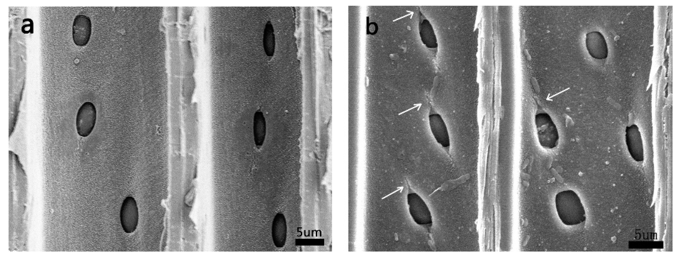
In the cross-field pit region of the low-intensity MW-treated sample (Figure 3), micro-cracks were observed at both ends of the cross-field pit apertures, propagating to nearby cell walls (Figure 3b). Lu et al. [25] found that steam pressure generated during a high-frequency vacuum drying can cause the formation of cracks near cross-field pits in Chinese fir. Similar damage was also observed by Zhang et al. [26] and Xia et al. [27] on sub-alpine fir (Abies lasiocarpa) and larch (Larix gmelinii) wood with steam explosion, and the degree of damage to cross-field pits increased with an increase in temperature or cycles of treatment. Furthermore, Muzamal et al. [28] used finite element software to simulate microstructural changes in Norway spruce (Picea abies) during steam explosion. The results showed that high stresses were visible at the poles of the elliptic cross-field pits under steam pressure, and the resultant stress concentration promoted the formation of cracks in these regions. These results demonstrate that cross-field pits were easily damaged by steam pressure.
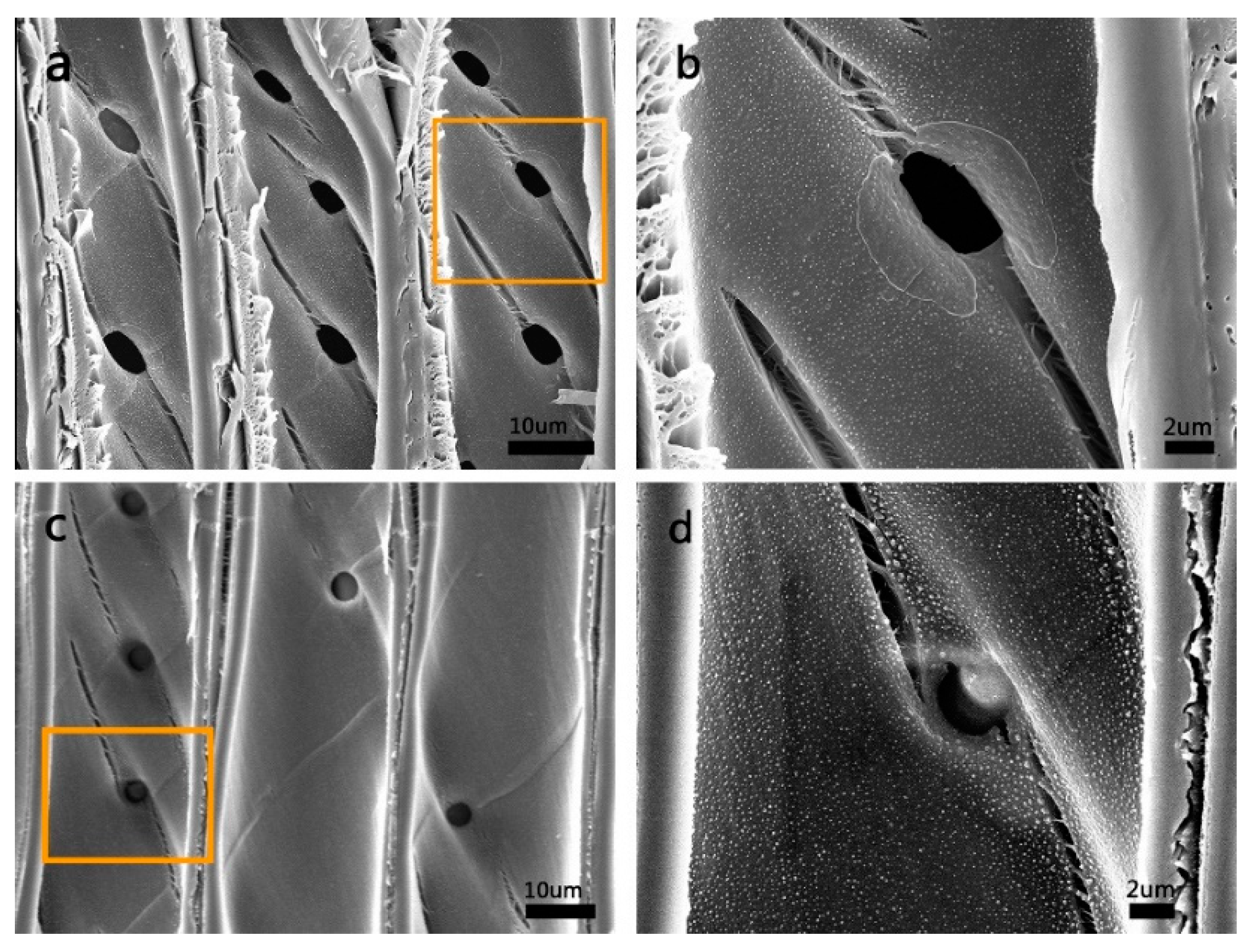
With an increase in MW intensity, cracks at both ends of the cross-field pit apertures also propagated further. The cracks reached 2–4 times as long as the long-axis diameter of the pit (Figure 4a). At the same time, similar cracks also appeared near the bordered pits (Figure 4c). By examining the amplified micrographs of cracks near cross-field pits and bordered pits (Figure 4b,d), it can be observed that the propagation direction of cracks was close to the micro-fibril orientation of the S2 layer in the cell wall (the micro-fibril angle of the S2 layer in Chinese fir wood lies from 25° to 30° to the fiber axis). This finding may be due to the S2 layer being considered to dominate the physical and chemical properties of the cell wall [29]. From a material viewpoint, the cell wall layer can be regarded as a fiber-reinforced composite material, in which cellulose micro-fibrils act as reinforcement and the matrix is a mixture of hemicellulose and lignin [30,31,32,33]. For fiber-reinforced composites, the values of strength and stiffness in the direction perpendicular to the fiber are lower than that in the parallel direction [28]. Therefore, it is more prone to break in the perpendicular direction under steam pressure. As a result, adjacent micro-fibrils separated and formed cracks that propagated along the direction of the micro-fibril orientation. In addition, the middle lamella between the tangential wall of tracheids was observed to be damaged, with detachments occurring (Figure 4a,c).
These results also indicated that MW treatment can cause the middle lamella between ray parenchyma cells and longitudinal tracheids to crack, resulting in the two components being separated (Figure 5). The width of cracks with low-intensity MW treatment was small, generally within 1–25 μm (Figure 5b). These changes may form new pathways for liquid flow and increase wood permeability, corroborating with He et al. [18]. After high-intensity MW treatment, the number of cracks increased and macro-cracks with a width of 100–130 μm appeared (Figure 5c). During MW treatment, water evaporation inside the wood resulted in a rapid increase in steam pressure. As the pressure exceeded the strength of the middle lamella between ray parenchyma cells and tracheids, the middle lamella cracked, and micro-cracks appeared. With the increasing steam pressure, micro-cracks propagated along the radial and longitudinal (RL) plane, resulting in detachments in nearby middle lamellas until macro-cracks formed. As high-intensity MW treatment can generate higher temperatures and pressure inside the wood compared to low-intensity, more middle lamellas cracked, and parts of the cracks gradually propagated to form macro-cracks.
In order to obtain a more comprehensive analysis of the microstructural changes in wood after MW treatment, the microscopic morphology of fracture surface of macro-cracks was observed. As shown in Figure 6, bordered pit membranes in tracheids fell off (Figure 6a), cross-field pit membranes disappeared and the ray parenchyma cells were severely damaged (Figure 6b). In addition, fracture of longitudinal tracheids also occurred (Figure 6c). As shown in Figure 4, high-intensity MW treatment can damage cell walls near the pits and generate large cracks in the tracheid walls, thereby seriously affecting the mechanical properties of tracheids and making tracheids more prone to breaking. During the crack propagation along the radial and longitudinal (RL) plane, the presence of cracks in tracheid walls also creates stress concentration and induces cell wall fracture.
The microstructural changes will have a significant effect on wood permeability. After low-intensity MW treatment, the bordered pit membranes in tracheids were damaged. In consequence, the radius and number of pit membrane openings were increased, accelerating the migration of moisture in the longitudinal and tangential directions. Detachments between ray parenchyma cells and longitudinal tracheids also provide new pathways for the radial migration of moisture. In addition, damage to the cell walls of tracheids can enhance the migration efficiency of moisture between cells. Thus, the longitudinal, radial and tangential permeability of Chinese fir wood can be increased. For the high-intensity MW-treated sample, damage to the microstructure was more severe than that after low-intensity MW treatment, and macro-cracks were generated under this condition. By observing and analyzing the tangential section and fracture surface of macro-cracks (Figure 5c and Figure 6), the macro-cracks can be used as pathways for moisture migration in longitudinal and radial directions. Furthermore, in the tangential direction, a significant reduction in resistance occurred when moisture passed through the fracture surface as the majority of pit membranes on the surface had disappeared. Therefore, the generation of macro-cracks is advantageous for moisture migration in wood. Compared with the low-intensity MW-treated sample, these changes will lead to a further increase in permeability in the high-intensity MW-treated sample.

3.2. Pore Size Distribution Variation of MW-Treated Wood

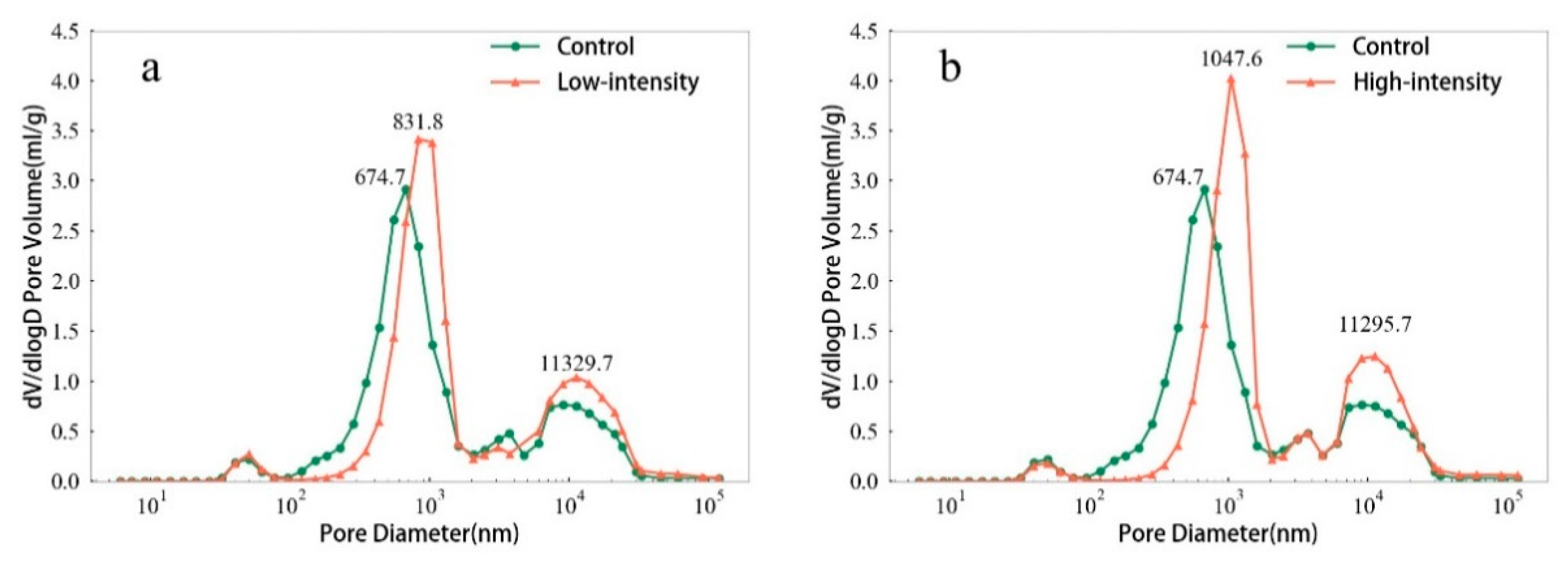
Microstructural changes can result in variation of the pore structure of wood. Figure 7 shows the pore volume calculated using the logarithm of the differential pore diameters, and the pore size distribution of Chinese fir before and after MW treatment can be analyzed. According to previous investigation, pore radii in wood were classified in the range of <0.1 µm for microvoids or cell wall capillaries, 0.1–5 µm for some small tracheid openings (diameter of margo capillaries ranging from 0.1 to 0.7 µm) and >5 µm for large lumens [34]. Figure 7 displays two distinct peaks in the curve of the control sample, with the first peak mainly corresponding to the diameter of margo capillaries and the second peak mainly corresponding to the tracheid lumens. For the low-intensity MW-treated sample (Figure 7a), pore diameter corresponding to margo capillaries in pit membranes became larger in comparison with the control (peak value of the control sample: 674.7 nm, peak value of the low-intensity MW-treated sample: 831.8 nm). In addition, the intensity of the curve in the tracheid lumen diameter range was enhanced, this may be because the bordered pit membranes were damaged after low-intensity MW treatment (Figure 2b), and the connectivity between tracheids was improved. The volume of mercury intruding into the tracheids therefore increased, resulting in an increase in the number of pores within the tracheid lumen diameter range. Micro-cracks between ray parenchyma cells and longitudinal tracheids may also affect the number of pores within the diameter range (Figure 5b). This result was consistent with Wang et al. [19]. The cited authors found an increase in number of micro-voids in the tracheid lumen diameter range on MW-treated Pinus sylvestris var. mongolica wood. After high-intensity MW treatment, pore diameter corresponding to margo capillaries further increased (peak value of the control sample: 674.7 nm, peak value of the high-intensity MW-treated sample: 1047.6 nm), as shown in Figure 7b. From peak height, increment due to high-intensity MW treatment was also the most significant, possibly being related to more severe damage to the bordered pit membranes under this treatment condition. Cracks near cross-field pits and bordered pits may also increase the number of pores within the diameter range (Figure 4). Moreover, the intensity of the curve in the tracheid lumen diameter range was higher than that of the control or low-intensity MW treatment sample, possibly being related to more severe damage to the bordered pit membranes under the effect of high-intensity MW and further enhancement of tracheid connectivity.
Overall, the damage to the wood microstructure caused by MW treatment can lead to an increase in pore diameter and the number of pores. Thus, new pathways for moisture migration were formed, resulting in an increase in permeability of the wood. In addition, the high-intensity MW treatment had a more significant effect on pore structure than the low-intensity MW treatment.

4. Conclusions

After low-intensity MW treatment, parts of the bordered pit membranes in tracheids were damaged, micro-fibrils on the margo were ruptured, while the torus basically remained intact. Micro-cracks were generated at both ends of the aperture of cross-field pits, propagating to the cell walls of tracheids. The middle lamella between ray parenchyma cells and longitudinal tracheids also cracked, resulting in the two components being separated with a crack width generally ranging between 1 and 25 μm. Damage to the microstructure after high-intensity MW treatment was more severe than that under low-intensity MW treatment. Ruptures were observed on the torus of bordered pit membranes. Cracks near the cross-field pits propagated further and similar damage also occurred near the bordered pits. An increase in cracks appeared between ray parenchyma cells and longitudinal tracheids, with some cracks propagating into macro-cracks with a width of 100–130 μm. Detachments in middle lamella between the tangential wall of tracheids were also observed. On the fracture surface of macro-cracks, the bordered pit membranes in tracheids fell off, cross-field pit membranes disappeared and the ray parenchyma cells were severely damaged, exhibiting fracture of the tracheid walls.
MIP test results showed that pore diameter corresponding to the margo capillaries in pit membranes became larger after low- and high-intensity MW treatment (peak value increased from 674.7 nm to 831.8 nm and 1047.6 nm, respectively). The number of pores in the tracheid lumen diameter range also significantly increased. As a result, new pathways for moisture migration were formed after MW treatment, which contribute to increased permeability of Chinese fir, thereby improving wood-drying efficiency and quality. However, the high-intensity MW treatment caused severe damage to the wood microstructure, and macro-cracks were generated, which may adversely affect the appearance quality and mechanical properties of wood.

Author Contributions

Conceptualization, Y.Z. and X.W.; methodology, Y.Z. and X.W.; software, X.W.; validation, Y.Z. and Z.F.; formal analysis, X.W. and F.Z.; investigation, X.W.; resources, F.F.; data curation, X.W. and X.G.; writing—original draft preparation, X.W.; writing—review and editing, Y.Z. and Z.F.; visualization, X.W.; supervision, Y.Z.; project administration, Y.Z.; funding acquisition, Y.Z. All authors have read and agreed to the published version of the manuscript.

Funding

This research was funded by the National Natural Science Foundation of China, grant number 31890772.

Acknowledgments

The authors express their gratitude to Junfeng Hou and Dong Wang for their assistance with this study.

Conflicts of Interest

The authors declare no conflict of interest.

References

  1. Bao, F.; Lv, J. A study on fluid permeability of important Chinese woods. Sci. Silvae Sin. 1992, 28, 237–246. (In Chinese) [Google Scholar]
  2. Guo, J.; Guo, X.; Xiao, F.; Xiong, C.; Yin, Y. Influences of provenance and rotation age on heartwood ratio, stem diameter and radial variation in tracheid dimension of cunninghamia lanceolata. Eur. J. Wood Wood Prod. 2018, 76, 669–677. [Google Scholar] [CrossRef]
  3. Torgovnikov, G.; Vinden, P. High-Intensity microwave wood modification for increasing permeability. For. Prod. J. 2009, 59, 84–92. [Google Scholar]
  4. Torgovnikov, G.; Vinden, P. Microwave wood modification technology and its applications. For. Prod. J. 2010, 60, 173–182. [Google Scholar] [CrossRef]
  5. Vinden, P.; Torgovnikov, G.; Hann, J. Microwave Modification of radiata pine railway sleepers for preservative treatment. Eur. J. Wood Wood Prod. 2011, 69, 271–279. [Google Scholar] [CrossRef]
  6. Harris, G.A.; Torgovnikov, G.; Vinden, P.; Brodie, G.I.; Shaginov, A. Microwave pretreatment of backsawn messmate boards to improve drying quality: Part 1. Dry. Technol. 2008, 26, 579–584. [Google Scholar] [CrossRef]
  7. Dömény, J.; Koiš, V.; Dejmal, A. Microwave radiation effect on axial fluid permeability in false heartwood of beech (Fagus Sylvatica L.). BioResources 2014, 9, 372–380. [Google Scholar] [CrossRef] [Green Version]
  8. He, X.; Xiong, X.; Xie, J.; Li, Y.; Wei, Y.; Quan, P.; Mou, Q.; Li, X. Effect of microwave pretreatment on permeability and drying properties of wood. BioResources 2017, 12, 3850–3863. [Google Scholar] [CrossRef] [Green Version]
  9. Balboni, B.M.; Ozarska, B.; Garcia, J.N.; Torgovnikov, G. Microwave treatment of eucalyptus macrorhyncha timber for reducing drying defects and its impact on physical and mechanical wood properties. Eur. J. Wood Wood Prod. 2018, 76, 861–870. [Google Scholar] [CrossRef]
  10. Zhang, Y.; Jia, K.; Cai, L.; Shi, S.Q. Acceleration of moisture migration in larch wood through microwave pre-treatments. Dry. Technol. 2013, 31, 666–671. [Google Scholar] [CrossRef]
  11. Dashti, H.; Shahverdi, M.; Taghiyari, H.R.; Salehpur, S.; Heshmati, S. Effects of steaming and microwave pretreatments on mass transfer characteristics of aleppe oak (quercus infectoria). BioResources 2012, 7, 3262–3273. [Google Scholar]
  12. Liu, H.; Wang, Q.; Yang, L.; Jiang, T.; Cai, Y. Modification of larch wood by intensive microwave irradiation. J. For. Res. 2005, 16, 237–240. [Google Scholar]
  13. Li, X.; Zhang, B.; Li, W.; Li, Y. Research on the effect of microwave pretreatment on moisture diffusion coefficient of wood. Wood Sci. Technol. 2005, 39, 521–528. [Google Scholar] [CrossRef]
  14. Plötze, M.; Niemz, P. Porosity and pore size distribution of different wood types as determined by mercury intrusion porosimetry. Eur. J. Wood Wood Prod. 2011, 69, 649–657. [Google Scholar] [CrossRef] [Green Version]
  15. Ding, W.D.; Koubaa, A.; Chaala, A.; Belem, T.; Krause, C. Relationship between wood porosity, wood density and methyl methacrylate impregnation rate. Wood Mater. Sci. Eng. 2008, 3, 62–70. [Google Scholar] [CrossRef]
  16. Martin-Sampedro, R.; Martin, J.A.; Eugenio, M.E.; Revilla, E.; Villar, J.C. Steam explosion treatment of Eucalyptus globulus wood: Influence of operational conditions on chemical and structural modifications. BioResources 2011, 6, 4922–4935. [Google Scholar]
  17. Schneider, A. Investigations on the suitability of mercury porosimetry for the evaluation of wood impregnability. Eur. J. Wood Wood Prod. 1983, 41, 101–107. [Google Scholar] [CrossRef]
  18. He, S.; Lin, L.; Fu, F.; Zhou, Y.; Fan, M. Microwave treatment for enhancing the liquid permeability of Chinese fir. BioResources 2014, 9, 1924–1938. [Google Scholar] [CrossRef] [Green Version]
  19. Wang, D.; Peng, L.; Zhu, G.; Fu, F.; Zhou, Y.; Song, B. Improving the sound absorption capacity of wood by microwave treatment. BioResources 2014, 9, 7504–7518. [Google Scholar] [CrossRef] [Green Version]
  20. Tong, Y.; Zhao, G. Structure of bordered pit membrane of Cunninghamia lanceolata tracheid. Sci. Silvae Sin. 2007, 43, 151–153. [Google Scholar]
  21. Washburn, E.W. Note on a method of determining the distribution of pore sizes in a porous material. Proc. Natl. Acad. Sci. USA 1921, 7, 115–116. [Google Scholar] [CrossRef] [PubMed] [Green Version]
  22. Muzamal, M.; Jedvert, K.; Theliander, H.; Rasmuson, A. Structural changes in spruce wood during different steps of steam explosion pretreatment. Holzforschung 2015, 69, 61–66. [Google Scholar] [CrossRef]
  23. Erickson, H.D. Permeability of southern pine wood-a review. Wood Sci. 1970, 2, 149–158. [Google Scholar]
  24. Petty, J.A. Permeability and structure of the wood of sitka spruce. Proc. R. Soc. Lond. B 1970, 175, 149–166. [Google Scholar]
  25. Lu, J.; Lin, Z.; Jiang, J.; Zhao, Y.; Jiang, J.; Gao, R.; Yin, Y. Effect of diferent drying methods on the liquid impregnation of Chinese fir plantation wood. Sci. Silvae Sin. 2006, 42, 86–90. (In Chinese) [Google Scholar]
  26. Zhang, Y.; Cai, L. Effects of steam explosion on wood appearance and structure of sub-alpine fir. Wood Sci. Technol. 2006, 40, 427–436. [Google Scholar] [CrossRef]
  27. Xia, J.; Zhang, Y.; Cai, J. Opening cell pathways of larch wood by steam explosion. J. Fujian Agric. For. Univ. 2013, 42, 543–547. (In Chinese) [Google Scholar]
  28. Muzamal, M. Structural Modifications in Spruce Wood during Steam Explosion Pretreatment. Ph.D. Thesis, Chalmers University of Technology, Gothenburg, Sweden, 2016. [Google Scholar]
  29. Donaldson, L.; Xu, P. Microfibril orientation across the secondary cell wall of radiata pine tracheids. Trees 2005, 19, 644–653. [Google Scholar] [CrossRef]
  30. Deng, Q.; Li, S.; Chen, Y. Mechanical properties and failure mechanism of wood cell wall layers. Comput. Mater. Sci. 2012, 62, 221–226. [Google Scholar] [CrossRef]
  31. Muzamal, M.; Gamstedt, E.K.; Rasmuson, A. Modeling wood fiber deformation caused by vapor expansion during steam explosion of wood. Wood Sci. Technol. 2014, 48, 353–372. [Google Scholar] [CrossRef]
  32. Qing, H.; Mishnaevsky, L. 3D hierarchical computational model of wood as a cellular material with fibril reinforced, heterogeneous multiple layers. Mech. Mater. 2009, 41, 1034–1049. [Google Scholar] [CrossRef]
  33. Yamamoto, H. Role of the gelatinous layer on the origin of the physical properties of the tension wood. J. Wood Sci. 2004, 50, 197–208. [Google Scholar] [CrossRef]
  34. Schneider, A. Analysing the porosity of wood with the mercury porosimeter. Eur. J. Wood Wood Prod. 1979, 37, 295–302. [Google Scholar] [CrossRef]
Figure 1. Scanning electron microscopy (SEM) micrographs of bordered pits under different microwave (MW) intensities: (a) Control, (b) low-intensity, (c) high-intensity.
Forests 11 00772 g001
Figure 2. SEM micrographs of bordered pit membranes under different MW intensities: (a) Control, (b) low-intensity, (c) high-intensity.
Forests 11 00772 g002
Figure 3. SEM micrographs of cross-field pit region in low-intensity MW-treated sample: (a) Control, (b) low-intensity MW-treated sample.
Forests 11 00772 g003
Figure 4. SEM micrographs of cross-field pit and bordered pit region in the high-intensity MW-treated sample: (a) Cracks near cross-field pits and detachments in middle lamella, (b) amplified crack near a cross-field pit, (c) cracks near bordered pits and detachments in middle lamella, (d) amplified crack near a bordered pit.
Forests 11 00772 g004
Figure 5. SEM micrographs of cracks between ray parenchyma cells and longitudinal tracheids under different MW intensities: (a) Control, (b) low-intensity, (c) high-intensity.
Forests 11 00772 g005
Figure 6. SEM micrographs of fracture surface of macro-cracks in the high-intensity MW-treated sample: (a) Bordered pit membranes fell off, (b) cross-field pit membranes disappeared and ray parenchyma cells were damaged, (c) longitudinal tracheids fractured.
Forests 11 00772 g006
Figure 7. Log differential intrusion versus pore diameter of samples under different intensities: (a) Low-intensity, (b) high-intensity.
Forests 11 00772 g007