Article

Comprehensive Transcriptome Analysis Revealed the Effects of the Light Quality, Light Intensity, and Photoperiod on Phlorizin Accumulation in Lithocarpus polystachyus Rehd.

1
College of Life Sciences, North China University of Science and Technology, Tangshan 063210, China
2
College of Pharmacy, North China University of Science and Technology, Tangshan 063210, China
3
College of Agriculture and Food Engineering, Baise University, Baise 533000, China
*
Authors to whom correspondence should be addressed.
Forests 2019, 10(11), 995; https://doi.org/10.3390/f10110995
Submission received: 8 October 2019 / Revised: 28 October 2019 / Accepted: 4 November 2019 / Published: 7 November 2019
(This article belongs to the Section Forest Ecophysiology and Biology)

Abstract

:
Lithocarpus polystachyus Rehd. is an important medicinal plant species grown in southern China, with phlorizin as its main active substance. The effects of light conditions on phlorizin biosynthesis in L. polystachyus remain unclear. Thus, we analyzed the transcriptomes of L. polystachyus plants cultivated under diverse light qualities, light intensities, and photoperiods. The light treatments resulted in 5977–8027 differentially expressed genes (DEGs), which were functionally annotated based on the gene ontology (GO) and Kyoto Encyclopedia of Genes and Genomes (KEGG) databases. Genes encoding transcription factors from 89 families were differentially expressed after the light treatments, implying these transcription factors are photoresponsive. Phenylalanine ammonia lyase (PAL) and 4-coumarate-CoA ligase (4CL) are the key enzymes for the accumulation of phlorizin. The transcription levels of PAL2, PAL, 4CL1 (DN121614), 4CLL7, and 4CL1 (DN102161) were positively correlated with phlorizin accumulation, suggesting that these genes are important for phlorizin biosynthesis. An ultra-high-performance liquid chromatography method was used to quantify the phlorizin content. Phlorizin accumulated in response to the green light treatment and following appropriate decreases in the light intensity or appropriate increases in the duration of the light exposure. The green light, 2000 lx, and 3000 lx treatments increased the PAL activity of L. polystachyus, but the regulatory effects of the light intensity treatments on PAL activity were relatively weak. This study represents the first comprehensive analysis of the light-induced transcriptome of L. polystachyus. The study results may form the basis of future studies aimed at elucidating the molecular mechanism underlying phlorizin biosynthesis in L. polystachyus. Moreover, this study may be relevant for clarifying the regulatory effects of light on the abundance of bioactive components in medicinal plants.

1. Introduction

Lithocarpus polystachyus Rehd. (i.e., sweet tea) is an evergreen tree species (family Fagaceae) that is mainly distributed in the provinces and autonomous regions south of the Yangtze River in China. Its tender leaves are often collected to produce a tea with health benefits. For example, it promotes fluid production, quenches thirst, and eliminates fatigue. A previous study revealed that a dihydrochalcone glucoside, phlorizin, is distributed in L. polystachyus leaves and is considered one of the main active substances in this plant species [1].
Phlorizin is an important flavonoid compound with various pharmacological effects (e.g., decreases blood sugar levels and protects against oxidation) that can reduce the risk of diabetic complications, lower the blood cholesterol level, inhibit the development of various cancers (e.g., lung cancer, colon cancer, and breast cancer), and synergistically promote the effects of anti-tumor drugs [2]. Therefore, enhancing phlorizin biosynthesis and accumulation by changing the growth environment has become a major research objective. Gosch et al. [3] described in detail the main phlorizin biosynthesis pathway in apples. Precursors for phlorizin biosynthesis are malonyl-CoA and p-coumaroyl-CoA. First, p-coumaroyl-CoA generates 4-hydrocinnamoyl-CoA through the NADPH pathway. Then, malonyl-CoA and 4-hydrocinnamoyl-CoA generate phloretin in a reaction catalyzed by chalcone synthetase. Finally, the glucosylation of phloretin requires the regulatory activities of various genes and enzymes. However, previous studies mostly involved apple, and there has been relatively little research on the mechanism underlying phlorizin biosynthesis in L. polystachyus.
Light is one of the most important environmental factors for plant growth and development. Specifically, plant growth is influenced by the light irradiance, wavelength, duration, and direction. Light is also used as an energy source for photosynthesis and can influence various physiological activities [4,5]. For example, the leaf area and anthocyanin content vary and the genes related to photosynthesis and pigment synthesis are differentially expressed in potato plantlets cultured under diverse spectral conditions [6]. Decreases in light intensity are accompanied by decreases in superoxide dismutase and peroxidase activities and the soluble protein content in Lithocarpus litseifolius leaves, whereas the chlorophyll a, chlorophyll b, total chlorophyll, and total flavonoid contents increase, thereby promoting flavonoid accumulation [7]. A previous study involving the sequencing of the transcriptome of ginkgo leaves harvested at midday and midnight revealed that the expression levels of genes related to flavonoid biosynthesis were lower in the leaves collected at midnight, as was the total flavonoid content [8]. There is considerable experimental evidence that changes in light quality, light intensity, and the photoperiod significantly affect the physiological activities and morphological characteristics of plant leaves.
A transcriptome analysis is an efficient method for detecting and clarifying the molecular mechanisms involved in physiological processes and enables the identification of important differentially expressed genes (DEGs). Additionally, RNA sequencing (RNA-seq) is a robust method for genome-wide transcriptome analyses [9]. The transcriptomes of various plant species under biotic or abiotic stress were analyzed, but there has been a relative lack of transcriptome research on plants under diverse light conditions, and there are few reports on the light-regulated genes and pathways of L. polystachyus.
In this study, the Illumina NovaSeq 6000 platform was used for a transcriptome sequencing analysis of L. polystachyus leaves exposed to different light qualities, light intensities, and photoperiods to clarify the expression trends of key genes for biological processes and to screen for genes related to phlorizin biosynthesis. Additionally, ultra-high-performance liquid chromatography (UPLC) was applied to determine the changes in phlorizin contents in response to various light treatments. The data presented herein may be useful for future studies aimed at characterizing phlorizin biosynthesis in L. polystachyus.

2. Materials and Methods

2.1. Experimental Material

Uniformly growing three-year-old L. polystachyus plants in Bama (county), Guangxi, China were selected and cultivated in the plant cultivation room of the College of Life Sciences, North China University of Science and Technology, and these plants are asexual reproduction individuals from the same plant. Specifically, the plants were grown in a 65 cm × 25 cm × 20 cm plastic basin containing soil from the region in which the plants were collected. After 15 days of adaptive growth under natural conditions, the plants were exposed to different monochromatic light treatments (Table 1) involving LED-T8/T5 light tubes (Aimite, China). The light treatment continued for 15 days, after which leaves were collected, quickly frozen in liquid nitrogen, and stored at −80 °C for subsequent analyses. For each light treatment, more than three leaves were collected and combined to form one replicate, with analyses completed with three replicates.

2.2. RNA Extraction and Illumina Sequencing

Total RNA was extracted and purified from L. polystachyus leaves with the RNAprep Pure Plant kit (Tiangen, Beijing, China), after which the RNA purity, concentration, and integrity were determined with the NanoDrop spectrophotometer (Thermo Fisher, Waltham, MA, USA) and 2100 Bioanalyzer (Agilent, Santa Clara, CA, USA). Magnetic beads with oligo (dT) were used to extract the mRNA from the total RNA. The mRNA was fragmented by an ion disruption and then used as the template for the synthesis of cDNA via reverse transcription. The resulting double-stranded cDNA was purified with AMPure XP beads and then processed (e.g., end repair, addition of a poly A tail, and PCR enrichment) to generate a cDNA library. The quality of the cDNA library was assessed with the 2100 Bioanalyzer. The high-quality library was sequenced with the second-generation sequencing technology of the Illumina NovaSeq 6000 system.

2.3. Transcript Splicing, Assembly, and Functional Annotation

The raw sequencing data were filtered to remove reads containing linkers and low-quality reads (i.e., reads in which the proportion of N is greater than 10% or the number of bases with a mass value Q ≤ 10 exceeds 50% of the whole read) to obtain high-quality sequencing data. Each library sequence was spliced using Trinity software (version 2.2.0) (http://trinityrnaseq.github.io/) [10]. Additionally, some short reads with overlapping regions were assembled into contigs. On the basis of the paired-end information, the distances separating contigs were determined by mapping the clean reads to obtain transcript sequences. Finally, the TGICL program (version 2.0.6) (https://sourceforge.net/projects/tgicl/) [11] was used to cluster transcripts into unigenes. The BLAST algorithm was used to compare the unigene sequences with sequences in the NCBI nonredundant (NR), Swiss-Prot, gene ontology (GO), Clusters of Orthologous Groups of proteins (COG), and Kyoto Encyclopedia of Genes and Genomes (KEGG) databases. Moreover, KOBAS v2.0 (http://kobas.cbi.pku.edu.cn) was applied to identify enriched KEGG pathways associated with the unigenes. Moreover, after predicting the amino acid sequences encoded by the unigenes, the HMMER (http://www.hmmer.org/) program was used to compare the sequences with those in the Pfam database to functionally annotate the unigenes. Sequence data generated for the present study was deposited into the SRA database. The bioproject ID assigned is: PRJNA575946 (https://www.ncbi.nlm.nih.gov/sra/PRJNA575946).

2.4. Screening of Differentially Expressed Genes

The RSEM (version 1.2.29) (http://deweylab.github.io/RSEM/) program was used to determine the read count for each gene in each sample. Gene expression levels were based on the number of fragments per kilobase of transcript per million mapped reads (FPKM). Then, EBSeq (version 1.12.0) (http://www.bioconductor.org/packages/release/bioc/html/EBSeq.html) was used for analyzing expression level differences, with a false discovery rate (FDR) < 0.01 and |log2 fold-change| ≥ 1 applied as the criteria for significant differences in expression. The fold-change represents the expression level ratio between two samples (groups) and the FDR is based on a corrected p-value. The GO database (http://www.geneontology.org/) was used to functionally annotate the DEGs with GO terms, whereas KOBAS 2.0 was used to detect the enriched KEGG pathways among the DEGs.

2.5. Quantitative Real-Time PCR

To verify the accuracy of the expression data based on the RNA-seq analysis, five genes were selected for a quantitative real-time (qRT)-PCR assay. Primer 5.0 (Premier Biosoft Interpairs, Palo Alto, CA, USA) [12] was used to design primers specific for the selected genes (Table 2), with a dissolution curve analysis used to determine specificity. The qRT-PCR assay was completed with the Talent qPCR Premix (SYBR Green) (Tiangen, Beijing, China) and the Applied Biosystems 7900HT Fast Real-Time PCR System (LifeTechnologies, Waltham, MA, USA). The L. polystachyus GAPDH gene served as the internal reference gene. The qRT-PCR program was as follows: 95 °C for 3 min; 40 cycles of 95 °C for 5 s and 60 °C for 15 s. The qRT-PCR assay was performed with three biological replicates and relative expression levels were calculated based on the 2−ΔΔCt method.

2.6. Determination of the Phlorizin Content and Phenylalanine Ammonia Lyase Activity

The phlorizin content of L. polystachyus leaves was determined based on a published method [13]. The UPLC system (Waters, Milford, MA, USA) comprised the ACQUITY UPLC BEN C18 column (2.1 mm × 50 mm, 1.7 μm) (Waters, Milford, MA, USA). Additionally, HPLC-grade acetonitrile and analytical-grade reagents were used for the UPLC. The phlorizin reference substance was accurately weighed, after which 10 μg reference substance was mixed with 1 mL ethanol. Approximately 0.2 g coarse ground L. polystachyus leaf powder was added to a beaker and mixed with 20 mL methanol. The resulting solution underwent an ultrasonic extraction for 20 min, after which it was filtered through a 0.22-μm microporous membrane to obtain the final product for analysis. The isocratic elution involved methanol (A) and 0.2% glacial acetic acid (B), with 75% A at a flow rate of 0.25 mL/min. The detection wavelength was 257 nm. The column temperature was 30 °C and the injection volume was 10 µL.
The phenylalanine ammonia lyase (PAL) activity in L. polystachyus leaves was determined spectrophotometrically with a Phenylalanine ammonia lyase Test kit (Keming, Suzhou, China). The data were analyzed with the SPSS 16.0 (IBM SPSS, Chicago, IL, USA) program. Additionally, Duncan’s multiple comparison method was used for assessing the significance of any differences (p ≤ 0.05, p ≤ 0.05 for significant differences and p ≤ 0.01 for extremely significant differences).

3. Results

3.1. Transcriptome Sequencing and Sequence Assembly

After removing the reads with a linker or low-quality reads, 20.51–24.27 million clean reads comprising 6.15–7.28 Gb were obtained. The GC content of the sequences for the 10 samples was about 45% and more than 93% of the bases had a Phred quality score greater than 30 (Table 3), indicating the quality of the sequencing data was acceptable.
The Trinity software was used to assemble the clean reads for the 10 samples. The initial assembly produced 206,515 transcripts, of which the longest and shortest transcripts were 15,707 bp and 201 bp, respectively. The average transcript length was 601 bp, with an N50 of 786 bp. Furthermore, the low-abundance transcripts were filtered to obtain 177,894 unigenes, with an average length of 560 bp and an N50 of 689 bp (Table 4). Unigenes comprising 200–300 bp accounted for the largest proportion of the total (66,284, 37.26%), whereas unigenes consisting of 200–1000 bp, 1000–2000 bp, and 2000–3000 bp accounted for 88.34% (157,153), 7.84% (13,960), and 2.56% (4559) of the total, respectively. Additionally, 2222 unigenes (1.25%) were longer than 3000 bp (Figure 1a).

3.2. Annotation and Classification of Transcripts

To predict unigene functions, the proteins encoded by the unigenes were functionally annotated based on seven protein databases (NR, GO, KEGG, KOG, COG, Pfam, and Swiss-Prot). A total of 76,726 unigenes (43.13% of all unigenes) were annotated according to matches to at least one of the databases. A total of 73,609 unigenes (41.37%) were annotated based on the NR database, which was the highest proportion of any of the databases. Regarding the other databases, 45,273 unigenes (25.45%) were annotated based on the GO database, 16,485 unigenes (9.27%) were annotated based on the KEGG database, 15,385 unigenes (8.65%) were annotated based on the COG database, 16,485 unigenes (9.27%) were annotated based on the KOG database, 34,632 unigenes (19.47%) were annotated based on the Pfam database, and 46,430 unigenes (26.1%) were annotated based on the Swiss-Prot database (Table S1).
An analysis of the sequence matches to the NR database revealed that the L. polystachyus unigenes were most similar to genes from Vitis vinifera (12.79%), Theobroma cacao (6.17%), Prunus mume (5.61%), Prunus persica (4.85%), and Citrus sinensis (4.34%) (Figure S1). Additionally, sequence matches to the GO database indicated 45,273 L. polystachyus unigenes were functionally classified into 50 groups (Figure 1b), which were mainly divided into three categories. Specifically, 56,180 unigenes were classified in the cellular component category, with “cell part” (17,191), “membrane part” (12,134), and “organelle” (7623) representing the main GO terms. The 55,048 unigenes in the molecular function category were mainly assigned the “binding” (24,667), “catalytical activity” (23,891), “transporter activity” (2673), and “structural molecular activity” (1023) GO terms. Finally, the 103,262 unigenes in the biological process category were mainly annotated with the “metabolic process” (27,865), “cellular process” (25,095), and “single-organism process” (17,490) GO terms. Because many unigenes were allocated to multiple categories, the total number of GO annotations exceeded the total number of unigenes.
The unigenes annotated based on the KEGG database were assigned to 128 KEGG pathways (Table S2), of which the biological pathways, including “carbon metabolism”, “plant-pathogen interaction”, “biosynthesis of amino acids”, and “protein processing in endoplasmic reticulum”, were the most common (Figure 1c). The unigenes annotated according to matches with the COG database were mainly divided into 25 COG classification groups. The “general function prediction only” group comprised the largest proportion of unigenes, followed by “signal transduction mechanisms”, “posttranslational modification, protein turnover, chaperones”, and “carbohydrate transport and metabolism”. Additionally, only nine unigenes were included in the “cell motility” group (Figure 1d).

3.3. Analysis of Transcript Abundance in L. polystachyus Plants Exposed to Various Light Conditions

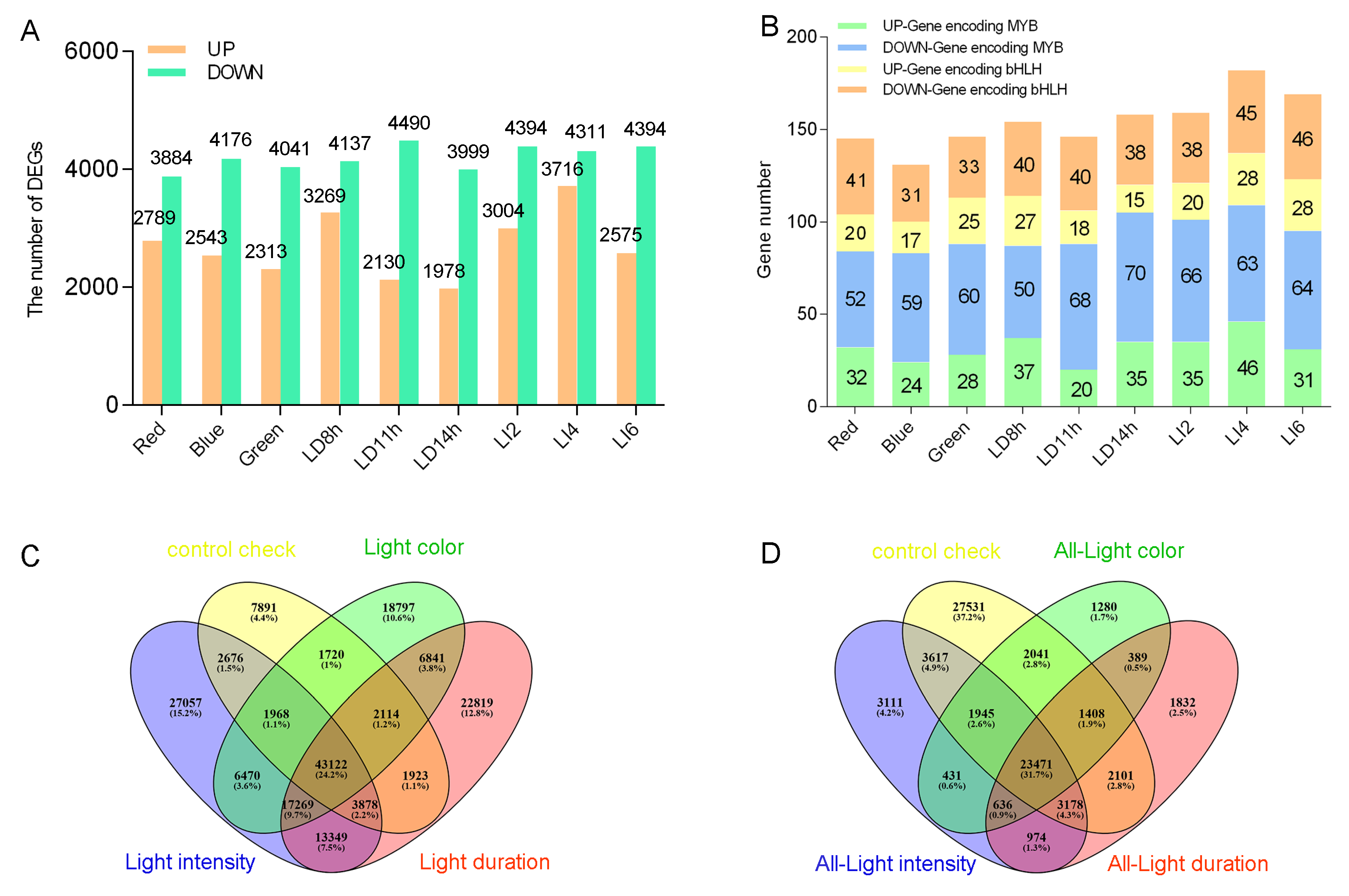
We examined the changes in unigene transcription induced by light conditions according to the FPKM values. The genes that were differentially expressed between the control and light-treated L. polystachyus plants were identified (FDR < 0.01 and |log2 fold-change| ≥ 1). A comparison with the control transcript levels revealed 5977–8027 DEGs, of which 1978–3716 were up-regulated and 3884–4490 were down-regulated in the light-treated plants (Figure 2a). Additionally, 7891 unigenes were expressed only in the control group (Figure 2c). In contrast, 1280 (light quality), 1832 (light duration), and 3111 (light intensity) unigenes were specifically expressed only in response to the corresponding light treatment, but were not expressed under control conditions or in response to the other light treatments (Figure 2d).
The GO annotations of the DEGs affected by light factors revealed the DEGs mainly belonged to two major categories: biological process and molecular function. Additionally, GO terms, such as “nucleotide binding”, “protein serine/threonine kinase activity”, “membrane”, “protein kinase activity”, “phosphorylation”, “protein phosphorylation”, and “polysaccharide binding’, were enriched among the DEGs of the nine light treatments. The “response to stimulus” and “kinase activity” GO terms were enriched among the DEGs identified in response to most treatments (Figure S2).
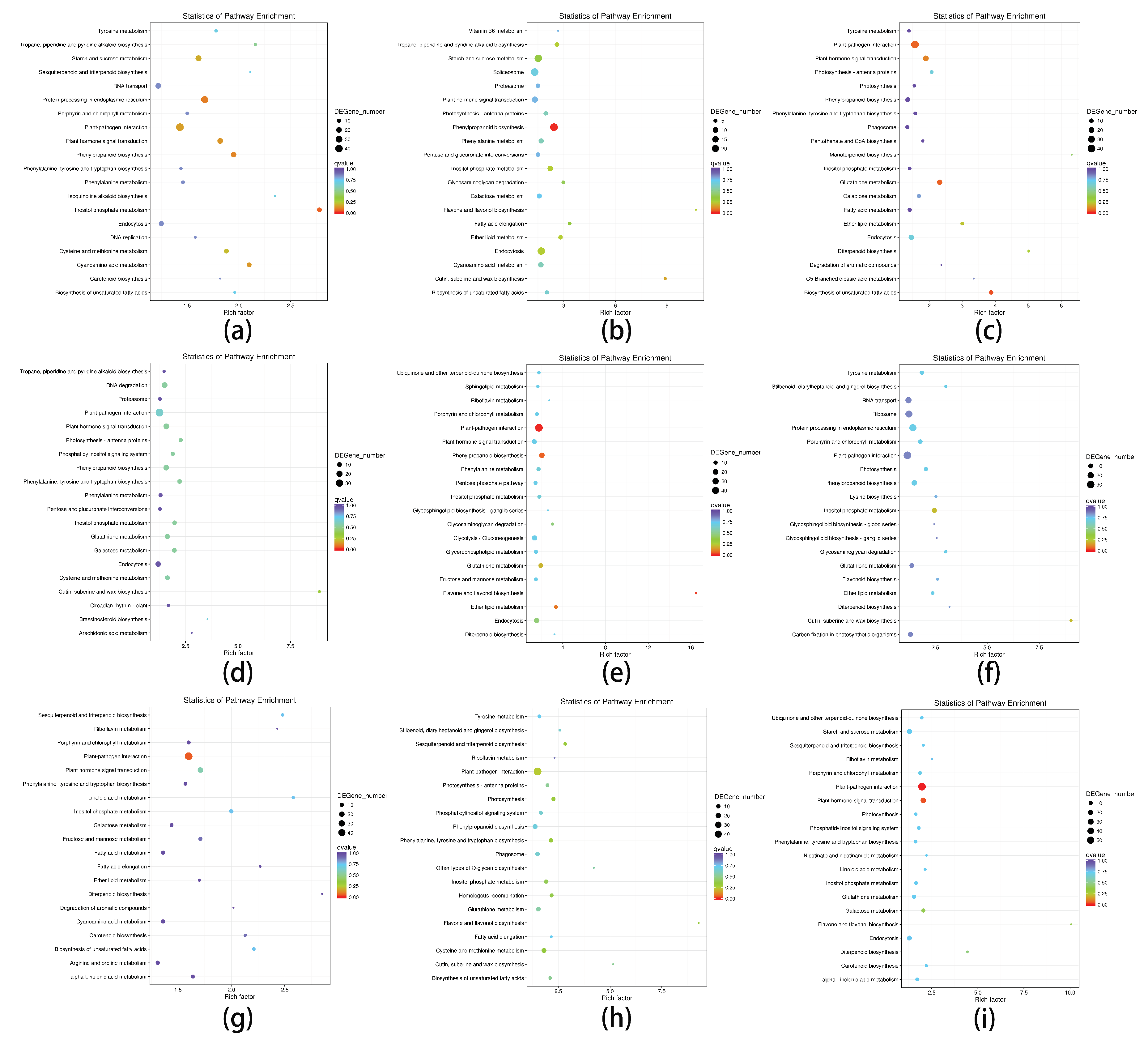
All DEGs were analyzed based on the KEGG database to identify the biological pathways affected by light factors. The “inositol phosphate metabolism” pathway was the most enriched pathway in response to the nine light treatments. “Phenylalanine, tyrosine and tryptophan biosynthesis”, “plant hormone signal transduction”, “galactose metabolism”, “porphyrin and chlorophyll metabolism”, “diterpenoid biosynthesis”, and other pathways were significantly enriched under most light treatments. The “plant-pathogen interaction” pathway was highly enriched among the DEGs for all light treatments, except for those associated with the blue light treatment, implying that blue light has a weaker effect on this pathway than the other light treatments. Moreover, plants were vulnerable to pathogenic bacteria under light-induced stress. The enrichment of the “Phenylpropanoid biosynthesis” pathway was lower in response to the three light intensity treatments than following the other light treatments, implying this pathway is less affected by changes in light intensity than by the other light treatments. In contrast, the “riboflavin metabolism” and “sesquiterpenes and triterpenoid biosynthesis” pathways were more enriched by the three light intensity treatments than by the other light conditions, suggesting these two pathways are considerably affected by light intensity changes. The “biosynthesis of unsaturated fatty acids” pathway was less enriched for the three photoperiod treatments than for the other treatments, possibly because of the insignificant effect of the light duration on this pathway (Figure 3).
A further analysis of the DEGs in each light treatment indicated that 437 unigenes were significantly differentially expressed (relative to the control level) in response to all light treatments (Figure S3), with 28 and 407 unigenes exhibiting up- and down-regulated expression, respectively. The expression levels of the other two unigenes (TRINITY_DN101595_c0_g1 and TRINITY_DN63337_c0_g1) were significantly up-regulated only in response to the 8-h and 14-h exposures to daylight, but were otherwise significantly down-regulated. The GO and KEGG annotations for these unigenes revealed that 83 are involved in biological processes, 79 are cellular components, and 93 are involved in molecular functions. The up-regulated genes were mainly related to the “endocytosis”, “ether lipid metabolism”, “glycerophospholipid metabolism”, and other metabolic pathways. In contrast, the down-regulated genes were primarily associated with “plant-pathogen interaction”, “spliceosome”, “glycolysis/gluconeogenesis”, and other pathways. The TRINITY_DN63337_c0_g1 gene was related to the “inositol phosphate metabolism” pathway, whereas TRINITY_DN101595_c0_g1 was not annotated (Table S3).

3.4. Differences in the Transcript Abundance of L. polystachyus Plants Exposed to Various Light Conditions

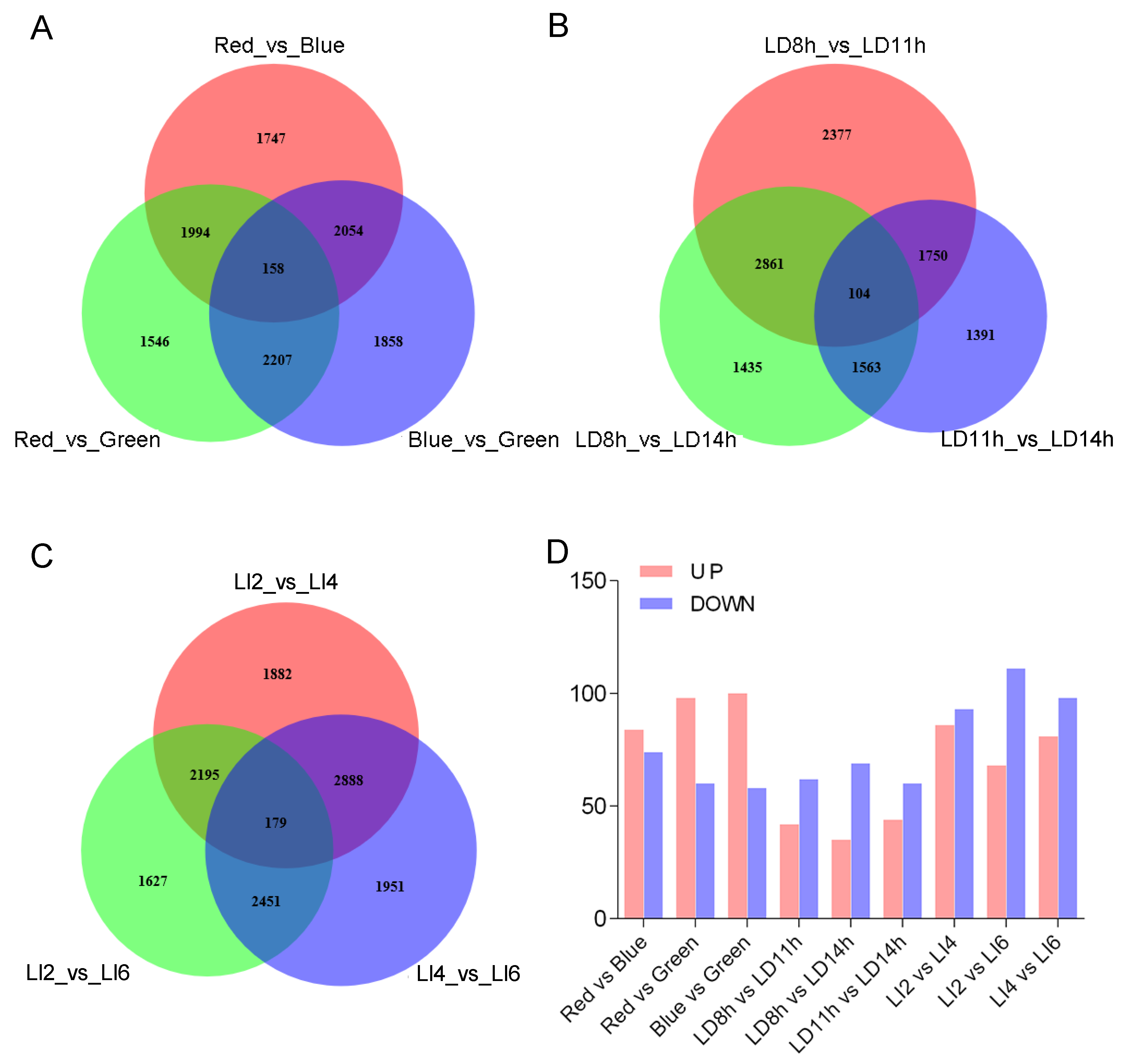
Unigene expression levels in response to the three treatments per light condition (i.e., light quality, duration, and intensity) were compared to screen for DEGs for each light condition. We detected 158 unigenes that were differentially expressed between pairs of light quality treatments (Figure 4a), whereas 104 unigenes were differentially expressed among the photoperiod treatments (Figure 4b) and 179 unigenes were differentially expressed among the light intensity treatments (Figure 4c). Details regarding the up- and down-regulated unigenes in pair-wise comparisons are presented in Figure 4d.
Of the 158 unigenes that were significantly differentially expressed in response to the light quality treatments, 38, 40, and 80 were most highly expressed in response to the red, blue, and green light treatments, respectively. Additionally, the expression of 44 unigenes were affected by the light qualities in the rank order green > blue > red (Table S4), indicating the green light treatment stimulated the expression of these unigenes. The GO annotations and KEGG enrichment analysis revealed that the 158 unigenes were mainly related to biological processes and molecular functions, but not cell components. Furthermore, the “galactose metabolism” and “starch and sucrose metabolism” pathways were significantly enriched among these unigenes.
Of the 104 unigenes that were significantly differentially expressed in response to the photoperiod treatments, 55, 18, and 31 were most highly expressed following the 8-h, 11-h, and 14-h exposures to daylight. The expression levels of 42 unigenes continually decreased in response to all light durations (Table S5). The significantly enriched GO terms among these DEGs included “brassinosteroid biosynthetic process”, “secondary metabolic process”, and “oxidation-reduction process”. The “plant-pathogen interaction” KEGG pathway was enriched among these DEGs.
Among the 179 genes that were significantly differentially expressed in response to the light intensity treatments, 70, 67, and 42 were most highly expressed following the 1000 lx, 2000 lx, and 3000 lx treatments, respectively. Additionally, 41 unigenes exhibited an expression level trend (highest to lowest) in the rank order 2000 lx > 1000 lx > 3000 lx, suggesting that moderate shading may enhance the expression of some of the unigenes (Table S6). Moreover, 74 DEGs were matched to sequences in the GO database mainly corresponding to the following GO terms: “membrane”, “sucrose alpha-glucosidase activity”, and “protein kinase activity”. Although 10 DEGs appeared to be associated with KEGG pathways, none of the pathways were significantly enriched.

3.5. Identification of Differentially Expressed Genes Associated with Light-Stress-Responsive Transcription Factors

Transcription factors (TFs) are important for regulating upstream signal transductions and activating the transcription of a series of downstream genes [14]. We screened the identified DEGs for TFs and identified 1659–2111 DEGs encoding TFs among all nine light treatments. The TFs belonged to 89 families, including AP2-EREBP, bHLH, MYB, MYB->MYB-related, NAC, bZIP, WRKY, GRAS, and HSF, which influence biotic and abiotic stress resistance as well as the regulation of plant development. The WRKY domain proteins accounted for the largest proportion of the TF unigenes (7.28%–9.60%), followed by the mTERF (5.38%–6.91%), MYB (4.26%–6.21%), C3H (3.96%–5.03%), C2H2 (3.68%–5.02%), AP2/ERF->AP2/ERF-ERF (3.98%–5.15%), and GNAT (2.33%–4.75%) families (Table S7).
Because a previous study proved that MYB and bHLH TFs can regulate flavonoid biosynthesis [15], we analyzed the changes in the number and expression level of genes encoding MYB and bHLH TFs (Figure 2b). For each light condition, we identified 83–109 DEGs encoding MYB TFs, with seven genes encoding MYB TFs exhibiting down-regulated expression in response to all light conditions. Moreover, some TF genes were differentially expressed in response to the different light conditions (Table S8). For example, the expression of the MYB4 TF gene was significantly up-regulated in response to the red and green light treatments as well as the 8-h and 14-h exposures to daylight, but was significantly down-regulated following all three light intensity treatments. Additionally, the expression of the MYB108 TF gene was significantly up-regulated following the red and blue light treatments as well as the 8-h and 11-h exposures to daylight and the 3000 lx treatment, but was down-regulated in response to 1000 lx. The MYB308 TF gene expression levels were up-regulated by the 14-h exposure to daylight and the red light treatment, but were down-regulated following the green light treatment and exposures to 1000 lx and 3000 lx. There were also genes encoding the same TF that were differentially regulated. For example, of the MYB308 TF genes, one exhibited up-regulated expression and one exhibited down-regulated expression following the 8-h exposure to daylight. Up- and down-regulated expression levels were also observed for the TT2 TF genes in response to the red and blue light treatments and the exposure to 2000 lx. Some TF genes were differentially expressed only in response to one light condition. For example, PhODO1 and AtMYB35 expression levels were significantly up-regulated only after the 14-h daylight treatment. In contrast, AmMYB306 expression was significantly down-regulated only by 2000 lx and PhODO1 expression was down-regulated in response to the green light treatment.
In each treatment group, the bHLH TF genes with up- and down-regulated expression respectively represented approximately 28.30%–43.10% and 56.90%–71.70% of the total number of bHLH TF genes. The up-regulated bHLH TF genes included those encoding bHLH041, bHLH87, bHLH51, and bHLH149, whereas the down-regulated genes encoded bHLH122, bHLH66, bHLH30, bHLH92, bHLH35, bHLH25, bHLH18, bHLH113, and other TFs. The expression levels of the genes encoding bHLH96 and other TFs were differentially up- and down-regulated under different light conditions (Table S9).

3.6. Expression of Genes Involved in Secondary Metabolite Synthesis and Photosynthesis

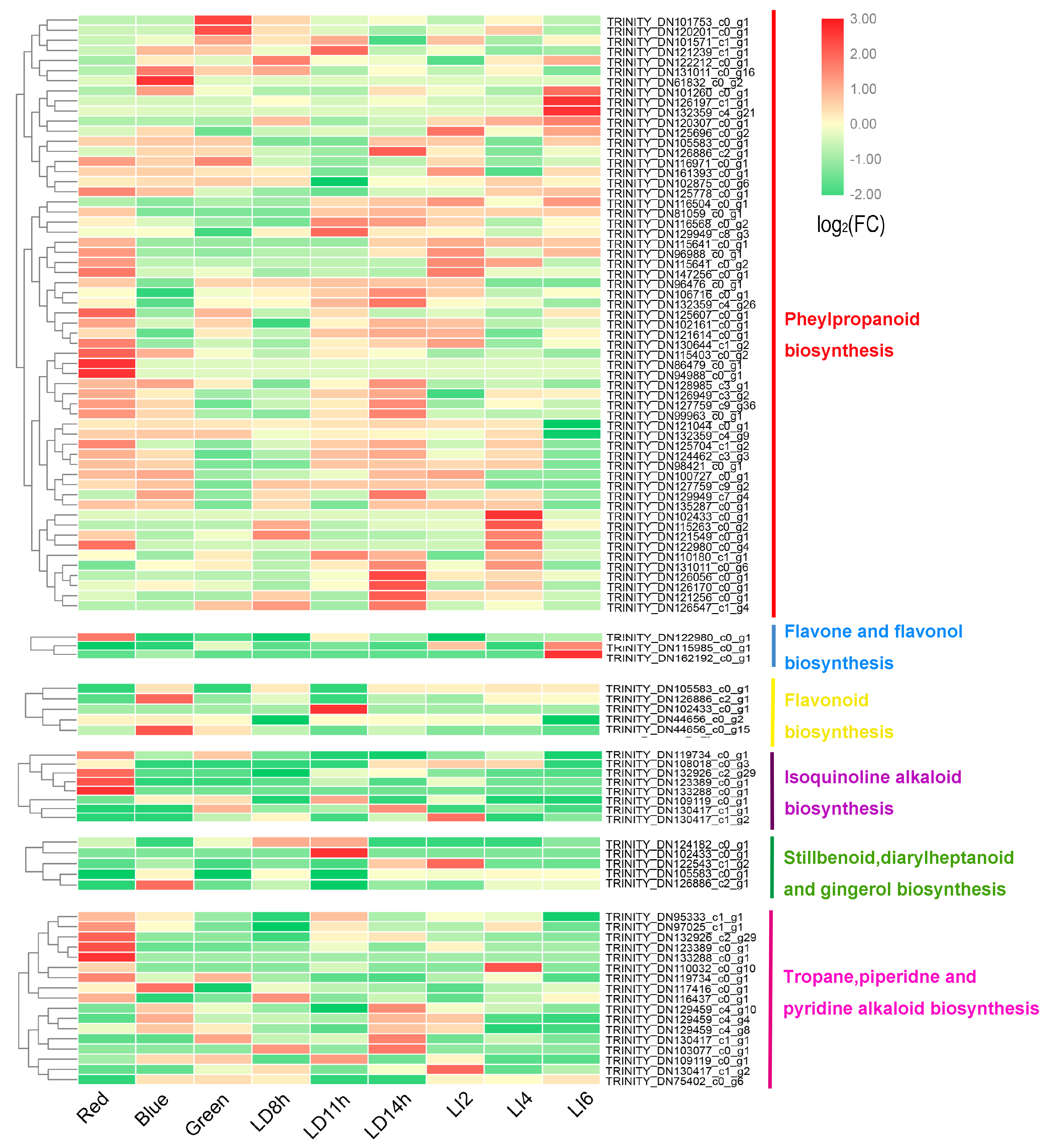
A KEGG pathway enrichment analysis revealed that the secondary metabolite contents changed in the light-treated samples. We further analyzed six common pathways for the synthesis of secondary metabolites that were significantly enriched among the DEGs. The “phenylpropanoid biosynthesis” (ko00940) and “tropane, piperidine and pyridine alkaloid biosynthesis” (ko00960) pathways were significantly enriched among the DEGs in all light treatments. The “isoquinoline alkaloid biosynthesis” (ko00950) pathway was significantly enriched among the DEGs identified following the other treatments, except for the 8-h exposure to daylight and the green light treatment. The “flavonoid biosynthesis” (ko00941) and “stilbenoid, diarylheptanoid and gingerol biosynthesis” (ko00945) pathways were significantly enriched among the DEGs following the blue light treatment, 14-h exposure to daylight, and the 2000 lx treatment, whereas the ‘flavone and flavonol biosynthesis’ (ko00944) pathway was significantly enriched among the DEGs of four light treatments (i.e., blue light, 11-h exposure to daylight, and the 2000 lx and 3000 lx light intensities) (Figure 5).
A total of 58 unigenes in the “phenylpropanoid biosynthesis” pathway were differentially expressed (relative to the control expression level) after one or more light treatments. The unigenes differentially expressed in response to multiple light conditions generally exhibited the same expression trend under different light conditions. For example, two unigenes (TRINITY_DN127759_C9_G36 and TRINITY_DN126949_c3_g2) encoding glycosyl hydrolases exhibited up-regulated expression after at least five light treatments. The expression of genes encoding methyltransferase, peroxidase 5, and cinnamyl alcohol dehydrogenase 2 (TRINITY_DN122980_c0_g4, TRINITY_DN126547_c1_g4, and TRINITY_DN132359_c4_g21, respectively) significantly decreased following at least six light treatments. However, the peroxidase 1 gene (TRINITY_DN121549_c0_g1) expression level was significantly down-regulated by the blue light treatment, 11-h and 14-h exposures to daylight, and the 3000 lx treatment, but was up-regulated in response to the 8-h exposure to daylight and the 2000 lx treatment. The expression of the cinnamyl alcohol dehydrogenase 2 gene (TRINITY_DN132359_c4_g26) was down-regulated by blue light, but was up-regulated by the 11-h and 14-h daylight treatments. Furthermore, 29 unigenes associated with the “phenylpropanoid biosynthesis” pathway were differentially expressed in response to only one light treatment.
In the “tropane, piperidine and pyridine alkaloid biosynthesis” pathway, the expression level of the unigene (TRINITY_DN132926_c2_g29) encoding nicotinamine amino transferase was significantly up-regulated by the light treatments, with the exception of the blue light treatment, the 8-h exposure to daylight, and the 3000 lx treatment. The expression of the unigene (TRINITY_DN110032_c0_g10) encoding tropinone reductase was significantly down-regulated by all light treatments, except for the red light treatment, the 11-h exposure to daylight, and the 2000 lx treatment. Eight unigenes were differentially expressed in response to only one light treatment. Additionally, seven unigenes in this pathway were also involved in the “isoquinoline alkaloid biosynthesis” pathway. Although the DEGs in the “flavonoid biosynthesis” pathway were mostly up-regulated by light, the leucoanthocyanidin dioxygenase isoform 2 gene (TRINITY_DN44656_c0_g15) expression level was significantly down-regulated by 2000 lx. Three other genes in this pathway (TRINITY_DN105583_c0_g1, TRINITY_DN126886_c2_g1, and TRINITY_DN102433_c0_g1) are also important for the “phenylpropanoid biosynthesis” and the “stilbenoid, diarylheptanoid and gingerol biosynthesis” pathways. The DEGs in the “flavone and flavonol biosynthesis” pathway exhibited the same trends regarding expression level changes due to the various treatments included in this study.
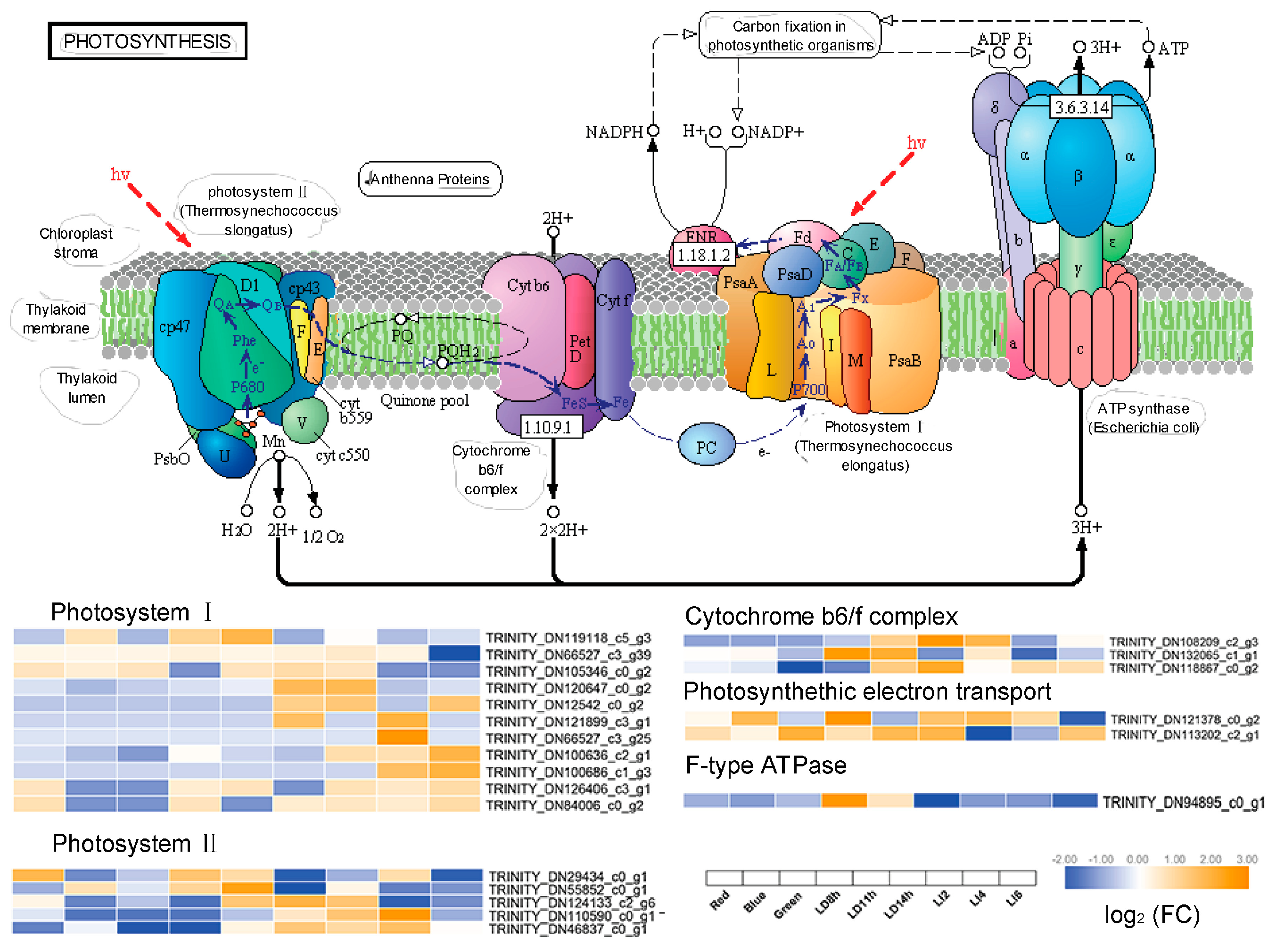
Photosynthesis in green plants is important for converting energy and maintaining the carbon–oxygen balance in the atmosphere. Light is the main environmental factor affecting plant photosynthesis. Therefore, besides the DEGs related to the synthesis of secondary metabolites, we also identified DEGs associated with the “photosynthesis” (ko00195) pathway (Figure 6). This KEGG pathway was not enriched among the DEGs due to the red light treatment and the 11-h exposure to daylight. The expression of unigenes related to photosystem I (PSI) was mostly up-regulated by light, whereas the expression of unigenes associated with photosystem II (PSII) exhibited the opposite trend. The TRINITY_DN119118_c5_g3 and TRINITY_DN121899_c3_g1 unigenes both encode an apolipoprotein of PSI P700 chlorophyll a. However, TRINITY_DN119118_c5_g3 expression was down-regulated by 2000 lx, whereas TRINITY_DN121899_c3_g1 exhibited the opposite trend. The expression of TRINITY_DN94895_c0_g1, which encodes an ATP synthetase, was significantly down-regulated by the 14-h exposure to daylight as well as the 2000 lx and 3000 lx treatments. Moreover, TRINITY_DN132065_c1_g1, which encodes cytochrome b6, and TRINITY_DN118867_c0_g2, which encodes the cytochrome b6-f complex, exhibited down-regulated expression in response to green light and 2000 lx, respectively. The expression levels of two unigenes (TRINITY_DN121378_C0_G2 and TRINITY_DN113202_c2_g1) involved in photosynthetic electron transport were up-regulated by the 8-h daylight treatment and down-regulated by 1000 lx.

3.7. Changes in the Expression of Phlorizin Biosynthesis-Related Genes in L. polystachyus under Various Light Conditions

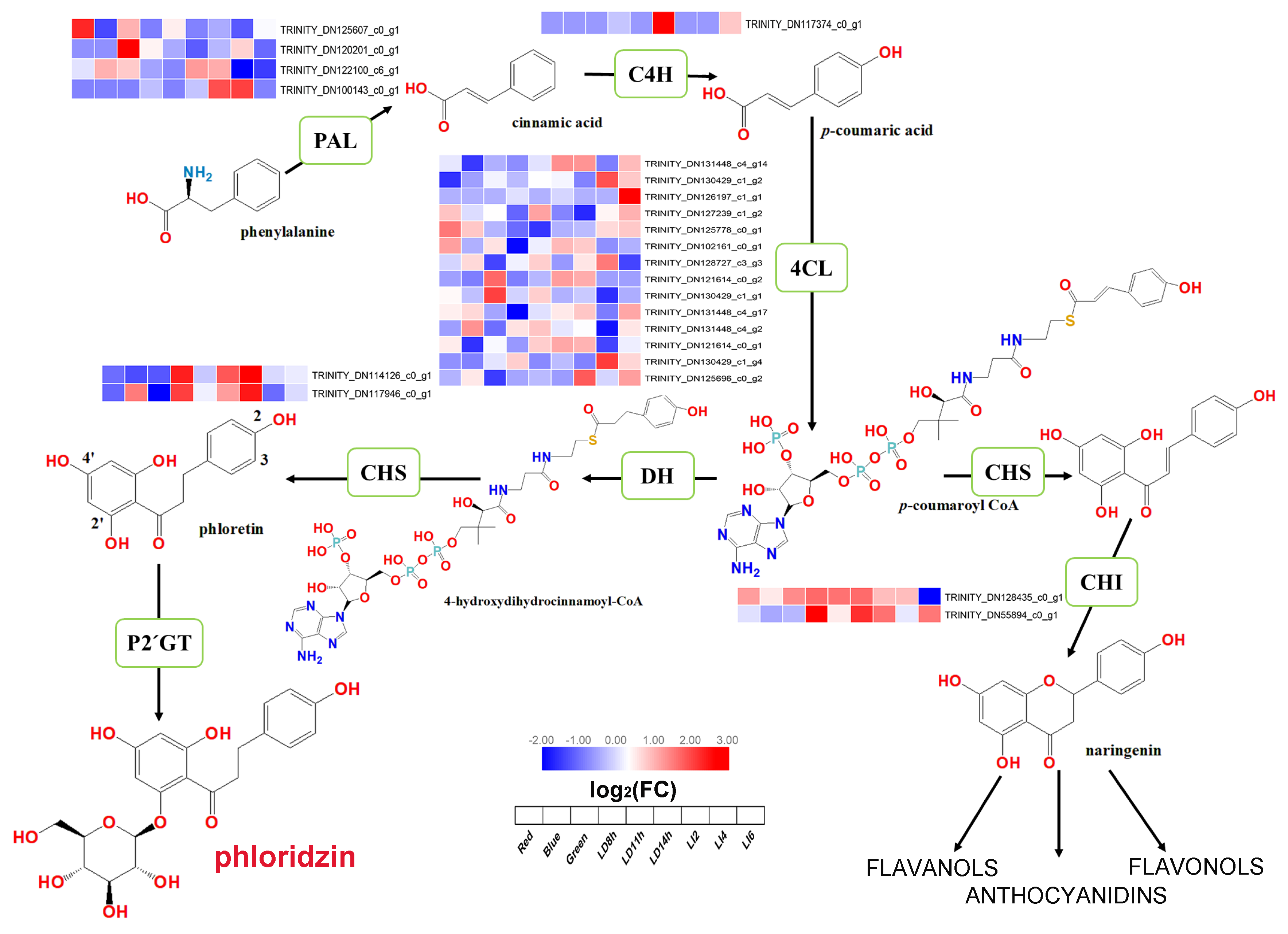
To identify the key genes involved in phlorizin biosynthesis, we analyzed the DEGs related to the phlorizin biosynthesis pathway, including 4 PAL, 1 C4H, 14 4CL, 2 CHS, and 2 CHI genes. These genes were differentially expressed following various light treatments (Figure 7). Compared with the control expression level, 4CLL9 (DN131448) exhibited up-regulated expression in response to the red light treatment, but CHS1 (DN114126) expression was significantly down-regulated. Following the blue light treatment, the PAL2 (DN125607), 4CL1 (DN121614), CHS1 (DN114126), and CHI3 (DN55894) expression levels were significantly down-regulated, whereas 4CLL9 expression was up-regulated, but its |log2 fold-change| was lower than that of CHS1. In response to the green light treatment, the PAL (DN120201) and 4CL (DN130429) expression levels were significantly up-regulated, but the |log2 fold-change| of 4CL was greater. Additionally, CHS1 and CHI3 expression levels were significantly down-regulated by green light, similar to their response to the blue light treatment. When exposed to daylight for 8 h, 4CLL7 (DN125778) and 4CL1 (DN102161) exhibited down-regulated expression, in contrast to the up-regulated expression of 4CLL9. An 11-h exposure to daylight resulted in down-regulated 4CLL7 expression, but up-regulated 4CL and 4CLL9 expression. The absolute value of the log2 fold-change of 4CLL7 was about 2 times that of the other two genes. The 4CLL9 expression level was significantly up-regulated by the 14-h exposure to daylight and the 1000 lx treatment. The 2000 lx treatment down-regulated the PAL2 and 4CL1 expression levels, but up-regulated 4CL expression. Following the 3000 lx treatment, three 4CL genes (DN125696, DN126197, and DN131448) exhibited significantly up-regulated expression, whereas CHI (DN128435) expression was significantly down-regulated. Significant changes in the expression level of 4CL gene occurred in each treatment group, indicating that 4CL gene is more susceptible to light than other genes.

3.8. Verification of Gene Expression Levels in a qRT-PCR Assay

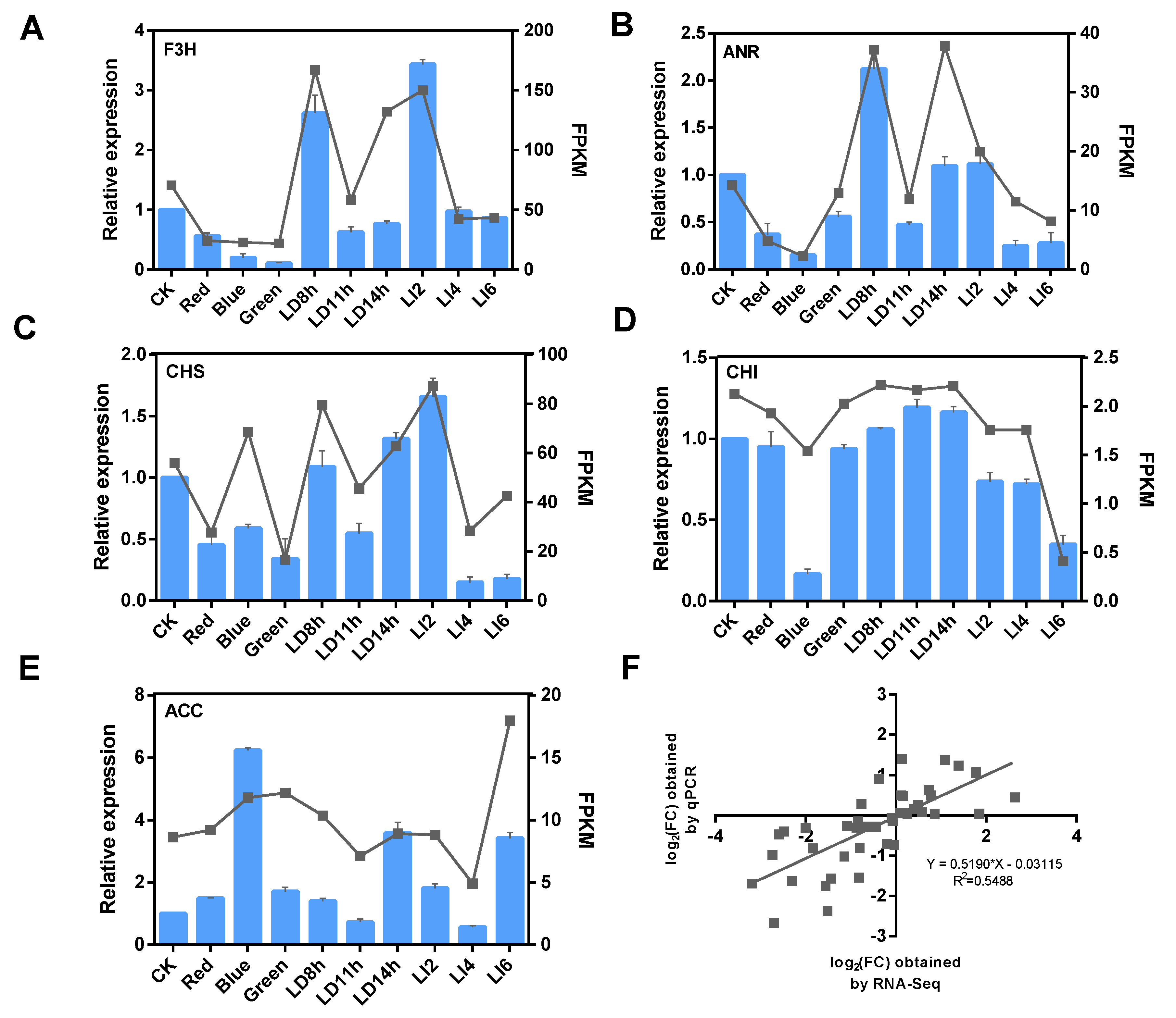
To confirm the accuracy and repeatability of the RNA-seq data, we randomly selected five DEGs in the phlorizin biosynthesis pathway and other secondary metabolism pathways for a qRT-PCR assay, by using new RNA and cDNA from the same plant sample used for sequencing. The results indicated the RNA-seq and qRT-PCR data produced consistent DEG expression levels for all analyzed light conditions. The log2 fold-changes based on the qRT-PCR and RNA-seq data were highly correlated [y = (0.5190)x − 0.03115; R2 = 0.5488] (Figure 8).

3.9. Changes to the Phlorizin Content and Phenylalanine Ammonia Lyase Activity

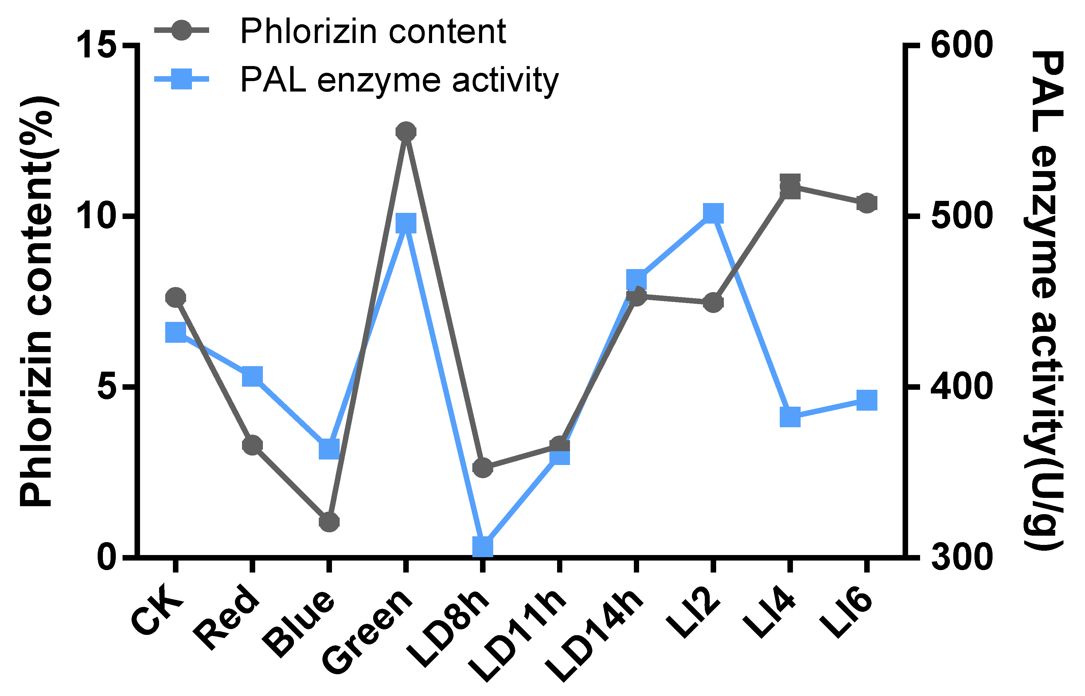
The three analyzed light conditions (i.e., light quality, duration, and intensity) induced significant changes to the phlorizin content (p ≤ 0.01; relative to the phlorizin content of the control plants). Specifically, the phlorizin content decreased by 1.28% and 3.41% following the red and blue light treatments, respectively. Additionally, the 8-h and 11-h exposures to daylight decreased the phlorizin content by 6.27% and 3.57%, respectively, whereas the 2000 lx and 3000 lx treatments decreased the phlorizin content by 2.47% and 1.98%, respectively. In contrast, the leaf phlorizin content increased by 3.20%, 1.56%, and 3.49% in response to green light, the 14-h exposure to daylight, and 1000 lx, respectively (Figure 9). Furthermore, the changes in the expression levels of several DEGs, such as PAL2, PAL, 4CL1 (DN121614), 4CLL7, and 4CL1 (DN102161), were consistent with the changes to the phlorizin content in response to different light treatments.
Phenylalanine ammonia lyase is widely distributed in plant species and a few microorganisms. It is the key rate-limiting enzyme for the production of secondary metabolites, such as flavonoids, in plants. We measured the PAL activity in L. polystachyus leaves following various light treatments. There were no significant differences in the PAL activities of the control plants and the plants exposed to 14 h of daylight or treated with a light intensity of 1000 lx. Additionally, PAL activity decreased significantly in response to the red and blue light treatments as well as the 8-h and 11-h exposures to daylight. However, the green light, 2000 lx, and 3000 lx treatments significantly increased the PAL activity (Figure 9). Regarding the effects of the light quality and photoperiod treatments, the changes to the PAL activity were consistent with the changes to the phlorizin content. In contrast, the three light intensity treatments induced different changes to the PAL activity and phlorizin content. The observed diversity in the effects of the light intensity treatments on PAL activity and the phlorizin content may be associated with the regulation of the transcription of L. polystachyus genes in leaves.

4. Discussion

Lithocarpus polystachyus is an important medicinal plant grown in southern China. It is widely used because it can efficiently lower blood sugar levels and exhibits anti-tumor activities. Flavonoids, including phlorizin, isolated from L. polystachyus are the most active components. Light affects the synthesis of flavonoids in most plants [16,17]. Therefore, we investigated the effects of light conditions on the molecular mechanism underlying the phlorizin biosynthesis pathway. The resulting data may be important for elucidating the accumulation of phlorizin in L. polystachyus. Additionally, to the best of our knowledge, this is the first study to comprehensively analyze the transcriptome of L. polystachyus leaves under various light conditions (i.e., light quality, light intensity, and photoperiod).
The Illumina system, which is a cost-effective next-generation sequencing platform for assembling transcriptomes, produces relatively short reads for a higher transcriptome coverage compared with other platforms [18]. We obtained more than 6 Gb of high-quality clean reads, which were assembled into 177,894 unigenes, with an average unigene length of 560 bp and an N50 of 689 bp. Additionally, homologs in publicly available databases were detected for 43.13% of the unigenes. Therefore, the generated sequence read archive data are important transcriptome resources for the discovery and functional annotation of L. polystachyus genes. The expression of light-sensing genes in plants can be induced or inhibited by altering the light conditions. In this study, a comparison of the transcriptomes from control and light-treated L. polystachyus plants revealed 1978–3716 up-regulated genes and 3884–4490 down-regulated genes. Additionally, 30% of the DEGs were not annotated in the analyzed public databases. These genes may be unique to L. polystachyus or homologs may not have been identified in other plant species. The DEGs for all light conditions were annotated with GO terms, including “nucleotide binding”, “protein serine/threonine kinase activity”, “protein kinase activity”, “phosphorylation”, “protein phosphorylation”, and “polysaccharide binding”. Thus, the expression of genes related to transcriptional regulation and signal transduction was highly regulated by various light treatments. Moreover, “membrane” was the most enriched GO term for all analyzed light conditions. Membrane transport and sensing systems are reportedly important for maintaining plant cell homeostasis under adverse environmental conditions, which up-regulate the expression of genes encoding membrane transporters, channelin, and receptor-like protein kinases [19]. In the current study, we also identified 437 unigenes that were differentially expressed (relative to the control levels) in response to diverse light stimuli. These unigenes included those encoding ribonucleases and DNA methyltransferases (i.e., known stress-responsive genes) [20,21]. Additionally, 19 unigenes were significantly expressed only in response to various light stimuli. These unigenes may not be expressed under natural light conditions or they are expressed at extremely low or negligible levels. Accordingly, these unigenes may be considered “light regulation-specific genes”.

4.1. Transcription Factors as Potential Regulators of Flavonoid Accumulation

Transcription factors play key roles in plant development and stress tolerance. Specifically, they regulate the expression of inducible genes by interacting with cis-acting elements in the promoter regions of stress-responsive genes [22]. The TFs function as part of the diverse transcriptional mechanisms mediating the stress signaling pathway and help regulate abiotic stress signals and the transcription of various genes [23]. The members of many TF families, including AP2/EREBP, bHLH, WRKY, MYB, NAC, and MYC, function as coordinating signals that are transduced when plants are exposed to various environmental stresses [24]. According to our results, the genes encoding TFs from 89 families were differentially expressed following various light treatments. The largest light-induced TF families were WRKY and mTERF.
Flavonoid biosynthesis is regulated by the transcription of a variety of TFs, especially R2R3-MYB, bHLH, and WD40 repeat proteins [25]. A previous study proved that MYB TFs can regulate the synthesis of flavonols by activating the expression of early biosynthetic genes, including PAL, C4H, CHS, CHI, and F3tH [26]. A recent study on Epimedium sagittatum revealed that EsMYBF1, which is a flavonol-specific regulator, increases the accumulation of kaempferol and quercetin by regulating the expression of CHS, CHI, F3H, and FLS [27]. However, we did not detect MYB12 or MYBF1 homologs among the DEGs, possibly because of the absence of the MYB12 gene in L. polystachyus. Alternatively, the MYB12 gene in L. polystachyus may be unresponsive to light. Additionally, we observed that the some MYBs were correlated with the phlorizin accumulation pattern induced by various light treatments, especially the homologs of the positive correlation genes. These findings imply the MYB TFs may help regulate phlorizin biosynthesis.
Two AtbHLH113 homologs identified in the Lonicera macranthoides transcriptome data are positively correlated with the accumulation of luteolin, suggesting both homologs are involved in the regulation of luteolin production [28]. A homolog of AtbHLH113 (TRINITY_DN113600_c0_g1) was also detected in our study. The expression of TRINITY_DN113600_c0_g1 was significantly down-regulated following the exposure to 3000 lx and the blue and green light treatments, but the phlorizin content was higher in the plants treated with green light. This accumulation pattern differs from that of related major metabolites and derivatives when the expression of this gene is up-regulated and down-regulated. An earlier investigation indicated that the overexpression of AtMYB12 in tomato activates the caffeoylquinic acid biosynthesis pathway and the down-regulated expression of SlMYB12 also leads to the accumulation of caffeic acid derivatives [29]. Interactions with MYB TFs may be necessary for regulatory activities. For example, three bHLH activators in Arabidopsis thaliana directly or indirectly interact with MYBs during the regulation of transcription by anthocyanins and proanthocyanidins [30]. Moreover, MdbHLH3 in apple and BobHLH1 in cauliflower interact with MdMYB10 and BoMYB2, respectively, thereby resulting in anthocyanin accumulation [31,32].

4.2. Effects of Different Light Treatments on the Enzymes of the Secondary Metabolism Pathways and Photosystems of L. polystachyus

Different light conditions can be used to stimulate pathways associated with secondary metabolism to increase the production of secondary metabolites [33]. In the current study, we identified DEGs related to six secondary metabolism pathways under various light conditions. These DEGs were mainly related to the phenylpropanoid biosynthesis pathway and encode glycosidases, methyltransferases, and peroxidases, implying these three enzymes are photosensitive. Flavonoid methyltransferase and flavonoid glycosyltransferase activities are reportedly inhibited or enhanced depending on the light conditions [34]. Moreover, phenolic compounds are the main products of phenylpropanoid biosynthesis and play various physiological roles related to abiotic stresses, while genes encoding peroxidases are involved in the oxidation of phenols in response to different stimuli [35].
In our study, we determined that most of the light-stimulated DEGs were significantly enriched in the “photosynthesis” pathway and almost all of the DEGs related to PSI were significantly up-regulated. Light stimulates PSI to varying degrees. Almost all of the DEGs associated with PSII were significantly down-regulated, possibly because the light stimuli were inhibitory toward PSII, with the exception of the red light treatment, the 11-h exposure to daylight, and the 1000 lx treatment. Photoinhibition refers to the phenomenon in which the photosynthetic activities decrease when the light energy exceeds the amount that can be used by the photosynthetic system [36]. Photoinhibition mainly occurs in PSII. Under natural conditions, light-suppression often occurs in the upper leaves of plants at noon on sunny days. The simultaneous exposure to sunlight glare and other environmental stress factors, such as low and high temperatures and drought, can intensify the photoinhibition, even under low-light conditions. Photoinhibition is a protective response of plants, but it is not a photoprotective mechanism [37,38].

4.3. Molecular Characteristics of the Enrichment of the Phlorizin Biosynthesis Pathway in L. polystachyus Leaves under Different Light Conditions

Light quality is an essential factor influencing plant growth and development. In addition to being an energy source for photosynthesis, light also acts as an inducer of plant growth, and it clearly affects flavonoid accumulation. Previous studies involving leaves indicated that blue light promotes the accumulation of flavonoids, whereas red light has the opposite effect [39,40]. However, the light used in these earlier investigations was composite light (blue/red and blue/red/green). In contrast, the current study was completed with single light sources. In response to the red and blue light treatments, the L. polystachyus phlorizin content decreased, but the opposite change in phlorizin content occurred following the green light treatment. These findings implied that the effects of single and complex lights differ regarding flavonoid accumulation in plants. Additionally, Oyaert et al. confirmed that excessive blue light inhibits plant growth, shortens internodes, and decreases branching, the leaf area, and the total dry weight [41]. The phlorizin content in L. polystachyus increased following the 1000 lx treatment, but decreased after the 2000 lx and 3000 lx treatments. These observations were consistent with the results of previous studies by Aimin Li [7]. Thus, appropriate shading or decreases in the light intensity can increase the flavonoid contents of the analyzed plants. The phlorizin content decreased in response to the 8-h and 11-h exposures to daylight, but increased following the 14-h daylight treatment. These results indicate that the phlorizin content may increase with increasing illumination time.
Phenylalanine ammonia lyase is a key enzyme in the synthesis of flavonoids, and its activity is regulated by ultraviolet light. Moreover, PAL is more light-responsive than other enzymes [42]. Therefore, we examined the PAL activity of L. polystachyus grown under different light conditions. An evaluation of the effects of the light quality and photoperiod treatments revealed that only the green light treatment significantly promoted PAL activity. Additionally, the changes in the PAL activity were consistent with the changes in the phlorizin content. The PAL activity did not change significantly in response to 1000 lx, but the corresponding phlorizin content increased significantly. The PAL activity of the L. polystachyus leaves exposed to 2000 lx increased significantly, but the phlorizin content decreased at this light intensity. Accordingly, we speculate that phlorizin biosynthesis under different light intensity treatments is mainly regulated by gene expression levels, but the specific underlying mechanism must be more thoroughly characterized.
We analyzed the transcriptomes of L. polystachyus leaves under different light conditions to elucidate the effects of light on phlorizin. In this study, the transcript levels of PAL2, PAL, 4CL1 (DN121614), 4CLL7, 4CL1 (DN102161), and other genes in response to light treatments were positively correlated with the corresponding phlorizin contents. Therefore, PAL and 4CL are considered key enzymes for the synthesis of phlorizin, whereas PAL2, PAL, 4CL1 (DN121614), 4CLL7, and 4CL1 (DN102161) are likely directly involved in the phlorizin biosynthesis pathway in L. polystachyus. The results of a study by Kong et al. confirmed that the production and activity of PAL and 4CL are positively correlated with luteolin biosynthesis in L. macranthoides [43]. Our results are consistent with these earlier findings. Furthermore, LjCHS1 and LjCHI2 are considered to be important for the luteolin biosynthesis in L. macranthoides [44]. We observed that CHS1 was differentially expressed in response to red and blue light treatments. The expression levels were consistent with the phlorizin accumulation, but were negatively correlated with the effects of the green light treatment. Additionally, the expression of 4CL (DN130429), 4CLL9 (DN131448), and two 4CLs in response to 3000 lx and other treatments was negatively correlated with the corresponding phlorizin contents. These results suggest these genes may not influence phlorizin accumulation in L. polystachyus or the L. polystachyus phlorizin biosynthesis pathway may be regulated by multiple gene families. For example, compared with the flavonoid biosynthesis pathway in A. thaliana, which is regulated by single-copy genes, the corresponding pathway in ginkgo appears to be controlled by multiple gene families, leading to complex networks [45]. A Southern blot analysis of GbCHS revealed multiple bands, indicating the presence of a multi-gene GbCHS family in ginkgo [46]. Further studies are needed to determine whether these genes are involved in phlorizin biosynthesis, either independently or synergistically.

5. Conclusions

In this study, we performed a comprehensive transcriptome analysis of L. polystachyus in response to various light stimuli to demonstrate that light quality, light intensity, and the photoperiod affect L. polystachyus gene expression levels, which indirectly affects phlorizin accumulation. The transcriptome analysis confirmed that PAL and 4CL are the key enzymes for the phlorizin biosynthesis pathway. We also identified five genes (two PAL and three 4CL genes) whose transcription levels were positively correlated with phlorizin accumulation. Regarding the effects of light quality, the phlorizin content and PAL activity increased in response to the green light treatment, but decreased following the blue and red light treatments. Additionally, increases in the photoperiod increased the phlorizin content and PAL activity, indicating that treatments that appropriately extend the duration of daylight may induce the accumulation of phlorizin. In the current study, the phlorizin content following the 1000 lx treatment was significantly higher than that of the control plants or the plants exposed to the other light intensities. However, the PAL activity induced by 1,000 lx was significantly lower than that induced by the other two light intensities. Therefore, appropriate decreases in light intensity may promote phlorizin accumulation. The regulation of gene transcription plays a leading role in the bioaccumulation due to various light intensities. The data presented herein may form the foundation for future studies of plant photoresponses and may be useful for clarifying the accumulation of medicinal components.

Supplementary Materials

The following are available online at https://www.mdpi.com/1999-4907/10/11/995/s1. Figure S1: NR database sequence alignment result graph, Figure S2: GO enrichment results of differentially expressed genes, Figure S3: Differentially expressed genes (DEGs) in different treatment groups, Table S1: unigenes function annotation statistics, Table S2: KEGG database annotation statistics, Table S3: Annotation information for genes differentially expressed in all treatment groups, Table S4: DEGs statistics of the treatment groups in the light quality treatment type, Table S5: DEGs statistics of the processing group in the photoperiod processing type, Table S6: DEGs statistics of the treatment group in the light intensity treatment type, Table S7: Statistics on the number of differentially expressed transcription factors, Table S8: Differentially expressed genes encoding MYB transcription factors, Table S9: Differentially expressed genes encoding BHLH transcription factors.

Author Contributions

Conceptualization, J.H. and Z.X.; experiments, Y.Z. and Z.W.; data analysis, H.G., L.L., and M.C.; writing—original draft preparation, Y.Z. and Y.L.; writing—review and editing, Z.X.

Funding

This work was primarily supported by the National Natural Science Foundation of China (Grant No. 31760221). Other support includes: Hebei Education Department sponsored scientific research projects(Grant No. ZD2019075); graduate student innovation fund of North China University of Science and Technology (Grant No. 2019S34); science fund for cultivating of North China University of Science and Technology (Grant No. SP201508).

Conflicts of Interest

The authors declare no conflict of interest.

References

  1. Wang, J.F.; Huang, Y.M.; Li, K.X.; Chen, Y.Y.; Vanegas, D.; McLamore, E.S.; Shen, Y.B. Leaf Extract from Lithocarpus polystachyus Rehd. Promote Glycogen Synthesis in T2DM Mice. PLoS ONE 2016, 11. [Google Scholar] [CrossRef] [PubMed]
  2. Mariadoss, A.V.A.; Vinyagam, R.; Rajamanickam, V.; Senthilkumar, V.; Venkatesan, S.; David, E. Pharmacological aspects and potential use of phloretin: A systemic review. Mini-Rev. Med. Chem. 2019, 19, 1060–1067. [Google Scholar] [CrossRef] [PubMed]
  3. Gosch, C.; Halbwirth, H.; Kuhn, J.; Miosic, S.; Stich, K. Biosynthesis of phloridzin in apple (Malus domestica Borkh.). Plant Sci. 2009, 176, 223–231. [Google Scholar] [CrossRef]
  4. Batista, D.S.; Felipe, S.H.S.T.; Silva, D.; Castro, K.M.; Mamedes-Rodrigues, T.C.; Miranda, N.A.; Ríos-Ríos, A.M.; Fortini, E.A.; Chagas, K.; Torres-Silva, G.; et al. Light quality in plant tissue culture: Does it matter? Vitr. Cell. Dev. Biol. 2018, 54, 195–215. [Google Scholar] [CrossRef]
  5. Plantenga, F.D.; Bergonzi, S.; Bachem, C.W.; Visser, R.G.; Heuvelink, E.; Marcelis, L.F. High light accelerates potato flowering independently of the FT-like flowering signal StSP3D. Environ. Exp. Bot. 2019, 160, 35–44. [Google Scholar] [CrossRef]
  6. Xu, J.M.; Yan, Z.M.; Xu, Z.G.; Wang, Y.H.; Xie, Z.Q. Transcriptome analysis and physiological responses of the potato plantlets in vitro under red, blue, and white light conditions. 3 Biotechnol. 2018, 8, 394. [Google Scholar] [CrossRef]
  7. Li, A.M.; Li, S.H.; Wu, X.J.; Zhang, J.; He, A.N.; Zhao, G.; Yang, X. Effect of Light Intensity on Leaf Photosynthetic Characteristics and Accumulation of Flavonoids in Lithocarpus litseifolius (Hance) Chun. (Fagaceae). Open J. For. 2016, 6, 445–459. [Google Scholar] [CrossRef]
  8. Ni, J.; Dong, L.X.; Jiang, Z.F.; Yang, X.L.; Chen, Z.Y.; Wu, Y.H.; Xu, M.J. Comprehensive transcriptome analysis and flavonoid profiling of Ginkgo leaves reveals flavonoid content alterations in day-night cycles. PLoS ONE 2018, 13, 0193897. [Google Scholar] [CrossRef]
  9. Wang, M.; Zhang, X.N.; Liu, J.H. Deep sequencing-based characterization of transcriptome of trifoliate orange (Poncirus trifoliata (L.) Raf) in response to cold stress. BMC Genom. 2015, 16, 555. [Google Scholar] [CrossRef] [PubMed]
  10. Singh, V.K.; Mangalam, A.K.; Dwivedi, S.; Naik, S. Primer premier: Program for design of degenerate primers from a protein sequence. Biotechniques 1998, 24, 318–319. [Google Scholar] [CrossRef]
  11. Grabherr, M.G.; Haas, B.J.; Yassour, M.; Levin, J.Z.; Thompson, D.A.; Amit, I.; Adiconis, X.; Fan, L.; Raychowdhury, R.; Zeng, Q.; et al. Full-length transcriptome assembly from RNA-Seq data without a reference genome. Nat Biotechnol. 2011, 29, 644–652. [Google Scholar] [CrossRef] [Green Version]
  12. Pertea, G.; Huang, X.; Liang, F.; Antonescu, V.; Sultana, R.; Karamycheva, S.; Lee, Y.; White, J.; Cheung, F.; Parvizi, B.; et al. TIGR Gene Indices clustering tools (TGICL): A software system for fast clustering of large EST datasets. Bioinformatics 2003, 19, 651–652. [Google Scholar] [CrossRef]
  13. Chen, Y.; Yin, L.Z.; Zhao, L.; Shu, G.; Yuan, Z.X.; Fu, H.L.; Lv, C.; Lin, J.C. Optimization of the ultrasound-assisted extraction of antioxidant phloridzin from Lithocarpus polystachyus Rehd. using response surface methodology. J. Sep. Sci. 2017, 40, 4329–4337. [Google Scholar] [CrossRef]
  14. Chen, S.Y.; Zhang, X.Q.; Ma, X.; Huang, L.K. Assessment of genetic diversity and differentiation of Elymus nutans indigenous to Qinghai-Tibet plateau using simple sequence repeats markers. Can. J. Plant Sci. 2013, 93, 1089–1096. [Google Scholar] [CrossRef]
  15. Li, S. Transcriptional control of flavonoid biosynthesis: Fine-tuning of the MYB-bHLH-WD40 (MBW) complex. Plant Signal. Behav. 2014, 9, e27522. [Google Scholar] [CrossRef]
  16. Neugart, S.; Krumbein, A.; Zrenner, R. Influence of light and temperature on gene expression leading to accumulation of specific flavonol glycosides and hydroxycinnamic acid derivatives in Kale (Brassica oleracea var. sabellica). Front. Plant Sci. 2016, 7, 326. [Google Scholar] [CrossRef]
  17. Liu, Y.; Fang, S.Z.; Yang, W.X.; Shang, X.L.; Fu, X.X. Light quality affects flavonoid production and related gene expression in Cyclocarya paliurus. J. Photochem. Photobiol. B 2018, 179, 66–73. [Google Scholar] [CrossRef] [PubMed]
  18. Xu, Y.J.; Gao, S.; Yang, Y.J.; Huang, M.Y.; Cheng, L.N.; Wei, Q.; Fei, Z.J.; Gao, J.P.; Hong, B. Transcriptome sequencing and whole genome expression profiling of chrysanthemum under dehydration stress. BMC Genom. 2013, 14, 662. [Google Scholar] [CrossRef] [PubMed]
  19. Wu, Y.; Wei, W.; Pang, X.; Wang, X.; Zhang, H.; Dong, B.; Xing, Y.; Li, X.; Wang, M. Comparative transcriptome profiling of a desert evergreen shrub, Ammopiptanthus mongolicus, in response to drought and cold stresses. BMC Genom. 2014, 15, 671. [Google Scholar] [CrossRef] [PubMed]
  20. Khraiwesh, B.; Zhu, J.K.; Zhu, J. Role of miRNAs and siRNAs in biotic and abiotic stress responses of plants. BBA-Gene Regul. Mech. 2012, 1819, 137–148. [Google Scholar] [CrossRef] [Green Version]
  21. Boyko, A.; Kovalchuk, I. Epigenetic control of plant stress response. Environ. Mol. Mutagen 2018, 49, 61–72. [Google Scholar] [CrossRef] [PubMed]
  22. Sazegari, S.; Niazi, A. Isolation and molecular characterization of wheat (Triticum aestivum) dehydration responsive element binding factor (DREB) isoforms. Aust. J. Crop Sci. 2012, 6, 1037–1044. [Google Scholar]
  23. Lata, C.; Yadav, A.; Prasad, M. Role of plant transcription factors in abiotic stress tolerance. In Abiotic Stress Response in Plants—Physiological, Biochemical and Genetic Perspectives; Shanker, A., Venkateshwarulu, B., Eds.; InTechOpen: London, UK, 2011. [Google Scholar]
  24. Liu, J.H.; Peng, T.; Dai, W.S. Critical cis-acting elements and interacting transcription factors: Key players associated with abiotic stress responses in plants. Plant Mol. Biol. Rep. 2014, 32, 303–317. [Google Scholar] [CrossRef]
  25. Hichri, I.; Barrieu, F.; Bogs, J.; Kappel, C.; Delrot, S.; Lauvergeat, V. Recent advances in the transcriptional regulation of the flavonoid biosynthetic pathway. J. Exp. Bot. 2011, 62, 2465–2483. [Google Scholar] [CrossRef] [PubMed] [Green Version]
  26. Pandey, A.; Misra, P.; Trivedi, P.K. Constitutive expression of Arabidopsis MYB transcription factor, AtMYB11, in tobacco modulates flavonoid biosynthesis in favor of flavonol accumulation. Plant Cell Rep. 2015, 34, 1515–1528. [Google Scholar] [CrossRef]
  27. Huang, W.; Khaidun, A.B.M.; Chen, J.; Zhang, C.; Lv, H.; Yuan, L.; Wang, Y. A R2R3-MYB Transcription Factor Regulates the Flavonol Biosynthetic Pathway in a Traditional Chinese Medicinal Plant, Epimedium sagittatum. Front Plant Sci. 2016, 7, 1089. [Google Scholar] [CrossRef]
  28. Chen, Z.X.; Liu, G.H.; Tang, N.; Li, Z.G. Transcriptome Analysis Reveals Molecular Signatures of Luteoloside Accumulation in Senescing Leaves of Lonicera macranthoides. Int. J. Mol. Sci. 2018, 19, 1012. [Google Scholar] [CrossRef] [PubMed]
  29. Adato, A.; Mandel, T.; Mintz-Oron, S.; Venger, I.; Levy, D.; Yativ, M.; Dominguez, E.; Wang, Z.; De Vos, R.C.H.; Jetter, R.; et al. Fruit-Surface Flavonoid Accumulation in Tomato is Controlled by a SIMYB12-Regulated Transcriptional Network. PLoS Genet. 2009, 5. [Google Scholar] [CrossRef] [PubMed]
  30. Xu, W.J.; Grain, D.; Bobet, S.; Le Gourrierec, J.; Thevenin, J.; Kelemen, Z.; Lepiniec, L.; Dubos, C. Complexity and robustness of the flavonoid transcriptional regulatory network revealed by comprehensive analyses of MYB-bHLH-WDR complexes and their targets in Arabidopsis seed. New Phytol. 2014, 202, 132–144. [Google Scholar] [CrossRef]
  31. Espley, R.V.; Hellens, R.P.; Putterill, J.; Stevenson, D.E.; Kutty-Amma, S.; Allan, A.C. Red colouration in apple fruit is due to the activity of the MYB transcription factor, MdMYB10. Plant J. 2007, 49, 414–427. [Google Scholar] [CrossRef] [PubMed] [Green Version]
  32. Chiu, L.W.; Zhou, X.J.; Burke, S.; Wu, X.L.; Prior, R.L.; Li, L. The Purple Cauliflower Arises from Activation of a MYB Transcription Factor. Plant Physiol. 2010, 154, 1470–1480. [Google Scholar] [CrossRef] [PubMed] [Green Version]
  33. Kuo, T.C.; Chen, C.H.; Chen, S.H.; Lu, I.H.; Chu, M.J.; Huang, L.C.; Lin, C.Y.; Chen, C.Y.; Lo, H.F.; Jeng, S.T.; et al. The effect of red light and far-red light conditions on secondary metabolism in agarwood. BMC Plant Biol. 2015, 15, 139. [Google Scholar] [CrossRef]
  34. Fu, B.; Ji, X.M.; Zhao, M.Q.; He, F.; Wang, X.L.; Wang, Y.D.; Liu, P.F.; Niu, L. The influence of light quality on the accumulation of flavonoids in tobacco (Nicotiana tabacum L.) leaves. J. Photochem. Photobiol. B 2016, 162, 544–549. [Google Scholar] [CrossRef] [PubMed]
  35. Deng, S.; Ma, J.; Zhang, L.; Chen, F.; Sang, Z.; Jia, Z.; Ma, L. De novo transcriptome sequencing and gene expression profiling of Magnolia wufengensis in response to cold stress. BMC Plant Biol. 2019, 19, 321. [Google Scholar] [CrossRef] [PubMed]
  36. Li, Y.Y.; Wang, X.W.; Ban, Q.Y.; Zhu, X.X.; Jiang, C.J.; Wei, C.L.; Bennetzen, J.L. Comparative transcriptomic analysis reveals gene expression associated with cold adaptation in the tea plant Camellia sinensis. BMC Genom. 2019, 20, 624. [Google Scholar] [CrossRef] [PubMed]
  37. Foyer, C.H.; Noctor, G. Leaves in the dark see the light. Science 1999, 284, 599–601. [Google Scholar] [CrossRef]
  38. Powlem, S.B. Photoinhibition of photosyntheain induced by viaible light. Annu. Rev. Plant Physiol. 1984, 35, 15–50. [Google Scholar] [CrossRef]
  39. Ouzounis, T.; Razi Parjikolaei, B.; Fretté, X.; Rosenqvist, E.; Ottosen, C.O. Predawn and high intensity application of supplemental blue light decreases the quantum yield of PSII and enhances the amount of phenolic acids, flavonoids, and pigments in Lactuca sativa. Front. Plant Sci. 2015, 6, 19. [Google Scholar] [CrossRef] [PubMed]
  40. Arcas, M.C.; Botia, J.M.; Ortuño, A.M.; Del Río, J.A. UV irradiation alters the levels of flavonoids involved in the defence mechanism of Citrus aurantium fruits against Peniillium digitatum. Eur. J. Plant Pathol. 2000, 106, 617. [Google Scholar] [CrossRef]
  41. Oyaert, E.; Volckert, E.; Debergh, P.C. Growth of Chrysanthemum under coloured plastic films with different light qualities and quantities. Sci. Hortic. 1999, 79, 195–205. [Google Scholar] [CrossRef]
  42. Zoratti, L.; Karppinen, K.; Luengo Escobar, A.; Häggman, H.; Jaakola, L. Light-controlled flavonoid biosynthesis in fruits. Front. Plant Sci. 2014, 5, 534. [Google Scholar] [CrossRef] [PubMed]
  43. Kong, D.X.; Li, Y.Q.; Bai, M.; He, H.J.; Liang, G.X.; Wu, H. Correlation between the dynamic accumulation of the main effective components and their associated regulatory enzyme activities at different growth stages in Lonicera japonica Thunb. Ind. Crops Prod. 2017, 96, 16–22. [Google Scholar] [CrossRef]
  44. Yuan, Y.; Wang, Z.Y.; Jiang, C.; Wang, X.M.; Huang, L.Q. Exploiting genes and functional diversity of chlorogenic acid and luteolin biosyntheses in Lonicera japonica and their substitutes. Gene 2014, 534, 408–416. [Google Scholar] [CrossRef] [PubMed]
  45. Winkel-Shirley, B. Flavonoid biosynthesis. A colorful model for genetics, biochemistry, cell biology, and biotechnology. Plant Physiol. 2001, 126, 485–493. [Google Scholar] [CrossRef] [PubMed]
  46. Pang, Y.Z.; Shen, G.A.; Wu, W.S.; Liu, X.F.; Lin, J.; Tan, F.; Snu, X.F.; Tang, K.X. Characterization and expression of chalcone synthase gene from Ginkgo biloba. Plant Sci. 2005, 168, 1525–1531. [Google Scholar] [CrossRef]
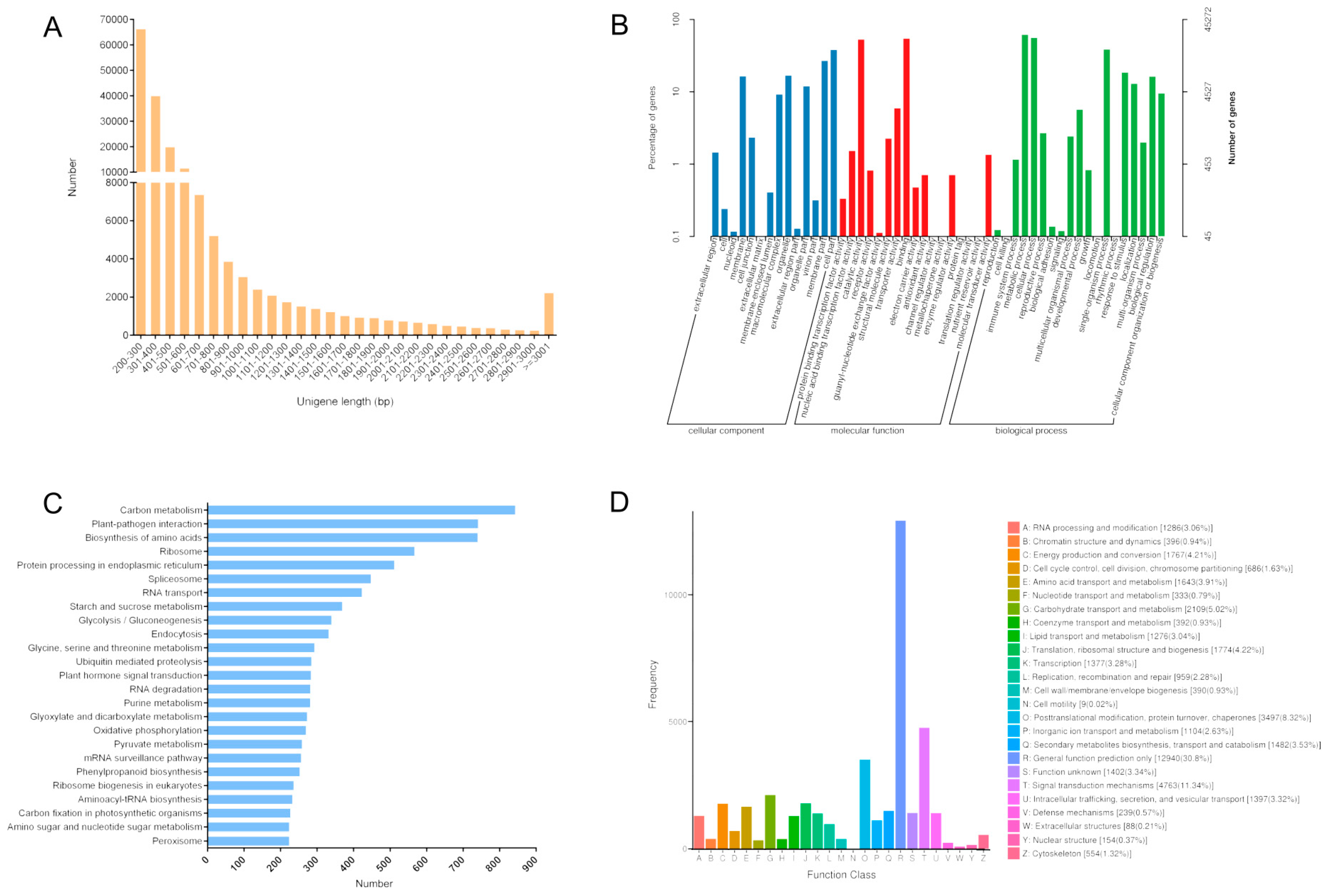
Figure 1. Unigene length and classification. (a) Distribution of unigene length. (b) Unigene functional annotations based on the gene ontology database. (c) Top 25 enriched KEGG pathways among the unigenes. (d) Classifications based on the Clusters of Orthologous Group database.
Forests 10 00995 g001
Figure 2. Differentially expressed genes in response to various light conditions. (a) Up-regulated and down-regulated genes in each treatment group. (b) Differentially expressed genes encoding MYB and bHLH transcription factors. (c) Venn diagram presenting the genes expressed in the control group and the genes expressed in response to at least one treatment for each light condition. (d) Venn diagram presenting the genes expressed in the control group and the genes expressed in response to all of the light treatments.
Forests 10 00995 g002
Figure 3. Pathways enriched among the identified DEGs. (ai) Enriched KEGG pathways among the DEGs in response to the red, blue, green, LD8h, LD11h, LD14h, LI2, LI4, and LI6 light treatments, respectively. Pathway names are provided on the vertical axis, whereas the enrichment factor is presented on the horizontal axis. The size of each point indicates the number of DEGs in a particular pathway, whereas the color of each point corresponds to the Q score range.
Forests 10 00995 g003
Figure 4. Differentially expressed genes (DEGs) in response to three light conditions. (a) Venn diagram presenting the DEGs in a pair-wise comparison of light quality treatments. (b) Venn diagram presenting the DEGs in a pair-wise comparison of photoperiod treatments. (c) Venn diagram presenting the DEGs in a pair-wise comparison of light intensity treatments. (d) Data regarding the up- and down-regulated genes in pair-wise comparisons of light conditions.
Forests 10 00995 g004
Figure 5. Expression profiles of the DEGs related to the secondary metabolism pathway.
Forests 10 00995 g005
Figure 6. Differentially expressed genes in the photosynthesis pathway.
Forests 10 00995 g006
Figure 7. Differentially expressed genes in the phlorizin accumulation pathway. The different colors in the heat map show unequal expression levels, and red and blue in the formula represent oxygen and nitrogen atoms, respectively.
Forests 10 00995 g007
Figure 8. Light-induced L. polystachyus gene expression determined by qRT-PCR. (ae) Expression levels of selected genes. The y-axis on the left indicates the relative expression levels determined by qRT-PCR (blue histogram), whereas the y-axis on the right indicates the expression levels based on the RNA-seq data (gray line chart). The x-axis indicates the light treatments. (f) Comparison between the log2 fold-changes calculated based on the RNA-seq and qRT-PCR data.
Forests 10 00995 g008
Figure 9. Changes to the phlorizin content and PAL activity due to various light treatments. The gray and blue lines correspond to the phlorizin content and PAL activity, respectively. The phlorizin content refers to the percentage of the extracted phlorizin mass to the dry weight of the sample.
Forests 10 00995 g009
Table 1. Lithocarpus polystachyus culture conditions during light treatments.
Sample NumberLight QualityLight Control Duration (Light/Dark)Light Intensity (μmol·m−2·s−1/lx)Temperature (℃)Soil Moisture (%)Number of Plants TreatedProcessing Type
CKNatural light12/12 h18–72/1000–400025 ± 27510Control group
RedRed12/12 h37.5/300025 ± 27510Light quality
BlueBlue
GreenGreen
LD8hWhite8/16 h37.5/300025 ± 27510photoperiod
LD11h11/13 h
LD14h14/10 h
LI2White12/12 h12.5/100025 ± 27510Light intensity
LI425/2000
LI637.5/3000
Table 2. Selected L. polystachyus partial gene sequences and qRT-PCR primer sequences.
GenesPrimer NamePrimer Sequence (5’–3’)Fragment Size/bp
acetyl coenzyme A carboxylase (ACC)DACCrt1S2AAGGCAAGCACTTCATCTCATCC200
DACCrt1X2ATAAGAATCTCGGCTATTGTTCC
chalcone synthase (CHS)DCHSrt2S1GCAGCAACAAAGGCAATCAAA186
DCHSrt2X1CACAGTCCCACCAGCAAAGCA
chalcone isomerase (CHI)DCHIrt2S1TTTATCAGGATCTTCTCAGGGAG144
DCHIrt2X1AATCAGTGTCTGGGTTTGTCTTC
anthocyanidin reductase (ANR)DANRrt1S2CACTGGGTTCGTCGCTTCTTT276
DANRrt1X2CCTTGGATCGCTGGCTTTATC
flavanone 3-hydroxylase (F3H)DF3Hrt1S1TATTTCGCATCATATTGCCACTC229
DF3Hrt1X1CAACTTTAGGACGCTCATCTTCG
glyceraldehyde-3-phosphate dehydrogenase (GAPDH)DGrtS1CCTTCCGTGTTCCTACCCCCAA133
DGrtX1TAGCCCAAGATGCCCTTCAGT
Table 3. Summary of sequencing reads after filtering.
Sample Total Claen Reads Total Claen Bases (bp) GC (%) Q20 (%) Q30 (%)
CK20,840,7766,252,232,80045.16%98.04%94.73%
Red20,510,9846,153,295,20045.01%97.96%94.56%
Blue20,951,2166,285,364,80045.26%98.03%94.74%
Green20,922,9756,276,892,50045.34%97.97%94.59%
LD8h20,725,9096,217,772,70045.40%98.11%94.90%
LD11h20,966,1976,289,859,10045.26%97.90%94.39%
LD14h20,793,4066,238,021,80045.29%97.98%94.60%
LI220,815,3196,244,595,70045.37%97.95%94.53%
LI420,753,4006,226,020,00046.00%97.96%94.54%
LI624,266,3087,279,892,40044.34%97.67%93.47%
Q20: percentage of bases with a Phred quality score greater than 20; Q30: percentage of bases with a Phred quality score greater than 30.
Table 4. Details regarding the transcriptome assembly and predicted unigenes.
TypeUnigenesTranscripts
Total_sequence number 177,894206,515
Total_sequence length99,790,703124,260,610
Maximum length (bp)15,70715,707
Minimum length (bp)201201
Average length (bp)560601
GC (%)39.19%39.33%
N40 (bp)9811111
N50 (bp)689786
N60 (bp)509572
The assembled transcripts were sorted by length (long to short) and N40, N50 and N60 represent the length of the sequence corresponding to the length of the transcript added to 40% of the total sequence length, 50% and 60%, respectively.