

In Vitro Handling Characteristics of a Particulate Bone Substitute for Ridge Preservation Procedures


Abstract
1. Introduction
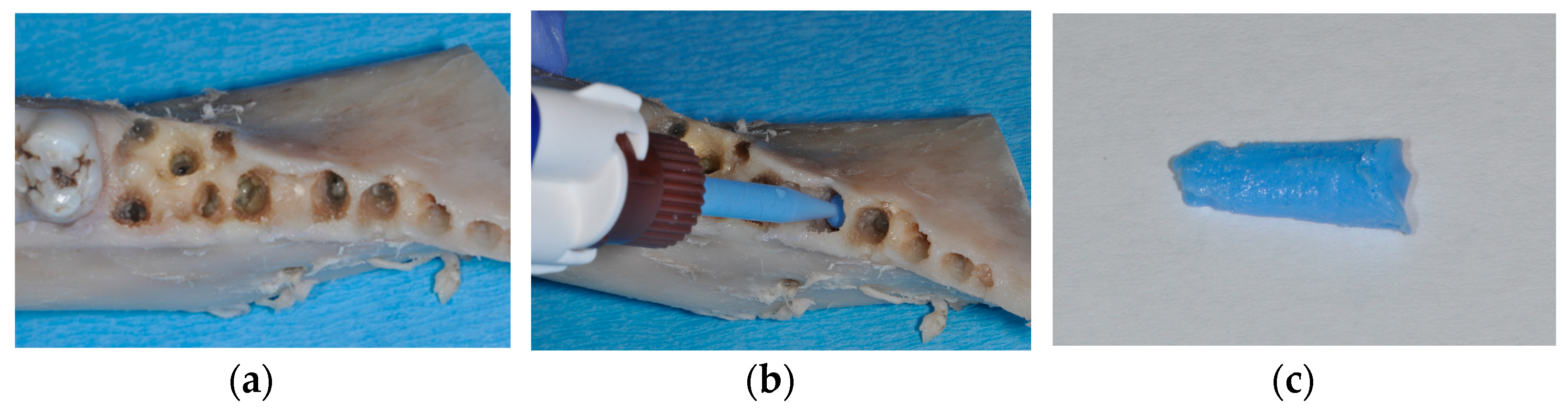
2. Materials and Methods
3. Results
4. Discussion
Author Contributions
Funding
Institutional Review Board Statement
Data Availability Statement
Acknowledgments
Conflicts of Interest
References
- Ganz, S.D.; Valen, M. Predictable synthetic bone grafting procedures for implant reconstruction: Part two. J. Oral Implantol. 2002, 28, 178–183. [Google Scholar] [CrossRef] [PubMed]
- McAllister, B.S.; Haghighat, K. Bone augmentation techniques. J. Periodontol. 2007, 78, 377–396. [Google Scholar] [CrossRef]
- Avila-Ortiz, G.; Gubler, M.; Romero-Bustillos, M.; Nicholas, C.L.; Zimmerman, M.B.; Barwacz, C.A. Efficacy of Alveolar Ridge Preservation: A Randomized Controlled Trial. J. Dent. Res. 2020, 99, 402–409. [Google Scholar] [CrossRef] [PubMed]
- Avila-Ortiz, G.; Chambrone, L.; Vignoletti, F. Effect of alveolar ridge preservation interventions following tooth extraction: A systematic review and meta-analysis. J. Clin. Periodontol. 2019, 21, 195–223. [Google Scholar] [CrossRef]
- Cardaropoli, G.; Araújo, M.; Hayacibara, R.; Sukekava, F.; Lindhe, J. Healing of extraction sockets and surgically produced—Augmented and non-augmented—Defects in the alveolar ridge. An experimental study in the dog. J. Clin. Periodontol. 2005, 32, 435–440. [Google Scholar] [CrossRef]
- Kunert-Keil, C.; Gredes, T.; Heinemann, F.; Dominiak, M.; Botzenhart, U.; Gedrange, T. Socket augmentation using a commercial collagen-based product—An animal study in pigs. Mater. Sci. Eng. C Mater. Biol. Appl. 2015, 46, 177–183. [Google Scholar] [CrossRef]
- Pereira, F.P.; Hochuli-Vieira, E.; Maté Sánchez de Val, J.E.; De Santis, E.; Salata, L.A.; Botticelli, D. Bone Ceramic® at Implants Installed Immediately into Extraction Sockets in the Molar Region: An Experimental Study in Dogs. Clin. Implant Dent. Relat. Res. 2016, 18, 360–368. [Google Scholar] [CrossRef]
- Kahnberg, K.E. Immediate implant placement in fresh extraction sockets: A clinical report. Int. J. Oral Maxillofac. Implants 2009, 24, 282–288. [Google Scholar]
- Roccuzzo, M.; Bonino, F.; Bonino, L.; Dalmasso, P. Surgical therapy of peri-implantitis lesions by means of a bovine-derived xenograft: Comparative results of a prospective study on two different implant surfaces. J. Clin. Periodontol. 2011, 38, 738–745. [Google Scholar] [CrossRef]
- Song, D.; Shujaat, S.; de Faria Vasconcelos, K.; Huang, Y.; Politis, C.; Lambrichts, I.; Jacobs, R. Diagnostic accuracy of CBCT versus intraoral imaging for assessment of peri-implant bone defects. BMC Med. Imaging 2021, 21, 23. [Google Scholar] [CrossRef]
- Hilgenfeld, T.; Juerchott, A.; Deisenhofer, U.K.; Krisam, J.; Rammelsberg, P.; Heiland, S.; Bendszus, M.; Schwindling, F.S. Accuracy of cone-beam computed tomography, dental magnetic resonance imaging, and intraoral radiography for detecting peri-implant bone defects at single zirconia implants-An in vitro study. Clin. Oral Implants Res. 2018, 29, 922–930. [Google Scholar] [CrossRef] [PubMed]
- Schlee, M.; Wang, H.L.; Stumpf, T.; Brodbeck, U.; Bosshardt, D.; Rathe, F. Treatment of Periimplantitis with Electrolytic Cleaning versus Mechanical and Electrolytic Cleaning: 18-Month Results from a Randomized Controlled Clinical Trial. J. Clin. Med. 2021, 10, 3475. [Google Scholar] [CrossRef] [PubMed]
- Schwarz, F.; Jepsen, S.; Obreja, K.; Galarraga-Vinueza, M.E.; Ramanauskaite, A. Surgical therapy of peri-implantitis. Periodontol. 2000 2022, 88, 145–181. [Google Scholar] [CrossRef]
- Misch, C.E.; Dietsh, F. Bone-grafting materials in implant dentistry. Implant Dent. 1993, 2, 158–167. [Google Scholar] [CrossRef] [PubMed]
- Wen, S.C.; Barootchi, S.; Wang, H.L.; Huang, W.X. Non-submerged reconstructive approach for peri-implantitis osseous defect, with removal of implant crowns: 1-year outcomes of a prospective case series study. J. Periodontol. 2022, 93, 1250–1261. [Google Scholar] [CrossRef]
- Amid, R.; Kheiri, A.; Kheiri, L.; Kadkhodazadeh, M.; Ekhlasmandkermani, M. Structural and chemical features of xenograft bone substitutes: A systematic review of in vitro studies. Biotechnol. Appl. Biochem. 2021, 68, 1432–1452. [Google Scholar] [CrossRef] [PubMed]
- Figueiredo, A.; Coimbra, P.; Cabrita, A.; Guerra, F.; Figueiredo, M. Comparison of a xenogeneic and an alloplastic material used in dental implants in terms of physico-chemical characteristics and in vivo inflammatory response. Mater. Sci. Eng. C Mater. Biol. Appl. 2013, 33, 3506–3513. [Google Scholar] [CrossRef] [PubMed]
- Hoang, T.N.; Mealey, B.L. Histologic comparison of healing after ridge preservation using human demineralized bone matrix putty with one versus two different-sized bone particles. J. Periodontol. 2012, 83, 174–181. [Google Scholar] [CrossRef]
- De Coster, P.; Browaeys, H.; De Bruyn, H. Healing of extraction sockets filled with BoneCeramic® prior to implant placement: Preliminary histological findings. Clin. Implant Dent. Relat. Res. 2011, 13, 34–45. [Google Scholar] [CrossRef]
- Turco, G.; Porrelli, D.; Marsich, E.; Vecchies, F.; Lombardi, T.; Stacchi, C.; Di Lenarda, R. Three-Dimensional Bone Substitutes for Oral and Maxillofacial Surgery: Biological and Structural Characterization. J. Funct. Biomater. 2018, 9, E62. [Google Scholar] [CrossRef]
- Baheiraei, N.; Nourani, M.R.; Mortazavi, S.M.J.; Movahedin, M.; Eyni, H.; Bagheri, F.; Norahan, M.H. Development of a bioactive porous collagen/β-tricalcium phosphate bone graft assisting rapid vascularization for bone tissue engineering applications. J. Biomed. Mater. Res. A 2018, 106, 73–85. [Google Scholar] [CrossRef] [PubMed]
- Karl, M.; Palarie, V.; Nacu, V.; Grobecker-Karl, T. A Pilot Animal Study Aimed at Assessing the Mechanical Quality of Regenerated Alveolar Bone. Int. J. Oral Maxillofac. Implants 2020, 35, 313–319. [Google Scholar] [CrossRef]
- Winter, W.; Krafft, T.; Steinmann, P.; Karl, M. Quality of alveolar bone--Structure-dependent material properties and design of a novel measurement technique. J. Mech. Behav. Biomed. Mater. 2011, 4, 541–548. [Google Scholar] [CrossRef] [PubMed]
- Delgado-Ruiz, R.; Romanos, G.E.; Alexandre Gerhke, S.; Gomez-Moreno, G.; Maté-Sánchez de Val, J.E.; Calvo-Guirado, J.L. Biological effects of compressive forces exerted on particulate bone grafts during socket preservation: Animal study. Clin. Oral Implants Res. 2018, 29, 792–801. [Google Scholar] [CrossRef]
- Pawlowsky, K.; Ernst, L.; Steitz, J.; Stopinski, T.; Kögel, B.; Henger, A.; Kluge, R.; Tolba, R. The Aachen Minipig: Phenotype, Genotype, Hematological and Biochemical Characterization, and Comparison to the Göttingen Minipig. Eur. Surg. Res. 2017, 58, 193–203. [Google Scholar] [CrossRef]
- Romanos, G.E.; Delgado-Ruiz, R.A.; Gómez-Moreno, G.; López-López, P.J.; Mate Sanchez de Val, J.E.; Calvo-Guirado, J.L. Role of mechanical compression on bone regeneration around a particulate bone graft material: An experimental study in rabbit calvaria. Clin. Oral Implants Res. 2018, 29, 612–619. [Google Scholar] [CrossRef]
- Low, K.L.; Tan, S.H.; Zein, S.H.; Roether, J.A.; Mouriño, V.; Boccaccini, A.R. Calcium phosphate-based composites as injectable bone substitute materials. J. Biomed. Mater. Res. B Appl. Biomater. 2010, 94, 273–286. [Google Scholar] [CrossRef]
- Bohner, M. Design of ceramic-based cements and putties for bone graft substitution. Eur. Cells Mater. 2010, 20, 1–12. [Google Scholar] [CrossRef]
- Klijn, R.J.; van den Beucken, J.J.; Félix Lanao, R.P.; Veldhuis, G.; Leeuwenburgh, S.C.; Wolke, J.G.; Meijer, G.J.; Jansen, J.A. Three different strategies to obtain porous calcium phosphate cements: Comparison of performance in a rat skull bone augmentation model. Tissue Eng. Part A 2012, 18, 1171–1182. [Google Scholar] [CrossRef]
- Steiner, C.; Karl, M.; Laschke, M.W.; Schupbach, P.; Venturato, A.; Gasser, A. Comparison of extraction sites versus artificial defects with xenogenic bone substitute in minipigs. Clin. Exp. Dent. Res. 2021, 7, 490–501. [Google Scholar] [CrossRef]
- Buser, D.; Hoffmann, B.; Bernard, J.P.; Lussi, A.; Mettler, D.; Schenk, R.K. Evaluation of filling materials in membrane--protected bone defects. A comparative histomorphometric study in the mandible of miniature pigs. Clin. Oral Implants Res. 1998, 9, 137–150. [Google Scholar] [CrossRef] [PubMed]
- Jensen, S.S.; Broggini, N.; Hjørting-Hansen, E.; Schenk, R.; Buser, D. Bone healing and graft resorption of autograft, anorganic bovine bone and beta-tricalcium phosphate. A histologic and histomorphometric study in the mandibles of minipigs. Clin. Oral Implants Res. 2006, 17, 237–243. [Google Scholar] [CrossRef] [PubMed]
- Donath, K.; Breuner, G. A method for the study of undecalcified bones and teeth with attached soft tissues. The Säge-Schliff (sawing and grinding) technique. J. Oral Pathol. 1982, 11, 318–326. [Google Scholar] [CrossRef] [PubMed]
- Indovina, A., Jr.; Block, M.S. Comparison of 3 bone substitutes in canine extraction sites. J. Oral Maxillofac. Surg. 2002, 60, 53–58. [Google Scholar] [CrossRef]
- Cochran, D.L.; Jones, A.A.; Sugita, R.; Brown, M.C.; Prasad, H.; Kay, G.W. Twelve-month evaluation of a novel mineral-organic adhesive material used to stabilize dental implants placed in oversized osteotomies in vivo in an animal model. Clin. Oral Implants Res. 2022, 33, 391–404. [Google Scholar] [CrossRef]
- Thousand, J.; Datar, J.; Font, K.; Powell, C.A. A root volume study of the adult dentition for ridge preservation purposes. Gen. Dent. 2017, 65, 21–23. [Google Scholar]


| Parameter | Mean | SD |
|---|---|---|
| Volume [cm3] | 0.120 | 0.041 |
| Mass Creos [g] | 0.155 | 0.054 |
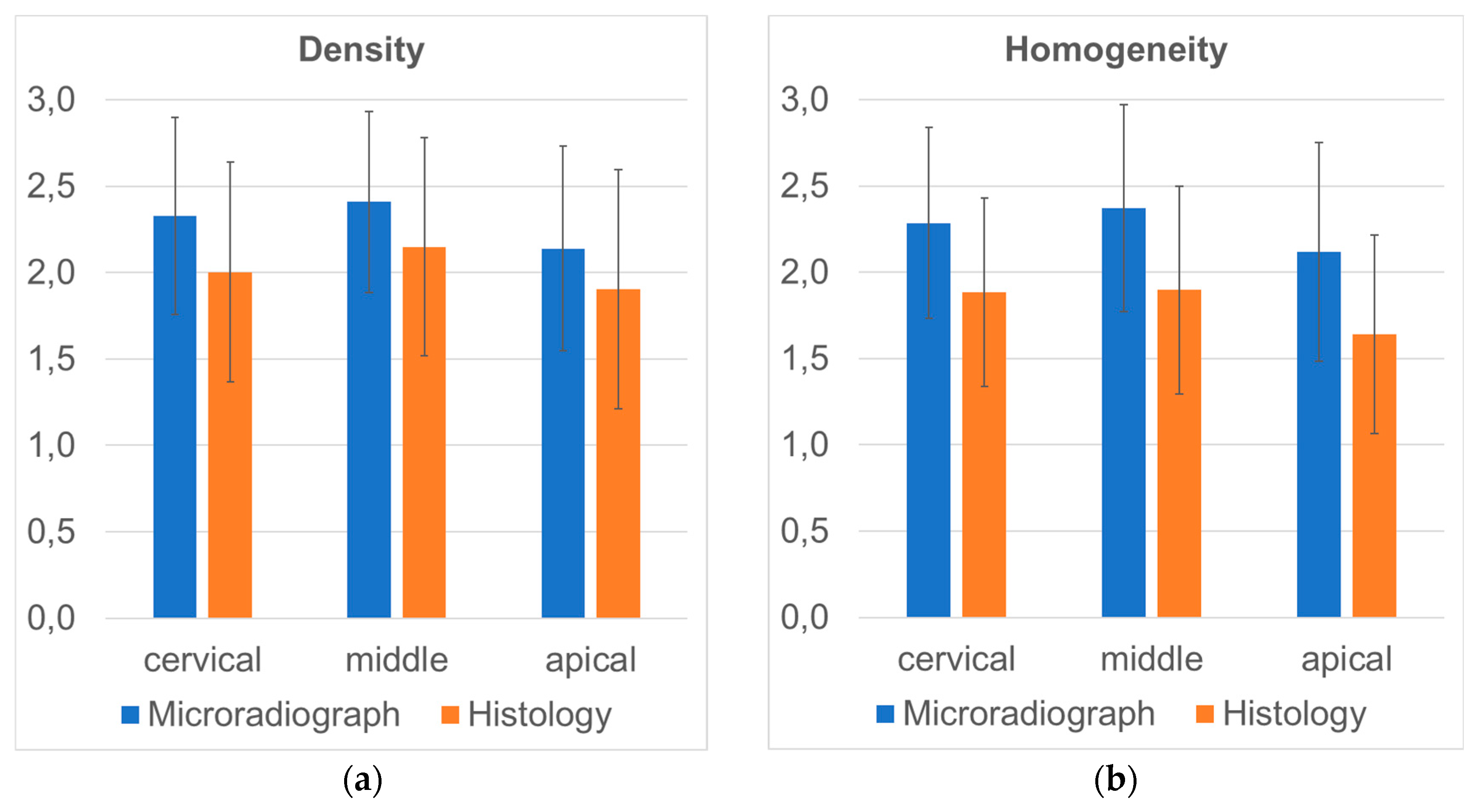
| Density—microradiograph | 2.291 | 0.395 |
| Density—histology | 2.018 | 0.504 |
| Homogeneity—microradiograph | 2.258 | 0.445 |
| Homogeneity—histology | 1.807 | 0.448 |
| Mass Creos | Density— Microradiograph | Density— Histology | Homogeneity— Microradiograph | Homogeneity— Histology | |
|---|---|---|---|---|---|
| Volume | 0.918 | 0.995 | 0.290 | 0.987 | 0.227 |
| Mass Creos | 0.913 | 0.262 | 0.959 | 0.263 | |
| Density— microradiograph | 0.279 | 0.323 | 0.230 | ||
| Density— histology | 0.267 | 0.449 | |||
| Homogeneity— microradiograph | 0.233 |
| Apical | Middle | Cervical | One Sample Wilcoxon Tests (Corrected p-Values) | ||||||
|---|---|---|---|---|---|---|---|---|---|
| MEAN | SD | MEAN | SD | MEAN | SD | apical—middle | apical—cervical | cervical—middle | |
| Density—microradiograph | 2.139 | 0.591 | 2.408 | 0.526 | 2.327 | 0.570 | <0.001 * | 0.100 | 0.200 |
| Density—histology | 1.903 | 0.692 | 2.149 | 0.629 | 2.003 | 0.637 | <0.001 * | 0.300 | 0.020 * |
| Homogeneity—microradiograph | 2.117 | 0.634 | 2.372 | 0.600 | 2.285 | 0.553 | <0.001 * | 0.040 * | 0.200 |
| Homogeneity—histology | 1.641 | 0.575 | 1.896 | 0.603 | 1.883 | 0.548 | <0.001 * | 0.002 * | 1.000 |
Disclaimer/Publisher’s Note: The statements, opinions and data contained in all publications are solely those of the individual author(s) and contributor(s) and not of MDPI and/or the editor(s). MDPI and/or the editor(s) disclaim responsibility for any injury to people or property resulting from any ideas, methods, instructions or products referred to in the content. |
© 2024 by the authors. Licensee MDPI, Basel, Switzerland. This article is an open access article distributed under the terms and conditions of the Creative Commons Attribution (CC BY) license (https://creativecommons.org/licenses/by/4.0/).
Share and Cite
Dahl, S.; Klär-Quarz, V.; Schulz, A.; Karl, M.; Grobecker-Karl, T. In Vitro Handling Characteristics of a Particulate Bone Substitute for Ridge Preservation Procedures. Materials 2024, 17, 313. https://doi.org/10.3390/ma17020313
Dahl S, Klär-Quarz V, Schulz A, Karl M, Grobecker-Karl T. In Vitro Handling Characteristics of a Particulate Bone Substitute for Ridge Preservation Procedures. Materials. 2024; 17(2):313. https://doi.org/10.3390/ma17020313
Chicago/Turabian StyleDahl, Samira, Virgilia Klär-Quarz, Annika Schulz, Matthias Karl, and Tanja Grobecker-Karl. 2024. "In Vitro Handling Characteristics of a Particulate Bone Substitute for Ridge Preservation Procedures" Materials 17, no. 2: 313. https://doi.org/10.3390/ma17020313
APA StyleDahl, S., Klär-Quarz, V., Schulz, A., Karl, M., & Grobecker-Karl, T. (2024). In Vitro Handling Characteristics of a Particulate Bone Substitute for Ridge Preservation Procedures. Materials, 17(2), 313. https://doi.org/10.3390/ma17020313