Review

Metal Chalcogenide–Hydroxide Hybrids as an Emerging Family of Two-Dimensional Heterolayered Materials: An Early Review

by
Yuri Mikhlin
1,2,*,
Maxim Likhatski
1,
Roman Borisov
1,3,
Denis Karpov
1,3 and
Sergey Vorobyev
1
1
Institute of Chemistry and Chemical Technology, Krasnoyarsk Science Center of the Siberian Branch of the Russian Academy of Sciences, Krasnoyarsk 660036, Russia
2
Department of Chemistry, Bar-Ilan University, Ramat Gan 52900, Israel
3
Institute of Nonferrous Metals and Materials Science, Siberian Federal University, Krasnoyarsk 660041, Russia
*
Author to whom correspondence should be addressed.
Materials 2023, 16(19), 6381; https://doi.org/10.3390/ma16196381
Submission received: 26 August 2023 / Revised: 19 September 2023 / Accepted: 19 September 2023 / Published: 24 September 2023
(This article belongs to the Special Issue Two-Dimensional Materials and Nano Devices)

Abstract

:
Two-dimensional (2D) materials and phenomena attract huge attention in modern science. Herein, we introduce a family of layered materials inspired by the minerals valleriite and tochilinite, which are composed of alternating “incompatible”, and often incommensurate, quasi-atomic sheets of transition metal chalcogenide (sulfides and selenides of Fe, Fe-Cu and other metals) and hydroxide of Mg, Al, Fe, Li, etc., stacked via electrostatic interaction rather than van der Waals forces. We survey the data available on the composition and structure of the layered minerals, laboratory syntheses of such materials and the effect of reaction conditions on the phase purity, morphology and composition of the products. The spectroscopic results (Mössbauer, X-ray photoelectron, X-ray absorption, Raman, UV-vis, etc.), physical (electron, magnetic, optical and some others) characteristics, a specificity of thermal behavior of the materials are discussed. The family of superconductors (FeSe)·(Li,Fe)(OH) having a similar layered structure is briefly considered too. Finally, promising research directions and applications of the valleriite-type substances as a new class of prospective multifunctional 2D materials are outlined.

1. Introduction

In this survey, we introduce a group of nature-inspired materials of valleriite, (Fe,Cu)S2·n(Mg,Fe,Al)(OH)2), type, with unusual chemical composition and a structure formed by alternating two-dimensional (2D) heterolayers, which are interesting for plentiful applications. Two-dimensional materials are attracting tremendous attention at present (Figure 1a), owing to their numerous unique properties [1,2,3,4,5,6,7,8,9,10,11,12,13,14,15,16,17,18,19,20]. Graphene is generally considered as the first and most studied 2D material, exhibiting giant electron mobility and conductivity, as well as extraordinary optical, thermal, mechanical and other properties (see, for example, reviews [2,3,4,5,6,7]). Since the seminal study by Geim, Novoselov et al. published in 2004 [1], many different 2D materials have been reported, although some of them were already well-known as natural minerals (molybdenite MoS2, clays, layered double hydroxides, etc.) or were synthesized in laboratory. Generally, the density of dangling bonds is very low on the atomic layers and the ones interact via weak van der Waals (vdW) forces. Like graphene, atomically thin layers can be prepared by using exfoliation of bulk materials, and a variety of chemical methods have been developed too. Let us briefly describe several examples of 2D materials, which may be compared with valleriites.
Two-dimensional transition metal dichalcogenides (TMDC) comprise an atomic plane of cations and chalcogen atoms above and below the plane, with the general formula MX2 (M is a d-metal, X is S, Se or Te) [10,11,12,13,14,15,16,17]. TMDCs exhibit metallic or semimetallic (VS2, NbS2, TaS2) and semiconducting characteristics (Mo and W sulfides and selenides, TiS2, HfS2, ZrS2, etc.). The direct band gap and excitonic effects arise upon the transition from multi- to monolayer in MoS2 and some other TMDCs. This promotes strong optical absorption in the UV-vis-NIR regions in conjunction with rather high electron mobility, which can be controlled by modifying the structure (including the number of stacked layers), and inspires their applications in 2D electronic and optoelectronic devices and photocatalysis [12,13,14,15,16,17]. The chemical and mechanical stability of TMDCs (especially of Mo and W) together with their high surface area make them interesting for tribology, electrode materials for Li ion batteries, (electro)catalysis, and so on. On the other hand, the low concentration of active sites requires additional chemical pre-treatment. To increase the electric conductivity and surface area, TMDCs are often utilized together with graphene and other carbon materials.
MXenes [18,19,20,21,22,23,24,25] are the family of 2D materials Mn+1XnTz or M1.33XnTz (where n = 1–4; M is a transition metal, X is carbon and/or nitrogen, T is a termination group such as -OH, =O or -F). They have been extensively studied since 2011, when Ti3C2 single layers were obtained in HF medium [18]. MXenes possess high electric conductivity, strong absorption in the near-IR region, and relatively high chemical resistance. In contrast to graphene and TMDC, -OH or =O terminations make them hydrophilic, extending the range of possible properties and applications, e.g., in sorption, catalysis, electrode materials and biomedicine. Exchanging the metal or specific surface groups allows them to influence electronic, optical (plasmonic) and other characteristics of MXenes. Theoretical calculations suggested that MXenes can be semiconductors, topological insulators and superconductors, for example in case of Nb2CTx with Se, S or NH terminations [25]. Nevertheless, preparation of magnetic or semiconducting MXenes, and even uniform surface termination on carbide/nitride MXenes, remains challenging.
(Nano)clay minerals (Figure 2) have a layered structure formed by tetrahedral sheets, in which the silicon–oxygen tetrahedra share three corners, while the fourth is connected with an adjacent octahedral sheet [26,27,28,29,30,31]. The octahedral sheet is typically composed of aluminum and/or magnesium in coordination with hydroxide ions, and with oxygen from the tetrahedra. The layers can have either a negative charge neutralized by interlayer aqueous cations (cationic clays like montmorillonite) or a positive charge balanced by variable anions in the interlayer space (anionic clays, including double-layered hydroxides). Natural clay minerals have been known for centuries, and their synthetic analogs are widely utilized due to their abundance, high specific area, sorption ability and chemical reactivity. Layered double hydroxides (LDHs) [32,33,34] have the common formula M1−x2+[Mx3+(OH)2(An−)x/n]x+·mH2O, where M2+ and M3+ are cations Mg2+, Ca2+, Fe2+, Fe3+, Al3+ and others within the positively charged brucite-like layers, and A stands for a variety of interlayered anions (CO32−, Cl, NO3, SO42−, etc.) (Figure 2c). The tunable chemical composition, reversible thermal and pH-sensitive (de)hydroxylation, high ionic 2D conductivity, anion exchange ability and composite formation stimulate the usage of LDHs as adsorbents, catalysts (often after incorporation of modifiers), energy storage materials, carriers for drugs and biological molecules, and many others. However, dielectric properties of LDHs, low stability toward the formation of defects, and non-ordered intercalates restrict their applications as electronic, magnetic and photosensitive materials.
Figure 1. (a) Number of publications in the domain of 2D nanomaterials [7] (licensed under CC BY 4.0). (b) Scheme of formation of heterostructured 2D materials [35]; permission from Springer Nature.
Materials 16 06381 g001
Two-dimensional phenomena take place not only in free-standing monolayers, but also in atomic layers deposited on a support and few-layer materials. A number of bulk properties are actually due to the inner 2D structure as, for example, superconductivity localized in certain crystallographic planes. New remarkable opportunities can arise if two or more different 2D crystals are combined in one vertical stack [35,36,37,38,39,40], producing several thick atomic layer heterostructures (Figure 1b). Arbitrary 2D layers interacting via vdW forces have been predicted theoretically, but only few heterostructures with a small lattice mismatch were stacked mechanically or synthesized by means of atomic layer deposition and similar complicated methods [38]. It is worth recalling that the heterostructures formed by two different (though rather similar, e.g., of AIIIBVI group and quasi-bulk) semiconductors have been widely used in many solid-state devices for decades. So, the development of new prospective 2D heterostructured materials is an important challenge.
Meanwhile, several naturally occurring valleriite-type minerals and synthetic “nature-inspired” metal chalcogenide–hydroxide materials, as well as superconductors composed of FeSe and (Li,Fe)OH molecular layers, are known at present. The layers are probably interacting not via vdW forces but the opposite electric charges; the distances between chalcogenide sheets detached by dielectric hydroxide ones as large as ~11.4 Å suggest a combination of 2D and 3D characteristics. These types of materials are mostly neglected in modern materials science and are restricted by surprisingly narrow considerations in superconductor physics and Earth and meteoritic sciences.
The aim of this article is to summarize the still very limited results on the natural and synthetic heterolayered materials available up to now in order to introduce those to researchers from various fields. First, the minerals of the valleriite family are considered in terms of the chemical composition and crystalline structure; the geological aspects are mainly omitted. A fairly full survey of laboratory syntheses of their analogues is presented. Then, up-to-date studies on the electronic structure and physical and chemical properties are given both for natural and synthetic materials. The superconductors are described concisely; the readers can refer for more detail to the relevant literature. Finally, we outline interesting characteristics of the materials currently found and some promising applications; this part is certainly incomplete, as much more work is required.

2. Natural Two-Dimensional Layered Minerals

Valleriite was first discovered by Blomstrand [41] in Sweden more than 150 years ago, but a lot of confusion regarding its composition, structure and discrimination with such minerals as chalcopyrite CuFeS2 and mackinawite FeS took place until the 1960s, when Evans and Allmann [42] managed to examine a single crystal of valleriite and establish that its lattice is constructed by alternating quasi-monolayers of Cu-Fe sulfide and brucite (Mg,Al)(OH)2. Later, tochilinite FeS·(Mg,Al)(OH)2 [43,44,45] and a number of similar heterolayered minerals were identified and their structures confirmed. Below, the terms “vallerite” or “valleriites” will mark both the specific substance formed by Mg-based hydroxide and Cu-Fe-S structural parts and a wider family of hydroxide-chalcogenide materials.

2.1. Brucite and Mackinawite

Before the discussion of the valleriite-group minerals, let us mention two related compounds. Brucite with the ideal chemical composition Mg(OH)2, formed in nature as a low-temperature hydrothermal mineral, is built with a sheet of Mg2+ cations between two sheets of hydroxide anions, with each Mg2+ being in the center of an octahedron of hydroxyls (Figure 3a) [27]. The layers are composed of the octahedra linked laterally by sharing the edges; the Mg–OH, OH–OH and O–H distances are 2.10 Å, 3.218 Å and 1.03 Å [46]. Important features of brucite are the ability to exchange Mg2+ for other cations, including Li+, Na+, Fe2+/Fe3+, Al3+, Ga3+; positive zeta potential up to very high pH; and easy dehydration [27,46,47,48,49].
Mackinawite, a metastable iron sulfide with a tetragonal layered structure [50,51,52,53,54,55], was discovered in Ni-Au ores in Mackinow mine, and first was erroneously identified as valleriite [50]; it also was detected in the products of steel corrosion in the presence of sulfide ions [51]. Mücke [56] specified its composition as (Fe,Ni,Co)1+xS (x ranges from −0.10 to +0.10). The crystal structure of mackinawite is constructed [52,54] by edge-connected and edge-supported FeS4 tetrahedra (space group P4/nmm) (Figure 3b). The layers are stacked by vdW forces, allowing for the intercalation of various species [57]. Iron cations are in a low-spin singlet Fe2+ state but easily transform to high-spin Fe2+-S and Fe3+-S centers, S-excessive composition and so forth under moderate oxidative conditions [58,59]. Iron selenide β-FeSe with the same tetragonal structure is a superconductor (TC~40 K) [60,61,62], and superconducting behavior has been reported for the tetragonal FeS at about 4 K [57].
Figure 3. Schematic representation of (a) brucite Mg(OH)2 structure viewed down the z axis; (b) the tetragonal structure and XRD pattern of mackinawite FeS (synthetic). Reproduced from [54] with permission from APS.
Materials 16 06381 g003

2.2. Valleriites

The chemical composition of natural valleriites notably vary for the samples examined; for example, Evans and Allmann [42] have reported the composition (Fe1.07Cu0.93) S2·1.526[(Mg0.68Al0.32)(OH)2], and Mücke [56] has suggested the formula (Fe,Cu)2S2·x[(Mg2+,Fe2+,Al3+y)(OH)2], where x = 1.24–2.25 and y = 0.00–0.26, for valleriites from five different locations. The proportion of the metal sulfide and brucite-like parts is not constant; the hydroxide layers contain variable quantities of Al, Fe and other cations, and there exist minerals with mainly Fe (ferrovalleriite) [63] or Cr (chromian valleriite) [64].
The crystalline lattice of valleriite [42] is formed by hexagonal brucite-based layers (space group P 3 ¯ m1) with the cell parameters a = 0.307 nm and c = 1.137 nm, and rhombohedral sulfide layers (R 3 ¯ m) with a = 0.379 nm and c = 0.341 nm (Figure 4). The inconsistency of the constant a for the sulfide and hydroxide layers causes a moiré picture upon stacking the layers (Figure 4a). In addition to the “three-layer” valleriite, Organova [65,66] identified a “single-layer” variety, in which both the layers are crystalized in a trigonal lattice (space group P 3 ¯ m1) with c = 1.137 nm. The strongest reflections in the X-ray diffraction patterns at 1.13–1.14 nm and 0.56–0.57 nm are explicit features of valleriite and related compounds. It has been suggested that some diffraction features may be due to ordering Fe cations in the hydroxide layers, but no super-lattices were found [66]. The arrangement of metal and sulfur atoms seems to be similar to that of nukundamite (Cu,Fe)4S4, a layered sulfide resembling CuS [67,68]. Statistical distribution of Cu and Fe atoms in the sulfide layers has been hypothesized but not confirmed experimentally. The X-ray absorption spectroscopy (Cu K- and Fe K-XANES and EXAFS) of the Cu,Fe sulfide part in comparison with chalcopyrite [69] concurs with X-ray and electron diffraction, including (Fe, Cu)-S bonds of 0.230 nm and 0.271 nm (the latter is absent in chalcopyrite), but could not distinguish Fe and Cu positions. Evans and Allmann [42] believed that the brucite layers are positively charged, as this is typical for Mg minerals, especially with Al3+ replacing Mg2+, and the sulfide layers bear a negative charge.
Although valleriite is not widespread, the ores with up to 20% of valleriite enriched in platinum group metals amount about 8% of total resources of the Noril’sk ore deposit in Siberia, Russia [70,71,72]; these are not in commercial exploitation due to the lack of processing technologies.

2.3. Tochilinite

Tochilinite was initially described as fibrous iron sulfide containing magnesium and water [43,73,74,75] before its valleriite-type layered structure was elucidated by Organova et al. [44,45,76]. The mineral found in many terrestrial locations has no industrial importance but is interesting because of its abundance in meteorites, particularly CM carbonaceous chondrites, and cosmic dust [77,78,79]. Tochilinite entities often have a tubular shape (Figure 5a). The composition of tochilinites can be described as 2Fe1−xn(Mg,Al,Fe)(OH)2, where 0.08 ≤ x ≤ 0.28 and 1.58 ≤ n ≤ 1.75 [44,45,76,80]. The mackinawite-like sulfide layers consist of Fe atoms located in a plane and coordinated with four sulfide anions; the distorted tetrahedra have a different orientation than in valleriite (compare Figure 3, Figure 4 and Figure 6). Iron occurs as singlet Fe2+ in the sulfide layers of natural tochilinites, probably forming under strongly reducing conditions [77,78,79,80], while in synthetic samples, as well as in hydroxide layers, both Fe2+/Fe3+ centers can be presented, as discussed in the next sections.
There are several known varieties of natural tochilinites, which differ in the ratio n of sulfide and hydroxide components and their mutual lattice arrangement, both commensurate and incommensurate [65]. Organova and co-workers [44,45,65] specified an isometric variety with the composition 6Fe0.9S·5(Mg0.71Fe0.29)(OH)2 crystallized in the space group C1. In acicular variety 6Fe0.8S·5[Mg0.7Fe0.3(OH)2], the sulfide sublattice has the space group P1, a = 0.834 nm, b = 0.854 nm, c = 1.074 nm, α = 87°20′, β = 94°30′, γ = 92°, and a brucite-based sublattice with the space group C1. A mineral with Fe completely replacing Mg in the hydroxide part (ferrotochilinite) was reported [80] to be monoclinic (the space group is C2/m, Cm or C2). The X-ray diffraction reflections with large d values, i.e., d001 = 1.083 nm and d002 = 0.539 nm, are signatures of the layered structures of tochilinite.

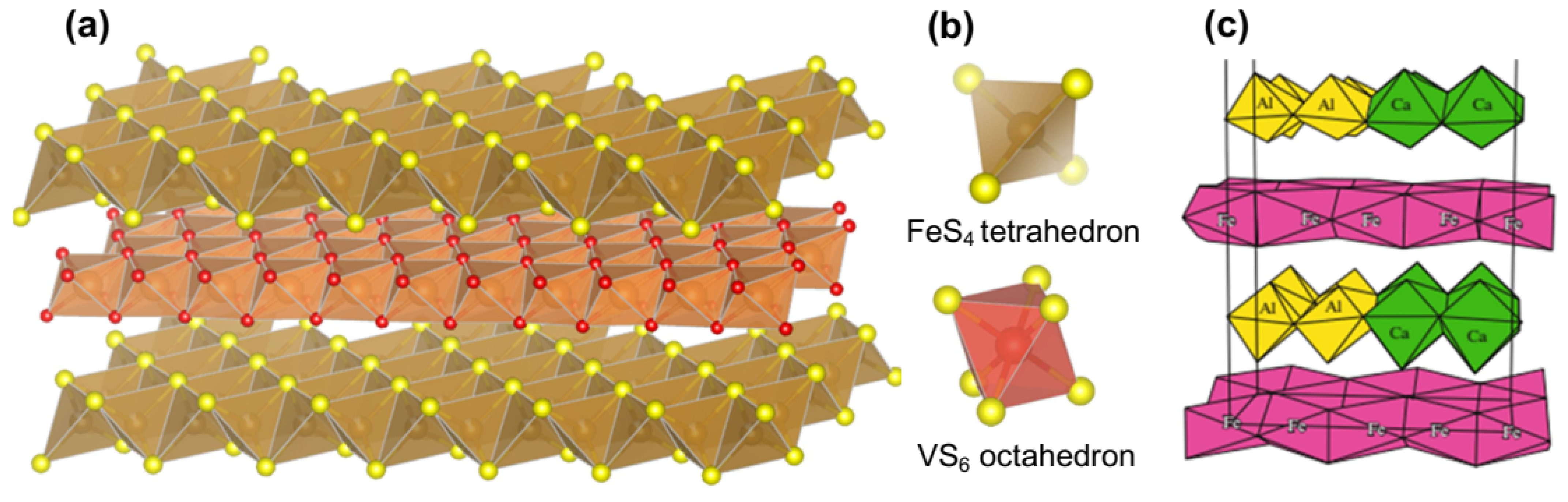
2.4. Other Minerals of Valleriite Group

A few valleriite-like minerals with different chemical compositions are known too. In 1973, haapalaite having the composition [Fe1.26Ni0.74S2]·1.61[Fe0.16Mg0.84(OH)2] and the hexagonal lattice constants of a = 0.364 nm and c = 3.402 nm was found in Outokumpu serpentines [81]. The authors suggested that Ni completely substitutes Cu in a valleriite-type structure; chemical bonding in the sulfide layers and other characteristics of haapalaite remain poorly studied.
Yushkinite [(Mg0.6Al0.3V0.1)(OH)2][V0.875S2] was discovered in 1984 [82] and has been re-examined recently [83]. The hydroxide and sulfide layers are revealed to be commensurate in the basal plane of the trigonal elementary cell (a = 0.325 nm, c = 1.14 nm, the space group P 3 ¯ m1). Interestingly, the sulfide layer of yushkinite is composed of VS6 octahedra but not tetrahedra.
Vyalsovite FeS·CaAl (OH)5 is a very rare hydrosulfide mineral found in 1989 in the Noril’sk ore provenance [84]. Soboleva et al. [85] concluded that its structure is formed by commensurate iron sulfide and Ca,Al hydroxide layers (elementary monoclinic cells with a = 0.5205, b = 2.140, c = 1.440 nm, β = 95°, space group Cm). The structural model suggests that the sulfide layers consist of FeS6 octahedra, in contrast to tochilinite, and Al and Ca sites in the hydroxide layers are ordered (Figure 6c).
Figure 6. (a) Structure of tochilinite, (b) coordination units FeS4 in tochilinite and VS6 in yushkinite, and (c) structure of vyalsovite [85], permission from Springer Publishing Company.
Materials 16 06381 g006
Pekov et al. [86] reported in 2014 on three new minerals of the valleriite group, which contain molybdenum and niobium in the sulfide layers. Nb is a major element in the sulfide blocks of ekplexite (after a Greek word for “surprise”) (Nb,Mo)S2·(Mg1−xAlx)(OH)2+x, while Mo prevails in kaskasite (Mo,Nb)S2·(Mg1−xAlx)(OH)2+x and manganokaskasite (Mo,Nb)S2·(Mn1−xAlx)(OH)2+x, with a share of Mo replaced with W. In the latter mineral, Mg2+ is completely substituted with Mn2+, and it is unclear whether solid solutions exist in the system. All three structures are incommensurate with trigonal sulfide (space group P 3 ¯ m1, a = b = 0.322–0.323 nm) and hydroxide (P3m1 or P321, a = 0.3066–0.3118 nm) sublattices, c = 1.144–1.161 nm; the replacement of Mg for Mn increases the hydroxide cell constants due to larger Mn2+ cation. Pekov and co-authors also summarized [80,86] the parameters of crystalline structures and X-ray powder diffraction data for all known vallerite-type minerals.
Chemical bonding in the natural layered materials remains insufficiently understood because of their complex and variable composition, contamination caused by fine intergrowth with different minerals, and the lack of big enough crystals, making it difficult to use spectroscopic techniques and to measure the physical and chemical properties. To overcome these problems, efforts to synthesize valleriite-type materials have been undertaken. Most of the synthetic work, however, has been aimed at understanding the mineral formation processes, and researchers have been interested in specifying the reaction products but not just preparing pure target substances.

3. Synthesis of Heterolayered Materials

3.1. Syntheses of Valleriites

The first laboratory preparation of valleriite was performed soon after the mineral crystalline structure was determined [42]. Iiishi et al. [87] obtained valleriite from either natural chalcopyrite or mixtures of Fe and Cu compounds with S and Mg and Al oxides in sealed gold or silver capsules at temperatures between 400 °C and 700 °C (water vapor pressure of 1000 Bar) over 5–20 days. Valleriite formed below 600 °C with chalcopyrite, brucite, pyrrhotite, covellite as by-products; the composition of valleriites was not firmly determined owing to its minor quantities. Later, Takeno and Moh [88] synthesized valleriite with Se fully or partially replacing S at 450 °C by using the same hydrothermal set-up. Preliminary prepared selenian chalcopyrite or cubanite or natural cubanite CuFe2S3 and Mg and Al hydroxides were starting reagents. Valleriite and selenian valleriite formed as submicrometer particles together with metal sulfides (selenides) and korshunskite Mg2Cl(OH)3·4H2O.
Hughes, Kakos et al. [89] conducted a successful hydrothermal synthesis of valleriite starting from co-precipitated Fe/Cu sulfides and Mg/Al hydroxide gels in the temperature range from 110 °C to 300 °C. The authors reported that valleriite formed if both hydrogen pressure (typically 10 Bar) and a large excess of sulfur (as ammonia sulfide) relative to CuFeS2 composition were applied (Figure 7). Almost pure valleriite phase was produced as platy crystals up to 200 nm in the lateral size and 15 nm thick using the atomic ratios of precursors Cu 2, Fe 2, S 14.6, Mg 2.08, Al 0.98 and initial pH of the mixture of 8.8 after heating at 110 °C for 25 days. X-ray diffraction, electron microscopy and X-ray photoelectron spectroscopy confirmed the structure consisting of alternating brucite-like (Mg,Al) hydroxide layers and Cu-Fe-S sulfide layers. EDS analysis of the valleriite prepared with different reagent ratios found the composition range of 1.67[Mg0.70Al0.30(OH)2]·[CuFeS2] to 1.35[Mg0.70Al0.30(OH)2]·[CuFeS2].
Chistyakova and co-workers [90,91,92] prepared valleriite from aqueous solutions with different proportions of Fe(II), Cu(II) and Mg sulfates mixed with solutions of Na2S taken in stoichiometric amounts. Valleriite emerged at 150 °C and 180 °C but not at 250 °C. Higher yields of valleriites were obtained for the Cu:Fe:Mg proportions of 1:2:2, 1:4:2, 3:1:4, but in all the experiments, valleriite was mixed with chalcopyrite and Fe (hydr)oxides by-products, and its content was lower than 50%, as determined by Mössbauer spectroscopy.
Recently, Mikhlin, Borisov et al. [93] developed a simple protocol for one-pot hydrothermal manufacturing of pure valleriite (Figure 8b,c) by mixing sulfates of iron, copper, magnesium, aluminum and, if necessary, other metals with sodium sulfide solutions and aqueous ammonia, precipitating Mg and Al hydroxides and supporting a slightly reducing atmosphere upon heating. The reaction was conducted at 160 °C during 8–100 h; the initial and final pHs were about 9 and ~12, respectively. The synthetic procedure typically involved a large excess of sulfide anions, with the initial atomic ratios S/(Fe + Cu) = 2–5, similar to the study in [89], but not hydrogen. The S excess could be almost excluded, but this resulted in minor impurity phases (mainly brucite) in some formulations. Valleriite was obtained as nanoflakes of 100–200 nm in the lateral size and 10–20 nm thickness (Figure 8d–f), which easily formed colloidal solutions during washing. Both solid precipitates and colloid particles were characterized using a set of techniques. It was shown that Mg in hydroxide layers of valleriite can be partially replaced with Al and Li, which decreases and increases, respectively, the content of Fe in the layers. The sulfide part can be doped with Co, Ni, Cr and some other metals, which partially entered hydroxide layers too.

3.2. Tochilinite-Type Materials

More efforts were made to synthesize tochilinite than valleriite because the former is interesting also for cosmochemistry. Kakos et al. [94] have utilized mixtures of FeS suspension prepared with iron (II) perchlorate and ammonium sulfide, and Mg/Al gels precipitated with ammonia. The mixtures were heated at 200 °C for two days under high hydrogen pressure (2 MPa). The products were tube-like and plate-like tochilinites (Figure 9a,b) with an average composition of 2Fe1−xS·1.7(Mg0.7Al0.3)(OH)2 that slightly varied in the particles of various morphology. Electron diffraction allowed one to distinguish four incommensurate structural modifications of the tochilinites: one, for plate-like crystals, was isostructural with valleriite, and three others were similar to plate-like and tubular natural tochilinites. The hexagonal hydroxide (“hydrotalcite”) layer was suggested to be charged negatively and the sulfide ones to be positive, contrary to valleriite [42,89]. Noteworthy, the authors neglected iron in the hydroxide layers both of valleriite [89] and tochilinite [94].
Kozerenko and co-workers [95] reported on a laboratory synthesis of ferrotochilinite as a product of interaction of Fe(II) hydroxides with H2S at 80 °C. Ferrotochilinite was obtained as a poor crystalline material mixed with magnetite and mackinawite (Figure 9d). To increase crystallinity, the reaction time was prolonged to more than 60 days and pH was enhanced. In an improved technique [96], a water suspension of ferrous hydroxide with admixture of metallic Mg (Fe:Mg = 4:1) was purged with gas H2S for precipitation of iron sulfides, and then tochilinite was synthesized at 120–140 °C during 10 to 45 days. The data were summarized and clarified in Ref. [97]. The formula of ferrotochilinite and more stable ferromagnesium tochilinite were specified as 2FeS·1.51(Fe2+, Fe3+)(OH)2 and 2FeS·1.54(Fe0.7Mg0.3)(OH)2. At pH 11.5–12.5, an additional phase containing Na and having tetrahedral lattice of hydroxide constituent was found; it decomposed above 100 °C with formation of mackinawite.
Chistyakova, Gubaidulina et al. [90,91] synthesized tochilinites, preferentially for Mössbauer spectroscopy studies, based on the method [96,97], i.e., via the interaction of Fe(II)-hydroxide with H2S at pH 11–11.5 at temperatures of 160 and 180 °C, varying the initial quantities of Mg. The yield of tochilinite increased with increasing content of Mg in the reaction mixture but stayed well below 50% as considerable impurities of magnetite, troilite and pyrite were produced.
Peng et al. [98,99,100] produced tochilinite and tochilinite–serpentine-intergrowth phases simulating the components of meteorites. The alloyed metal particles of Fe, Mg and Al [98], or more complex mixtures [100], were preliminary prepared. The precursors were then reacted with aqueous ammonium sulfide or elemental sulfur at pH 13–14 below 200 °C. Tochilinite was obtained as nanoflakes and tubes with the content in the solid products less than 40%. “Sodium-tochilinite”, which has a tochilinite structure with Na hydroxide layers containing Fe, was synthesized from Fe metallic powder and sodium sulfide at 98–150 °C during 30–15 days [99]. The authors identified four varieties of Na-tochilinite, which were essentially disordered and not quite stable. It was reported that the sodium hydroxide layers can be completely removed with water or weak acids, leaving mackinawite-like Fe1−xS phase. Less stable varieties of Na-tochilinite entered reactions with a number of such substances as NH3, N2H4, N2, 2,2′-bipyridine and 1,10-phenanthroline, forming several types of intercalates, mainly replacing sodium hydroxide layers.
Vacher et al. [79] have obtained tochilinite along with cronstedtite and other minerals under conditions modeling their formation in CM chondrite meteorites, starting from metallic iron and mineral assemblages. Iron-rich tochilinite surrounded the grains of metallic Fe reacted in S-bearing solutions at 80–120 °C; content of Mg in the hydroxide layers increased with temperature.
Tochilinites with various hydroxide layers have recently been synthesized by Bolney et al. [101], applying the hydrothermal method developed for mackinawite [102]. For the preparation of magnesium tochilinite with the composition Fe0.76S·0.86[Fe2+0.01Fe3+0.56Mg2+0.43(OH)2.01], a water suspension of FeS nanoparticles obtained at 80 °C using elemental S and Fe at the atomic ratio of 1 [102] was mixed with a suspension of powdered magnesium and FeO(OH) and heated at 160 °C for 3 days. Aluminum tochilinite Fe0.89S·0.85[Fe2+0.55Fe3+0.11Al3+0.33(OH)1.84(O)0.16] was prepared from a finely ground mixture of elemental Fe, S and Al in water at 130 °C over 3 days. Ferrotochilinite Fe0.71S·0.79[Fe2+0.25Fe3+0.73Mg2+0.01Al3+0.01(OH)1.98(O)0.02] was synthesized similarly from iron and sulfur taken in a mass ratio 3:1. In all the cases, excess of Fe was used to support hydrogen pressure (along with metallic Mg and Al), and to suppress the formation of pyrrhotite and pyrite. By-products (magnetite) and unreacted Fe metal were separated by a magnet or/and washed out (brucite and others). The tochilinites were submicrometer platelets of a few nm in thickness with admixtures of nanotubes (Figure 10, upper photos). The tochilinite samples were monophase after the purification; the XRD patterns were dominated by two strong reflections, the d-values of which at approximately 11 Å and 5.5 Å were assigned to the (001) and (002) peaks, as previously reported for natural tochilinite. All other diffraction peaks had little intensity, which might be caused by preferred orientation or turbostratic stacking of the layers [101]. The abovementioned distribution and oxidation state of Fe were derived from Mössbauer spectra, which suggest that singlet Fe2+ cations occur in the sulfide layers akin to natural minerals, and all the hydroxide layers comprise high numbers of Fe2+/Fe3+ centers.
Tochilinites with magnesium hydroxide layers, including doped with Al and Li, were prepared [103] using aqueous solutions of metal sulfates, sodium sulfide and ammonia in a simple hydrothermal method similar to that developed for valleriite [93]. The iron sulfide and Mg, Al, Li hydroxides were loaded in an autoclave with Teflon liner and heated at slow rotation (8 rpm), typically at 160 °C for ~40 h. The reaction media were purged with Ar before sealing the reactor, but no reductants (elemental metals, hydrogen, etc.) were employed, in contrast to other works. The atomic ratios of S and Fe precursors varied from 1 to 10, and usually, a large excess of sodium sulfide was used (and removed during decantation washing of the solid products), allowing one to obtain pure tochilinite with no admixtures of iron sulfide or (hydr)oxide phases. The samples prepared with nearly stoichiometric initial amounts of sulfide contained minor impurities of brucite. Tochilinite formed as nanoflakes of 100–200 nm in the lateral dimensions and 10–15 nm thickness (no tubular particles were observed), which were insignificantly affected by precursor ratios, temperature and reaction time. It was found that Al and Li entered Mg hydroxide layers proportionally to their contents in the reaction mixtures, decreasing and increasing, respectively, the concentrations of Fe in the hydroxide part of tochilinite; characteristic examples can be described by the formula Fe0.77S·1.14[(MgFe0.2)(OH)2.22], Fe0.85S·0.87[(MgAl0.25Fe0.17)(OH)2.75] and Fe0.7S·0.81[(MgLi0.3Fe0.5)(OH)1.5O0.05]. The synthetic tochilinites were examined using, in addition to TEM, EDS, XRD and electron diffraction, a number of experimental techniques described below.

3.3. Layered Superconductors of FeSe·(Li,Fe)(OH) Group

Superconductivity below TC = 8.5 K discovered in tetragonal iron selenide β-FeSe by Hsu et al. [60] in 2008 attracted new interest to layered iron chalcogenide materials. The critical temperature TC depends on the substitution of Se with Te or S, stoichiometry of the iron chalcogenide and ordering the Fe vacancy system, the interlayer distance and other factors. Moreover, single-layer FeSe films grown on SrTiO3 (001) surfaces exhibited the temperature TC as high as 109 K [104]. A number of superconductors with metal cations or molecules intercalated between the FeSe sheets weakly bonded by vdW forces, including AxFe2−ySe2, where A stands for K, Rb, Cs, were synthesized [105,106].
Lu and coworkers [107] reported in 2015 on a hydrothermal synthesis of polycrystalline (Li0.8Fe0.2)OHFeSe with Tc~40 K, constructed from alternating layers of the distorted FeSe tetrahedra and Li-based hydroxide. Interestingly, the authors initially missed the existence of (Li,Fe)OH sheets and considered the spacer layer as LiFeO2 [108]. The structure strongly resembling tochilinite was obtained using metallic Fe powder, selenourea, in large excess in relation to FeSe, and a big amount of LiOH, that is, under essentially reducing and strongly alkaline conditions, at 160 °C over 3–10 days. In contrast to FeSe intercalated with alkaline metals, ammonia and their complexes, these materials are principally stable in air. With some variations, this method was then utilized by many researchers for the preparation of [(Li1−xFe)OH][(Fe1−yLiy)Se], [Li0.85Fe0.15OH][FeS] and other candidate superconductors with slightly varying compositions of hydroxide and chalcogenide layers, as powders with the crystallites on the order of several decades of μm [109,110,111,112,113,114] (Figure 11).
Large, 1–2 cm, single crystals of FeSe (Li1−xFex)OH and similar compounds were synthesized using the “ion exchange” procedure with AxFe2−ySe2 (A = K, Rb, and Cs) single crystals, grown by self-flux, Bridgman, or other methods, as precursors [115,116]. In a typical procedure, the precursor crystals and the reaction mixture of selenourea, Fe powder (now with excess of Fe) and LiOH were loaded into a Teflon-lined autoclave and heated to 120–200 °C for several days. Using a similar protocol, Guo et al. [117] manufactured single crystals of layered Fe1−xS (NaOH); the authors did not mention Fe in the sodium hydroxide part, and admitted its sensitivity toward ambient air.
The above studies of layered superconductors (more references and discussion can be found, for example, in [118] and the literature cited above) are very focused on structural, magnetic and other phenomena related to superconductivity, taking into consideration only few chemical compositions and almost ignoring additional characteristics and applications of this class of materials. Certainly, the advances in the syntheses must be employed for preparation of other heterolayered substances.

3.4. Summary on the Synthesis

Brief results on the synthesis of valleriite-type materials are collected in Table 1. The total number of such studies is not large, except for the preparation of the superconducting materials (only few examples are shown). Mostly, the target of the research was to shed light onto mineral formation mechanisms, and the layered hybrids were mixed with other solid products, sometimes unintentionally, because of imperfect synthetic protocols. Pure valleriite phases were successfully fabricated using an excess of sulfur or/and selenium [89,93]. The hydrogen pressure and highly reducing conditions [89] are not mandatory for valleriites, but they likely influence some structural peculiarities and properties, which needs further investigation. Tochilinites were produced under such conditions too; the hydrogen atmosphere is essential for obtaining low-spin Fe2+ centers in the chalcogenide layers. The syntheses of FeSe(Li,Fe)OH-type superconductors [107,109] were effectively performed using metallic iron and selenourea (thiourea for FeS layers), which react slowly and probably impede nucleation of iron chalcogenides. This causes the growth of rather big particles, up to several decades of micrometers (often single crystalline), instead of nanoflakes and nanotubes for chalcogenide anions as precursors. The synthesis of large single crystals is possible via the multistep “ion exchange” procedure [115,116], which has not been applied for valleriites yet.

4. Spectroscopic Characterization and Properties

4.1. Mössbauer, XPS, X-ray Absorption Spectra of Valleriite

Whereas the crystalline structures of layered materials are rather well understood [42,63,64], a number of major questions regarding the chemical bonding and state of elements, particularly Fe, and their physical and chemical properties are still unresolved. The spectroscopic studies of natural minerals and synthetic samples are rare and often the results are not unambiguous.
57Fe Mössbauer spectroscopy, X-ray photoelectron spectroscopy or their combination have been applied to study the oxidation and spin state of iron atoms in sulfide (chalcogenide) and hydroxide blocks. Hughes et al. [89] concluded from XPS spectra that the submicrometer particles of Al-containing valleriite synthesized under hydrogen pressure contain Cu+ and Fe3+ centers. Waanders and Pollak [119] published room-temperature Mössbauer spectra acquired from two natural valleriite crystals replacing chalcopyrite and associated with magnetite, and having the compositions [Cu0.94Fe1.06S2]·1.4[Mg0.8Fe0.20(OH)2] and [Cu1.02Fe0.98S2]·1.72[Mg0.69Fe0.05Al0.26(OH)2], respectively. The spectra consist of central signals fitted with four doublets, one of which (isomer shifts δ of 0.28 or 0.22 mm/s and quadrupole splitting Δ of ~0.3 mm/s) was assigned to Fe3+ centers in hydroxide layers (22% and 10% of total iron in these valleriites, respectively). The others were attributed to two Fe3+ centers (δ = 0.25–0.4 mm/s and the quadrupole splitting Δ = 0.74–0.89 mm/s) and Fe2+ centers with δ = 0.4–0.42 mm/s and Δ = 1.12 and 1.32 mm/s (about 32% of total iron) orderly arranged in tetrahedral positions in sulfide layers. Chistyakova et al. [90] reported Mössbauer spectra of several natural and synthetic valleriites notably contaminated, but with impurity phases, particularly chalcopyrite. The authors suggested that doublets δ = 0.36 ± 0.03 mm/s and ε = Δ/2 = 0.30 mm/s, and δ = 0.39 ÷ 0.54 mm/s and ε = 0.46 ÷ 0.56 mm/s are due to Fe in sulfide layers of valleriite, while quadrupole doublets with parameters δ ≈ 1.1 mm/s and ε ≈ 1.2 mm/s could correspond to iron positions in the hydroxide layers [92].
The interpretation of the above spectra of the samples containing several Fe species both in valleriite and foreign phases is difficult and contradictive since it is based at the room-temperature Mössbauer spectra only. In Ref. [69], Mössbauer spectra were acquired at 293 K, 78 K and 4.2 K and compared with the results of X-ray absorption spectroscopy (particularly, Fe-K- and Cu K-edge XANES and EXAFS) and XPS [69,120] from two natural valleriites intergrown with pyrrhotite Fe1−xS and magnetite, as well as with chalcopyrite. The joint analysis of the spectra suggested that Cu+ and Fe3+ centers occur in the sulfide part of valleriites (hyperfine Mössbauer parameters δ ≈ 0.4 mm/s and Δ ≈ 0.6 mm/s at room temperature), and Fe3+ cations predominate in the hydroxide part. Room-temperature Mössbauer and XPS spectra [121] for valleriite associated mainly with serpentines generally agreed with this interpretation. Upon cooling down to 4.2 K [69], the central paramagnetic doublets almost disappeared, giving rise to a series of hyperfine Zeeman sextets with internal hyperfine magnetic fields H of ~270, 320 and 490 kOe emerging both in sulfide and hydroxide layers of valleriites; no transitions into the antiferromagnetic state typical for Cu-Fe sulfides were observed.
Further insight was gained by exploring synthetic valleriites free of foreign phases (Figure 12) [93]. It was derived from the photoelectron Fe 2p (fitted using multiplet line sets) and Fe 3p spectra that Fe3+ cations predominate both in Cu-Fe sulfide and Mg-based hydroxide layers. The quantity of hydroxide” Fe can be diminished by adding Al (spectrum a vs. spectra b and c) or increased by adding Li, which enter the hydroxide module during the hydrothermal synthesis, in the range from 10% to 45% of total iron; this also influenced the share of O2− anions (compare O 1s spectra a and b). In addition, XPS found a higher content of sulfur (as monosulfide and minor polysulfide) than EDS analysis, suggesting that some excessive sulfide anions used in the synthesis were adsorbed on the nanoflake surfaces.
These findings were used to better understand Mössbauer spectra. It was confirmed that the room-temperature doublets with δ = 0.3–0.4 mm/s and Δ = 0.5–0.6 mm/s, and δ~0.3–0.4 mm/s and Δ of 1–1.2 mm/s should be assigned to Fe3+ centers in the tetrahedral coordination with S and octahedral coordination with OH groups, respectively. Several sextets with distinct hyperfine fields at low temperatures are indicative of distinct Fe sites both in sulfide and hydroxide sheets. The sextets with H of 300–330 kOe were attributed to sulfide layers, and those with H~270 kOe were ascribed to Fe-OH centers. The assignment of smaller sextets with H~500 kOe (spectra b, c) to Fe-S centers was probably erroneous, since those are likely due to Fe3+-(OH)xO2−y centers involving some O2− anions seen in O 1s spectra, as it was later established for tochilinite [103]. Magnetic measurements revealed generally paramagnetic behavior that becomes more complex if the content of Fe in the hydroxide layers decreases (Figure 12, samples b and c).

4.2. Mössbauer and XPS Investigation of Tochilinite

There is agreement in the literature that Fe cations in sulfide layers of natural tochilinite [77,78,79,80,90,91,101,122] are in the low-spin Fe2+ state, similar to mackinawite [55,56,57,58,59,102], exhibiting a narrow Mössbauer doublet with isomer shifts δ in the range from 0.4 to 0.45 mm/s and a small quadrupole splitting Δ (0.18 mm/s to 0.24 mm/s). The maxima are retained at cryogenic temperatures. The hyperfine parameters for Fe2+ and Fe3+ octahedral centers in hydroxide layers were determined to be δ of ~1.2 mm/s and Δ = 2.2–2.3 mm/s, and δ of ~0.3 mm/s and Δ = 0.7–1 mm/s, respectively. These findings allowed for the use of Mössbauer spectroscopy for the analysis of concentrations of iron in tochilinites. Many authors have taken for granted that iron occurs in hydroxide layers as Fe2+, but this seems to be incorrect. Particularly, Bolney et al. [101] found with EDS analysis and Mössbauer spectroscopy that in tochilinites prepared under essentially reducing conditions, Fe3+ cations predominated in the Mg-based hydroxide layers; preferentially, Fe2+ centers were found in Al-containing layers [Fe2+0.55Fe3+0.11Al3+0.33(OH)1.84O0.16] and a (Fe2+0.25Fe3+0.73)(OH)1.98O0.02 proportion was reported for ferrotochilinite. X-ray photoelectron Fe 2p spectra, which should have a characteristic strong narrow peak from singlet Fe2+at 707 eV, were rarely reported. Moreover, Zhang et al. [111] published the XPS spectra of single crystals of Fe1−xS (NaOH), which did not show such a peak and were explained in terms of (Fe3+, Fe2+)-S centers.
Tochilinite-type materials synthesized without metallic iron [103] have Mössbauer spectra resembling those of valleriite, that is, asymmetric doublets in room-temperature spectra and at least three Zeeman sextets at 4 K. The doublets with the isomer shift δ = 0.3–0.4 mm/s and quadrupole splitting Δ = 0.4–0.6 mm/s are due to Fe3+-S centers, and the ones with δ ≥ 0.4 mm/s and Δ = 1 ± 0.2 mm/s originate from high-spin Fe3+ cations in the octahedral OH environment, whereas the quantities of Fe2+-OH centers (δ~1 mm/s, Δ > 2 mm/s [90,91,92]) are minor. However, the signals of Fe2+ cations in tetrahedral coordination to S and those of Fe3+-6OH centers having similar parameters are difficult to resolve. The sextets (hyperfine fields of ~290, 350 and 480 kOe) indicating magnetic ordering at low temperatures were attributed to high-spin Fe2+-4S together with Fe3+-(6OH), centers Fe3+-4S, and Fe3+(OH)x(O)y centers, respectively, in hydroxide layers. Photoelectron Fe 2p spectra are not quite clearly fitted too due to the co-existence of comparable numbers of high-spin Fe3+ and Fe2+centers in the sulfide part. It was established, nevertheless, that the hydroxide layers preferentially contain Fe3+ cations, whose amount also depends on the addition of Al and Li. Combined analysis of Mössbauer, XPS and UV-vis spectra (see below) suggested that comparable quantities of high-spin Fe2+ and Fe3+ cations are presented in the sulfide layers of such tochilinites.

4.3. Raman, IR Spectroscopy, UV-Vis-NIR Spectroscopy

Raman spectroscopy is an important instrument for the characterization of 2D materials [123]. Several Raman spectra are available in the literature for natural valleriite-type minerals [63,124,125] and synthetic valleriites [93]. The spectra below 500 cm−1 originate from sulfide sheets; the strongest maxima at about 300 cm−1 both for tochilinite and valleriite are due to sulfur-only, almost independent on cations, vibrations of A1g symmetry, while others are due Cu-S and Fe-S vibrations [93,126,127,128]. Full interpretation and application of Raman spectra for studying the chalcogenide–hydroxide structures are still required. The same is true for IR spectroscopy, mainly characterizing the hydroxide part [48,63,80,124,125], and optical reflection spectra, which are commonly used as a tool for identification of minerals.
The absorption UV-vis-NIR spectra [93] collected from valleriite flakes in the colloidal solutions have maxima centered at 500–700 nm and redshifted for the samples with higher contents of Fe in the hydroxide layers (Figure 13c,f). Such maxima commonly observable for metallic nanoparticles may be attributed to localized surface plasmon resonances (LSPR) [129]. This, however, looks unlikely, as the Tauc plots, as well as reflection electron energy loss spectra (REELS) [93], suggest that valleriites are semiconductors with the indirect band gap of 0.4–0.5 eV. The gap width is close to that for chalcopyrite CuFeS2, the nanoparticles of which exhibit similar optical spectra with the maxima attributed to the quasi-static dielectric resonance [130,131] due to the “intermediate” narrow minority-spin Fe 3d band rather than LSPR.
UV-vis-NIR absorption spectra of tochilinite colloids are more complicated [103], showing broad maxima A and a set of narrower blueshifted peaks B–D. It was suggested that feature A can be interpreted as the all-dielectric Mie resonance in insulating and semiconducting nanomaterials [15,132,133]. This is associated with light scattering (both electric and magnetic components) by subwavelength particles with a high refractive index. The measurements of dielectric properties (Figure 14) [103] imply that this can be the case for tochilinites and possibly for valleriite too. On the other hand, the spectral features at shorter wavelengths likely reflect the ligand-to-metal charge transfer, involving orbitals of sulfur anions and Fe 3d states, similar to Fe-S clusters in proteins [134,135]. The clusters denoted as [1Fe-0S]2+/3+ with Fe2+ (spin number S = 2) or Fe3+ (S = 5/2) in tetrahedral environment of sulfur [134] better agree with the data of Mössbauer spectroscopy and XPS, thus suggesting that both Fe2+ and Fe3+ cations with insignificant alignment of neighboring electron spins exist in the tochilinite produced in not strongly reducing media [103].

4.4. Physical and Chemical Properties

Some basic physical properties determined experimentally or calculated are available for minerals in the literature; for example, density of 3.1–3.2 g/cm3 and Mohs hardness 1.0–1.5 are reported for valleriite, and the densities are ~3 g/cm3 and 3.47 g/cm3 (calculated) for tochilinite and ferrovalleriite, respectively [63]. However, critically important electronic characteristics of the materials have been very poorly studied up to now, leaving aside superconductors. Valleriite and tochilinite appear to be narrow-gap semiconductors with the indirect band gaps of the sulfide layers of about 0.5 eV and 0.3 eV, as determined from optical absorption spectra and REELS [93,103], as well as the solid-state impedance measurement for tochilinite [103].
Magnetic measurements in combination with Mössbauer spectroscopy (Figure 12) revealed that synthetic valleriite and tochilinite [93,103] are paramagnetic at room temperature and 4 K, but magnetic ordering takes place at low temperatures. The behavior is more complicated if the quantity of iron in hydroxide layers is reduced. This suggests a kind of interlayer magnetic interaction in the materials. It is worth mentioning that FeSe (Li,Fe)OH superconductors demonstrate superconductivity in chalcogenide layers and low-temperature ferromagnetism [107,109].
Only selected chemical properties of valleriites have been reported. The Mg-based materials are quite stable in ambient air and under moderate heating, as it was demonstrated for natural and synthetic samples [93,101,121,136,137]. Valleriite is more resistant than tochilinite. A thorough examination of the thermal behavior of synthetic valleriite nanoflakes was conducted [136] using TG/DSC methods in inert and oxidative atmosphere up to 1000 °C (Figure 15). Dehydroxylation of the hydroxide layers was found to peak at 413 °C, and metal sulfide sheets start to degrade below 500 °C, converting to bulk Cu5FeS4, CuFeS2 and FeS in inert atmosphere. The exothermic reactions in oxidative media proceed with a mass increase as sulfur oxides form and react with magnesium hydroxide layers to yield MgSO4 (the main maximum at 495 °C). Samples doped with Al, which decreases the content of Fe in hydroxide layers, notably impeded endothermic decay of valleriite in argon; on the contrary, the rate of oxidation increased. This was explained by high thermal resistance across the stacked sheets having very different composition (masses of elements and phonon frequencies), so the large number of Fe atoms in the hydroxide sheets, comparable with that in sulfide layers, promote the phonon exchange and heat transfer between the layers, and vice versa.
Several studies have been devoted to chemical reactions of valleriite-containing materials related with mineral processing, including flotation [137], leaching and sulfidation with aqueous SO2 [72], deposition of precious metals from aqueous solutions [138], and bacterial leaching [139]. Some special features of the valleriite reactivity were observed, but more research is needed.
For colloidal solutions of valleriite and tochilinite synthesized using excessive sodium sulfide [93,103], zeta potentials of the particles were negative, about −30 mV, over a wide pH range. This implies a negative charge of the hydrophilic hydroxide layers, which are expected to be exposed in aqueous media, and a positive charge of sulfide layers. The effect of the precursor proportion Fe/Mg and Al or Li doping was less significant. Tochilinite manufactured with the initial ratio Fe/S close to 1 exhibits slightly negative potentials, thus underlying the role of sulfide anions in the synthesis.

5. New Characteristics and Potential Applications of Chalcogenide–Hydroxide 2D Materials

The layered chalcogenide–hydroxide materials comprise very different, chemically incompatible components, which often have incommensurate lattices. The compositions both of chalcogenide and hydroxide layers widely vary, and they are referred to as composites or hybrids, or heterostructures. Meanwhile, the alternating layers are ordered on a molecular level and are spatially homogeneous, despite some packing defects and imperfection within the layers. This allows one to consider valleriites as individual chemical substances rather than a combination of two compounds.
The large distances, on the order of 11 Å in comparison with 6.5 Å in MoS2 [11] and 5.0 Å in mackinawite FeS [54], and the absence of short-range forces between the metal sulfide sheets separated by dielectric hydroxide layers, and vice versa, suggest probable two-dimensional phenomena in the layers. On the other hand, long-range (electrostatic, magnetic) interactions controllable via characteristics of distinct layers, e.g., Fe2+/Fe3+ content in the hydroxide ones, could affect the 2D properties. The interplay of 2D and 3D factors opens ways for engineering new materials and tuning their properties, and promises novel non-trivial effects.
In contrast to vdW heterostructures, the layers are generally believed to interact via the opposite electrostatic charges. This is not directly confirmed yet, and there are contradicting opinions regarding their signs [42,44,89,93,94,103], which stem, among other things, from errors in determining the concentrations of Fe2+/Fe3+ cations in both layers, and covalent metal–sulfur and S-S bonding. Zeta potentials of synthetic valleriite [93] and tochilinite [103] are indicative of the negative charge of magnesium-based hydroxide layers, despite the replacement of Mg2+ with Fe3+ and Al3+ cations, maybe, owing to the formation of O2− anions. It has been speculated that electroneutrality of FeSe layers and H-bonding between the layers take place in FeSe(Li,Fe)OH superconductors. However, Chen et al. [112] calculated using density functional theory that Fe atoms in (Li0.8Fe0.2)OH layers promote electron density transfer to FeSe and attraction between the chalcogenide and hydroxide sheets. One should expect that the charge signs and amplitudes, affected by the total material composition and the state of Fe, can be tuned upon the synthesis and post-synthetic treatment.
The charge difference is an apparent directing force of self-assembly of valleriites, which also can be controlled using, for instance, excessive sulfide anions or redox potential of reaction media. At present, hydrothermal synthesis with elemental metals and chalcogens or their simple compounds as precursors is the main route to powder materials composed of submicrometer flakes, tubes or micrometer plate-like crystals (Table 1). Large single crystals of few materials have been prepared via the protocol combining thermic synthesis of precursors and hydrothermal ion exchange reaction [115,116]. Further research and application of valleriites require fabrications of films, coatings and devices, in which the nanoflakes can be utilized as building blocks. Also, it would be interesting to prepare mono- and few-layer valleriites using different techniques, e.g., atomic layer deposition and chemical bath deposition. An intriguing question is whether components other than chalcogenides and hydroxides can be joined in 2D heterostructures; it is worth recalling, for example, a mineral koenenite consisting of chloride [(Na4(Ca,Mg)2Cl12]4− and hydroxide [Mg7Al4(OH)22]4+ layers [140].
The preliminary study of magnetic properties revealed paramagnetic character of valleriites and tochilinites along with magnetic ordering at 4 K, distinguishing several Fe sites in both layers. High Fe concentrations in hydroxide sheets were found to promote paramagnetic behavior, probably due to some 3D magnetic exchange interaction. Two-dimensional materials containing different 3d transition and rare-earth metals both in hydroxide and chalcogenide layers have already been obtained. It is worth remembering that ferromagnetism was observed in the layered FeSe·(Li,Fe)(OH) together with superconductivity.
Valleriite and tochilinite appear to be narrow-gap semiconductors, with their electronic properties mostly determined by the metal sulfide layers. It is likely that a wider gap or metallic conductivity can be obtained by varying the composition of the chalcogenide part; however, experimental studies of conductivity and other physical parameters are still absent. Again, we leave aside FeSe-based superconductors and a potential superconductivity in valleriites, although the independent control of the layer characters paves new ways in this direction.
Fascinating optical properties were found in valleriite and tochilinite nanoflakes, particularly an intense absorption in the visible region that is likely due to all-dielectric resonances in submicrometer particles, which are of great interest for new-generation nanophotonics and solar energy conversion, photocatalysis and sensors. The additional absorption maxima below 500 nm belonging to Fe-S charge transfer suggest a ladder of Fe 3d states in the fundamental gap akin to Fe-S clusters in proteins, which can be promising in catalysis, photo- and electrocatalysis, electrochemical energy storage and so forth.
A few available studies aimed at examination of reactivity of valleriites have demonstrated, first of all, their relative stability in ambient air, aqueous media (not acidic) and under heating, especially in inert atmosphere. The interesting effect of impeded heat transport across the molecular layers (but not in the plane) as a function of concentration of Fe in the hydroxide sheets was found.
In general, the little research conducted on these type of materials was mainly directed either on the problems of mineral origination on Earth and in meteorites, or on superconductivity. The studies were restricted within the methods and chemical compositions of the materials of interest; to the best of our knowledge, no theoretical simulation of valleriites has been performed yet (if not mentioning the FeSe-related superconductors). Consequently, we tried to demonstrate in this review that the 2D heterolayered chalcogenide–hydroxide materials are a big new class of solid substances with unique characteristics, many of which are still waiting to be discovered, explored and utilized.

Author Contributions

Conceptualization, writing, review and editing, Y.M.; writing, original draft preparation, M.L., R.B., S.V. and D.K.; funding acquisition, Y.M. and M.L. All authors have read and agreed to the published version of the manuscript.

Funding

This research was funded by the Russian Science Foundation, project 22-13-00321.

Institutional Review Board Statement

Not applicable.

Informed Consent Statement

Not applicable.

Data Availability Statement

No new data were created or analyzed in this study. Data sharing is not applicable to this article.

Conflicts of Interest

The authors declare no conflict of interest. The funders had no role in the design of the study; in the collection, analyses, or interpretation of data; in the writing of the manuscript; or in the decision to publish the results.

References

  1. Novoselov, K.S.; Geim, A.K.; Morozov, S.V.; Jiang, D.; Zhang, Y.; Dubonos, S.V.; Grigorieva, I.V.; Firsov, A.A. Electric Field Effect in Atomically Thin Carbon Films. Science 2004, 306, 67–69. [Google Scholar] [CrossRef]
  2. Geim, A.K. Graphene: Status and Prospects. Science 2009, 324, 1530–1534. [Google Scholar] [CrossRef]
  3. Urade, A.R.; Lahiri, I.; Suresh, K.S. Graphene Properties, Synthesis and Applications: A Review. JOM 2023, 75, 614–630. [Google Scholar] [CrossRef] [PubMed]
  4. Razaq, A.; Bibi, F.; Zheng, X.; Papadakis, R.; Jafri, S.H.M.; Li, H. Review on Graphene-, Graphene Oxide-, Reduced Graphene Oxide-Based Flexible Composites: From Fabrication to Applications. Materials 2022, 15, 1012. [Google Scholar] [CrossRef]
  5. Xia, Y.; Gao, W.; Gao, C. A Review on Graphene-Based Electromagnetic Functional Materials: Electromagnetic Wave Shielding and Absorption. Adv. Funct. Mater. 2022, 32, 2204591. [Google Scholar] [CrossRef]
  6. Saeed, M.; Palacios, P.; Wei, M.D.; Baskent, E.; Fan, C.Y.; Uzlu, B.; Wang, K.T.; Hemmetter, A.; Wang, Z.; Neumaier, D.; et al. Graphene-Based Microwave Circuits: A Review. Adv. Mater. 2022, 34, 2108473. [Google Scholar] [CrossRef]
  7. Derakhshi, M.; Daemi, S.; Shahini, P.; Habibzadeh, A.; Mostafavi, E.; Ashkarran, A.A. Two-Dimensional Nanomaterials beyond Graphene for Biomedical Applications. J. Funct. Biomater. 2022, 13, 27. [Google Scholar] [CrossRef] [PubMed]
  8. Roy, S.; Zhang, X.; Puthirath, A.B.; Meiyazhagan, A.; Bhattacharyya, S.; Rahman, M.M.; Babu, G.; Susarla, S.; Saju, S.K.; Tran, M.K.; et al. Structure, Properties and Applications of Two-Dimensional Hexagonal Boron Nitride. Adv. Mater. 2021, 33, 2101589. [Google Scholar] [CrossRef]
  9. Moon, S.; Kim, J.; Park, J.; Im, S.; Kim, J.; Hwang, I.; Kim, J.K. Hexagonal Boron Nitride for Next-Generation Photonics and Electronics. Adv. Mater. 2023, 35, 2204161. [Google Scholar] [CrossRef]
  10. Tedstone, A.A.; Lewis, D.J.; O’Brien, P. Synthesis, properties, and applications of transition metal-doped layered transition metal dichalcogenides. Chem. Mater. 2016, 28, 1965–1974. [Google Scholar] [CrossRef]
  11. He, Z.; Que, W. Molybdenum disulfide nanomaterials: Structures, properties, synthesis and recent progress on hydrogen evolution reaction. Appl. Mater. Today 2016, 3, 23–56. [Google Scholar] [CrossRef]
  12. Mak, K.F.; Shan, J. Photonics and optoelectronics of 2D semiconductor transition metal dichalcogenides. Nat. Photonics 2016, 10, 216–226. [Google Scholar] [CrossRef]
  13. Gao, T.; Zhang, Q.; Li, L.; Zhou, X.; Li, L.; Li, H.; Zhai, T. 2D ternary chalcogenides. Adv. Optical Mater. 2018, 6, 1800058. [Google Scholar] [CrossRef]
  14. Long, M.; Wang, P.; Fang, H.; Hu, W. Progress, Challenges, and Opportunities for 2D Material Based Photodetectors. Adv. Funct. Mater. 2019, 29, 1803807. [Google Scholar] [CrossRef]
  15. Verre, R.; Baranov, D.G.; Munkhbat, B.; Cuadra, J.; Käll, M.; Shegai, T. Transition metal dichalcogenide nanodisks as high-index dielectric Mie nanoresonators. Nat. Nanotechnol. 2019, 14, 679–683. [Google Scholar] [CrossRef]
  16. Monga, D.; Sharma, S.; Shetti, N.P.; Basu, S.; Reddy, K.R.; Aminabhavi, T.M. Advances in transition metal dichalcogenide-based two-dimensional nanomaterials. Mater. Today Chem. 2021, 19, 100399. [Google Scholar] [CrossRef]
  17. Yang, R.; Fan, Y.; Zhang, Y.; Mei, L.; Zhu, R.; Qin, J.; Hu, J.; Chen, Z.; Ng, Y.H.; Voiry, D.; et al. 2D Transition Metal Dichalcogenides for Photocatalysis. Ang. Chem.Int. Ed. 2023, 62, e202218016. [Google Scholar] [CrossRef]
  18. Naguib, M.; Kurtoglu, M.; Presser, V.; Lu, J.; Niu, J.; Heon, M.; Hultman, L.; Gogotsi, Y.; Barsoum, M.W. Two-Dimensional Nanocrystals Produced by Exfoliation of Ti3AlC2. Adv. Mater. 2011, 23, 4248–4253. [Google Scholar] [CrossRef]
  19. Gogotsi, Y.; Anasori, B. The Rise of MXenes. ACS Nano 2019, 13, 8491–8494. [Google Scholar] [CrossRef]
  20. Verger, L.; Natu, V.; Carey, M.; Barsoum, M.W. MXenes: An Introduction of Their Synthesis, Select Properties, and Applications. Trends Chem. 2019, 1, 656–669. [Google Scholar] [CrossRef]
  21. Cheng, Y.W.; Dai, J.H.; Zhang, Y.M.; Song, Y. Two-Dimensional, Ordered, Double Transition Metal Carbides (MXenes): A New Family of Promising Catalysts for the Hydrogen Evolution Reaction. J. Phys. Chem. C 2018, 122, 28113–28122. [Google Scholar] [CrossRef]
  22. Vahid Mohammadi, A.; Rosen, J.; Gogotsi, Y. The world of two-dimensional carbides and nitrides (MXenes). Science 2021, 372, eabf1581. [Google Scholar] [CrossRef]
  23. Jiang, J.; Bai, S.; Zou, J.; Liu, S.; Hsu, J.P.; Li, N.; Zhu, G.; Zhuang, Z.; Kang, Q.; Zhang, Y. Improving stability of MXenes. Nano Res. 2022, 15, 6551–6567. [Google Scholar] [CrossRef]
  24. Kim, H.; Alshareef, H.N. MXetronics: MXene-Enabled Electronic and Photonic Devices. ACS Mater. Lett. 2020, 2, 55–70. [Google Scholar] [CrossRef]
  25. Kamysbayev, V.; Filatov, A.S.; Hu, H.; Rui, X.; Lagunas, F.; Wang, D.; Klie, R.F.; Talapin, D.V. Covalent surface modifications and superconductivity of two-dimensional metal carbide MXenes. Science 2020, 369, 979–983. [Google Scholar] [CrossRef]
  26. Cavani, F.; Trifirò, F.; Vaccari, A. Hydrotalcite-type anionic clays: Preparation, properties and applications. Catal. Today 1991, 11, 173–301. [Google Scholar] [CrossRef]
  27. Hofmeister, W.; Von Platen, H. Crystal Chemistry and Atomic Order in Brucite-related Double-layer Structures. Crystallogr. Rev. 1992, 3, 3–26. [Google Scholar] [CrossRef]
  28. Kloprogge, J.T.; Hartman, H. Clays and the Origin of Life: The Experiments. Life 2022, 12, 259. [Google Scholar] [CrossRef] [PubMed]
  29. Tronto, J.; Bordonal, A.C.; Naal, Z.; Valim, J.B. Conducting polymers/layered double hydroxides intercalated nanocomposites. In Materials Science—Advanced Topics; Mastai, Y., Ed.; IntechOpen: London, UK, 2013; ISBN 978-953-51-6345-9. [Google Scholar] [CrossRef]
  30. Zhang, T.; Wang, W.; Zhao, Y.; Bai, H.; Wen, T.; Kang, S.; Song, G.; Song, S.; Komarneni, S. Removal of heavy metals and dyes by clay-based adsorbents: From natural clays to 1D and 2D nanocomposites. Chem. Eng. J. 2020, 420, 127574. [Google Scholar] [CrossRef]
  31. Awad, A.M.; Shaikh, S.M.R.; Jalab, R.; Gulied, M.H.; Nasser, M.S.; Benamor, A.; Adham, S. Adsorption of Organic Pollutants by Natural and Modified Clays: A Comprehensive Review. Sep. Purif. Technol. 2019, 228, 115719. [Google Scholar] [CrossRef]
  32. Mohapatra, L.; Parida, K. A review on the recent progress, challenges and perspective of layered double hydroxides as promising photocatalysts. J. Mater. Chem. A 2016, 4, 10744–10766. [Google Scholar] [CrossRef]
  33. Fan, G.; Li, F.; Evans, D.G.; Duan, X. Catalytic applications of layered double hydroxides: Recent advances and perspectives. Chem. Soc. Rev. 2014, 43, 7040–7066. [Google Scholar] [CrossRef] [PubMed]
  34. Meng, X.; Han, J.; Lu, L.; Qiu, G.; Wang, Z.L.; Sun, C. Fe2+-doped layered double (Ni, Fe) hydroxides as efficient electrocatalysts for water splitting and self-powered electrochemical systems. Small 2019, 15, 1902551. [Google Scholar] [CrossRef] [PubMed]
  35. Geim, A.K.; Grigorieva, I.V. Van der Waals heterostructures. Nature 2013, 499, 419–425. [Google Scholar] [CrossRef] [PubMed]
  36. Novoselov, K.S.; Mishchenko, A.; Carvalho, A.; Castro Neto, A.H. 2D materials and van der Waals heterostructures. Science 2016, 353, aac9439. [Google Scholar] [CrossRef]
  37. Li, M.-Y.; Chen, C.-H.; Shi, Y.; Li, L.-J. Heterostructures based on two-dimensional layered materials and their potential applications. Mater. Today 2016, 19, 322–335. [Google Scholar] [CrossRef]
  38. Jit, S.; Das, S. (Eds.) 2D Nanoscale Heterostructured Materials; Elsevier: Amsterdam, The Netherlands, 2020; ISBN 9780128176788. [Google Scholar]
  39. Pomerantseva, E.; Gogotsi, Y. Two-dimensional heterostructures for energy storage. Nat. Energy 2017, 2, 17089. [Google Scholar] [CrossRef]
  40. Wang, J.; Malgras, V.; Sugahara, Y.; Yamauchi, Y. Electrochemical energy storage performance of 2D nanoarchitectured hybrid materials. Nat. Commun. 2021, 12, 3563. [Google Scholar] [CrossRef]
  41. Blomstrand, C.W. On some new Swedish minerals and the composition of pyrrhotite. Öfversigt Af Kongl. Vetensk. Akad. Forh. 1870, 27, 19–27. [Google Scholar]
  42. Evans, H.T.; Allmann, R. The crystal structure and crystal chemistry of valleriite. Z. Flir Krist. 1968, 127, 73–93. [Google Scholar] [CrossRef]
  43. Harris, D.C.; Vaughan, D.J. Two fibrous iron sulfides and valleriite from Cupros, with new data on valleriite. Am. Mineral. 1972, 57, 1037–1053. [Google Scholar]
  44. Organova, N.I.; Drits, V.A.; Dmitrik, A.L. Structural study of tochilinite: Part I. The isometric variety. Sov. Phys. Crystallogr. 1973, 17, 667–671. [Google Scholar]
  45. Organova, N.I.; Drits, V.A.; Dmitrik, A.L. Structural study of tochilinite: Part II. Acicular variety: Unusual diffraction patterns. Sov. Phys. Crystallogr. 1974, 18, 606–609. [Google Scholar]
  46. Kato, Y.; Takahashi, R.; Sekiguchi, T.; Ryu, J. Study on medium-temperature chemical heat storage using mixed hydroxides. Int. J. Refrig. 2009, 32, 661–666. [Google Scholar] [CrossRef]
  47. Kurosawa, R.; Takeuchi, M.; Ryu, J. Comparison of the Effect of Coaddition of Li Compounds and Addition of a Single Li Compound on Reactivity and Structure of Magnesium Hydroxide. ACS Omega 2019, 4, 17752–17761. [Google Scholar] [CrossRef]
  48. Kondo, A.; Kurosawa, R.; Ryu, J.; Matsuoka, M.; Takeuchi, M. Investigation on the Mechanisms of Mg(OH)2 Dehydration and MgO Hydration by Near-Infrared Spectroscopy. J. Phys. Chem. C 2021, 125, 10937–10947. [Google Scholar] [CrossRef]
  49. Kosmulski, M. Isoelectric points and points of zero charge of metal (hydr)oxides: 50 years after Parks’ review. Adv. Colloid Interf. Sci. 2016, 238, 1–61. [Google Scholar] [CrossRef]
  50. Milton, C.; Milton, D.J. Nickel-gold ore of the Mackinaw Mine, Snohomish County, Washington. Econ. Geol. 1958, 53, 426–447. [Google Scholar] [CrossRef]
  51. Meyer, F.H.; Riggs, O.L.; McGlasson, R.L.; Sudbury, J.D. Corrosion products of mild steel in hydrogen sulfide environments. Corrosion 1958, 14, 69–75. [Google Scholar] [CrossRef]
  52. Kouvo, O.; Vuorelainen, Y.; Long, J.V.P. A tetragonal iron sulfide. Am. Mineral. 1963, 48, 511–524. [Google Scholar]
  53. Evans, H.T.; Milton, C.; Chao, E.C.T.; Adler, I.; Mead, C.; Ingram, B.; Berner, R.A. Valleriite and the new iron sulfide, mackinawite. US Geol. Surv. Prof. Pap. 1964, 475, 64–69. [Google Scholar]
  54. Borg, C.K.H.; Zhou, X.; Eckberg, C.; Campbell, D.J.; Saha, S.R.; Paglione, J.; Rodrigue, E.E. Strong anisotropy in nearly ideal tetrahedral superconducting FeS single crystals. Phys. Rev. B 2016, 93, 094522. [Google Scholar] [CrossRef]
  55. Bertaut, E.F.; Burlet, P.; Chappert, J. Sur l’absence d’ordre magnetique dans la forme quadratique de FeS. Solid State Commun. 1965, 3, 335–338. [Google Scholar] [CrossRef]
  56. Mücke, A. Review on Mackinawite and Valleriite: Formulae, Localities, Associations and Intergrowths of the Minerals, Mode of Formation and Optical Features in Reflected Light. J. Earth Sci. Clim. Change 2017, 8, 11. [Google Scholar] [CrossRef]
  57. Harmer, C.P.; Pak, C.H.; Greenfield, J.T.; Adeyemi, A.N.; Gamage, E.H.; Kovnir, K. Non-innocent intercalation of diamines into tetragonal FeS superconductor. ACS Appl. Energy Mater. 2021, 4, 42–46. [Google Scholar] [CrossRef]
  58. Boursiquot, S.; Mullet, M.; Abdelmoula, M.; Génin, J.-M.; Ehrhardt, J.-J. The dry oxidation of tetragonal FeS1-x mackinawite. Phys. Chem. Min. 2001, 28, 600–611. [Google Scholar] [CrossRef]
  59. Schröder, C.; Wan, M.; Butler, I.B.; Tait, A.; Peiffer, S.; McCammon, C.A. Identification of Mackinawite and Constraints on Its Electronic Configuration Using Mössbauer Spectroscopy. Minerals 2020, 10, 1090. [Google Scholar] [CrossRef]
  60. Hsu, F.C.; Luo, J.Y.; The, K.W.; Chen, T.K.; Huang, T.W.; Wu, P.M.; Lee, Y.C.; Huang, Y.L.; Chu, Y.Y.; Yan, D.C.; et al. Superconductivity in the PbO-type structure α-FeSe. Proc. Natl. Acad. Sci. USA 2008, 105, 14262–14264. [Google Scholar] [CrossRef]
  61. Mizuguchi, Y.; Takano, Y. Review of Fe chalcogenides as the simplest Fe-based superconductor. J. Phys. Soc. Jpn. 2010, 79, 102001. [Google Scholar] [CrossRef]
  62. Paglione, J.; Greene, R.L. High-temperature superconductivity in iron-based materials. Nat. Phys. 2010, 6, 645–658. [Google Scholar] [CrossRef]
  63. Pekov, I.V.; Sereda, E.V.; Yapaskurt, V.O.; Polekhovsky, Y.S.; Britvin, S.N.; Chukanov, N.V. Ferrovalleriite, 2(Fe,Cu)S·1.5Fe(OH)2: Validation as a mineral species and new data. Geol. Ore Depos. 2013, 55, 637–647. [Google Scholar] [CrossRef]
  64. Nickel, E.H.; Hudson, D.R. The replacement of chrome spinel by chromian valleriite in sulphide-bearing ultramafic rocks in Western Australia. Contrib. Mineral. Petrol. 1976, 55, 265–277. [Google Scholar] [CrossRef]
  65. Organova, N.I. Crystallochemistry of modulated and incommensurate structures in minerals. Int. Geol. Rev. 1986, 28, 802–814. [Google Scholar] [CrossRef]
  66. Organova, N.I.; Genkin, A.D.; Dmitrik, A.L.; Evstigneeva, T.L.; Laputina, I.P. On Structural Features and Isomorphism of Minerals of the Vallerite Group: Isomorphism in Minerals; Nauka: Moscow, Russia, 1975; pp. 150–162. (In Russian) [Google Scholar]
  67. Rice, C.M.; Atkin, D.; Bowles, J.F.; Criddle, A.J. Nukundamite, a new mineral, and idaite. Mineral. Mag. 1979, 43, 193–200. [Google Scholar] [CrossRef]
  68. Sugaki, A.; Shima, H.; Kitakaze, A.; Mizota, T. Hydrothermal synthesis of nukundamite and its crystal structure. Am. Mineral. 1981, 66, 398–402. [Google Scholar]
  69. Mikhlin, Y.L.; Likhatski, M.N.; Bayukov, O.A.; Knyazev, Y.V.; Velikanov, D.A.; Tomashevich, Y.V.; Romanchenko, A.S.; Vorobyev, S.A.; Volochaev, M.V.; Meira, D.M. Valleriite, a Natural Two-Dimensional Composite: X-ray Absorption, Photoelectron, and Mössbauer Spectroscopy, and Magnetic Characterization. ACS Omega 2021, 6, 7533–7543. [Google Scholar] [CrossRef]
  70. Genkin, A.D.; Distler, V.V.; Gladyshev, G.D. Sul’fidnye Medno-Nikelevye Rudy Noril’skikh Mestorozhdenii (Sulphidic Copper-Nickel Ores of Noril’sk Deposits); Nauka: Moscow, Russia, 1981. (In Russian) [Google Scholar]
  71. Dodin, D.A. Metallogeniya Taimyro-Noril’skogo Regiona (Metallogeny of Taimyr-Noril’sk Region); Nauka: St. Petersburg, Russia, 2002. (In Russian) [Google Scholar]
  72. Laptev, Y.V.; Shevchenko, V.S.; Urakaev, F.K. Sulphidation of Valleriite in SO2 Solutions. Hydrometallurgy 2009, 98, 201–205. [Google Scholar] [CrossRef]
  73. Chernyshov, N.M.; Molotkov, S.P. Some features of sulfide copper-nickel ore occurrences in the southeastern part of the Voronezh anteclise. In Proceedings of the Third Meeting on the Problems of Studying the Voronezh Anteclise, Voronezh, Russia, 7–11 April 1966. (In Russian). [Google Scholar]
  74. Jambor, J.L. Coalingite from the Muskox Intrusion, Northwest Territories. Am. Mineral. 1969, 54, 437–447. [Google Scholar]
  75. Clark, A.H. A probable second occurance of Jambors “fibrous iron sulfide”, Cornwall, England. Am. Mineral. 1970, 55, 283–286. [Google Scholar]
  76. Organova, N.I.; Genkin, A.D.; Drits, V.A.; Molotkov, S.P.; Kuz’mina, O.V.; Dmitrik, A.L. Tochilinite, a new iron and magnesium sulfide–hydroxide. Zap. Vsesoyuznogo Mineral. Obs. 1971, 100, 477–487. (In Russian) [Google Scholar]
  77. MacKinnon, I.D.R.; Zolensky, M.E. Proposed structures for poorly characterized phases in C2M carbonaceous chondrite matrix. Nature 1984, 309, 240–242. [Google Scholar] [CrossRef]
  78. Zolensky, M.E.; Mackinnon, J.D.R. Microstructures of cylindrical tochilinites. Am. Miner. 1986, 71, 1201–1209. [Google Scholar]
  79. Vacher, L.G.; Truche, L.; Faure, F.; Tissandier, L.; Mosser-Ruck, R.; Marrocchi, Y. Deciphering the conditions of tochilinite and cronstedtite formation in CM chondrites from low temperature hydrothermal experiments. Meteor. Planet. Sci. 2019, 54, 1870–1889. [Google Scholar] [CrossRef]
  80. Pekov, I.V.; Sereda, E.V.; Polekhovsky, Y.S.; Britvin, S.N.; Chukanov, N.V.; Yapaskurt, V.O.; Bryzgalov, I.A. Ferrotochilinite, 6FeS·5Fe(OH)2, a new mineral from the Oktyabr’sky deposit, Noril’sk district, Siberia, Russia. Geol. Ore Depos. 2013, 55, 567–574. [Google Scholar] [CrossRef]
  81. Muhma, M.; Vuorelainen, Y.; Häkli, T.A.; Papunen, H. Haapalaite, a new nickel-iron sulphide of the valleriite type from East Finland. Bull. Geol. Soc. Finl. 1973, 45, 103–106. [Google Scholar]
  82. Makeev, A.B.; Evstigneeva, T.L.; Troneva, N.V.; Vyalsov, L.N.; Gorshkov, A.I.; Trubkin, N.V. Yushkinite V1−xS·n[(Mg,Al)(OH)2]—A new mineral. Mineral. Zhurnal 1984, 6, 91–98. [Google Scholar]
  83. Soboleva, S.V.; Evstigneeva, T.E.; Boeva, N.M.; Bortnikov, N.S. Crystal Structure of Yushkinite [(Mg0.60Al0.30V0.10)Σ1(OH)2][V0.875S2]: An Example of a Commensurate Combination of Brucite and Sulfide Layers. Dokl. Earth Sci. 2020, 491, 210–213. [Google Scholar] [CrossRef]
  84. Evstigneeva, T.L.; Genkin, A.D.; Sandomirskaya, S.M.; Trubkin, N.V. Vyalsovite, a new sulfide-hydroxide of iron, calcium, and aluminum. Am. Mineral. 1992, 77, 201–206. [Google Scholar]
  85. Soboleva, S.V.; Boeva, N.M.; Evstigneeva, T.E.; Bortnikov, N.S. The Crystal Structure of Vyalsovite FeCaAlS(OH)5: First Example of the Commensurate Combination of Iron Sulfide and Hydroxide Layers. Dokl. Earth Sci. 2022, 503, 164–167. [Google Scholar] [CrossRef]
  86. Pekov, I.V.; Yapaskurt, V.O.; Polekhovsky, Y.S.; Vigasina, M.F.; Siidra, O.I. Ekplexite (Nb,Mo)S2∙(Mg1−xAlx)(OH)2+x, kaskasite (Mo,Nb)S2∙(Mg1−xAlx)(OH)2+x and manganokaskasite (Mo,Nb)S2∙(Mn1−xAlx)(OH)2+x, three new valleriite-group mineral species from the Khibiny alkaline complex, Kola peninsula, Russia. Mineral. Mag. 2014, 78, 663–679. [Google Scholar] [CrossRef]
  87. Iishi, K.; Kato, T.; Takeno, S. Syntheses of valleriite. Am. Mineral. 1970, 55, 2107–2110. [Google Scholar]
  88. Takeno, S.; Moh, G.H. Syntheses of selenian valleriite. Mineral. Petrol. 1994, 50, 209–218. [Google Scholar] [CrossRef]
  89. Hughes, A.E.; Kakos, G.A.; Turney, T.W.; Williams, T.B. Synthesis and structure of valleriite, a layered metal hydroxide/sulfide composite. J. Solid State Chem. 1993, 104, 422–436. [Google Scholar] [CrossRef]
  90. Chistyakova, N.I.; Gubaidulina, T.V.; Rusakov, V.S. Mössbauer investigations of natural and synthetic tochilinite and valleriite. Czech. J. Phys. 2006, 56, E123–E131. [Google Scholar] [CrossRef]
  91. Gubaidulina, T.V.; Chistyakova, N.I.; Rusakov, V.S. Mössbauer study of layered iron hydroxysulfides: Tochilinite and valleriite. Bull. Russ. Acad. Sci. Phys. 2007, 71, 1269–1272. [Google Scholar] [CrossRef]
  92. Chistyakova, N.I.; Rusakov, V.S.; Gubaidulina, T.V.; Gapochka, A.M.; Bychkov, A.Y. Mössbauer investigations of synthetic valleriit. Hyperfine Interact. 2012, 208, 99–104. [Google Scholar] [CrossRef]
  93. Mikhlin, Y.L.; Borisov, R.V.; Vorobyev, S.A.; Tomashevich, Y.V.; Romanchenko, A.S.; Likhatski, M.N.; Karacharov, A.A.; Bayukov, O.A.; Knyazev, Y.V.; Velikanov, D.A.; et al. Synthesis and characterization of nanoscale composite particles formed by 2D layers of Cu–Fe sulfide and Mg-based hydroxide. J. Mater. Chem. A 2022, 10, 9621–9634. [Google Scholar] [CrossRef]
  94. Kakos, G.A.; Turney, T.W.; Williams, T.B. Synthesis and structure of tochilinite: A layered metal hydroxide/sulfide composite. J. Solid State Chem. 1994, 108, 102–111. [Google Scholar] [CrossRef]
  95. Kozerenko, S.V.; Organova, N.J.; Fadeev, V.V.; Magazina, L.O.; Kolpakova, N.N.; Kopneva, L.A. Tochilinite produced in laboratory. Lunar Planet. Sci. Conf. 1996, 27, 695–696. [Google Scholar]
  96. Moroz, L.V.; Kozerenko, S.V.; Fadeev, V.V. The reflectance spectrum of synthetic tochilinite. Lunar Planet. Sci. Conf. 1997, 28, 983. [Google Scholar]
  97. Kozerenko, S.V.; Fadeev, V.V.; Organova, N.I.; Rusakov, V.S.; Chistyakova, N.I.; Kolpakova, N.N.; Senin, V.G. Synthesis, formation conditions and crystallochemistry of tochilinites-iron, magnesium and sodium hydroxide-sulfides. Exp. Geosci. 2001, 10, 57–58. [Google Scholar]
  98. Peng, Y.; Xu, L.; Xi, G.; Zhong, C.; Lu, J.; Meng, Z.; Li, G.; Zhang, S.; Zhang, G.; Qian, Y. An experimental study on the hydrothermal preparation of tochilinite nanotubes and tochilinite-serpentine-intergrowth nanotubes from metal particles. Geochim. Cosmochim. 2007, 71, 2858–2875. [Google Scholar] [CrossRef]
  99. Peng, Y.; Xi, G.; Zhong, C.; Wang, L.; Lu, J.; Sun, X.; Zhu, L.; Han, Q.; Chen, L.; Shi, L.; et al. An experimental study on the preparation of tochilinite-originated intercalation compounds comprised of Fe1−xS host layers and various kinds of guest layers. Geochim. Cosmochim. Acta 2009, 73, 4862–4878. [Google Scholar] [CrossRef]
  100. Peng, Y.; Jing, Y. Hydrothermal preparation of analogous matrix minerals of CM carbonaceous chondrites from metal alloy particles. Earth Planet. Sci. Lett. 2014, 408, 252–262. [Google Scholar] [CrossRef]
  101. Bolney, R.; Grosch, M.; Winkler, M.; van Slageren, J.; Weigand, W.; Robl, C. Facile Synthesis and Characterization of Pure Tochilinite-like Materials from Nanoparticulate FeS. Z. Für Anorg. Und Allg. Chem. 2022, 648, e202200219. [Google Scholar] [CrossRef]
  102. Bolney, R.; Grosch, M.; Winkler, M.; van Slageren, J.; Weigand, W.; Robl, C. Mackinawite formation from elemental iron and sulfur. RSC Adv. 2021, 11, 32464–32475. [Google Scholar] [CrossRef]
  103. Mikhlin, Y.L.; Borisov, R.V.; Likhatski, M.N.; Bajukov, O.A.; Knyazev, Y.V.; Zharkov, S.M.; Vorobyev, S.A.; Tomashevich, Y.V.; Ivaneeva, A.D.; Karacharov, A.A.; et al. Facile synthesis and selected characteristics of two-dimensional material composed of iron sulfide and magnesium-based hydroxide layers (tochilinite). New J. Chem. 2023, 47, 11869–11881. [Google Scholar] [CrossRef]
  104. Ge, J.-F.; Liu, Z.-L.; Liu, C.; Gao, C.-L.; Qian, D.; Xue, Q.-K.; Liu, Y.; Jia, J.-F. Superconductivity above 100 K in single-layer FeSe films on doped SrTiO3. Nat. Mater. 2015, 14, 285–289. [Google Scholar] [CrossRef]
  105. Krzton-Maziopa, A.; Svitlyk, V.; Pomjakushina, E.; Puzniak, R.; Conder, K. Superconductivity in alkali metal intercalated iron selenides. J. Phys. Cond. Matt. 2016, 28, 293002. [Google Scholar] [CrossRef]
  106. Krzton-Maziopa, A. Intercalated Iron Chalcogenides: Phase Separation Phenomena and Superconducting Properties. Front. Chem. 2021, 9, 640361. [Google Scholar] [CrossRef]
  107. Lu, X.F.; Wang, N.Z.; Wu, H.; Wu, Y.P.; Zhao, D.; Zeng, X.Z.; Luo, X.G.; Wu, T.; Bao, W.; Zhang, G.H.; et al. Coexistence of superconductivity and antiferromagnetism in (Li0.8Fe0.2)OHFeSe. Nat. Mater. 2015, 14, 325–329. [Google Scholar] [CrossRef]
  108. Lu, X.F.; Wang, N.Z.; Zhang, G.H.; Luo, X.G.; Ma, Z.M.; Lei, B.; Huang, F.Q.; Chen, X.H. Superconductivity in LiFeOFeSe with anti-PbO-type spacer layers. Phys. Rev. B 2014, 89, 020507. [Google Scholar] [CrossRef]
  109. Pachmayr, U.; Nitsche, F.; Luetkens, H.; Kamusella, S.; Brückner, F.; Sarkar, R.; Klauss, H.H.; Johrendt, D. Coexistence of 3d-Ferromagnetism and Superconductivity in [(Li1−xFex) OH](Fe1−yLiy)Se. Angew. Chem. Int Ed. 2015, 54, 293–297. [Google Scholar] [CrossRef]
  110. Pachmayr, U.; Johrendt, D. [(Li0.8Fe0.2) OH]FeS and the ferromagnetic superconductors [(Li0.8Fe0.2) OH]Fe(S1−xSex)(0 < x ≤ 1). Chem. Commun. 2015, 51, 4689–4692. [Google Scholar] [CrossRef]
  111. Zhang, X.; Lai, X.; Yi, N.; He, J.; Chen, H.; Zhang, H.; Lin, J.; Huang, F. Synthesis, crystal structure and physical properties of [Li0.85Fe0.15OH][FeS]. RSC Adv. 2015, 5, 38248–38253. [Google Scholar] [CrossRef]
  112. Chen, W.; Zeng, C.; Kaxiras, E.; Zhang, Z. Dual role of Fe dopants in enhancing stability and charge transfer in (Li0.8Fe0.2)OHFeSe superconductors. Phys. Rev. B 2016, 93, 064517. [Google Scholar] [CrossRef]
  113. Zhou, X.; Eckberg, C.; Wilfong, B.; Liou, S.C.; Vivanco, H.K.; Paglione, J.; Rodriguez, E.E. Superconductivity and magnetism in iron sulfides intercalated by metal hydroxides. Chem. Sci. 2017, 8, 3781–3788. [Google Scholar] [CrossRef]
  114. Du, Z.; Yang, X.; Altenfeld, D.; Gu, Q.; Yang, H.; Eremin, I.; Hirschfeld, P.J.; Mazin, I.I.; Lin, H.; Zhu, X.; et al. Sign reversal of the order parameter in (Li1−xFex) OHFe1−yZnySe. Nat. Phys. 2018, 14, 134–139. [Google Scholar] [CrossRef]
  115. Dong, X.; Jin, K.; Yuan, D.; Zhou, H.; Yuan, J.; Huang, Y.; Hua, W.; Sun, J.; Zheng, P.; Hu, W.; et al. (Li0.84Fe0.16) OHFe0.98Se superconductor: Ion-exchange synthesis of large single-crystal and highly two-dimensional electron properties. Phys. Rev. B 2015, 92, 064515. [Google Scholar] [CrossRef]
  116. Yu, G.; Zhang, G.Y.; Ryu, G.H.; Lin, C.T. Structure and superconductivity of (Li1−xFex)OHFeSe single crystals grown using AxFe2−ySe2 (A = K, Rb, and Cs) as precursors. J. Phys. Condens. Matter 2016, 28, 015701. [Google Scholar] [CrossRef]
  117. Guo, M.; Lai, X.; Deng, J.; He, L.; Hao, J.; Tan, X.; Ren, Y.; Jian, J. NaOH-Intercalated Iron Chalcogenides (Na1−xOH)Fe1−yX (X = Se, S): Ion-Exchange Synthesis and Physical Properties. Inorg. Chem. 2021, 60, 8742–8753. [Google Scholar] [CrossRef] [PubMed]
  118. Zhang, G.Y.; Chou, M.M.C.; Lin, C.T. (Li1−xFex)OHFeSe superconductors: Crystal growth, structure, and electromagnetic properties. Crystals 2017, 7, 167. [Google Scholar] [CrossRef]
  119. Waanders, F.B.; Pollak, H. Mössbauer Spectroscopy to Characterize Iron Sulphides. S. Afr. J. Sci. 1999, 95, 387–390. [Google Scholar]
  120. Mikhlin, Y.L.; Romanchenko, A.S.; Tomashevich, E.V.; Volochaev, M.N.; Laptev, Y.V. XPS and XANES study of layered mineral valleriite. J. Struct. Chem. 2017, 58, 1137–1143. [Google Scholar] [CrossRef]
  121. Mikhlin, Y.L.; Likhatski, M.N.; Romanchenko, A.S.; Vorobyev, S.A.; Tomashevich, Y.V.; Fetisova, O.Y. Valleriite-containing ore from Kingash deposit (Siberia, Russia): Mössbauer and X-ray photoelectron spectroscopy characterization, thermal and interfacial properties. J. Sib. Fed. Univ. Chem. 2022, 15, 303–317. [Google Scholar] [CrossRef]
  122. Burns, R.G.; Fisher, D.S. Nanophase mixed-valence iron minerals in meteorites identified by cryogenic Mössbauer spectroscopy. Hyperfine Interact. 1994, 91, 571–576. [Google Scholar] [CrossRef]
  123. Zhang, X.; Tan, Q.-H.; Wu, J.-B.; Shi, W.; Tan, P.-H. Review on the Raman spectroscopy of different types of layered materials. Nanoscale 2016, 8, 6435–6450. [Google Scholar] [CrossRef]
  124. Available online: https://rruff.info/valleriite/names/asc/R080119 (accessed on 25 August 2023).
  125. Available online: https://rruff.info/chalcopyrite/names/asc/R050018 (accessed on 25 August 2023).
  126. Ohrendorf, F.W.; Haeuseler, H. Lattice dynamics of chalcopyrite type compounds. Part I. Vibrational frequencies. Cryst. Res. Technol. 1999, 34, 339–349. [Google Scholar] [CrossRef]
  127. Genchev, G.; Erbe, A. Raman Spectroscopy of Mackinawite FeS in Anodic Iron Sulfide Corrosion Products. J. Electrochem. Soc. 2016, 163, C333–C340. [Google Scholar] [CrossRef]
  128. Baum, A.; Milosavljević, A.; Lazarević, N.; Radonjić, M.M.; Nikolić, B.; Mitschek, M.; Inanloo Maranloo, Z.; Šćepanović, M.; Grujić-Brojčin, M.; Stojilović, N.; et al. Phonon anomalies in FeS. Phys. Rev. B 2018, 97, 054306. [Google Scholar] [CrossRef]
  129. Agrawal, A.; Cho, S.H.; Zandi, O.; Ghosh, S.; Johns, R.W.; Milliron, D.J. Localized surface plasmon resonance in semiconductor nanocrystals. Chem. Rev. 2018, 118, 3121–3207. [Google Scholar] [CrossRef]
  130. Gaspari, R.; Della Valle, G.; Ghosh, S.; Kriegel, I.; Scotognella, F.; Cavalli, A.; Manna, L. Quasi-static resonances in the visible spectrum from all-dielectric intermediate band semiconductor nanocrystals. Nano Lett. 2017, 17, 7691–7695. [Google Scholar] [CrossRef]
  131. Lee, S.; Ghosh, S.; Hoyer, C.E.; Liu, H.; Li, X.; Holmberg, V.C. Iron-content-dependent, quasi-static dielectric resonances and oxidative transitions in bornite and chalcopyrite copper iron sulfide nanocrystals. Chem. Mater. 2021, 33, 1821–1831. [Google Scholar] [CrossRef]
  132. Kuznetsov, A.I.; Miroshnichenko, A.E.; Brongersma, M.L.; Kivshar, Y.S.; Lukanchuk, B. Optically resonant dielectric nanostructures. Science 2016, 354, 6314. [Google Scholar] [CrossRef] [PubMed]
  133. Zywietz, U.; Evlyukhin, A.B.; Reinhardt, C.; Chichkov, B.N. Laser printing of silicon nanoparticles with resonant optical electric and magnetic responses. Nat. Commun. 2014, 5, 3402. [Google Scholar] [CrossRef]
  134. Valer, L.; Rossetto, D.; Scintilla, S.; Hu, Y.J.; Tomar, A.; Nader, S.; Betinol, I.O.; Mansy, S.S. Methods to identify and characterize iron–sulfur oligopeptides in water. Can. J. Chem. 2022, 100, 475–483. [Google Scholar] [CrossRef]
  135. Kubas, A. Characterization of charge transfer excited states in [2Fe–2S] iron–sulfur clusters using conventional configuration interaction techniques. Theor. Chem. Acc. 2020, 139, 120. [Google Scholar] [CrossRef]
  136. Likhatski, M.N.; Borisov, R.V.; Fetisova, O.Y.; Ivaneeva, A.D.; Karpov, D.V.; Tomashevich, Y.V.; Karacharov, A.A.; Vorobyev, S.A.; Mazurova, E.V.; Mikhlin, Y.L. Specificity of thermal stability and reactivity of two-dimensional layered Cu-Fe sulfide-Mg-based hydroxide compounds (valleriites). ACS Omega 2023. [Google Scholar] [CrossRef]
  137. Li, R.; Cui, L. Investigations on valleriite from Western China: Crystal chemistry and separation properties. Int. J. Miner. Process. 1994, 41, 271–283. [Google Scholar] [CrossRef]
  138. Romanchenko, A.; Likhatski, M.; Mikhlin, Y. X-ray Photoelectron Spectroscopy (XPS) Study of the Products Formed on Sulfide Minerals Upon the Interaction with Aqueous Platinum (IV) Chloride Complexes. Minerals 2018, 8, 578. [Google Scholar] [CrossRef]
  139. Karacharov, A.A.; Borisov, R.V.; Mikhlin, Y.L.; Likhatski, M.N.; Teremova, M.I.; Gurevich, Y.L. The Study of Bacterial Leaching of Synthetic Valleriite-Containing Materials. J. Sib. Fed. Univ. Chem. 2023, 16, 300–311. (In Russian) [Google Scholar]
  140. Allmann, R.; Lohse, H.N.; Hellner, E. Die kristallstruktur des koenenits, eine doppelschichtstruktur mit zwei inkommensurablen teilgittern. Z. Fur Krist. 1968, 126, 7–22. [Google Scholar] [CrossRef]
Figure 2. Typical crystal structures of layered clay minerals: (a) montmorillonite, a 2:1 type smectite, with layers consisting of two tetrahedral sheets and one octahedral sheet separated by the interlayer space with hydrated cations; (b) kaolinite, a 1:1 type clay mineral, with layers consisting of one tetrahedral and one octahedral sheet. Reproduced from Ref. [28] with permission from the Royal Society of Chemistry (RSC). (c) Schematic LDH structure [29]. © Distributed by Creative Commons Attribution 3.0 License.
Materials 16 06381 g002
Figure 4. Schemes of crystalline structures of (a,b) “single-layer” and (c) “three-layer” valleriites, and (d) coordination units in the hydroxide and sulfide layers.
Materials 16 06381 g004
Figure 5. SEM images of (a) tochilinite fibers from Cornwall, Pennsylvania [78] (permission from the Mineralogical Society of America), and (b) ferrotochilinite from Noril’sk, Russia [80] (the scale length is probably 40 μm but not 40 mm) (permission from Springer Nature).
Materials 16 06381 g005
Figure 7. (a) Phase diagram showing hydrothermal formation of valleriite and other compounds, and (b,c) TEM micrographs of valleriite particles: (c) shows enlarged area A. Lines bounding the different phase regions were not determined accurately and are given as guides only [89]. Permission from Elsevier.
Materials 16 06381 g007
Figure 8. (a) Atomic structure of the Cu-Fe sulfide and (Mg,Fe,Al)(OH)2 sheets in valleriite (slightly tilted for better view), (b) X-ray diffraction patterns of valleriite samples synthesized a—without Al, b—with Al and c, d—various initial ratios of Fe, Cu, Mg precursors. XRD data in panel (c) illustrate the effect of addition of e, f—Cr, g—Co and h—La. Reflections of Mg and La hydroxides are marked as Mg (a) and La (h). TEM images (d,e,g,h) and particle size distributions (f,i) are given for the samples a and b, respectively [93]. Permission from RSC.
Materials 16 06381 g008
Figure 9. (a,b) TEM images of plate-like and tubular tochilinite particles prepared under hydrogen pressure and phase diagrams for the formation of (c) Mg,Fe-tochilinite [94] (permission from Elsevier) and (d) ferrotochilinite [95] during the interaction of H2S with metal hydroxides.
Materials 16 06381 g009
Figure 10. Upper panels: (a) SEM and (b) TEM images of Fe0.76S⋅0.86 [Fe2+0.01Fe3+0.56Mg2+0.43(OH)2.01] prepared using elemental Fe and S [101]. Lower panels: (c,d) typical TEM images, (e) electron diffractogram and (f) X-ray diffraction patterns of tochilinites prepared using atomic proportions of reagents Fe 2, Mg 1.5, S 15 (black), Fe 2, Mg 1.5, S 15, Al 0.5 (red), and Fe 2, Mg 1.5, S 15, Li 0.5 (green); asterisks designate reflections of brucite [103]. Reproduced with permission from RSC.
Materials 16 06381 g010
Figure 11. Left panels: (a) FESEM of [Li0.85Fe0.15OH][FeS] single crystals. (b) EDX results of the single crystals. (c) Elemental distribution of the Fe, O and S elements in the [Li0.85Fe0.15OH][FeS] single crystal. (d) X-ray diffraction of [Li0.85Fe0.15OH][FeS] [111] (permission from RSC). Right panels: (e) a schematic illustration of structural changes in the process of the hydrothermal ionic exchange reaction with the starting materials of big matrix crystals of K0.8Fe1.6Se2, LiOH·H2O, Fe and CH4N2Se. (f,g) The XRD patterns of (00l) type for the K0.8Fe1.6Se2 and the (Li0.84Fe0.16)OH·Fe0.98Se crystals, respectively, demonstrating their crystal orientations along (001) planes. The insets show the corresponding photographs of the crystals. Reproduced from [115] with permissions of APS.
Materials 16 06381 g011
Figure 12. Upper panels: X-ray photoelectron spectra, lower panels: Mössbauer spectra, temperature (FC and ZFC) and field dependences (hysteresis loops at 4.2 K) of magnetization and reciprocal susceptibility 1/χ of valleriite. The samples were synthesized with different proportions of Fe and Cu precursors without Al (a) and with Al (b), (c), and with Cr (d) [93]. Permission from RSC.
Materials 16 06381 g012
Figure 13. Left panels: The hydrodynamic diameters Dh (a,d), zeta potentials (b,e), and UV-vis-NIR absorption spectra (c,f) of valleriite hydrosols spontaneously formed during washing (upper panels (ac) and a zeta potential—pH plot for the sample b5 in an insert) and prepared by means of sonification of corresponding residues in aqueous 2 mM SDS solution (df). In upper panels, the samples were synthesized (32 h) using the initial precursor ratios: a3, a5—Al 0.5, Fe 2, Cu 2, Mg 2, S 14; b3, b5—Al 0, Fe 2, Cu 2, Mg 2, S 14, with indexes 3 and 5 standing for the number of the washing stage in which the particular sol was formed. In lower panels, hydrosol sample a contained no Al; b, c—Al-containing, e—Cr-doped [93]. Right panels. (g): UV-vis-NIR absorption spectra of aqueous colloids of tochilinite synthesized hydrothermally using the proportions of reagents a—Fe 2, Mg 1.5, S 15, b—Fe 2, Mg 1.5, S 15, Al 0.5, c—Fe 2, Mg 1.5, S 15, Li 0.5, d—Fe 2, Mg 1.5, S 15, Al 0.5, Li 0.5; and dispersed in water. (h): Simplified diagram of the electron levels for Fe2+ cations assuming tetrahedral coordination (the levels for Fe3+ are not shown), S ligands, and tentative optical charge-transfer transitions in the sulfide layer of tochilinite [103]. Permissions from RSC.
Materials 16 06381 g013
Figure 14. (a): Imaginary part of dielectric permittivity for tochilinites prepared with the atomic ratios of precursors a—Fe 2, Mg 2, S 15, b—Fe 2, Mg 2, S 15, Al 0.5, c—Fe 2, Mg 2, S 15, Li 0.5. (b): dielectric loss tangents at various frequencies as a function of temperature for the sample b; arrow marks increasing frequency. (c): peak frequency vs. reciprocal temperature (Arrhenius plot) for the sample b. Reproduced from Ref. [103] with permission from RSC.
Materials 16 06381 g014
Figure 15. TGA and DSC profiles acquired in Ar (left panels (a,b)) and 20% O2 + 80% N2 media (right panels (c,d)) for synthetic valleriite samples prepared with no additives (a,c) or using Al (b,d) as a modifier decreasing the content of Fe in hydroxide layers. Adapted from [136]. © Distributed by Creative Commons Attribution 3.0 License.
Materials 16 06381 g015
Table 1. Summary of hydrothermal syntheses of heterolayered materials.
Products of SynthesisStarting MaterialsReaction ConditionsReference(s)
Valleriite, chalcopyrite, brucite, pyrrhotite, covellite, boehmiteCuFeS2, MgO, γ-Al2O31000 bar, 5–20 days, 400–700 °CK. Iiishi et al. [87] (1970)
Selenian valleriite, valleriite, metal sulfides (selenides), korshunskiteCuFeSe2, CuFe2S3, MgO and Al2O31000 bar, 10–19 days, 450 °CS. Takeno and G. Moh [88] (1994)
Valleriite as thin platy crystals (100–200 nm)Fe2+/Cu2+, excessive (NH4)2S
Mg2+/Al3+ + 25% NH4OH
pH 8.5–9.5,
H2 pressure of 10 Bar,
1–25 days, 110–300 °C
A. Hughes et al. [89] (1993)
Valleriite (<50%), chalcopyrite, Fe (hydr)oxidesFeSO4, CuSO4 and MgSO4 solutions, H2S (or Na2S), NaOHVarying ratio Cu:Fe:Mg
30 days, 150 °C, 180 °C
N.Chistyakova et al. [90,91,92] (2006), (2006), (2012)
Valleriite, valleriite doped with Al, Li, Ni, Cr, Co, La, 100–200 nm flakes 10–20 nm thickSulfates of Fe, Cu and doping elements, excessive Na2S, sulfates of Mg, Al, NH4OH160 °C, 10–80 h, initial pH 9.5, final pH 12–12.5Y. Mikhlin et al. [93] (2022)
Tochilinite as plate- and tube-like sub-μm particlesAqueous Fe2+, excess of (NH4)2S
(Mg,Al)-hydroxide gel, NH4OH
pH 8.5–9.5,
H2 pressure of 2 MBar
2 days, 200 °C
G. Kakos et al. [94] (1994)
Fe-tochilinite, magnetite, mackinawite, pyrrhotiteAqueous Fe oxyhydroxide, H2S, NaOHpH 7.8 and 11.5, 30–150 days
80 °C
S. Kozerenko et al. [95] (1996)
Tochilinite, Fe-tochilinite, magnetite, mackinawiteAqueous Fe2+, metallic Mg, NaOH, H2SpH > 12
10–45 days
120–140 °C
L. Moroz et al. [96] (1997), S. Kozerenko et al. [97] (2001)
Tochilinite (low yield), magnetite, troilite and pyriteFe(OH)2, H2S, MgMedium-alkaline
no data of synthesis times
160–180 °C
N. Chistyakova, T. Gubaidulina et al. [90,91] (2006, 2007)
Tochilinite (<40%) in the mixtures characteristic of meteoritesFeMgAl alloy particles, aqueous solutions of Na2S or (NH4)2S, or S0, NaOHN2 or Ar atmosphere, pH 13–14, 4–120 (typically 40–60) days, 105–160 °CY. Peng et al. [98,99,100] (2007, 2009, 2014)
Mg,Fe-Tochilinite, Al,Fe-tochilinite, Na-tochilinite, sub-μm platelets few nm thick, nanotubesElemental Fe and S (Fe/S ≥ 1), Al, Mg, Na hydroxides3–4 days, H2
130–160 °C
R. Bolney et al. [101] (2022)
Tochilinite, Al-, Li-doped, flakes ~150 nm and 10–20 nm thickaqueous sulfates of Fe, Mg, Al, Na2S, NH4OHpH after synthesis 12–12.5
40 h, 160 °C
Y. Mikhlin et al. [103] (2023)
FeSe·(Li,Fe)OH powder (μm crystals)Fe metal powder, excess of selenourea, LiOH3–10 days
160 °C
X.F. Lu [107] (2015)
FeS·(Li,Fe)OH, powder of ~10–50 μmFe metal powder, thiourea, LiOH3 days, 200 °CX Zhang et al. [111] (2015)
FeSe·(Li,Fe)OH, 1–2 cm single crystalsAxFe2−ySe2 (A = K, Rb, Cs) single crystal precursors, Fe metal powder, selenourea, LiOH2 days
120–200 °C
X. Dong et al. [115] (2015), G. Yu et al. [116] (2016)
Disclaimer/Publisher’s Note: The statements, opinions and data contained in all publications are solely those of the individual author(s) and contributor(s) and not of MDPI and/or the editor(s). MDPI and/or the editor(s) disclaim responsibility for any injury to people or property resulting from any ideas, methods, instructions or products referred to in the content.

