Article

A Flavonoid-Rich Extract of Sambucus nigra L. Reduced Lipid Peroxidation in a Rat Experimental Model of Gentamicin Nephrotoxicity

by
Rodica Ana Ungur
1,†,
Ileana Monica Borda
1,*,
Răzvan Andrei Codea
2,*,
Viorela Mihaela Ciortea
1,†,
Bogdana Adriana Năsui
3,†,
Sevastița Muste
4,
Orsolya Sarpataky
2,
Miuța Filip
5,†,
Laszlo Irsay
1,†,
Elena Cristina Crăciun
6,
Simona Căinap
7,†,
Delia Bunea Jivănescu
8,
Anca Lucia Pop
9,
Victoria Emilia Singurean
10,†,
Maria Crișan
10,
Oana Bianca Groza
10,† and
Georgiana Smaranda Martiș (Petruț)
4
1
Department of Medical Specialties, Faculty of Medicine, “Iuliu Hațieganu” University of Medicine and Pharmacy, 8 Victor Babeș Street, 400012 Cluj-Napoca, Romania
2
Faculty of Veterinary Medicine, University of Agricultural Science and Veterinary Medicine, 3-5 Mănăștur Street, 400372 Cluj-Napoca, Romania
3
Department of Community Health, Faculty of Medicine, “Iuliu Hatieganu” University of Medicine and Pharmacy, 8 Victor Babeș Street, 400012 Cluj-Napoca, Romania
4
Food Engineering Department, University of Agricultural Sciences and Veterinary Medicine, 64 Calea Floresti, 400509 Cluj-Napoca, Romania
5
Raluca Ripan Institute for Research in Chemistry, Babeş-Bolyai University, 30 Fântânele Street, 400294 Cluj-Napoca, Romania
6
Department of Pharmaceutical Biochemistry and Clinical Laboratory, Faculty of Pharmacy, “Iuliu Hatieganu” University of Medicine and Pharmacy, 6 Pasteur Street, 400349 Cluj-Napoca, Romania
7
Department of Mother and Child, Faculty of Medicine, “Iuliu Hatieganu” University of Medicine and Pharmacy, 8 Victor Babeș Street, 400012 Cluj-Napoca, Romania
8
Department of Internal Medicine, Faculty of Medicine, “Iuliu Hatieganu” University of Medicine and Pharmacy, 8 Victor Babeș Street, 400012 Cluj-Napoca, Romania
9
Department of Clinical Laboratory, Food Safety, Nutrition, “Carol Davila” University of Medicine and Pharmacy, 6 Traian Vuia Street, 020945 Bucharest, Romania
10
Department of Morphological Sciences, Faculty of Medicine, “Iuliu Hatieganu” University of Medicine and Pharmacy, 8 Victor Babeș Street, 400012 Cluj-Napoca, Romania
*
Authors to whom correspondence should be addressed.
These authors contributed equally to this work.
Materials 2022, 15(3), 772; https://doi.org/10.3390/ma15030772
Submission received: 11 December 2021 / Revised: 3 January 2022 / Accepted: 12 January 2022 / Published: 20 January 2022

Abstract

:
The use of gentamicin (GM) is limited due to its nephrotoxicity mediated by oxidative stress. This study aimed to evaluate the capacity of a flavonoid-rich extract of Sambucus nigra L. elderflower (SN) to inhibit lipoperoxidation in GM-induced nephrotoxicity. The HPLC analysis of the SN extract recorded high contents of rutin (463.2 ± 0.0 mg mL−1), epicatechin (9.0 ± 1.1 µg mL−1), and ferulic (1.5 ± 0.3 µg mL−1) and caffeic acid (3.6 ± 0.1 µg mL−1). Thirty-two Wistar male rats were randomized into four groups: a control group (C) (no treatment), GM group (100 mg kg−1 bw day−1 GM), GM+SN group (100 mg kg−1 bw day−1 GM and 1 mL SN extract day−1), and SN group (1 mL SN extract day−1). Lipid peroxidation, evaluated by malondialdehyde (MDA), and antioxidant enzymes activity—superoxide dismutase (SOD), catalase (CAT), and glutathione peroxidase (GPX)—were recorded in renal tissue after ten days of experimental treatment. The MDA level was significantly higher in the GM group compared to the control group (p < 0.0001), and was significantly reduced by SN in the GM+SN group compared to the GM group (p = 0.021). SN extract failed to improve SOD, CAT, and GPX activity in the GM+SN group compared to the GM group (p > 0.05), and its action was most probably due to the ability of flavonoids (rutin, epicatechin) and ferulic and caffeic acids to inhibit synthesis and neutralize reactive species, to reduce the redox-active iron pool, and to inhibit lipid peroxidation. In this study, we propose an innovative method for counteracting GM nephrotoxicity with a high efficiency and low cost, but with the disadvantage of the multifactorial environmental variability of the content of SN extracts.

1. Introduction

Gentamicin (GM) is an aminoglycoside antibiotic with high efficacy in treating infections caused by Gram-negative bacteria, but with limited use due to its ototoxic and nephrotoxic effects. However, GM is still commonly used in pediatric clinical practice and the UK National Institute of Health and Care Excellence (NIHCE) guidelines recommend the use of GM (in combination with penicillin) as a first-line therapy in neonates with suspected early-onset sepsis [1]. On the other hand, the occurrence of carbapenemase-producing and/or colistin-resistant Enterobacteriaceaes requires the use of antibiotic combinations that include GM, a drug that was proven effective on these bacteria [2]. These aspects create the premise for increasing GM use and require solutions to counteract its ototoxic and nephrotoxic effects.
GM induces nephrotoxicity by concentrating in the proximal renal tubules, in the endosomal and lysosomal vacuoles [3], and in the Golgi complex [4], causing oxidative stress (OS), inflammatory and vascular responses, and finally, acute tubular necrosis. Several days after starting GM therapy, it may lead to non-oliguric renal failure, a slow rise in serum creatinine, and urinary hypo-osmolarity Renal OS is determined by the imbalance between reactive oxygen and nitrogen species (ROS, RNS) and antioxidant defense mechanisms (enzymatic and non-enzymatic).
In GM’s case, OS is caused by the increased production of reactive species (RS) and decreased enzymatic antioxidant defense, which both lead to the accumulation of free radicals and oxidative cascade reactions. In particular, due to the high lipid content, the kidney has an increased sensitivity to RS, leading to lipid peroxidation in the renal tissue [5].
To date, numerous studies have proven the nephroprotective effects of compounds with antioxidant and anti-inflammatory effects synthesized by plants and animals [6,7,8].
Of these natural compounds, flavonoids play an important role in prophylactic and curative intervention for both acute kidney injury (AKI) and chronic kidney disease (CKD) [9].
In a previously published study, we demonstrated nephroprotective effects, in terms of renal function and morphology, for Sambucus nigra L. elderflower extract rich in flavonoids, obtained from the wild specimens of European elder Sambucus nigra L. [10].
The elderflowers’ active principles consist of many polyphenolic compounds, including flavonoids such as kaempferol, quercetin, hyposide, astragalin, isoquercitrin, and rutin, chlorogenic acids and derivates, sterols, triterpene acids, free fatty acids, tannins, alkanes, mucilage, and sugar [11]. Previous studies have shown that Sambucus nigra L. elderflower infusion had an antioxidant activity higher than that of Sambucus nigra L. elderberry infusion [12], and the flower’s methanolic extract contained higher levels of phenolic compounds compared to the flower’s water extract [13]. The major phenolic constituents found in elderflower extracts were hydroxycinnamic acids (HCAs) and flavonol glycosides (Figure 1). Almost 75% of all the flavonols was represented by quercetin-3-rutinoside (rutin) [13], which has been proven to have beneficial effects in kidney pathology due to its antioxidant, anti-inflammatory, and antifibrotic activity [14,15].
The main HCAs isolated from elderflowers were represented by caffeic acid (CA) and its derivates, p-coumaric acid, and ferulic acid (FA) [17]. Previous studies have also demonstrated the nephroprotective effects of HCAs, due to their antioxidant and anti-inflammatory activity [18,19].
Because elderflowers have a high rutin content with proven antioxidant effects, the aim of the present was to check the possible protective effects of Sambucus nigra L. elderflower (SN) extract, obtained from elderflowers harvested in 2018, against lipid peroxidation in rats with experimentally GM-induced nephrotoxicity. In order to achieve this goal, we analyzed the effect of SN extract on the lipid peroxidation and on the activity of the main antioxidant enzymes in kidney tissue exposed to OS induced by GM. We concluded that SN extract inhibited lipid peroxidation, without improving enzymatic antioxidant activity in renal tissue.
In this study, we propose an innovative method of fighting against GM nephrotoxicity, with a high efficiency and low cost, but with the disadvantage of the multifactorial environmental variability of the content of SN extracts.

2. Materials and Methods

2.1. Collection and Processing of Elderflowers

To conduct the study, we used organic material represented by elderflowers (Sambucus nigra L. species) harvested in 2018 from the local spontaneous flora near Cluj-Napoca, Romania. Sambucus nigra L. inflorescences harvested without stalks were preserved by freezing at −18 °C in vacuum polyethylene packaging.

2.2. Quantitative Analysis of Phenolic Compounds in Elderflowers

The phenolic compounds, catechin, epicatechin, rutin (quercetin-3-O-rutinoside) and quercetin, vanillic acid, p-coumaric acid, and FA, were purchased from Sigma-Aldrich Co. (St. Louis, MO, USA). Analytical grade water was obtained from a Milli-Q Ultrapure water purification system (Millipore, Bedford, MA, USA). The formic acid and methanol were purchased from Merck (Darmstadt, Germany).
The preparation of samples for phenolic compounds analysis was done according to a modified version of the method proposed by Mikulic-Petkovsek et al. [20]. Ethanol was used as a solvent in which 1 g of crushed frozen SN sample was added and homogenized. The mixture was kept for 24 h at 2–3 °C, then the solvent was removed on a rotary vacuum evaporator (Laborota® 4010 Digital, Heidolph, Schwabach, Germany). The resulting extract was recovered in 10 mL ethanol, filtered through a 0.45 μm Millipore® filter, and kept frozen at −20 °C until analysis.
The phenolic compounds were assessed by the HPLC method described by Filip et al. [21]. Several studies have demonstrated that high-performance liquid chromatography (HPLC) is the most efficient technique for the qualitative and quantitative determination of polyphenolic compounds in plants, due to its high selectivity and precision [12,22,23,24,25,26,27,28].
Analyses were carried out on a Jasco Chromatograph (Jasco® International Co., Ltd., Tokyo, Japan), equipped with an intelligent HPLC pump, an intelligent column thermostat, an intelligent UV/VIS detector, a ternary gradient unit, and an injection valve that was equipped with a 20 µL sample loop (Rheodyne®, Thermo Fisher Scientific, Waltham, MA, USA). Experimental data processing was performed with ChromPass® software (version v1.7, Jasco International Co., Ltd., Tokyo, Japan).
Separation was carried out on a LiChrosorb® RP-C18 column (25 × 0.46 cm) at 22 °C and UV detection at 270 nm. The mobile phase was a mixture of methanol (A, HPLC grade) and 0.1% formic acid solution (Millipore ultrapure water), and a gradient method was applied: 0–10 min, linear gradient 10–25% A; 10–25 min, linear gradient 25–30% A; 25–50 min, linear gradient 35–50% A; 50–70 min, isocratic 50% A, at the flow rate of 1 mL min−1. The injection volume was always 20 µL. The identification of the compounds was performed by comparing their elution times with those of the standard compounds (Sigma-Aldrich®), analyzed under the same HPLC conditions. The HPLC calibration curves were obtained with four concentration levels between 120 μg mL−1 and 11.25 μg mL−1, and the regression factors R2 were higher than 0.998 [21]. The recoveries were 98–101.2% and the relative standard deviations were ≤3.18% (n = 6). Limits of detection (LOD) and quantification (LOQ) were determined based on signal-to-noise, as follows: for catechin (1 and 3.3 µg mL−1), epicatechin (1.1 and 3.63 µg mL−1), vanillic acid (0.43 and 1.42 µg mL−1), caffeic acid (0.97 and 3.2 µg mL−1), p-coumaric acid (0.8 and 2.64 µg mL−1), ferulic acid (0.45 and 1.48 µg mL−1), rutin (0.38 and 1.26 µg mL−1), quercetin (1.1 and 3.63 µg mL−1), and luteolin (1.19 and 3.93 µg mL−1).
The stock solution of standards (1 mg mL−1 of each) was prepared in methanol and stored at 4 °C.

2.3. Protocol for Obtaining Elderflower Extract for In Vivo Administration

The “mother tincture” for in vivo administration was obtained from 100 g of finely ground frozen elderflower sample to which 1200 mL ethanol was added. After a rest of 24 h, a volume of 102.4 mL was separated to dryness with the vacuum rotary evaporator (Laborota® 4010 Digital, Heidolph, Schwabach, Germany), the resulting residue being recovered in 32 mL of normal saline solution (sodium chloride, NaCl 0.9%), to obtain the SN extract for in vivo administration [29,30,31,32,33]. The amount of antioxidant compounds in the elderflower extract administered in vivo was calculated per 1 g of elderflower, according to the laws of proportionality (the rule of three).

2.4. Experimental Design

GM, a broad-spectrum aminoglycoside antibiotic, known for its nephrotoxic potential by oxidative mechanisms initiated in the proximal renal tubules where it generates ROS cascade, was used to induce nephrotoxicity.
The experiment was carried out at the University of Agricultural Science and Veterinary Medicine Cluj-Napoca, Romania, following Directive 2010/63/EU and national legislation (Law no. 43/2014). It was approved by the Committee for Bioethics and authorized by the State Veterinary Authority (No. 70/30.05.2017). The animals were caged at a controlled temperature (21–22 °C) and humidity (40–60%), with a 12/12 h light/dark cycle. Standard laboratory animal forage and water were freely available.
The study was conducted over ten days, in accordance with previous experimental models [34,35], and with the recommendations made by the medical guidelines for GM administration [36,37]. The experimental animals (32 male rats, adults from the Wistar line), with an average weight of 195 ± 10 g, were randomized into four groups, each composed of eight individuals. Rats in the control group (C) were injected with 1 mL of standard saline solution (0.9% NaCl) intraperitoneally daily and received 1 mL of normal saline solution (0.9% NaCl) by gavage daily. Rats from the GM group were injected with 100 mg kg−1 day−1 GM intraperitoneally daily and received 1 mL of normal saline solution (0.9% NaCl) by gavage daily. Rats from the GM + SN group were injected with 100 mg kg−1 bw day−1 GM intraperitoneally daily and received 1 mL of SN extract for in vivo administration (corresponding to 17.67 mg kg−1 bw rutin, 137.36 μg kg−1 bw CA, and 57.23 μg kg−1 bw FA) by gavage daily. Rats in the SN group received 1 mL of SN extract for in vivo administration by gavage daily and were injected with 1 mL of normal saline solution (0.9% NaCl) intraperitoneally daily.
On the 11th day, all animals were euthanized in the isoflurane euthanasia chamber.
After previous disinfection, the abdominal cavity was carefully dissected, and the right kidney was taken from each animal and frozen at −80 °C. After unfreezing, kidney samples were mechanically homogenized, separately, in the sterile 0.05 M phosphate-buffered saline (PBS) (pH = 7.5), and then the total amount of proteins was assessed by the Bradford method in each homogenate [38].

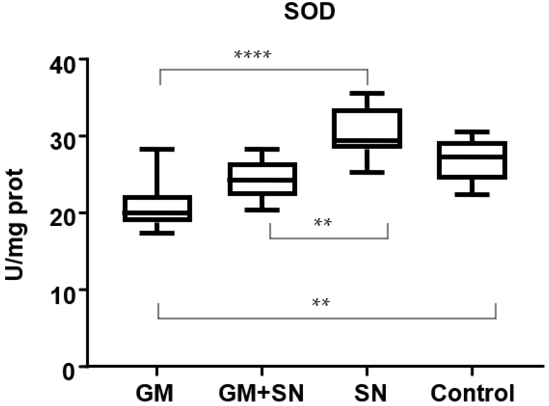
2.5. Analysis of Lipid Peroxidation

For quantifying lipid peroxidation, malondialdehyde (MDA) was assessed using 2-thiobarbituric acid by the fluorimetric method described by Conti, using a synchronous technique with excitation at 534 nm and emission at 548 nm [39]. The MDA values were expressed in nmol/mg protein.

2.6. Analysis of Antioxidant Enzyme Activity

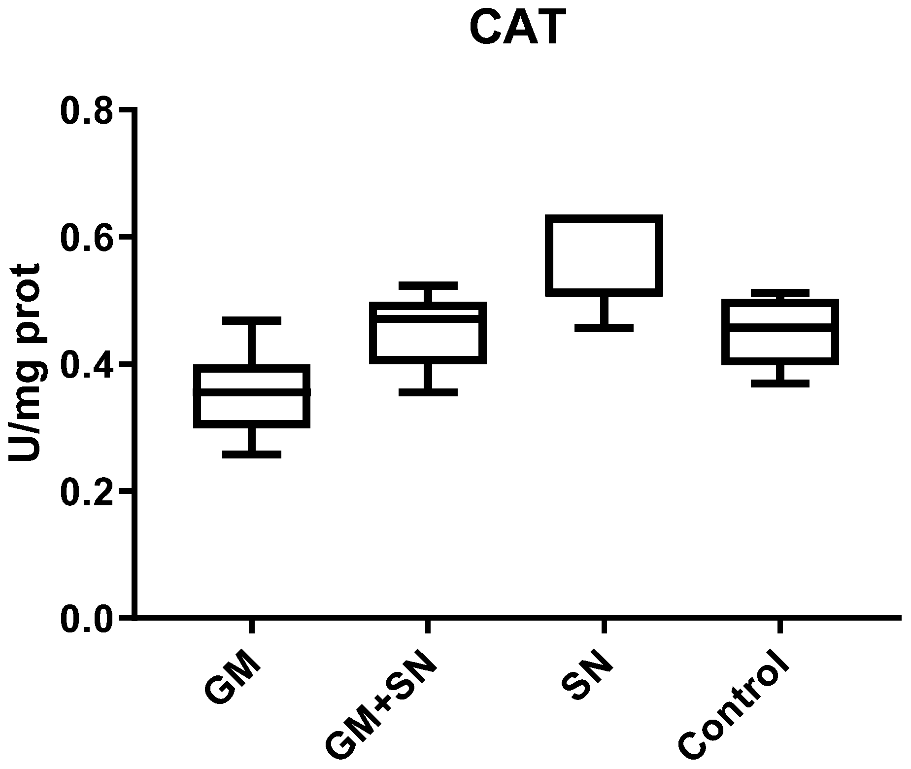
2.6.1. Superoxide Dismutase (SOD)

SOD activity was determined by using the cytochrome c reduction test, described by Flohe and Otting [40], into a mixture containing cytochrome c solution (2 μM in 50 mM phosphate buffer, pH 7.8), xanthine (5 μM), and cyanide (2 mM), in order to inhibit Cu and Zn SOD. Xanthine oxidase (0.2 U mL−1 in 0.1 mM ethylenediaminetetraacetic acid (EDTA)) was added to initiate the reaction. Increased absorbance at 550 nm, recorded for 5 min, indicated cytochrome c reduction. One unit of SOD activity is defined as the amount of enzyme able to inhibit by 50% the rate of cytochrome c reduction in specific conditions. Results were expressed in U/mg protein.

2.6.2. Glutathione Peroxidase (GPX)

We measured GPX activity according to a slightly modified Flohe and Gunzler method by using a reaction mixture consisting of 10 mM glutathione (GSH), 2.4 U mL−1 glutathione reductase, and 1.5 mM NADPH, 1.5 mM H2O2 in 0.1 mM phosphate buffer (pH = 7.0) [41]. The reaction mixture was incubated at 37 °C, and the decrease in absorbance at 340 nm was recorded for 6 min.
The enzyme activity was defined as the amount of GPX that induces a GSH decrease by 10% from the initial concentration in one minute, at 37 °C and pH = 7, and was expressed in U/mg protein.

2.6.3. Catalase (CAT)

According to the Pippenger method, we assayed CAT activity in the kidney based on absorbance modifications for a 10 mM H2O2 solution in 0.05 M potassium phosphate buffer (pH = 7.4) at 240 nm [42]. The enzyme quantity that produced a 0.43 reduction in absorbance per 3 min at 25 °C was defined as 1 unit (U) of CAT activity and was expressed as U/mg protein.

2.7. Statistical Analysis

All data were reported as mean ± SEM. The Gaussian distribution was checked by a D’Agostino and Pearson omnibus normality test. A one-way analysis of variance (ANOVA), followed by Bonferroni’s multiple comparison test procedure, was performed. Statistical significance was set at p < 0.05. Statistical values and graphs were obtained using GraphPad Prism version 5.0 for Windows (GraphPad® Software, San Diego, CA, USA).

3. Results and Discussion

In the human body, ROS and RNS are normally synthesized in small quantities and play numerous roles in cell division and death, angiogenesis and vascular reactivity, adaptation to hypoxia and ischemia, antimicrobial defense, and hormonal synthesis. The best-known ROS are superoxide anion (O2•−), hydrogen peroxide (H2O2), and the hydroxyl radical (HO). RNS are represented by nitric oxide (NO) and peroxy-nitrite (ONOO), the latter being formed by the reaction of NO with O2•− [43].
RS production is modulated by metabolic, hormonal, pro-inflammatory, and environmental factors [44,45,46,47], and, in normal circumstances, is quickly neutralized by enzymatic (mainly SOD, CAT, GPX), and non-enzymatic defense systems (GSH, melatonin, uric acid, bilirubin, vitamins C and E, plant polyphenols) [48].
OS appears when there is a hyper-production of RS or a reduction of antioxidant defense. OS causes lipid peroxidation, deoxyribonucleic acid (DNA) damage, protein denaturation, and damage to the mitochondria and cell membranes.
Due to its high content of long-chain polyunsaturated fatty acids targeted by ROS, the kidney is particularly vulnerable to OS and lipid peroxidation caused by nephrotoxic substances or drugs [5].
Previous studies demonstrated that GM induced renal OS by increasing RS synthesis and by reducing antioxidant defense. In this process, iron molecules take part in the ample and complex cascade of OS response to GM. After GM administration, RS are synthesized in the kidney, and iron is released by RS action and catalyzes further RS synthesis.
GM forms with iron the iron–GM complexes that further increase the synthesis of O2•−, H2O2, and HO, the main ROS that cause OS [49].
On the other hand, redox active iron is able to initiate and perpetuate lipid peroxidation in cells [50]. Further H2O2 and renal lipid oxidation products can enhance ROS generation in mitochondria [51]. Additionally, ROS promotes intracellular iron increase by releasing iron from mitochondria, lysosomes, and ferritin [52].
In kidney tissue, ROS cause damage to the phospholipid membranes, proteins, DNA, mitochondria, and cytochrome c release, resulting in the necrosis or apoptosis of epithelial cells in the proximal renal tubules and vascular and mesangial changes [49,53,54,55,56].
Previous studies have shown that neutralizing HO radicals can reduce OS and GM’s nephrotoxic effects [49].
Several experimental studies assessed the effects of antioxidant compounds, and in 2018 Vargas reported that numerous flavonoids had nephroprotective effects, including against GM-induced nephrotoxicity [9], due to chemical structure and anti-oxidative activity.
The anti-oxidative activity of flavonoids is due to three main mechanisms: (1) the suppression of ROS synthesis by inhibiting synthesis enzymes or by chelating metal ions (iron, copper) involved in free radical generation; (2) the scavenging of ROS and RNS by donating hydrogen atoms to hydroxyl, peroxyl, and peroxynitrite radicals; and (3) the protection or upregulation of antioxidant defense.
Flavonoids protect lipids against oxidative damage by the same mechanisms involved in OS decreasing. Particularly, a 3′4′-catechol structure in the B ring of flavonoids enhances their capacity to inhibit lipid peroxidation. Rutin and epicatechin flavonoids have free-radical scavenging properties and powerfully inhibit lipid peroxidation [57].
Herbal extracts rich in rutin have been shown to have nephroprotective effects and have been intensively studied in recent years. To date, several studies have communicated the nephroprotective effect of rutin-rich extracts obtained from cactus cladodes (Opuntia ficus-indica) [58] and mulberry [59].
To our knowledge, the antioxidant effects of elderflower extract (also rich in rutin) on nephrotoxicity have not been studied to date. Moreover, elderflower extracts demonstrated their superiority to elderberry extracts in antioxidant potential assessed by the 2,2-diphenyl-1-picrylhydrazyl radical (DPPH) or ferric reducing antioxidant power (FRAP) assays, and confirmed the significant relationships obtained between the antioxidant properties and total phenolic and flavonoids [12,60].

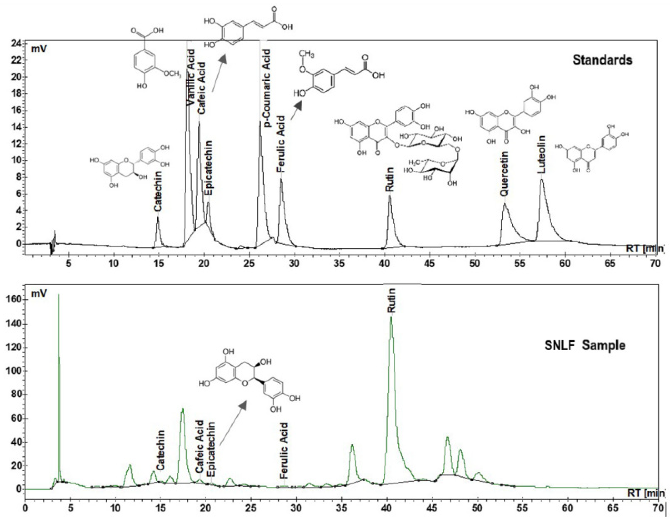
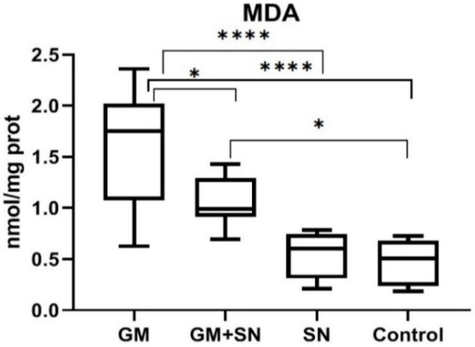
3.1. Quantitative Evaluation of Phenolic Compounds in the Analyzed SN Ethanolic Extract

The main phenolic compounds identified in the analyzed SN ethanolic extract by HPLC-UV were catechin, epicatechin, rutin, CA, and FA (Table 1). Of these compounds, rutin recorded the highest concentration.
Figure 2 presents the HPLC-UV chromatograms of the standards mixture of phenolic compounds and the analyzed SN extract in ethanol.
As with other species, elderflowers may show variations in composition depending on the harvesting year, elderberry cultivar, exposure to pollutants, or electromagnetic fields [61,62,63,64]. Thus, it is particularly important to determine the composition of plant extracts used in each experimental model and to correlate the therapeutic effects with the main compounds found in the studied sample. The analyzed SN ethanolic extract had a concentration of 463.2 µg mL−1 rutin, 9 µg mL−1 epicatechin, 3.9 µg mL−1 catechin, 3.6 µg mL−1 CA, and 1.5 µg mL−1 FA, and other components in low concentrations.
A total of 1 g of elderflower was found in 10 mL of SN extract analyzed by HPLC and 1 mL of extract analyzed by HPLC contained 0.1 g of elderflower. The 1 mL of SN extract analyzed by HPLC contained 463.2 µg rutin, 9 µg epicatechin, 3.9 µg catechin, 3.6 µg CA, and 1.5 µg FA (see Table 1). Each experimental animal (average weight 195 g) received by gavage 1 mL of SN extract for in vivo administration, containing 0.744 g elderflower (a 134.4 mL final volume of SN extract for in vivo administration contained 100 g elderflower), which contained 3.44 mg rutin, 66.96 µg epicatechin, 29.01 µg catechin, 11.16 µg FA, and 26.78 µg CA, safe doses for in vivo administration. By converting the amounts to mg kg−1 bw, the result is that each experimental animal received 17.67 mg kg−1 bw rutin, 343.4 µg kg−1 bw epicatechin, 148.8 µg kg−1 bw catechin, 137.36 µg kg−1 bw CA, and 57.23 µg kg−1 bw FA.
In a previous study that used an elderflower extract with a composition similar to that used in the present study, we demonstrated the lack of nephrotoxicity and the improvement of renal function [10].
In another study, it was proven that cyanogenic glycosides, potentially toxic compounds, were present in low levels (adequate for human consumption) in the elderflowers collected at a foothill [65].
For rutin, the predicted median lethal dose (LD50) was calculated at >5000 mg kg−1 bw, corresponding to a toxicity class of five and indicating the safety of oral rutin administration [66]. The LD50 for CA was calculated at >100 mg kg−1 bw for birds [67], and intraperitoneally CA lower than 1250 mg kg−1 bw was non-lethal in rats [68]. FA has a low toxicity [69] and the LD50 was appreciated at 2445 mg kg−1 bw in male and 2113 mg kg−1 bw in female rats [70]. Epicatechin has an LD50 at 1000 mg kg−1 bw for intraperitoneal administration in mice [71]. In rats, the LD50 for oral administration of catechin was found at 2.428 mol kg−1 bw and oral rat chronic toxicity was 2.500 mg kg−1 bw day−1 [72]. All the LD50 communicated for the phenolic compounds identified in our study were much larger than the amounts to which the experimental animals were exposed.

3.2. Influence of SN Extract Administered In Vivo on Oxidative Stress Parameters

3.2.1. In Vivo Influence of SN Extract on Lipid Peroxidation

In the kidney, the level of lipid peroxidation is used as a marker for the severity of kidney damage. Increased lipid peroxidation and the subsequent formation of advanced lipid peroxidation end products trigger pro-inflammatory pathways and activate the receptor for advanced glycation end products, with the subsequent activation of ROS and cytokine production [51]. MDA is the main marker of lipid peroxidation. Its presence confirms the presence of tissue OS. Previous studies have shown that an increased MDA level in the renal tissue, induced by GM, was associated with morphological and functional impairment [73,74,75,76]. Consistent with these results, in our study GM administration led to a considerable increase in lipid peroxidation.
The MDA level was significantly higher in the GM group compared to the control (C) group (p < 0.0001). In the GM+SN group, the SN extract for in vivo administration prevented GM-induced MDA growth. Thus, the MDA level was 1.6 nmol/mg protein in the GM group, compared to 1.1 nmol/mg protein in the GM+SN group and 0.5 nmol/mg protein in the C group. The reduction of the MDA level was 34.9% in the GM+SN group compared to the GM group, which is statistically significant (p = 0.021). Nevertheless, for the protocol applied in this experimental model, the SN extract for in vivo administration did not completely eliminate the effects of GM. Thus, the MDA level was significantly higher in the GM+SN group than in the C group (p = 0.028) (Figure 3).
In this study, we demonstrated for the first time the effect of SN extract for in vivo administration to significantly reduce lipid peroxidation, induced by experimental exposure to GM, in the renal tissues.
In GM-induced nephropathy, numerous plant extracts and antioxidant compounds have been found to decrease lipid peroxidation and to provide nephroprotection. Some of them have been analyzed by Casanova in a systematic analysis. In this analysis, calcium dobesilate and Zingiber officinale (ginger) extract showed the best nephroprotective profile associated with antioxidant activity. Antioxidant and nephroprotective activity was also proven for melatonin, erdosteine, rosiglitazone, garlic, S-allylcysteine, b-sitosterol, L-carnitine, D-carnitine curcumin, Spirulina platensis, soybean extract, grape seed extract, olive leaf extract, Zingiber officinale extract, Sida rhomboidea leaf extract, Sonchus asper extract, Morchella esculenta mycelium extract, and Nigella sativa oil. Arabic gum, carvedilol, and rosmarinic acid failed in nephroprotection despite their antioxidant activity [6]. Apart from the compounds analyzed by Casanova, the MDA level was also alleviated in GM-induced nephrotoxicity experimental models by Zataria multiflora hydroalcoholic extract [77], Pimpinella anisum L. ethanolic extract [78], Pistacia atlantica leaf hydroethanolic extract [79], pomegranate extract [80], Helichrysum plicatum DC. subsp. plicatum extract [81], Ginkgo biloba extract [82], grape seed extract [83], crude extract and solvent fractions of Euclea divinorum leaves [84], beet root (Beta vulgaris L.) ethanolic extract [85], aqueous extract of root of Boerhavia diffusa [86], Crocus sativus (known as saffron) [87], aqueous garlic extract [88], Riceberry bran extract [89], Tephrosia purpurea (L.) Pers. leaves ethanolic extract [90], hydroalcoholic Malva sylvestris extract [74], Urtica dioica methanolic leaf extract [91], ethyl acetate fraction from Rotula aquatic [92], Origanum vulgare L. extract [93], wild bilberries (Vaccinium myrtillus L.) extract [94], and ginger (Zingiber officinale) and turmeric (Curcuma longa) rhizomes administrated as dietary supplements [95]. The antioxidant activity of elderflower extract was compared to that of elderberry and elder leaves and was found to be highest in flowers [16,59]. Elderflower extract also showed the highest inhibition of DPPH in comparison with rutin, neutralized reactive hydroxyl radicals more effectively than rutin and quercetin, and showed metal-chelative properties stronger than those of rutin and quercetin [96,97].
In these circumstances, we believe that a flavonoid-rich SN extract for in vivo administration, which acts by reducing MDA in renal parenchyma, could be considered a useful nephroprotective agent for combating histological lesions caused by GM.

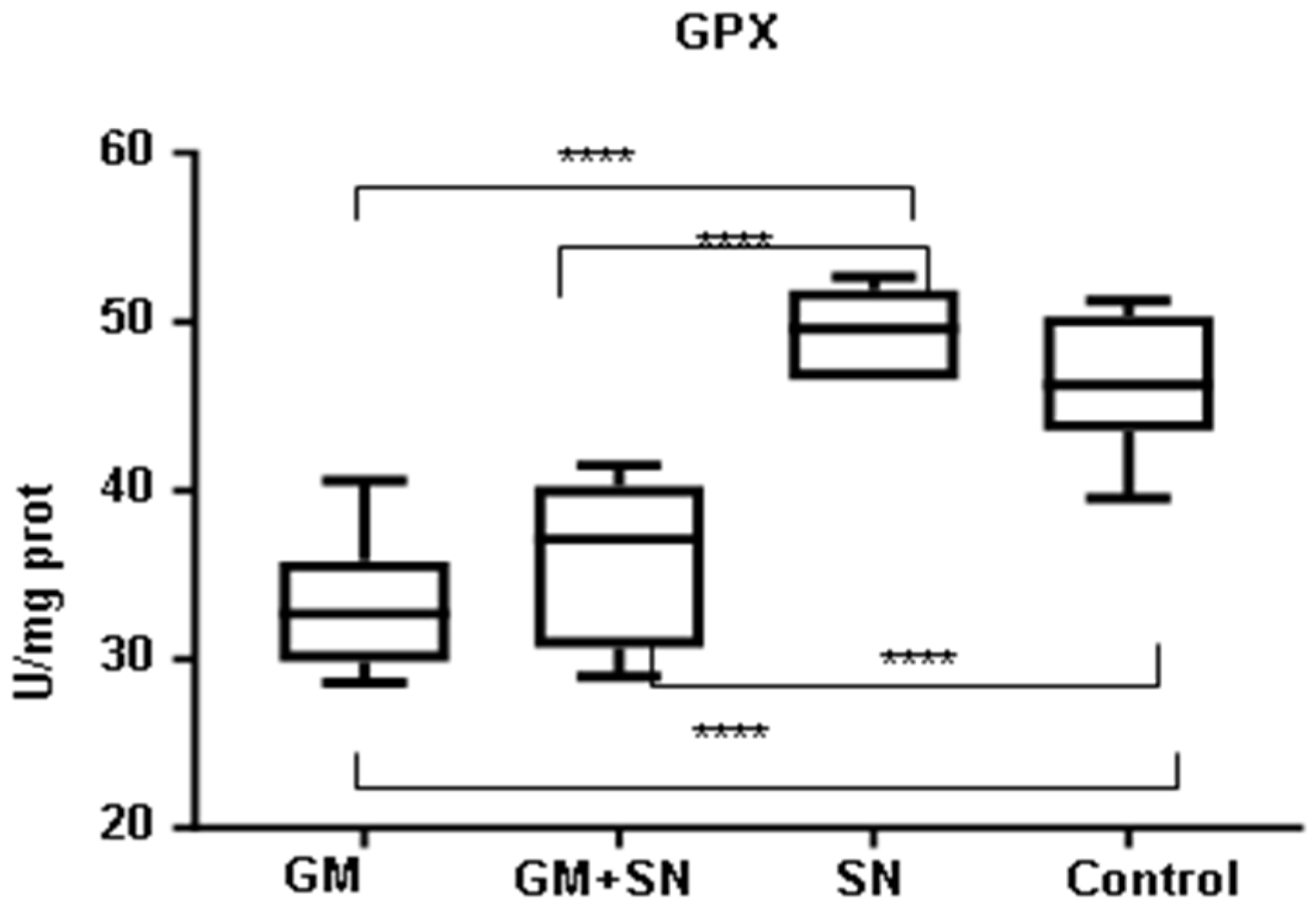
3.2.2. In Vivo Influence of SN Extract on Superoxide Dismutase, Catalase, and Glutathione Peroxidase

GM induces synthesis of an increased amount of RS, both ROS (O2•−, H2O2 and OH) and RNS (NO and ONOO), in the renal parenchyma [98].
In the kidney, RS have different effects on inducible antioxidant enzymes, such as SOD, CAT, and GPX, depending on their amount and action period [99,100,101,102]. Thus, in short-term stimulation, RS can induce the synthesis of antioxidant enzymes, confirmed by the studies published by Medić and Martínez-Salgado that showed increased SOD activity after exposing rats to GM for 7 days and exposing renal mesangial cells to GM for 8 h [103,104], respectively. Dobashi et al. reported an increase in GPX gene expression after the exposure of rat kidney cells (KNRK) to a NO donor [102]. On the other hand, a long exposure (8–10 days) led to exhaustion of the synthesis of these enzymes, as demonstrated by studies in which exposure to GM decreased SOD, CAT, and GPX activity [105,106].
Some studies have shown statistically insignificant changes in SOD and CAT after exposure to GM [103,107,108]. Thus, it is even more difficult to interpret the seemingly contradictory results of the studies in the literature.
Consistent with previously published data reporting reduced SOD and GPX activity as an effect of GM [108], in our study GM administration produced a significant decrease in SOD and GPX activity in the renal tissue in the group with GM compared to the control group.
Our results show that the SOD level decreased significantly (p < 0.01) by 19.3% in the GM group compared to the untreated C group. In the GM+SN treated group, the renal SOD level was reduced by only 9.8% compared to the C group. SN extract for in vivo administration treatment did not significantly increase the renal SOD level after the induction of nephrotoxicity with GM: the renal SOD level increased by 23.2% in the GM+SN group compared to the GM group (p > 0.05). The SOD level was 13.3% higher in the SN group than in the C group (p > 0.05) (Figure 4).
The CAT level recorded no statistically significant difference between groups.
The CAT value was not significantly influenced by the administration of SN extract in the GM+SN group compared to the GM group (p = 0.7367) or in the SN group compared to the C group (p = 0.9889), respectively (Figure 5).
Concerning GPX, its level was significantly reduced (by 29.1%) in the GM group compared to the C group (p < 0.0001). GPX was increased in the SN group compared to the C group, but the increase was not statistically significant (p = 0.5622) (Figure 6). Similar to the SOD activity, the GPX activity increased significantly only when comparing the SN group with the GM+SN group (<0.0001) and with the GM group (p < 0.0001) (Figure 4 and Figure 6).
SOD is the first defense line against O2•−. Its main role is to prevent the transformation of O2•− into ONOO and HO, highly reactive compounds that fragment DNA and membrane lipid structures.
SOD deficiency allows the combination of O2•− with NO, with two major negative consequences. On the one hand, NO loses the function of a signaling molecule and with it the beneficial effects of regulating blood flow in the renal cortex, and on the other hand generates ONOO, an extremely aggressive compound for renal structures.
ONOO disrupts the mitochondrial respiratory chain and induces lipid peroxidation, protein nitrosylation, enzymatic inhibition [109], and the apoptosis of renal cells [110].
In the presence of free ions of iron (Fe+2) and copper (Cu+2), another harmful compound, O2•−, which is not neutralized by SOD, can generate HO•− radicals, fragmenting DNA, proteins, and lipids [111].
SOD activity must be coordinated with that of CAT and GPX, participating in the neutralization of H2O2 generated by SOD action.
CAT intervenes to remove H2O2 by reactions resulting in harmless molecules of water.
GPX is an enzyme with a major role in the antioxidant defense in kidneys, as it is able to participate in the neutralization of both H2O2 and organic peroxides [112]. GPX is considered an enzyme with strong activity in neutralizing fatty acid hydroperoxides [113].
GPX deficiency allows H2O2 to diffuse into tissues and act as an RS. The main mechanism by which H2O2 acts at the renal level is the inhibition of pexophagy, which allows for the structural recovery of peroxisomes and ROS generation limitation at the peroxisomal level [114]. In the renal cortex, H2O2 mediates iron release from the mitochondria by GM [115].
In our study, a less expected result was in the CAT level changes. In the GM exposed group, CAT was lower (but not significantly) than in the control group, and after the administration of SN, CAT did not record a significant increase in the GM+SN group compared to the GM group. These results can be explained by the fact that changes in CAT expression are influenced by the type of analyzed tissue and its basal enzymatical antioxidant capacity [116].
Another unexpected result of our study was that SN failed to restore the enzymatic activity of GPX and SOD. These had only a statistically insignificant increase in the GM+SN group compared to the GM group.
In a previous study, Su reported an increase in GPX activity after rutin administration in mice exposed to intense physical exertion [117]. Moreover, rutin administered in rats at a dose of 150 mg kg−1 for 8 days restored GPX activity, reduced after 80 mg kg−1 bw day−1 GM, simultaneously with the reduction of OS, the improvement of renal function, and the reduction of histological lesions [118]. In our study, GPX recorded only an insignificant increase after the administration of SN. This result can be explained by the fact that the GM dose was higher and the rutin amount in SN was lower than those in the abovementioned studies.
The failure of GPX increase could also be explained by the presence of compounds that interfere with antioxidant enzymes activity. Simos has shown that the flavanols catechin and epicatechin, also present in our elderflower extract, could reduce GPX activity [119].
In a previous GM-induced experimental model of nephrotoxicity in rats, pre-treatment (6 days) and concomitant treatment (8 days) with 150 mg kg−1 of rutin attenuated nephrotoxicity induced by 80 mg kg−1 GM by increasing SOD, CAT, and GPX activity [118].
In our study, the amount of rutin administered to each animal was much smaller (17.67 mg kg−1) and administration was done concomitantly with GM, which allowed some amount of SOD to be consumed in redox reactions. Another explanation could be a differentiated stimulation of SOD isoform synthesis.
The kidney tissue contains three isoforms of SOD. SOD1 and SOD3 are located in the cytosol and the extracellular space, respectively. SOD2 (Mn-SOD) is found in the mitochondria and plays a vital role in keeping O2•− at low levels in the mitochondrial matrix by conversion to H2O2 [120,121].
The SOD1 isoform accounts for up to 80% of the total SOD activity in the mammalian kidney [122]. SOD2 ablation results in a more severe pathological phenotype than SOD1 ablation by worsening OS [51].
In a previous study, Sitarek demonstrated that Leonurus sibiricus L. plant extract, which contains catechin, quercetin, rutin, CA, and FA, increased encoding SOD2 gene expression in Chinese hamster ovary cells exposed to H2O2 [123]. These five compounds were also found in the elderflower extract we studied. Therefore, we believe that the studied SN especially stimulates the synthesis of SOD2, essential for reducing OS at the mitochondrial level and for reducing lipid peroxidation, without generating a statistically significant increase in the level of total SOD activity.

3.3. Non-Enzymatic Antioxidant Effects of Bioflavonoids from Elderflower Extract and Their Main Compound—Rutin

The main compound from the analyzed SN extract in our study was rutin, a bioflavonoid, glycoside of quercetin, with antioxidant and anti-inflammatory effects [118]. The mechanisms by which rutin reduces tissue OS are correlated with its hydrogen donor capacity, which confers on it the function of scavenger for ROS and RNS, and with the chelating property of metals that confers the ability to neutralize iron ions and to form inactive, stabile iron compounds [117].
GM causes an increase in mitochondrial H2O2 synthesis at the renal level in a dose dependent manner [124] and induces a H2O2-mediated iron mobilization from mitochondria [115]. In an environment with H2O2, free iron catalyzes the production of high levels of ROS: O2•−, HO, and OH by the Haber–Weiss reaction, initiated by the Fenton reaction [125].
Normally, most cellular iron is safely stored in organelles—lysosomes, mitochondria—or in ferritin, but in chronic or acute stress, a labile iron pool (defined as a chelatable and redox-active pool of iron, comprising both ionic forms of iron: Fe2+ and Fe3+) increases [126]. Free iron is a key player in the Haber–Weiss reaction, using H2O2 and O2·− to produce more toxic HO radicals and catalyzing lipid peroxidation and the oxidative damage of proteins that are strongly associated with kidney disease and its severity [51,127].
In fact, iron, similar to ROS, functions as an inducible stress intracellular messenger that serves to regulate cell death responses. Iron chelators can act as powerful antioxidants [128]; Eid et al. communicated in 2017 that intracellular iron chelators were found to function as ROS scavengers [52].
It was proven that elderflower extract has metal-chelative properties stronger than those of rutin and quercetin standards, their main effect being to prevent the initiation of HO radicals synthesis [100,129].
If we accept that elderflower extract protects the kidney against gentamicin-induced OS by chelating iron ions, and if we take into account that the chelating properties of the whole elderflower extract are superior to those of rutin and quercetin standards, we can consider that elder flower extract with its full polyphenol content is preferable for controlling renal OS to the detriment of standardized extracts that use only one type of polyphenols.
Flavonoids can act as antioxidants per se, independently from constitutively enzymatic or non-enzymatic antioxidant cell systems, and could have an additive effect to the endogenous scavenging compounds [130].
In a previous study, Robak demonstrated that many flavonoids inhibited lipid peroxidation, mainly via the scavenging of O2•− anions, whereas other non-flavonoid antioxidant compounds act on free radical reactions via HO radicals scavenging [131].
Rutin inhibits lipid peroxidation, working as an ROS scavenger by donating hydrogen atoms to all RS involved in the peroxidation of lipids: O2•− anions, singlet oxygen, HO radicals, and peroxy radicals. Peroxyl radicals scavenging by rutin is essential for preventing lipid peroxidation by interrupting the propagation of free radical chain reactions [132]. Rutin also works as fast-acting antioxidant by its ability to intervene as a terminator for lipid peroxidation by chelating metal ions [29].
In GM administration, rutin most likely intervenes from the initial stages of OS generation by blocking the iron ions released from mitochondria and preventing the formation of pro-oxidant iron–GM complexes. Consequently, ROS (O2•−, H2O2, and OH) generating cascade reactions are considerably limited. Additionally, by its ROS scavenger function, rutin neutralizes H2O2 released by GM from mitochondria, causing a blockade of H2O2-dependent iron release and a further reduction in the OS, lipid peroxidation, and MDA synthesis induced by redox-active iron.
In our study, rutin’s action of neutralizing ROS, chelating iron ions, and blocking GM–iron complexes formation most likely represents the main mechanism that explains the reduction in MDA levels, without antioxidant enzymes increase, in the group of rats that received GM and SN extract for in vivo administration with a high rutin content.
We also believe that co-occurring flavonoids work synergistically with rutin to antagonize ROS and to inhibit lipid peroxidation.
Together, rutin, epicatechin, and catechin have been proved to have a HO scavenging capacity 100–300 times higher than that of mannitol, a typical HO scavenger. Moreover, all flavonoids, except epicatechin, had an inhibitory effect on xanthine oxidase involved in the synthesis of O2•− anions [133].
Another property of rutin involved in lipid peroxidation decrease is its ability to inhibit NO production. The involvement of NO in the initiation of lipid peroxidation was reported for all situations when it acts in the presence of O2•− anions to form peroxynitrite, a powerful oxidant able to initiate lipid peroxidation. To inhibit NO production, rutin acts together with CA, another strong inhibitor of NO synthesis [134].

3.4. Role of Caffeic Acid and Ferulic Acid

A positive correlation between CA derivatives and FA derivatives and antioxidant activity was demonstrated in many studies [135,136].
CA is a compound that belongs to the HCAs class and can chelate ferrous ions (Fe2+) [137]. Fe2+ accelerates lipid oxidation by breaking down H2O2 and lipid peroxides to reactive free radicals via the Fenton reaction [138].
Due to the chelating properties of Fe2+, CA acts as a compound with an inhibitory effect on lipid peroxidation and MDA synthesis.
Another recognized property of CA involved in lipid peroxidation reduction is the ability to inhibit NO synthesis [134].
FA present in SN extract for in vivo administration is a phenolic compound that possesses a free radical scavenging capacity. The carboxylic acid group acts as an anchor for the lipid bilayer and confers FA properties against lipid peroxidation. Because FA scavenges O2•− and inhibits lipid peroxidation induced by O2•−, FA activity is considered similar to that of SOD [139].
Tannic acid is another natural polyphenol with antioxidant, antimicrobial, and anti-inflammatory properties, but is also able to influence the structure and conformation of fibrinogen to form fibrin. In comparison with tannic acid, which could potentially be dangerous for renal fibrosis development and the poor evolution of kidney diseases [140,141,142], in elderflower extract, antifibrotic and nephroprotective effects against gentamicin-induced nephrotoxicity have been proven [10].

3.5. New Research Direction for Elderflowers Extract

Our study is far from completely presenting and explaining all the therapeutic effects of elderflower extract.

3.5.1. Action Mechanism of Active Compounds

In the future, the antioxidant mechanisms for rutin, epicatechin, catechin, FA, and CA, administered alone or in combination, must be evaluated in experimental models of nephrotoxicity to gentamicin. The ability of phenolic compounds to scavenge free radicals must be evaluated using the 2,2′-azino-bis(3-ethylbenzothiazoline-6-sulfonic acid) (ABTS•+) radical cation-based test (for cation radicals) or 2,2-diphenyl-1-picrylhydrazyl (DPPH) radical-based test (for stable radicals). For the antioxidant activity assay, a wide variety of methods may be used, including chemical-based methods such as the cupric ions reducing power assay or FRAP and biological assays, such as cellular antioxidant activity. The peroxyl radical, superoxide radical anion, hydrogen peroxide, hydroxyl radical scavenging assay, and singlet oxygen quenching assay must by performed for specific antioxidant activity [143]. The near hydrogen atom transfer mechanism, sequential proton loss–electron transfer, and single electron transfer–proton transfer must be also studied with density functional theory methods [144]. For finding a relationship between structural features of compounds and their activities, the development of a quantitative structure–activity relationship model should be considered [145].
Another question logically arising is whether active compounds act synergically. In a previous study, it was demonstrated that rutin pretreatment could attenuate GM-induced nephrotoxicity by increasing GSH levels and SOD, CAT, and GPx activity, as well as by reducing MDA level [118]. In an experimental model, catechin prevented a GM-induced decrease in renal GSH, concomitantly improving renal function [146]. Epicatechin was not studied in GM-induced nephrotoxicity, but it was found to alleviate mitochondrial structural changes and ROS level caused by cisplatin in the renal cortex of mice [147], alterations observed also in GM-induced nephrotoxicity [148]. Beneficial effects in kidney injury caused by GM have been also proven for HCA. Pretreatment with caffeic acid phenethyl ester was effective in preventing the rise of MDA level induced by GM [149], and FA reduced MDA level and OS by increasing SOD activity in GM-induced nephrotoxicity [150]. In another study, phenolic natural compounds presented better antioxidant capacities when they were tested in a mixture compared to the antioxidant activities displayed individually by each of them in a GM nephrotoxicity experimental model, suggesting synergistic antioxidant effects [151]. Another study showed that the extract of Sambucus nigra L. elderflowers had metal-chelative properties stronger than those of isolated rutin and quercetin standards, preventing the initiation of the synthesis of hydroxyl radicals. A similar superiority of Sambucus nigra L. elderflower extract compared to rutin and quercetin was found for radical scavenging and the elimination of hydroxyl radicals [96]. In our study, all phenolic antioxidant compounds were administered at lower amounts compared to those in the previous studies, leading us to consider that the synergistic effect of these compounds merits study in the future.
Other research directions that deserve to be developed refer to the effects of elderflower extract on NO and inducible nitric oxide synthase (iNOS), caspase-3, and light chain 3B (LC3B), because previous studies demonstrated that rutin pre-treatment attenuated nephrotoxicity induced by OS after GM administration by inhibiting iNOS, cleaving caspase-3, and LC3B [118].

3.5.2. Interference between SN Extract and Antimicrobial Effect of GM

Even if mechanisms for antimicrobial effects of GM are not fully elucidated, some authors have suggested that oxidation reduction reactions are involved in bacterial death [152]. In this context, a logical question to be raised is whether the combination of GM with antioxidants is beneficial for the antibacterial effect.
Contradictory results are communicated for the association between GM and antioxidants. The simultaneous administration of GM and luteolin enhanced the antibacterial activity of GM against S. aureus and E. coli. [153]. Quercetin, another antioxidant, did not substantially modify the antibacterial activity of gentamicin against E. coli and contributed to the enhancement of GM activity against S. aureus. In a previous study, Miroshnichenko demonstrated that other antioxidants, ascorbic acid, methylethylpyridinol, and N-acetylcysteine, reduced the activity of GM in vitro and in vivo without decreasing nephrotoxic effects [154]. On the other hand, antioxidant and antimicrobial activities have been demonstrated for many natural compounds from plants. Many of these compounds are phenols found in medicinal plants (Curcuma longa L., Kalanchoe delagoensis L., Asparagus aethiopicus L., Senna alexandrina L., Citrullus colocynthis L., Gasteria pillansii L., Brassica juncea and Cymbopogon citratus, L. glaucescens, Convolvulus austro-aegyptiacus, and Convolvulus pilosellifolius) and in marine plants (Ecklonia cava) [155]. For many culinary herbs rich in flavonoids, concomitant antimicrobial and antioxidant properties, useful in food preservation, have been proven [156]. Krawitz demonstrated that standardized elderberry extract possesses antimicrobial activity against both Gram-negative bacteria (Branhamella catarrhalis) and Gram-positive bacteria (Streptococcus pyogenes and group C and G Streptococci) [157].
In a previous study, it was proven that elderflower extract had an inhibitory activity against a wide range of nosocomial pathogens, namely Gram-positive (Staphylococcus sp., B. cereus) and Gram-negative (Salmonella Poona, P. aeruginosa) pathogens, and the highest inhibitory activity towards methicillin-resistant Staphylococcus aureus (MRSA) [158]. To our knowledge, no studies have been conducted to date on GM and elderflower extract association. Because the inhibitory or additive/stimulative effects of the association between GM and elderflower extract are dependent on the concentration of the partners, the bacteria and host organism, this aspect must be studied in the future.

3.5.3. Pharmacological Characteristics of SN Extract

The EC50 is the concentration of a drug that gives a half-maximal response and is a fundamental concept in pharmacology. Because SN extract’s composition may differ due to environmental factors, it is important to find the EC50 for the whole extract and for each active antioxidant compound, and to verify if the EC50 is reached in all normal environmental conditions (temperature, altitude, rainfall, sunlight). These findings will contribute to the optimal use of elderflower in the food and pharmacological industry.
In order to improve efficiency, curves for time-dependent activity of the whole SN extract should also be calculated in the future.
At the same time, it is worth studying Sambucus nigra effects in other conditions where OS is an important pathogenetic mechanism, e.g., in metabolic, cardiovascular, and neurologic diseases.

3.5.4. Efficiency of SN Extract in Other Nephrotoxicity Models

The nephroprotective effects of elderberry should also be studied as they relate to the use of other nephrotoxic compounds, such as alcohol, non-steroidal anti-inflammatory drugs (NSAIDS), chemotherapeutics, immunosuppressants, and contrast agents [159,160,161].
While effective alternatives have been proposed for the use of NSAIDS, unfortunately this is not the case for chemotherapeutics, immunosuppressants, and contrast agents [162,163,164].

3.5.5. Antiviral Effect in SARS-CoV-2 Infection

In the context of the SARS-CoV-2 pandemic, elderberry fruit extract has been discussed as a potential antiviral drug [165], but also as a possible enhancer of the inflammatory reaction [166]. In a previous study, we demonstrated the renal antifibrotic effect of elder flower extract (Sambucus nigra L. species) [10], with a higher bioflavonoid content than elderberry fruits. On the other hand, OS, which can be reduced by elderflower extract, are one of the main mechanisms involved in pulmonary fibrosis secondary to SARS-CoV-2 infection. Considering these aspects, it is worth studying the type of elder plant extract and the optimal time of administration in SARS-CoV-2 infection.

3.6. Elderflowers—Promising Medical and Nutritional Intervention in Kidney Diseases

For a long time, elderflowers and berries have been widely used in nutrition and traditional therapy, and more recently in the food industry. As natural flavoring components, they can be found in alcoholic and nonalcoholic beverages, sparkling, bitter, and white wine, fruit brandies, and various spirits, as well as in tea and products such as yoghurt or ice cream [129].
Elderflowers can be used both as standardized extracts and as functional foods. Moreover, Sambucus nigra L. flowers contain even higher amounts of phenolic compounds than elderberries and leaves [16], and thus usually also have a higher antioxidant activity. As both characteristics are maintained at thermal processing, elderflowers can be widely used in food and pharmaceutical industries [129].
Our study validated the hypothesis that SN extract rich in polyphenolic compounds may provide nephroprotection in GM-induced nephrotoxicity, due to the antioxidant action. This characteristic recommends it to be studied further, in order to use it for medical purposes and in the food industry.

3.7. Limits of the Study

Our study demonstrated only the ability of elderberry flower extract to inhibit GM-induced lipid peroxidation, without demonstrating its action mechanism. Flavonoid-rich content was proved for the elderflower extract, but the action mechanism was only taken from other studies and not demonstrated for our experimental model. Another limit of this study was the multifactorial environmentally induced variability of elderflowers composition, which means that the results of the present study can only be extrapolated to similar extracts.

4. Conclusions

In our experimental model, elderflower extract rich in rutin, CA, and FA significantly reduced GM-induced lipid peroxidation, quantified by MDA levels, in the renal tissue.
Because we did not observe a statistically significant increase in the levels of the antioxidant enzymes SOD, CAT, and GPX, we are entitled to believe that the antioxidant effect of elderflower extract was due to the reduced production of RS and the direct inhibition of lipid peroxidation by rutin, epicatechin, FA, and CA, found in significant quantities in the SN extract. Rutin and CA, which have the property of chelating iron ions and reducing labile, redox-active pools of iron, were probably the main factors responsible for the decrease of RS synthesis after the iron release from mitochondria, lysosomes, and protein complexes caused by renal exposure to GM.
In this context, we believe that elderflower extract could be considered a useful nephroprotective agent against lipoperoxidation caused by GM.

Author Contributions

Conceptualization, R.A.U., R.A.C., D.B.J. and G.S.M.; methodology, I.M.B., R.A.C., V.M.C., B.A.N., M.F., E.C.C., S.C., V.E.S., O.B.G. and G.S.M.; software, R.A.C. and S.M.; validation, I.M.B., V.M.C., M.F., S.C. and A.L.P.; formal analysis, R.A.C., V.M.C., B.A.N., O.S., E.C.C., S.C. and M.C.; investigation, R.A.C., S.M., O.S., M.F., E.C.C., V.E.S. and G.S.M.; resources, O.S., M.F., D.B.J. and M.C.; data curation, I.M.B., R.A.C., V.M.C., B.A.N., S.M., M.F., A.L.P. and V.E.S.; writing—original draft preparation, R.A.U., I.M.B. and M.F.; writing—review and editing, R.A.U., I.M.B. and B.A.N.; visualization, B.A.N., L.I., S.C., A.L.P. and G.S.M.; supervision, R.A.U., D.B.J. and M.C.; project administration, R.A.U. and L.I.; funding acquisition, R.A.U., O.B.G. and G.S.M. V.M.C., B.A.N., M.F., L.I., S.C., V.E.S. and O.B.G. had contributions equal to that of the first author. All authors have read and agreed to the published version of the manuscript.

Funding

This research received no external funding.

Institutional Review Board Statement

The study was conducted according to the guidelines of the Declaration of Helsinki, and was approved by the https://www.mdpi.com/ethics Ethics Committee of the State Veterinary Authority (No. 70/30.05.2017).

Informed Consent Statement

Not applicable.

Data Availability Statement

The data presented in this study are openly available in FigShare at https://doi.org/10.6084/m9.figshare.18515198.

Acknowledgments

The publication was supported by funds from the National Research Development Projects to finance excellence (PFE)-14/2022-2024 granted by the Romanian Ministry of Research and Innovation.

Conflicts of Interest

The authors declare no conflict of interest.

Abbreviations

ANOVAOne-way analysis of variance
CATCatalase
CACaffeic acid
DPPH2,2-Diphenyl-1-picrylhydrazyl radical
EDTAEthylenediaminetetraacetic acid
EC50Half maximal effective concentration
FAFerulic acid
FRAPFerric reducing antioxidant power
GMGentamicin
GPXGlutathione peroxidase
GSHGlutathione
HCAsHydroxycinnamic acids
HPLCHigh performance liquid chromatography
iNOSInducible nitric oxide synthase
MDA Malondialdehyde
NADPH Nicotinamide adenine dinucleotide phosphate
NIHCEUK National Institute of Health and Care Excellence
OSOxidative stress
PBS Phosphate-buffered saline solution
RNSReactive nitrogen species
ROSReactive oxygen species
RSReactive species
SEMStandard error of the mean
SN Sambucus nigra L. elderflowers
SODSuperoxide dismutase

References

  1. McWilliam, S.J.; Antoine, D.J.; Smyth, R.L.; Pirmohamed, M. Aminoglycoside-induced nephrotoxicity in children. Pediatr. Nephrol. 2016, 32, 2015–2025. [Google Scholar] [CrossRef]
  2. Cebrero-Cangueiro, T.; Marín, R.; Labrador-Herrera, G.; Smani, Y.; Cordero, E.; Pachón, J.; Pachón-Ibáñez, M.E. In vitro Activity of Pentamidine Alone and in Combination With Aminoglycosides, Tigecycline, Rifampicin, and Doripenem against Clinical Strains of Carbapenemase-Producing and/or Colistin-Resistant Enterobacteriaceae. Front. Cell. Infect. Microbiol. 2018, 8, 363. [Google Scholar] [CrossRef] [PubMed] [Green Version]
  3. Silverblatt, F.J.; Kuehn, C. Autoradiography of gentamicin uptake by the rat proximal tubule cell. Kidney Int. 1979, 15, 335–345. [Google Scholar] [CrossRef] [Green Version]
  4. Sandoval, R.; Leiser, J.; Molitoris, B.A. Aminoglycoside antibiotics traffic to the Golgi complex in LLC-PK1 cells. J. Am. Soc. Nephrol. 1998, 9, 167–174. [Google Scholar] [CrossRef]
  5. Ozbek, E. Induction of Oxidative Stress in Kidney. Int. J. Nephrol. 2012, 2012, 465897. [Google Scholar] [CrossRef] [Green Version]
  6. Casanova, A.G.; Vicente-Vicente, L.; Hernández-Sánchez, M.T.; Pescador, M.; Prieto, M.; Martínez-Salgado, C.; Morales, A.I.; López-Hernández, F.J. Key role of oxidative stress in animal models of aminoglycoside nephrotoxicity revealed by a systematic analysis of the antioxidant-to-nephroprotective correlation. Toxicology 2017, 385, 10–17. [Google Scholar] [CrossRef] [PubMed]
  7. Elgebaly, H.A.; Mosa, N.M.; Germoush, M.O.; Brahim, A.C. The Nephro—Protective Effects of Olive Oil and Bee Honey against Gentamicin—Induced Nephrotoxicity in Rabbits. Aljouf Univ. Med. J. 2016, 300, 1–7. [Google Scholar] [CrossRef]
  8. Codea, R.; Mircean, M.; Nagy, A.; Sarpataky, O.; Sevastre, B.; Stan, R.L.; Hangan, A.C.; Popovici, C.; Neagu, D.; Purdoiu, R.C.; et al. Melatonine and erythropoietin prevents gentamicin induced nephrotoxicity in rats. Farmacia 2019, 67, 392–397. [Google Scholar] [CrossRef]
  9. Vargas, F.; Romecín, P.; Guillen, A.I.G.; Wangesteen, R.; Vargas-Tendero, P.; Paredes, M.D.; Atucha, N.M.; García-Estañ, J. Flavonoids in Kidney Health and Disease. Front. Physiol. 2018, 9, 394. [Google Scholar] [CrossRef] [Green Version]
  10. Ungur, R.; Buzatu, R.; Lacatus, R.; Purdoiu, R.C.; Petrut, G.; Codea, R.; Sarpataky, O.; Biris, A.; Popovici, C.; Mircean, M.; et al. Evaluation of the Nephroprotective Effect of Sambucus nigra Total Extract in a Rat Experimental Model of Gentamicine Nephrotoxicity. Rev. Chim. 2019, 70, 1971–1974. [Google Scholar] [CrossRef]
  11. Mahboubi, M. Sambucus nigra (black elder) as alternative treatment for cold and flu. Adv. Tradit. Med. 2020, 21, 405–414. [Google Scholar] [CrossRef]
  12. Viapiana, A.; Wesolowski, M. The Phenolic Contents and Antioxidant Activities of Infusions of Sambucus nigra L. Mater. Veg. 2017, 72, 82–87. [Google Scholar] [CrossRef] [PubMed] [Green Version]
  13. Mikulic-Petkovsek, M.; Samoticha, J.; Eler, K.; Stampar, F.; Veberic, R. Traditional Elderflower Beverages: A Rich Source of Phenolic Compounds with High Antioxidant Activity. J. Agric. Food Chem. 2015, 63, 1477–1487. [Google Scholar] [CrossRef] [PubMed]
  14. Gong, B.; Gou, X.; Han, T.; Qi, Y.; Ji, X.; Bai, J. Protective effects of rutin on kidney in type 1 diabetic mice. Pak. J. Pharm. Sci. 2020, 33, 597–603. [Google Scholar]
  15. Khajevand-Khazaei, M.-R.; Mohseni-Moghaddam, P.; Hosseini, M.; Gholami, L.; Baluchnejadmojarad, T.; Roghani, M. Rutin, a quercetin glycoside, alleviates acute endotoxemic kidney injury in C57BL/6 mice via suppression of inflammation and up-regulation of antioxidants and SIRT1. Eur. J. Pharmacol. 2018, 833, 307–313. [Google Scholar] [CrossRef] [PubMed]
  16. Dawidowicz, A.L.; Wianowska, D.; Baraniak, B. The antioxidant properties of alcoholic extracts from Sambucus nigra L. (antioxidant properties of extracts). LWT Food Sci. Technol. 2006, 39, 308–315. [Google Scholar] [CrossRef]
  17. Bhattacharya, S.; Christensen, K.B.; Olsen, L.C.B.; Christensen, L.P.; Grevsen, K.; Færgeman, N.J.; Kristiansen, K.; Young, J.F.; Oksbjerg, N. Bioactive Components from Flowers of Sambucus nigra L. Increase Glucose Uptake in Primary Porcine Myotube Cultures and Reduce Fat Accumulation in Caenorhabditis elegans. J. Agric. Food Chem. 2013, 61, 11033–11040. [Google Scholar] [CrossRef]
  18. Salem, A.M.; Ragheb, A.S.; Hegazy, M.G.A.; Matboli, M.; Eissa, S. Caffeic Acid Modulates miR-636 Expression in Diabetic Nephropathy Rats. Indian J. Clin. Biochem. 2018, 34, 296–303. [Google Scholar] [CrossRef] [PubMed]
  19. Sanjeev, S.; Bidanchi, R.M.; Murthy, M.K.; Gurusubramanian, G.; Roy, V. Influence of ferulic acid consumption in ameliorating the cadmium-induced liver and renal oxidative damage in rats. Environ. Sci. Pollut. Res. 2019, 26, 20631–20653. [Google Scholar] [CrossRef]
  20. Mikulic-Petkovsek, M.; Ivancic, A.; Schmitzer, V.; Veberic, R.; Stampar, F. Comparison of major taste compounds and antioxidative properties of fruits and flowers of different Sambucus species and interspecific hybrids. Food Chem. 2016, 200, 134–140. [Google Scholar] [CrossRef]
  21. Filip, M.; Silaghi-Dumitrescu, L.; Prodan, D.; Sarosi, C.; Moldovan, M.; Cojocaru, I. Analytical Approaches for Characterization of Teeth Whitening Gels Based on Natural Extracts. Key Eng. Mater. 2017, 752, 24–28. [Google Scholar] [CrossRef]
  22. Huang, H.-S.; Yu, H.-S.; Yen, C.-H.; Liaw, E.-T. HPLC-DAD-ESI-MS Analysis for Simultaneous Quantitation of Phenolics in Taiwan Elderberry and Its Anti-Glycation Activity. Molecules 2019, 24, 3861. [Google Scholar] [CrossRef] [Green Version]
  23. Mota, A.H.; Andrade, J.M.; Rodrigues, M.J.; Custódio, L.; Bronze, M.R.; Duarte, N.; Baby, A.; Rocha, J.; Gaspar, M.M.; Simões, S.; et al. Synchronous insight of in vitro and in vivo biological activities of Sambucus nigra L. extracts for industrial uses. Ind. Crop. Prod. 2020, 154, 112709. [Google Scholar] [CrossRef]
  24. Koleva, P.; Tsanova-Savova, S.; Paneva, S.; Velikov, S.; Savova, Z. Polyphenols content of selected medical plants and food supplements present at Bulgarian market. Pharmacia 2021, 68, 819–826. [Google Scholar] [CrossRef]
  25. Kim, T.; Shin, H.; Park, S.; Kim, H.; Chung, D. Development and Validation of a Method for Determining the Quercetin-3-O-glucuronide and Ellagic Acid Content of Common Evening Primrose (Oenothera biennis) by HPLC-UVD. Molecules 2021, 26, 267. [Google Scholar] [CrossRef] [PubMed]
  26. Saad, W.M.M.; Ridwan, R.; Lasim, N.S.M.; Rapi, N.L.M.; Salim, F. Determination and quantification of p-coumaric acid in pineapples (Ananas comosus) extracts using gradient mode RP-HPLC. Pharmacogn. Res. 2019, 11, 67. [Google Scholar] [CrossRef]
  27. Sá, R.R.; Matos, R.A.; Silva, V.C.; Caldas, J.D.C.; Sauthier, M.C.D.S.; dos Santos, W.N.L.; Magalhães, H.I.F.; Junior, A.D.F.S. Determination of bioactive phenolics in herbal medicines containing Cynara scolymus, Maytenus ilicifolia Mart ex Reiss and Ptychopetalum uncinatum by HPLC-DAD. Microchem. J. 2017, 135, 10–15. [Google Scholar] [CrossRef]
  28. Moura, H.F.S.; Dias, F.D.S.; e Souza, L.B.S.; de Magalhães, B.E.A.; Tannus, C.D.A.; de Carvalho, W.C.; Brandão, G.C.; dos Santos, W.N.L.; Korn, M.G.A.; dos Santos, D.C.M.B.; et al. Evaluation of multielement/proximate composition and bioactive phenolics contents of unconventional edible plants from Brazil using multivariate analysis techniques. Food Chem. 2021, 363, 129995. [Google Scholar] [CrossRef]
  29. Han, Y.; Lu, J.S.; Xu, Y.; Zhang, L.; Hong, B.F. Rutin ameliorates renal fibrosis and proteinuria in 5/6-nephrectomized rats by anti-oxidation and inhibiting activation of TGFβ1-smad signaling. Int. J. Clin. Exp. Pathol. 2015, 8, 4725–4734. [Google Scholar]
  30. Yagmurca, M.; Yasar, Z.; Bas, O. Effects of quercetin on kidney injury induced by doxorubicin. Bratisl Lek List. 2015, 116, 486–489. [Google Scholar] [CrossRef] [Green Version]
  31. Hsieh, C.-L.; Peng, C.-C.; Chen, K.-C.; Peng, R.Y. Rutin (Quercetin Rutinoside) Induced Protein-Energy Malnutrition in Chronic Kidney Disease, but Quercetin Acted Beneficially. J. Agric. Food Chem. 2013, 61, 7258–7267. [Google Scholar] [CrossRef] [PubMed]
  32. Peng, C.-C.; Hsieh, C.-L.; Ker, Y.-B.; Wang, H.-Y.; Chen, K.-C.; Peng, R.Y. Selected nutraceutic screening by therapeutic effects on doxorubicin-induced chronic kidney disease. Mol. Nutr. Food Res. 2012, 56, 1541–1558. [Google Scholar] [CrossRef]
  33. Al-Rejaie, S.S.; Abuohashish, H.M.; Alkhamees, O.A.; Aleisa, A.M.; Alroujayee, A.S. Gender difference following high cholesterol diet induced renal injury and the protective role of rutin and ascorbic acid combination in Wistar albino rats. Lipids Heal. Dis. 2012, 11, 41. [Google Scholar] [CrossRef] [Green Version]
  34. Bledsoe, G.; Shen, B.; Yao, Y.-Y.; Hagiwara, M.; Mizell, B.; Teuton, M.; Grass, D.; Chao, L.; Chao, J. Role of Tissue Kallikrein in Prevention and Recovery of Gentamicin-Induced Renal Injury. Toxicol. Sci. 2008, 102, 433–443. [Google Scholar] [CrossRef] [PubMed] [Green Version]
  35. Thibault, N.; Grenier, L.; Simard, M.; Bergeron, M.G.; Beauchamp, D. Attenuation by daptomycin of gentamicin-induced experimental nephrotoxicity. Antimicrob. Agents Chemother. 1994, 38, 1027–1035. [Google Scholar] [CrossRef] [Green Version]
  36. Medscape. Drugs & Diseases—Gentamicin. Available online: https://reference.medscape.com/drug/gentak-garamycin-gentamicin-342517 (accessed on 27 December 2021).
  37. Prescribers’ Digital Reference—Gentamicin Sulfate—Drug Summary. Available online: https://www.pdr.net/drug-summary/Gentamicin-Injection-10-mg-mL-gentamicin-sulfate-3300 (accessed on 27 December 2021).
  38. Bradford, M.M. A rapid and sensitive method for the quantitation of microgram quantities of protein utilizing the principle of protein-dye binding. Anal. Biochem. 1976, 72, 248–254. [Google Scholar] [CrossRef]
  39. Conti, M.; Morand, P.C.; Levillain, P.; Lemonnier, A. Improved fluorometric determination of malonaldehyde. Clin. Chem. 1991, 37, 1273–1275. [Google Scholar] [CrossRef]
  40. Flohé, L. Superoxide dismutase assays. Methods Enzym. 1984, 105, 93–104. [Google Scholar] [CrossRef]
  41. Titheradge, M.A. The Enzymatic Measurement of Nitrate and Nitrite. Methods Mol. Biol. 1998, 100, 83–91. [Google Scholar] [CrossRef]
  42. Pippenger, C.E.; Browne, R.W.; Armstrong, D. Regulatory Antioxidant Enzymes. Methods Mol. Biol. 1998, 108, 299–314. [Google Scholar] [CrossRef] [PubMed]
  43. Pizzino, G.; Irrera, N.; Cucinotta, M.; Pallio, G.; Mannino, F.; Arcoraci, V.; Squadrito, F.; Altavilla, D.; Bitto, A. Oxidative Stress: Harms and Benefits for Human Health. Oxidative Med. Cell. Longev. 2017, 2017, 8416763. [Google Scholar] [CrossRef]
  44. Chainy, G.B.N.; Sahoo, D.K. Hormones and oxidative stress: An overview. Free Radic. Res. 2020, 54, 1–26. [Google Scholar] [CrossRef] [PubMed]
  45. Tan, B.L.; Norhaizan, M.E.; Liew, W.-P.-P. Nutrients and Oxidative Stress: Friend or Foe? Oxidative Med. Cell. Longev. 2018, 2018, 9719584. [Google Scholar] [CrossRef] [PubMed]
  46. Gangwar, R.S.; Bevan, G.H.; Palanivel, R.; Das, L.; Rajagopalan, S. Oxidative stress pathways of air pollution mediated toxicity: Recent insights. Redox Biol. 2020, 34, 101545. [Google Scholar] [CrossRef] [PubMed]
  47. Ungur, R.; Dronca, M.; Crăciun, E.C.; Rusu, R.L.; Văleanu, M.; Onac, I.; Borda, I.M.; Laszlo, I. Improvement of total antioxidant status, a possible bioeffect of the ultrasound therapy—A pilot study. Rev. Romana Med. Lab. 2011, 19, 177–183. [Google Scholar]
  48. Mironczuk-Chodakowska, I.; Witkowska, A.; Zujko, M.E. Endogenous non-enzymatic antioxidants in the human body. Adv. Med. Sci. 2018, 63, 68–78. [Google Scholar] [CrossRef]
  49. Walker, P.D.; Shah, S.V. Evidence suggesting a role for hydroxyl radical in gentamicin-induced acute renal failure in rats. J. Clin. Investig. 1988, 81, 334–341. [Google Scholar] [CrossRef] [PubMed]
  50. Mccord, J.M. Iron, free radicals, and oxidative injury. J. Nutr. 2004, 134, 3171S–3172S. [Google Scholar] [CrossRef]
  51. Ratliff, B.B.; Abdulmahdi, W.; Pawar, R.; Wolin, M.S. Oxidant Mechanisms in Renal Injury and Disease. Antioxid. Redox Signal 2016, 25, 119–146. [Google Scholar] [CrossRef] [PubMed] [Green Version]
  52. Eid, R.; Arab, N.T.; Greenwood, M.T. Iron mediated toxicity and programmed cell death: A review and a re-examination of existing paradigms. Biochim. Biophys. Acta BBA-Bioenerg. 2017, 1864, 399–430. [Google Scholar] [CrossRef]
  53. Morales, A.I.; Detaille, D.; Prieto, M.; Puente, A.; Briones, E.; Arévalo, M.; Leverve, X.; Lopez-Novoa, J.M.; El-Mir, M.-Y. Metformin prevents experimental gentamicin-induced nephropathy by a mitochondria-dependent pathway. Kidney Int. 2010, 77, 861–869. [Google Scholar] [CrossRef]
  54. Nakazawa, T.; Miyanoki, Y.; Urano, Y.; Uehara, M.; Saito, Y.; Noguchi, N. Effect of vitamin E on 24(S)-hydroxycholesterol-induced necroptosis-like cell death and apoptosis. J. Steroid Biochem. Mol. Biol. 2016, 169, 69–76. [Google Scholar] [CrossRef] [PubMed]
  55. Negrette-Guzmán, M.; García-Niño, W.R.; Tapia, E.; Zazueta, C.; Huerta-Yepez, S.; León-Contreras, J.C.; Hernández-Pando, R.; Aparicio-Trejo, O.E.; Madero, M.; Pedraza-Chaverri, J. Curcumin Attenuates Gentamicin-Induced Kidney Mitochondrial Alterations: Possible Role of a Mitochondrial Biogenesis Mechanism. Evid.-Based Complement. Altern. Med. 2015, 2015, 917435. [Google Scholar] [CrossRef] [PubMed]
  56. Helal, M.G.; Zaki, M.M.A.F.; Said, E. Nephroprotective effect of saxagliptin against gentamicin-induced nephrotoxicity, emphasis on anti-oxidant, anti-inflammatory and anti-apoptic effects. Life Sci. 2018, 208, 64–71. [Google Scholar] [CrossRef]
  57. Kumar, S.; Pandey, A.K. Chemistry and Biological Activities of Flavonoids: An Overview. Sci. World J. 2013, 2013, 162750. [Google Scholar] [CrossRef] [PubMed] [Green Version]
  58. Hfaiedh, M.; Brahmi, D.; Zourgui, M.N.; Zourgui, L. Phytochemical analysis and nephroprotective effect of cactus (Opuntia ficus-indica) cladodes on sodium dichromate-induced kidney injury in rats. Appl. Physiol. Nutr. Metab. 2019, 44, 239–247. [Google Scholar] [CrossRef] [PubMed]
  59. Lee, D.; Yu, J.S.; Lee, S.R.; Hwang, G.S.; Kang, K.S.; Park, J.G.; Kim, H.Y.; Kim, K.H.; Yamabe, N. Beneficial Effects of Bioactive Compounds in Mulberry Fruits against Cisplatin-Induced Nephrotoxicity. Int. J. Mol. Sci. 2018, 19, 1117. [Google Scholar] [CrossRef] [Green Version]
  60. Marțiș, G.S.; Mureșan, V.; Marc, R.M.; Mureșan, C.C.; Pop, C.R.; Buzgău, G.; Mureșan, A.E.; Ungur, R.A.; Muste, S. The Physicochemical and Antioxidant Properties of Sambucus nigra L. and Sambucus nigra Haschberg during Growth Phases: From Buds to Ripening. Antioxidants 2021, 10, 1093. [Google Scholar] [CrossRef] [PubMed]
  61. Ferreira, S.S.; da Silva, P.M.C.; Silva, A.; Nunes, F.M. Effect of harvesting year and elderberry cultivar on the chemical composition and potential bioactivity: A three-year study. Food Chem. 2019, 302, 125366. [Google Scholar] [CrossRef] [PubMed]
  62. Topolska, J.; Kostecka-Gugała, A.; Ostachowicz, B.; Latowski, D. Selected metal content and antioxidant capacity of Sambucus nigra flowers from the urban areas versus soil parameters and traffic intensity. Environ. Sci. Pollut. Res. 2019, 27, 668–677. [Google Scholar] [CrossRef]
  63. Singureanu, V.; Ungur, R.; Onac, I.; Kovacs, M.H.; Moldovan, G.; Singureanu, V. Automatic Germination Evaluation and Qualitative Analysis of Essential Oil of Mentha ×piperita L. under the Influence of High Frequency Pulsatile Electro-magnetic and Ultrasound Pulsatile Fields. Not. Bot. Horti Agrobot. 2015, 43, 146–152. [Google Scholar] [CrossRef] [Green Version]
  64. Onac, I.; Singureanu, V.; Moldovan, G.; Ungur, R. High Frequency Pulsatile Electromagnetic Fields and Ultrasound Pulsatile Fields Impact on Germination Dynamic at Ocimum basilicum L. and O. basilicum var. purpurascens Benth., Observed with Open Source Software. Not. Bot. Horti Agrobot. 2016, 44, 41–47. [Google Scholar] [CrossRef] [Green Version]
  65. Senica, M.; Stampar, F.; Veberic, R.; Mikulic-Petkovsek, M. The higher the better? Differences in phenolics and cyanogenic glycosides in Sambucus nigra leaves, flowers and berries from different altitudes. J. Sci. Food Agric. 2016, 97, 2623–2632. [Google Scholar] [CrossRef]
  66. Muhammad, A.; Arthur, D.E.; Babangida, S.; Erukainure, O.; Malami, I.; Sani, H.; Abdulhamid, A.W.; Ajiboye, I.O.; Saka, A.A.; Hamza, N.M.; et al. Modulatory role of rutin on 2,5-hexanedione-induced chromosomal and DNA damage in rats: Validation of computational predictions. Drug Chem. Toxicol. 2018, 43, 113–126. [Google Scholar] [CrossRef] [PubMed]
  67. Schafer, E.W.; Bowles, W.A.; Hurlbut, J. The acute oral toxicity, repellency, and hazard potential of 998 chemicals to one or more species of wild and domestic birds. Arch. Environ. Contam. Toxicol. 1983, 12, 355–382. [Google Scholar] [CrossRef] [PubMed]
  68. Zieger, E.; Tice, R. Chlorogenic Acid and Caffeic Acid: Review of Toxicological Literature. Integrated Laboratory System. Available online: https://ntp.niehs.nih.gov/ntp/htdocs/chem_background/exsumpdf/chlorogenicacid_508.pdf (accessed on 29 December 2021).
  69. Zduńska, K.; Dana, A.; Kolodziejczak, A.; Rotsztejn, H. Antioxidant Properties of Ferulic Acid and Its Possible Application. Ski. Pharmacol. Physiol. 2018, 31, 332–336. [Google Scholar] [CrossRef] [PubMed]
  70. Tada, Y.; Tayama, K.; Aoki, N. Acute oral toxicity of ferulic acid, natural food additive, in rats. Ann. Rep. Tokyo Metr. Lab. 1999, 50, 311–313. [Google Scholar]
  71. Shin, H.A.; Shin, Y.S.; Kang, S.U.; Kim, J.-H.; Oh, Y.-T.; Park, K.H.; Lee, B.H.; Kim, C.-H. Radioprotective effect of epicatechin in cultured human fibroblasts and zebrafish. J. Radiat. Res. 2013, 55, 32–40. [Google Scholar] [CrossRef] [Green Version]
  72. Yusuf, A.J.; Abdullahi, M.I.; Musa, A.M.; Abubakar, H.; Amali, A.M.; Nasir, A.H. Potential inhibitors of SARS CoV-2 from Neocarya macrophylla: Chemoinformatic and Molecular modeling studies against three key targets. Turk. J. Pharm. Sci. 2021, 2, 100038. [Google Scholar] [CrossRef]
  73. Otunctemur, A.; Ozbek, E.; Cekmen, M.; Cakir, S.S.; Dursun, M.; Polat, E.C.; Somay, A.; Ozbay, N. Protective Effect of Montelukast Which Is Cysteinyl-Leukotriene Receptor Antagonist on Gentamicin-Induced Nephrotoxicity and Oxidative Damage in Rat Kidney. Ren. Fail. 2013, 35, 403–410. [Google Scholar] [CrossRef]
  74. Yarijani, Z.M.; Najafi, H.; Shackebaei, D.; Madani, S.H.; Modarresi, M.; Jassemi, S.V. Amelioration of renal and hepatic function, oxidative stress, inflammation and histopathologic damages by Malva sylvestris extract in gentamicin induced renal toxicity. Biomed. Pharmacother. 2019, 112, 108635. [Google Scholar] [CrossRef]
  75. El-Kashef, D.H.; El-Kenawi, A.; Suddek, G.M.; Salem, H. Protective effect of allicin against gentamicin-induced nephrotoxicity in rats. Int. Immunopharmacol. 2015, 29, 679–686. [Google Scholar] [CrossRef]
  76. Khattab, H.A.; Wazzan, M.A.; Al-Ahdab, M.A. Nephroprotective potential of artichoke leaves extract against gentamicin in rats: Antioxidant mechanisms. Pak. J. Pharm. Sci. 2016, 29, 1775–1782. [Google Scholar] [PubMed]
  77. Hajihashemi, S.; Jafarian, T.; Ahmadi, M.; Rahbari, A.; Ghanbari, F. Ameliorative Effects of Zataria Multiflora Hydro-Alcoholic extract on Gentamicin Induced Nephrotoxicity in Rats. Drug Res. 2018, 68, 387–394. [Google Scholar] [CrossRef] [PubMed]
  78. Changizi-Ashtiyani, S.; Seddigh, A.; Najafi, H.; Hossaini, N.; Avan, A.; Akbary, A.; Manian, M.; Nedaeinia, R. Pimpinella anisum L. ethanolic extract ameliorates the gentamicin—Induced nephrotoxicity in rats. Nephrology 2017, 22, 133–138. [Google Scholar] [CrossRef] [PubMed]
  79. Heidarian, E.; Jafari-Dehkordi, E.; Valipour, P.; Ghatreh-Samani, K.; Ashrafi-Eshkaftaki, L. Nephroprotective and Anti-Inflammatory Effects of Pistacia atlantica Leaf Hydroethanolic Extract against Gentamicin-Induced Nephrotoxicity in Rats. J. Diet. Suppl. 2017, 14, 489–502. [Google Scholar] [CrossRef]
  80. Cekmen, M.; Otunctemur, A.; Ozbek, E.; Cakir, S.S.; Dursun, M.; Polat, E.C.; Somay, A.; Ozbay, N. Pomegranate Extract Attenuates Gentamicin-Induced Nephrotoxicity in Rats by Reducing Oxidative Stress. Ren. Fail. 2012, 35, 268–274. [Google Scholar] [CrossRef] [Green Version]
  81. Yildirim, B.A.; Kordali, S.; Kapakin, K.A.T.; Yildirim, F.; Senocak, E.A.; Altun, S. Effect of Helichrysum plicatum DC. subsp. plicatum ethanol extract on gentamicin-induced nephrotoxicity in rats. J. Zhejiang Univ. Sci. B 2017, 18, 501–511. [Google Scholar] [CrossRef] [Green Version]
  82. Naidu, M.; Shifow, A.A.; Kumar, K.V.; Ratnakar, K. Ginkgo biloba extract ameliorates gentamicin-induced nephrotoxicity in rats. Phytomedicine 2000, 7, 191–197. [Google Scholar] [CrossRef]
  83. El-Ashmawy, I.M.; El-Nahas, A.F.; Salama, O.M. Grape Seed Extract Prevents Gentamicin-Induced Nephrotoxicity and Genotoxicity in Bone Marrow Cells of Mice. Basic Clin. Pharmacol. Toxicol. 2006, 99, 230–236. [Google Scholar] [CrossRef]
  84. Feyissa, T.; Asres, K.; Engidawork, E. Renoprotective effects of the crude extract and solvent fractions of the leaves of Euclea divinorum Hierns against gentamicin-induced nephrotoxicity in rats. J. Ethnopharmacol. 2013, 145, 758–766. [Google Scholar] [CrossRef]
  85. El Gamal, A.; Alsaid, M.S.; Raish, M.; Al-Sohaibani, M.; Al-Massarani, S.M.; Ahmad, A.; Hefnawy, M.; Al-Yahya, M.; Basoudan, O.A.; Rafatullah, S. Beetroot (Beta vulgaris L.) Extract Ameliorates Gentamicin-Induced Nephrotoxicity Associated Oxidative Stress, Inflammation, and Apoptosis in Rodent Model. Mediat. Inflamm. 2014, 2014, 983952. [Google Scholar] [CrossRef] [Green Version]
  86. Sawardekar, S.; Patel, T. Evaluation of the effect of Boerhavia diffusa on gentamicin-induced nephrotoxicity in rats. J. Ayurveda Integr. Med. 2015, 6, 95–103. [Google Scholar] [CrossRef] [PubMed] [Green Version]
  87. Ajami, M.; Eghtesadi, S.; Pazoki-Toroudi, H.; Habibey, R.; Ebrahimi, S.A. Effect of crocus sativus on gentamicin induced nephrotoxicity. Biol. Res. 2010, 43, 83–90. [Google Scholar] [CrossRef] [PubMed]
  88. Galal, H.M.; El-Rady, N.M.A. Aqueous garlic extract supresses experimental gentamicin induced renal pathophysiology mediated by oxidative stress, inflammation and Kim-1. Pathophysiology 2019, 26, 271–279. [Google Scholar] [CrossRef] [PubMed]
  89. Arjinajarn, P.; Pongchaidecha, A.; Chueakula, N.; Jaikumkao, K.; Chatsudthipong, V.; Mahatheeranont, S.; Norkaew, O.; Chattipakorn, N.; Lungkaphin, A. Riceberry bran extract prevents renal dysfunction and impaired renal organic anion transporter 3 (Oat3) function by modulating the PKC/Nrf2 pathway in gentamicin-induced nephrotoxicity in rats. Phytomedicine 2016, 23, 1753–1763. [Google Scholar] [CrossRef]
  90. Jain, A.; Nahata, A.; Singhai, A.K. Effect of Tephrosia purpurea (L.) Pers. Leaves on Gentamicin-Induced Nephrotoxicity in Rats. Sci. Pharm. 2013, 81, 1071–1087. [Google Scholar] [CrossRef] [PubMed] [Green Version]
  91. Hajihashemi, S.; Ahmadi, M.; Chehrei, A.; Ghanbari, F. Ameliorative effect of cotreatment with the methanolic leaf extract of Urtica dioica on acute kidney injury induced by gentamicin in rats. Avicenna J. Phytomed. 2019, 10, 273–286. [Google Scholar] [CrossRef]
  92. Vysakh, A.; Abhilash, S.; Kuriakose, J.; Midhun, S.J.; Jyothis, M.; Latha, M.S. Protective effect of Rotula aquatica Lour against gentamicin induced oxidative stress and nephrotoxicity in Wistar rats. Biomed. Pharmacother. 2018, 106, 1188–1194. [Google Scholar] [CrossRef]
  93. Raeeszadeh, M.; Rezaee, M.; Akbari, A.; Khademi, N. The comparison of the effect of Origanum vulgare L. extract and vitamin C on the gentamycin-induced nephrotoxicity in rats. Drug Chem. Toxicol. 2021, 1–8. [Google Scholar] [CrossRef]
  94. Veljković, M.; Pavlović, D.R.; Stojiljković, N.; Ilić, S.; Jovanovic, I.; Ulrih, N.P.; Rakić, V.; Velickovic, L.J.; Sokolovic, D. Bilberry: Chemical Profiling, in Vitro and in Vivo Antioxidant Activity and Nephroprotective Effect against Gentamicin Toxicity in Rats. Phytother. Res. 2016, 31, 115–123. [Google Scholar] [CrossRef]
  95. Ademiluyi, A.O.; Oboh, G.; Ogunsuyi, O.B.; Akinyemi, A. Attenuation of gentamycin-induced nephrotoxicity in rats by dietary inclusion of ginger (Zingiber officinale) and turmeric (Curcuma longa) rhizomes. Nutr. Heal. 2012, 21, 209–218. [Google Scholar] [CrossRef] [PubMed]
  96. Stoilova, I.; Wilker, M.; Stoyanova, A.; Krastanov, A.; Stanchev, V. Antioxidant activity of extract from elder flower (Sambucus nigra L.). Herba Pol. 2007, 53, 45–54. [Google Scholar]
  97. Młynarczyk, K.; Walkowiak-Tomczak, D.; Staniek, H.; Kidoń, M.; Łysiak, G.P. The Content of Selected Minerals, Bioactive Compounds, and the Antioxidant Properties of the Flowers and Fruit of Selected Cultivars and Wildly Growing Plants of Sambucus nigra L. Molecules 2020, 25, 876. [Google Scholar] [CrossRef] [PubMed] [Green Version]
  98. Morsy, M.A.; Ibrahim, S.A.; Amin, E.F.; Kamel, M.Y.; Rifaai, R.A.; Hassan, M.K. Sildenafil Ameliorates Gentamicin-Induced Nephrotoxicity in Rats: Role of iNOS and eNOS. J. Toxicol. 2014, 1–7. [Google Scholar] [CrossRef] [Green Version]
  99. Miao, L.; Clair, D.K.S. Regulation of superoxide dismutase genes: Implications in disease. Free Radic. Biol. Med. 2009, 47, 344–356. [Google Scholar] [CrossRef] [Green Version]
  100. Yoshioka, Y.; Kitao, T.; Kishino, T.; Yamamuro, A.; Maeda, S. Nitric Oxide Protects Macrophages from Hydrogen Peroxide-Induced Apoptosis by Inducing the Formation of Catalase. J. Immunol. 2006, 176, 4675–4681. [Google Scholar] [CrossRef] [Green Version]
  101. Rindler, P.M.; Plafker, S.M.; Szweda, L.I.; Kinter, M. High Dietary Fat Selectively Increases Catalase Expression within Cardiac Mitochondria. J. Biol. Chem. 2013, 288, 1979–1990. [Google Scholar] [CrossRef] [Green Version]
  102. Dobashi, K.; Asayama, K.; Nakane, T.; Kodera, K.; Hayashibe, H.; Nakazawa, S. Induction of glutathione peroxidase in response to inactivation by nitric oxide. Free. Radic. Res. 2001, 35, 319–327. [Google Scholar] [CrossRef]
  103. Medić, B.; Stojanović, M.; Rovčanin, B.; Kekic, D.; Škodrić, S.R.; Jovanović, G.B.; Vujović, K.S.; Divac, N.; Stojanović, R.; Radenković, M.; et al. Pioglitazone attenuates kidney injury in an experimental model of gentamicin-induced nephrotoxicity in rats. Sci. Rep. 2019, 9, 13689. [Google Scholar] [CrossRef]
  104. Martínez-Salgado, C.; Eleno, L.; Tavares, P.; Rodríguez-Barbero, A.; García-Criado, J.; Bolaños, J.P.; López-Novoa, J.M. Involvement of reactive oxygen species on gentamicin-induced mesangial cell activation. Kidney Int. 2002, 62, 1682–1692. [Google Scholar] [CrossRef] [PubMed] [Green Version]
  105. Mahmoud, A.M.; Ahmed, O.; Galaly, S.R. Thymoquinone and curcumin attenuate gentamicin-induced renal oxidative stress, inflammation and apoptosis in rats. EXCLI J. 2014, 13, 98–110. [Google Scholar] [CrossRef] [PubMed]
  106. Ghaznavi, H.; Fatemi, I.; Kalantari, H.; Hosseini Tabatabaei, S.M.T.; Mehrabani, M.; Gholamine, B.; Kalantar, M.; Mehrzadi, S.; Goudarzi, M. Ameliorative effects of gallic acid on gentamicin-induced nephrotoxicity in rats. J. Asian Nat. Prod. Res. 2018, 20, 1182–1193. [Google Scholar] [CrossRef] [PubMed]
  107. Yazar, E.; Elmas, M.; Altunok, V.; Sivrikaya, A.; Oztekin, E.; Birdane, Y.O. Effects of aminoglycoside antibiotics on renal an-tioxidants, malondialdehyde levels, and some serum biochemical parameters. Can. J. Vet. Res. 2003, 67, 239–240. [Google Scholar]
  108. Ramsammy, L.; Ling, K.-Y.; Josepovitz, C.; Levine, R.; Kaloyanides, G.J. Effect of gentamicin on lipid peroxidation in rat renal cortex. Biochem. Pharmacol. 1985, 34, 3895–3900. [Google Scholar] [CrossRef]
  109. Randjelovic, P.; Veljkovic, S.; Stojiljkovic, N.; Sokolovic, D.; Ilic, I. Gentamicinnephrotoxicity in animals: Current knowledge and future perspectives. EXCLI J. 2017, 16, 388–399. [Google Scholar] [PubMed]
  110. Pan, H.; Shen, K.; Wang, X.; Meng, H.; Wang, C.; Jin, B. Protective Effect of Metalloporphyrins against Cisplatin-Induced Kidney Injury in Mice. PLoS ONE 2014, 9, e86057. [Google Scholar] [CrossRef] [PubMed]
  111. Nakanishi, T.; Kuragano, T.; Nanami, M.; Nagasawa, Y.; Hasuike, Y. Misdistribution of iron and oxidative stress in chronic kidney disease. Free. Radic. Biol. Med. 2018, 133, 248–253. [Google Scholar] [CrossRef] [PubMed]
  112. El-Far, M.A.; Bakr, M.A.; Farahat, S.E.; El-Fattah, E.A.A. Glutathione peroxidase activity in patients with renal disorders. Clin. Exp. Nephrol. 2005, 9, 127–131. [Google Scholar] [CrossRef]
  113. Banning, A.; Deubel, S.; Kluth, D.; Zhou, Z.; Brigelius-Flohé, R. The GI-GPx Gene Is a Target for Nrf2. Mol. Cell. Biol. 2005, 25, 4914–4923. [Google Scholar] [CrossRef] [Green Version]
  114. Amiri, F.S. Intracellular organelles in health and kidney disease. Nephrol. Ther. 2019, 15, 9–21. [Google Scholar] [CrossRef] [PubMed]
  115. Ueda, N.; Guidet, B.; Shah, S.V. Gentamicin-induced mobilization of iron from renal cortical mitochondria. Am. J. Physiol. 1993, 265, F435–F439. [Google Scholar] [CrossRef] [PubMed]
  116. Glorieux, C.; Zamocky, M.; Sandoval, J.M.; Verrax, J.; Calderon, P.B. Regulation of catalase expression in healthy and cancerous cells. Free Radic. Biol. Med. 2015, 87, 84–97. [Google Scholar] [CrossRef] [PubMed]
  117. Su, K.-Y.; Yu, C.Y.; Chen, Y.-W.; Huang, Y.-T.; Chen, C.-T.; Wu, H.-F.; Chen, Y.-L.S. Rutin, a Flavonoid and Principal Component of Saussurea involucrata, Attenuates Physical Fatigue in a Forced Swimming Mouse Model. Int. J. Med. Sci. 2014, 11, 528–537. [Google Scholar] [CrossRef] [PubMed] [Green Version]
  118. Kandemir, F.M.; Ozkaraca, M.; Yildirim, B.A.; Hanedan, B.; Kirbas, A.; Kilic, K.; Aktas, E.; Benzer, F. Rutin attenuates gentamicin-induced renal damage by reducing oxidative stress, inflammation, apoptosis, and autophagy in rats. Ren. Fail. 2015, 37, 518–525. [Google Scholar] [CrossRef] [PubMed]
  119. Simos, Y.V.; Verginadis, I.I.; Toliopoulos, I.K.; Velalopoulou, A.P.; Karagounis, I.V.; Karkabounas, S.C.; Evangelou, A.M. Effects of catechin and epicatechin on superoxide dismutase and glutathione peroxidase activity, in vivo. Redox Rep. 2012, 17, 181–186. [Google Scholar] [CrossRef] [PubMed] [Green Version]
  120. Zelko, I.N.; Mariani, T.J.; Folz, R.J. Superoxide dismutase multigene family: A comparison of the CuZn-SOD (SOD1), Mn-SOD (SOD2), and EC-SOD (SOD3) gene structures, evolution, and expression. Free Radic. Biol. Med. 2002, 33, 337–349. [Google Scholar] [CrossRef]
  121. Kitada, M.; Xu, J.; Ogura, Y.; Monno, I.; Koya, D. Manganese Superoxide Dismutase Dysfunction and the Pathogenesis of Kidney Disease. Front. Physiol. 2020, 11, 755. [Google Scholar] [CrossRef] [PubMed]
  122. Marklund, S.L. Extracellular superoxide dismutase and other superoxide dismutase isoenzymes in tissues from nine mammalian species. Biochem. J. 1984, 222, 649–655. [Google Scholar] [CrossRef] [Green Version]
  123. Sitarek, P.; Skała, E.; Wysokińska, H.; Wielanek, M.; Szemraj, J.; Toma, M.; Śliwiński, T. The Effect of Leonurus sibiricus Plant Extracts on Stimulating Repair and Protective Activity against Oxidative DNA Damage in CHO Cells and Content of Phenolic Compounds. Oxidative Med. Cell. Longev. 2015, 2016, 5738193. [Google Scholar] [CrossRef] [Green Version]
  124. Walker, P.D.; Shah, S.V. Gentamicin enhanced production of hydrogen peroxide by renal cortical mitochondria. Am. J. Physiol. Physiol. 1987, 253, C495–C499. [Google Scholar] [CrossRef] [PubMed]
  125. Liochev, S.I.; Fridovich, I. The Haber-Weiss cycle—70 years later: An alternative view. Redox Rep. 2002, 7, 55–57. [Google Scholar] [CrossRef] [PubMed]
  126. Kakhlon, O.; Cabantchik, Z.I. The labile iron pool: Characterization, measurement, and participation in cellular processes. Free Radic. Biol. Med. 2002, 33, 1037–1046. [Google Scholar] [CrossRef]
  127. Zhou, D.R.; Eid, R.; Miller, K.A.; Boucher, E.; Mandato, C.A.; Greenwood, M.T. Intracellular second messengers mediate stress inducible hormesis and Programmed Cell Death: A review. Biochim. Biophys. Acta BBA-Bioenerg. 2019, 1866, 773–792. [Google Scholar] [CrossRef] [PubMed]
  128. Dinte, E.; Vostinaru, O.; Samoila, O.; Sevastre, B.; Bodoki, E. Ophthalmic Nanosystems with Antioxidants for the Prevention and Treatment of Eye Diseases. Coatings 2020, 10, 36. [Google Scholar] [CrossRef] [Green Version]
  129. Młynarczyk, K.; Walkowiak-Tomczak, D.; Łysiak, G.P. Bioactive properties of Sambucus nigra L. as a functional ingredient for food and pharmaceutical industry. J. Funct. Foods 2018, 40, 377–390. [Google Scholar] [CrossRef] [PubMed]
  130. Nijveldt, R.J.; van Nood, E.; van Hoorn, D.E.C.; Boelens, P.G.; van Norren, K.; van Leeuwen, P.A.M. Flavonoids: A review of probable mechanisms of action and potential applications. Am. J. Clin. Nutr. 2001, 74, 418–425. [Google Scholar] [CrossRef]
  131. Robak, J.; Gryglewski, R.J. Flavonoids are scavengers of superoxide anions. Biochem. Pharmacol. 1988, 37, 837–841. [Google Scholar] [CrossRef]
  132. Dugas, J.A.J.; Castañeda-Acosta, J.; Bonin, G.C.; Price, K.L.; Fischer, N.H.; Winston, G.W. Evaluation of the Total Peroxyl Radical-Scavenging Capacity of Flavonoids: Structure−Activity Relationships. J. Nat. Prod. 2000, 63, 327–331. [Google Scholar] [CrossRef]
  133. Hanasaki, Y.; Ogawa, S.; Fukui, S. The correlation between active oxygens scavenging and antioxidative effects of flavonoids. Free Radic. Biol. Med. 1994, 16, 845–850. [Google Scholar] [CrossRef]
  134. Ho, G.T.T.; Wangensteen, H.; Barsett, H. Elderberry and Elderflower Extracts, Phenolic Compounds, and Metabolites and Their Effect on Complement, RAW 264.7 Macrophages and Dendritic Cells. Int. J. Mol. Sci. 2017, 18, 584. [Google Scholar] [CrossRef] [Green Version]
  135. Tajner-Czopek, A.; Gertchen, M.; Rytel, E.; Kita, A.; Kucharska, A.Z.; Sokół-Łętowska, A. Study of Antioxidant Activity of Some Medicinal Plants Having High Content of Caffeic Acid Derivatives. Antioxidants 2020, 9, 412. [Google Scholar] [CrossRef]
  136. Karamać, M.; Koleva, L.; Kancheva, V.D.; Amarowicz, R. The Structure–Antioxidant Activity Relationship of Ferulates. Molecules 2017, 22, 527. [Google Scholar] [CrossRef] [Green Version]
  137. Genaro-Mattos, T.; Maurício, D.Q.; Rettori, D.; Alonso, A.; Hermes-Lima, M. Antioxidant Activity of Caffeic Acid against Iron-Induced Free Radical Generation—A Chemical Approach. PLoS ONE 2015, 10, e0129963. [Google Scholar] [CrossRef] [Green Version]
  138. Latunde-Dada, G.O. Ferroptosis: Role of lipid peroxidation, iron and ferritinophagy. Biochim. Biophys. Acta BBA-Gen. Subj. 2017, 1861, 1893–1900. [Google Scholar] [CrossRef] [PubMed] [Green Version]
  139. Srinivasan, M.; Sudheer, A.R.; Menon, V.P. Ferulic Acid: Therapeutic Potential Through Its Antioxidant Property. J. Clin. Biochem. Nutr. 2007, 40, 92–100. [Google Scholar] [CrossRef] [PubMed] [Green Version]
  140. Deng, L.; Qi, Y.; Liu, Z.; Xi, Y.; Xue, W. Effect of tannic acid on blood components and functions. Colloids Surf. B Biointerfaces 2019, 184, 110505. [Google Scholar] [CrossRef] [PubMed]
  141. Mörtberg, J.; Blombäck, M.; Wallén, Å.; He, S.; Jacobson, S.H.; Spaak, J. Increased fibrin formation and impaired fibrinolytic capacity in severe chronic kidney disease. Blood Coagul. Fibrinolysis 2016, 27, 401–407. [Google Scholar] [CrossRef] [PubMed]
  142. Pan, W.; Qi, X.; Xiang, Y.; You, S.; Cai, E.; Gao, T.; Tong, X.; Hu, R.; Shen, J.; Deng, H. Facile formation of injectable quaternized chitosan/tannic acid hydrogels with antibacterial and ROS scavenging capabilities for diabetic wound healing. Int. J. Biol. Macromol. 2022, 195, 190–197. [Google Scholar] [CrossRef] [PubMed]
  143. Gulcin, İ. Antioxidants and antioxidant methods: An updated overview. Arch. Toxicol. 2020, 94, 651–715. [Google Scholar] [CrossRef] [Green Version]
  144. Jing, J.; He, Y.; Wang, Y.; Zeng, M. Inhibitory effects of Portulaca oleracea L. and selected flavonoid ingredients on heterocyclic amines in roast beef patties and Density Function Theory calculation of binding between heterocyclic amines intermediates and flavonoids. Food Chem. 2020, 336, 127551. [Google Scholar] [CrossRef]
  145. Spiegel, M.; Kapusta, K.; Kołodziejczyk, W.; Saloni, J.; Żbikowska, B.; Hill, G.A.; Sroka, Z. Antioxidant Activity of Selected Phenolic Acids–Ferric Reducing Antioxidant Power Assay and QSAR Analysis of the Structural Features. Molecules 2020, 25, 3088. [Google Scholar] [CrossRef]
  146. Sardana, A.; Kalra, S.; Khanna, D.; Balakumar, P. Nephroprotective effect of catechin on gentamicin-induced experimental nephrotoxicity. Clin. Exp. Nephrol. 2014, 19, 178–184. [Google Scholar] [CrossRef]
  147. Tanabe, K.; Tamura, Y.; Lanaspa, M.A.; Miyazaki, M.; Suzuki, N.; Sato, W.; Maeshima, Y.; Schreiner, G.F.; Villarreal, F.J.; Johnson, R.J.; et al. Epicatechin limits renal injury by mitochondrial protection in cisplatin nephropathy. Am. J. Physiol. Physiol. 2012, 303, F1264–F1274. [Google Scholar] [CrossRef] [Green Version]
  148. Huang, H.; Jin, W.W.; Huang, M.; Ji, H.; Capen, D.E.; Xia, Y.; Yuan, J.; Păunescu, T.G.; Lu, H.A.J. Gentamicin-Induced Acute Kidney Injury in an Animal Model Involves Programmed Necrosis of the Collecting Duct. J. Am. Soc. Nephrol. 2020, 31, 2097–2115. [Google Scholar] [CrossRef] [PubMed]
  149. Akyol, S.; Ugurcu, V.; Altuntas, A.; Hasgul, R.; Cakmak, O.; Akyol, O. Caffeic Acid Phenethyl Ester as a Protective Agent against Nephrotoxicity and/or Oxidative Kidney Damage: A Detailed Systematic Review. Sci. World J. 2014, 2014, 561971. [Google Scholar] [CrossRef] [Green Version]
  150. Erseçkin, V.; Mert, H.; Irak, K.; Yildirim, S.; Mert, N. Nephroprotective effect of ferulic acid on gentamicin-induced nephrotoxicity in female rats. Drug Chem. Toxicol. 2020, 1–7. [Google Scholar] [CrossRef]
  151. Laaroussi, H.; Bakour, M.; Ousaaid, D.; Ferreira-Santos, P.; Genisheva, Z.; El Ghouizi, A.; Aboulghazi, A.; Teixeira, J.A.; Lyoussi, B. Protective Effect of Honey and Propolis against Gentamicin-Induced Oxidative Stress and Hepatorenal Damages. Oxidative Med. Cell. Longev. 2021, 2021, 9719906. [Google Scholar] [CrossRef] [PubMed]
  152. Kohanski, M.A.; Dwyer, D.J.; Hayete, B.; Lawrence, C.A.; Collins, J.J. A Common Mechanism of Cellular Death Induced by Bactericidal Antibiotics. Cell 2007, 130, 797–810. [Google Scholar] [CrossRef] [PubMed] [Green Version]
  153. Bustos, P.; Ponzio, R.D.; Páez, P.L.; Cabrera, J.L.; Virgolini, M.B.; Ortega, M.G. Flavonoids as protective agents against oxidative stress induced by gentamicin in systemic circulation. Potent protective activity and microbial synergism of luteolin. Food Chem. Toxicol. 2018, 118, 294–302. [Google Scholar] [CrossRef] [PubMed]
  154. Miroshnichenko, A.G.; Perfiliev, V.Y.; Lysenko, I.V.; Zhernakova, N.I. Interaction between some antibiotics and antioxidant. Res. Results Pharmacol. 2017, 3, 100–112. [Google Scholar] [CrossRef]
  155. Angiolella, L.; Sacchetti, G.; Efferth, T. Antimicrobial and Antioxidant Activities of Natural Compounds. Evid.-Based Complement. Altern. Med. 2018, 2018, 1945179. [Google Scholar] [CrossRef] [PubMed]
  156. Faleiro, M.L.; Miguel, G. Antimicrobial and Antioxidant Activities of Natural Compounds: Enhance the Safety and Quality of Food. Foods 2020, 9, 1145. [Google Scholar] [CrossRef]
  157. Krawitz, C.; Abu Mraheil, M.; Stein, M.; Imirzalioglu, C.; Domann, E.; Pleschka, S.; Hain, T. Inhibitory activity of a standardized elderberry liquid extract against clinically-relevant human respiratory bacterial pathogens and influenza A and B viruses. BMC Complement. Altern. Med. 2011, 11, 16. [Google Scholar] [CrossRef] [Green Version]
  158. Hearst, C.; Mccollum, G.; Nelson, D.W.; Ballard, L.M.; Millar, B.C.; Goldsmith, C.E.; Rooney, P.J.; Loughrey, A.C.; Moore, J.E.; Rao, J.R. Antibacterial activity of elder (Sambucus nigra L.) flower or berry against hospital pathogens. J. Med. Plants Res. 2010, 4, 1805–1809. [Google Scholar]
  159. Năsui, B.A.; Ungur, R.A.; Talaba, P.; Varlas, V.N.; Ciuciuc, N.; Silaghi, C.A.; Silaghi, H.; Opre, D.; Pop, A.L.l. Is Alcohol Consumption Related to Lifestyle Factors in Romanian University Students? Int. J. Environ. Res. Public Health 2021, 18, 1835. [Google Scholar] [CrossRef]
  160. Popović, B.; Šutić, I.; Marković, N.B. Nephrotoxic drugs. Acta Med. Croat. 2016, 70, 309–314. [Google Scholar]
  161. Irsay, L.; Checicheș, A.; Perja, D.; Borda, I.M.; Dogaru, G.; Onac, I.; Ungur, R.; Ciortea, V. Pharmacological pain management in patients with chronic kidney disease. Balneo Res. J. 2019, 10, 12–16. [Google Scholar] [CrossRef]
  162. Matzkin, E.G.; Curry, E.J.; Kong, Q.; Rogers, M.; Henry, M.; Smith, E.L. Efficacy and Treatment Response of Intra-articular Corticosteroid Injections in Patients With Symptomatic Knee Osteoarthritis. J. Am. Acad. Orthop. Surg. 2017, 25, 703–714. [Google Scholar] [CrossRef]
  163. Bannuru, R.R.; Osani, M.C.; Vaysbrot, E.E.; Arden, N.K.; Bennell, K.; Bierma-Zeinstra, S.M.A.; Kraus, V.B.; Lohmander, L.S.; Abbott, J.H.; Bhandari, M.; et al. OARSI guidelines for the non-surgical management of knee, hip, and polyarticular osteoarthritis. Osteoarthr. Cartil. 2019, 27, 1578–1589. [Google Scholar] [CrossRef] [Green Version]
  164. Ungur, R.A.; Florea, A.; Tăbăran, A.F.; Scurtu, I.C.; Onac, I.; Borda, I.M.; Irsay, L.; Ciortea, V.M.; Dronca, E.; Zdrenghea, M.T.; et al. Chondroprotective effects of pulsed shortwave therapy in rabbits with experimental osteoarthritis. Rom. J. Morphol. Embryol. 2017, 58, 465–472. [Google Scholar] [PubMed]
  165. Alschuler, L.; Weil, A.; Horwitz, R.; Stamets, P.; Chiasson, A.M.; Crocker, R.; Maizes, V. Integrative considerations during the COVID-19 pandemic. Explore 2020, 16, 354–356. [Google Scholar] [CrossRef] [PubMed]
  166. Barak, V. The effect of Sambucol, a black elderberry-based, natural product, on the production of human cytokines: I. Inflammatory cytokines. Eur. Cytokine Netw. 2001, 12, 290–296. [Google Scholar] [PubMed]
Figure 1. Chemical structures of the main flavonols from Sambucus nigra: rutin (1); isoquercitrin (2); astragaline (3) and antocyanins: cyanidin-3-sambubioside (4); cyanidin-3-glucoside (5) (Created with BioRender.com) [16].
Materials 15 00772 g001
Figure 2. HPLC-UV chromatograms of standards mixture of studied phenolic compounds and analyzed SN ethanolic extract.
Materials 15 00772 g002
Figure 3. Malondialdehyde (MDA) level in the investigated groups (* p < 0.05, **** p < 0.0001). GM—gentamicin group, GM+SN—gentamicin and Sambucus nigra group, SN—Sambucus nigra group, Control—control group (no treatment).
Materials 15 00772 g003
Figure 4. Superoxide dismutase (SOD) level in the investigated groups (** p < 0.01, **** p < 0.0001). GM—gentamicin group, GM+SN—gentamicin and Sambucus nigra group, SN—Sambucus nigra group, Control—control group (no treatment).
Materials 15 00772 g004
Figure 5. Catalase (CAT) level in the investigated groups. GM—gentamicin group, GM+SN—gentamicin and Sambucus nigra group, SN—Sambucus nigra group, Control—control group (no treatment).
Materials 15 00772 g005
Figure 6. Glutathione peroxidase (GPX) level in the investigated groups (**** p < 0.0001). GM—gentamicin group, GM+SN—gentamicin and Sambucus nigra group, SN—Sambucus nigra group, Control—control group (no treatment).
Materials 15 00772 g006
Table 1. The main phenolic compounds identified by HPLC-UV in the analyzed SN ethanolic extract.
Polyphenolic CompoundsAmount of Polyphenolic Compounds (μg mL−1)
X 1 ± St. Dev. 2±St. Dev. 2
Flavanols
    Catechin3.9 ± 0.3
    Epicatechin9.0 ± 1.1
Flavonols
    Quercetin-3-O-rutinoside (rutin)463.2 ± 0.0
Hydroxycinnamic acids
    Caffeic acid3.6 ± 0.1
    Ferulic acid 1.5 ± 0.3
1 X —mean value, 2 St. Dev.—standard deviation.
Publisher’s Note: MDPI stays neutral with regard to jurisdictional claims in published maps and institutional affiliations.

