
Tooth Bleaching of Discolorations Caused by Hydraulic Cements in Regenerative Endodontic Treatment: A 3-Year In Vitro Study

 , ,
, ,
Abstract
1. Introduction
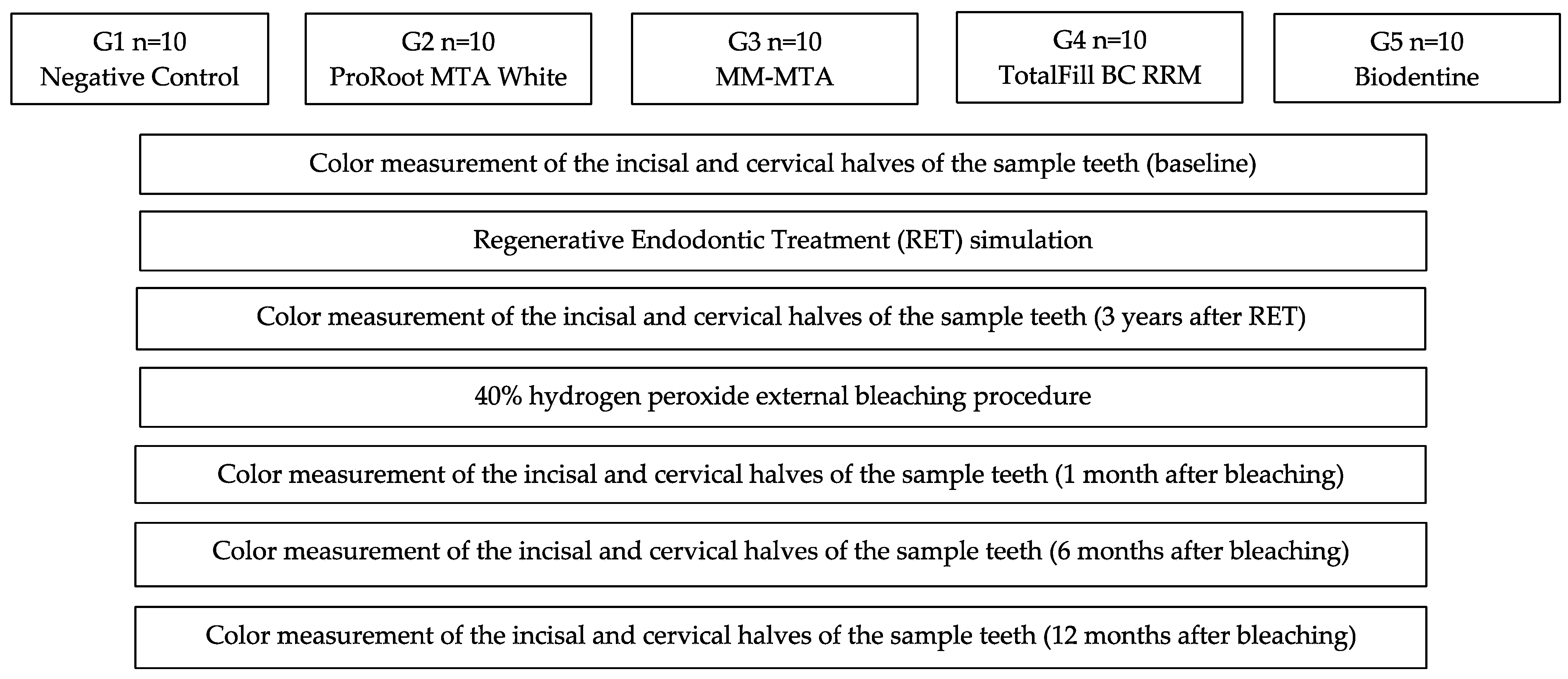
2. Materials and Methods
2.1. Sample Selection and Preparation
2.2. Regenerative Endodontic Treatment Simulation
2.3. Tooth Whitening Process Simulation
2.4. Tooth Color Evaluation
2.5. Statistical Analysis
3. Results
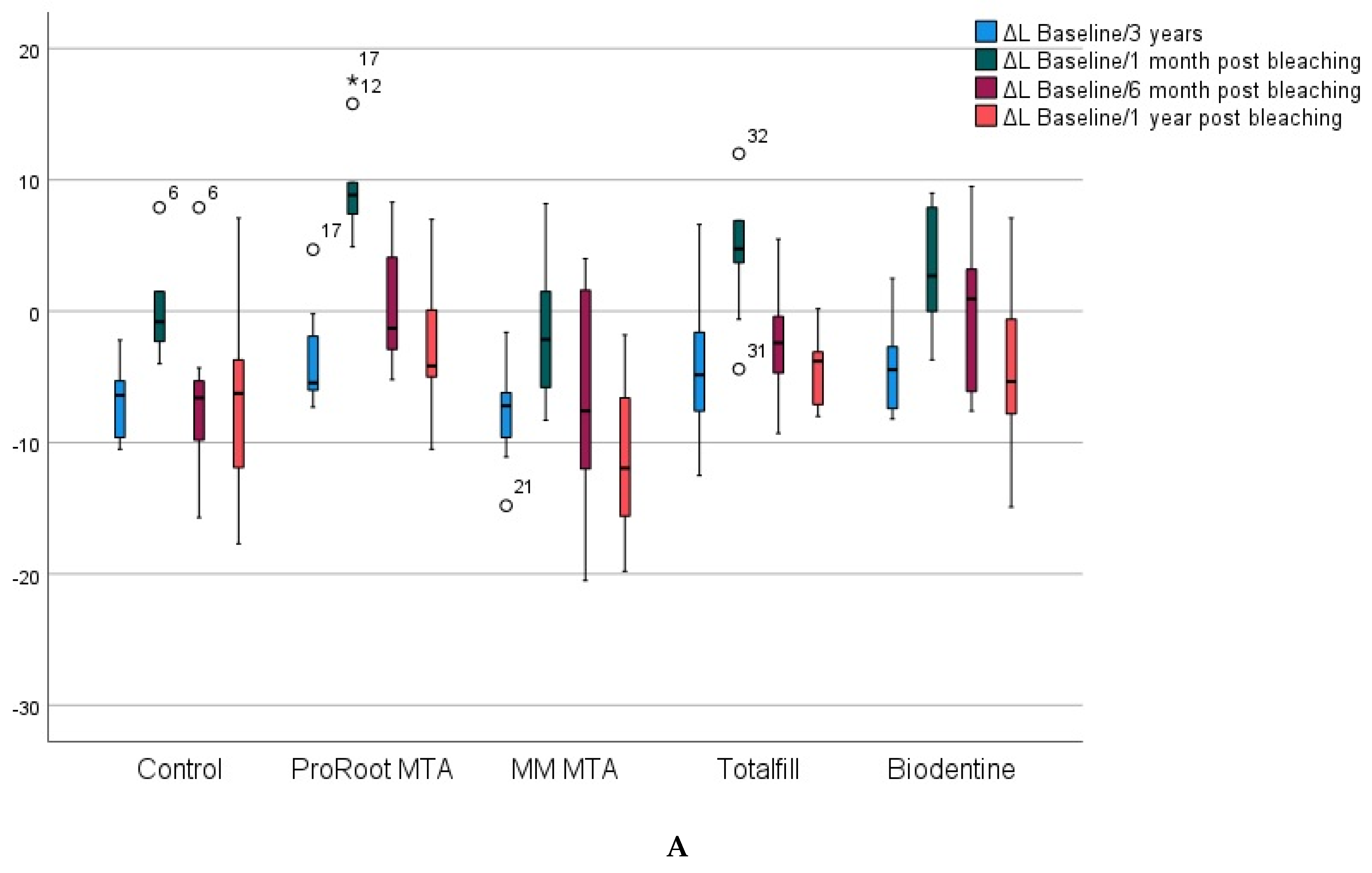
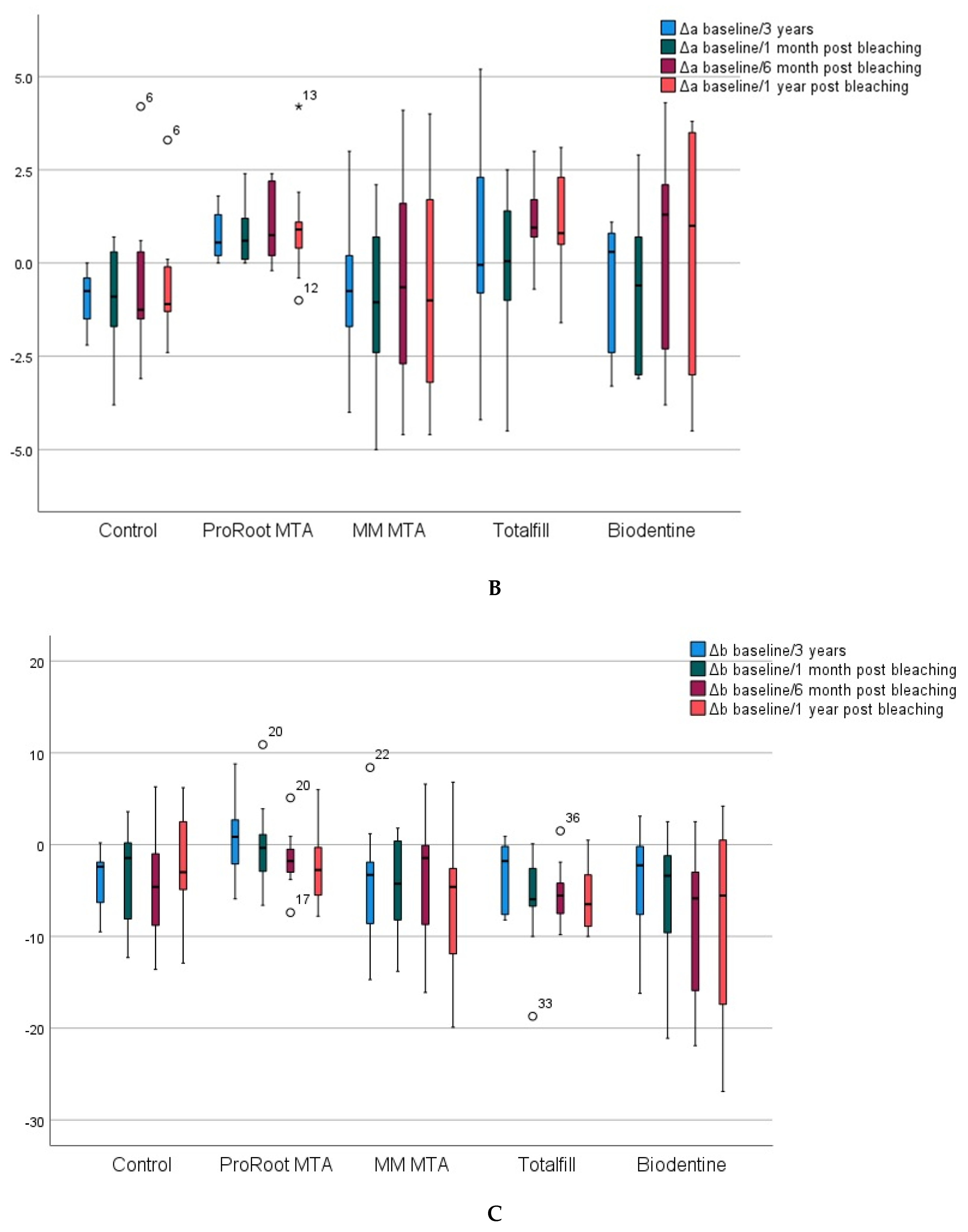
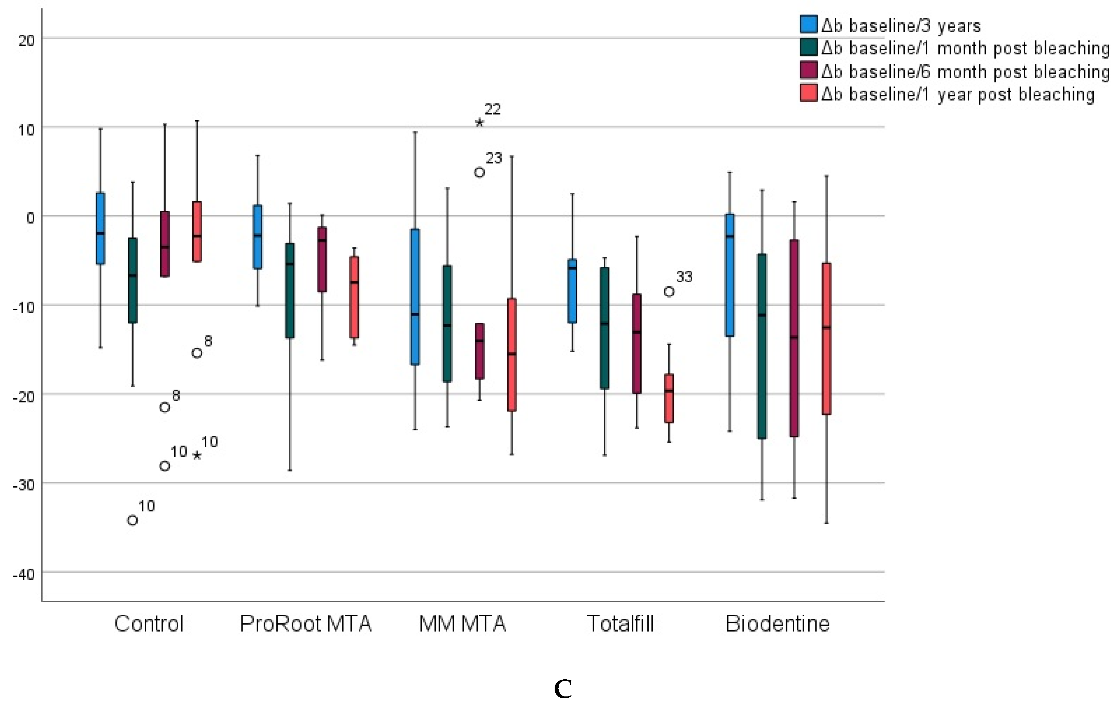
3.1. Inicisal Half
3.2. Cervical Half
4. Discussion
5. Conclusions
Author Contributions
Funding
Institutional Review Board Statement
Informed Consent Statement
Data Availability Statement
Conflicts of Interest
References
- Shaik, I.; Tulli, M.; Unnam, P.; Karunakaran, S.; Vaddi, D.S.; Jabeen, R.; Tiwari, R. Regenerative Endodontic Therapy in the Management of Nonvital Immature Permanent teeth: A Systematic Review and Meta-analysis. J. Pharm. Bioallied Sci. 2021, 13, S36–S42. [Google Scholar] [CrossRef] [PubMed]
- Galler, K.M.; Krastl, G.; Simon, S.; Van Gorp, G.; Meschi, N.; Vahedi, B.; Lambrechts, P. European Society of Endodontology position statement: Revitalization procedures. Int. Endod. J 2016, 49, 717–723. [Google Scholar] [CrossRef] [PubMed]
- Guzman, S.; Caccia, M.; Cortes, O.; Bolarin, J.M.; Requena, A.; Garcia-Godoy, A.; Garcia-Godoy, F.; Boj, J.R. Human root dentin microhardness and degradation by triple antibiotic paste and calcium hydroxide. Am. J. Dent. 2022, 35, 205–211. [Google Scholar]
- Sanz, J.L.; Forner, L.; Almudéver, A.; Guerrero-Gironés, J.; Llena, C. Viability and Stimulation of Human Stem Cells from the Apical Papilla (hSCAPs) Induced by Silicate-Based Materials for their Potential Use in Regenerative Endodontics: A Systematic Review. Materials 2020, 13, 974. [Google Scholar] [CrossRef] [PubMed]
- Możyńska, J.; Metlerski, M.; Lipski, M.; Nowicka, A. Tooth Discoloration Induced by Different Calcium Silicate-based Cements: A Systematic Review of In Vitro Studies. J. Endod. 2017, 43, 1593–1601. [Google Scholar] [CrossRef] [PubMed]
- Fagogeni, I.; Metlerska, J.; Lipski, M.; Falgowski, T.; Maciej, G.; Nowicka, A. Materials used in regenerative endodontic procedures and their impact on tooth discoloration. J. Oral. Sci. 2019, 61, 379–385. [Google Scholar] [CrossRef] [PubMed]
- Felman, D.; Parashos, P. Coronal tooth discoloration and white mineral trioxide aggregate. J. Endod. 2013, 39, 484–487. [Google Scholar] [CrossRef]
- Shokouhinejad, N.; Nekoofar, M.H.; Pirmoazen, S.; Shamshiri, A.R.; Dummer, P.M. Evaluation and Comparison of Occurrence of Tooth Discoloration after the Application of Various Calcium Silicate-based Cements: An Ex Vivo Study. J Endod. 2016, 42, 140–144. [Google Scholar] [CrossRef]
- Žižka, R.; Šedý, J.; Gregor, L.; Voborná, I. Discoloration after Regenerative Endodontic Procedures: A Critical Review. Iran Endod. J. 2018, 13, 278–284. [Google Scholar]
- Santos, L.G.; Felippe, W.T.; Souza, B.D.; Konrath, A.C.; Cordeiro, M.M.; Felippe, M.C. Crown discoloration promoted by materials used in regenerative endodontic procedures and effect of dental bleaching: Spectrophotometric analysis. J. Appl. Oral Sci. 2017, 25, 234–242. [Google Scholar] [CrossRef]
- Camilleri, J.; Borg, J.; Damidot, D.; Salvadori, E.; Pilecki, P.; Zaslansky, P.; Darvell, D.W. Colour and chemical stability of bismuth oxide in dental materials with solutions used in routine clinical practice. PLoS ONE 2020, 15, e0240634. [Google Scholar] [CrossRef] [PubMed]
- Akbulut, M.B.; Terlemez, A.; Akman, M.; Buyukerkmen, B.; Guneser, M.B.; Eldeniz, A.U. Tooth discoloration effects of calcium silicate based barrier materials used in revascularization and treatment with internal bleaching. J. Dent. Sci. 2017, 12, 347–353. [Google Scholar] [CrossRef] [PubMed]
- Palma, P.J.; Marques, J.A.; Falacho, R.I.; Correia, E.; Vinagre, A.; Santos, J.M.; Ramos, J.C. Six-Month Color Stability Assessment of Two Calcium Silicate-Based Cements Used in Regenerative Endodontic Procedures. J. Funct. Biomater. 2019, 10, 14. [Google Scholar] [CrossRef] [PubMed]
- Fagogeni, I.; Falgowski, T.; Metlerska, J.; Lipski, M.; Górski, M.; Nowicka, A. Efficiency of Teeth Bleaching after Regenerative Endodontic Treatment: A Systematic Review. J. Clin. Med. 2021, 10, 316. [Google Scholar] [CrossRef]
- Paravina, R.D.; Pérez, M.M.; Ghinea, R. Acceptability and perceptibility thresholds in dentistry: A comprehensive review of clinical and research applications. J. Esthet. Restor. Dent. 2019, 31, 103–112. [Google Scholar] [CrossRef] [PubMed]
- Yasa, B.; Arslan, H.; Akcay, M.; Kavrik, F.; Hatirli, H.; Ózkan, B. Comparison of bleaching efficacy of two bleaching agents on teeth discoloured by different antibiotic combinations used in revascularization. Clin. Oral Investig. 2014, 19, 1437–1442. [Google Scholar] [CrossRef]
- Kirchhoff, A.L.; Raldi, D.P.; Salles, A.C.; Cunha, R.S.; Mello, L. Tooth discolouration and internal bleaching after the use of triple antibiotic paste. Int. Endod. J. 2015, 48, 1181–1187. [Google Scholar] [CrossRef] [PubMed]
- Lriboz, E.; Óztürk, B.A.; Korklü, S.; Tarcin, B.; Berker, Y.G.; Óvecoglu, H.S. Comparison of intracoronal bleaching methods on teeth discolored by different antibiotic pastes. Niger. J. Clin. Pract. 2017, 20, 700–706. [Google Scholar]
- Fundaoğlu Küçükekenci, F.; Çakici, F.; Küçükekenci, A.S. Spectrophotometric analysis of discoloration and interna! bleaching after use of different antibiotic pastes. Clin. Oral Investig. 2018, 23, 161–167. [Google Scholar] [CrossRef]
- Saoud, T.M.; Martin, G.; Chen, Y.H.; Chen, K.L.; Chen, C.A.; Songtrakul, K.; Malek, M.; Sigurdsson, A.; Lin, L.M. Treatment of mature per-manent teeth with necrotic pulps and apical periodontitis using regenerative endodontic procedures: A case series. J. Endod. 2016, 42, 57–65. [Google Scholar] [CrossRef]
- Plascencia, H.; Cruz, Á.; Díaz, M.; Jiménez, A.L.; Solís, R.; Bernal, C. Root canal filling after revascu-larization/revitalization. J. Clin. Pediatr. Dent. 2016, 40, 445–449. [Google Scholar] [CrossRef] [PubMed]
- González, M.; García-Bernal, D.; Oñate-Sánchez, R.E.; Martínez, C.M.; Moraleda, J.M.; Rodríguez-Lozano, F.J.; Forner, L. Comparison of diffusion, cytotoxicity and tissue inflammatory reactions of four commercial bleaching products against human dentalpulp stem cells. Sci. Rep. 2019, 23, 7743. [Google Scholar]
- Yang, W.C.; Tsai, L.Y.; Hsu, Y.H.; Teng, N.C.; Yang, J.C.; Hsieh, S.C. Tooth discoloration and the effects of internal bleaching on the novel endodontic filling material SavDen® MTA. J. Formos. Med. Assoc. 2021, 120, 476–482. [Google Scholar] [CrossRef]
- Ghinea, R.; Pérez, M.M.; Herrera, L.J.; Rivas, M.J.; Yebra, A.; Paravina, R.D. Color difference thresholds in dental ceramics. J. Dent. 2010, 38, e57–e64. [Google Scholar] [CrossRef] [PubMed]


| Composition | Application | ||
|---|---|---|---|
| G1 | Control negativo | No cement. Cotton pellet. | - |
| G2 | ProRoot MTA White (Dentsply Maillefer, Ballaigues, Switzerland) | Powder: bismuth oxide, tricalcium silicate, dicalcium silicate, calcium dialuminate, and calcium sulfate dehydrated. Liquid: distilled water | Manually mixed powder/liquid (1:3 ratio) |
| G3 | MM-MTA (Micro-Mega sur MedicalExpo, BESANCON Cedex, France). | Powder: tricalcium silicate, dicalcium silicate, tricalcium aluminate, bismuth oxide, calcium sulfate dehydrate, and magnesium oxide. Liquid: calcium carbonate | Automixed |
| G4 | TotalFill BC RRM (FKG Dentaire, La Chaux-de-Fonds, Switzerland). | Paste: Calcium silicate, zirconium oxide, tantalum oxide, calcium phosphate monobasic, and fillers | Pre-mixed material and ready to apply |
| G5 | Biodentine (Septodont, Saint-Maur-des-Fossés, France). | Powder: tricalcium silicate dicalcium silicate, calcium carbonate, iron oxide, and zirconium oxide. Liquid: Water, calcium chloride, and soluble polymer (polycarboxylate). | Automixed |
| Baseline/3 Years | 3 Years/1 Month Post Bleaching | 3 Years/6 Months Post Bleaching | 3 Years/1 Year Post Bleaching | Baseline/1 Year Post Bleaching | |
|---|---|---|---|---|---|
| ΔE00 | ΔEab | ΔE00 | ΔEab | ΔE00 | |
| Control | 4.23 | 7.73 | 4.57 | 6.82 | 4.63 |
| ProRoot MTA | 2.70 | 4.03 | 8.48 | 9.44 | 3.03 |
| MM MTA | 5.40 | 8.63 | 4.13 | 6.14 | 4.38 |
| TotalFill | 3.16 | 5.20 | 5.73 | 7.03 | 2.77 |
Publisher’s Note: MDPI stays neutral with regard to jurisdictional claims in published maps and institutional affiliations. |
© 2022 by the authors. Licensee MDPI, Basel, Switzerland. This article is an open access article distributed under the terms and conditions of the Creative Commons Attribution (CC BY) license (https://creativecommons.org/licenses/by/4.0/).
Share and Cite
Llena, C.; Iglesias-Diaz, M.; Ciscar-Muñoz, P.; Bataller-Martínez, A.B.; Melo, M.; Sanz, J.L. Tooth Bleaching of Discolorations Caused by Hydraulic Cements in Regenerative Endodontic Treatment: A 3-Year In Vitro Study. Materials 2022, 15, 7845. https://doi.org/10.3390/ma15217845
Llena C, Iglesias-Diaz M, Ciscar-Muñoz P, Bataller-Martínez AB, Melo M, Sanz JL. Tooth Bleaching of Discolorations Caused by Hydraulic Cements in Regenerative Endodontic Treatment: A 3-Year In Vitro Study. Materials. 2022; 15(21):7845. https://doi.org/10.3390/ma15217845
Chicago/Turabian StyleLlena, Carmen, Manuel Iglesias-Diaz, Paula Ciscar-Muñoz, Ana Belén Bataller-Martínez, María Melo, and José Luis Sanz. 2022. "Tooth Bleaching of Discolorations Caused by Hydraulic Cements in Regenerative Endodontic Treatment: A 3-Year In Vitro Study" Materials 15, no. 21: 7845. https://doi.org/10.3390/ma15217845
APA StyleLlena, C., Iglesias-Diaz, M., Ciscar-Muñoz, P., Bataller-Martínez, A. B., Melo, M., & Sanz, J. L. (2022). Tooth Bleaching of Discolorations Caused by Hydraulic Cements in Regenerative Endodontic Treatment: A 3-Year In Vitro Study. Materials, 15(21), 7845. https://doi.org/10.3390/ma15217845