Abstract
Fe-Co alloys are the most important soft magnetic materials, which are successfully used for a wide range of applications. In this work, the magnetic properties of lanthanide-substituted (Fe0.65Co0.35)0.95(RE2O3)0.05 (RE = La, Nd, and Sm) nanoparticles, prepared by mechanical alloying, are reported. Our comprehensive studies (X-ray diffraction, Mössbauer spectroscopy, scanning electron microscopy with X-ray energy dispersive spectrometry, SQUID magnetometry and differential scanning calorimetry) have revealed different properties, depending on the dopant type. The RE2O3 addition led to a decrease in the crystallite size and to an increase in the internal microstrain. Moreover, because of the high grain fragmentation tendency of RE2O3, the cold welding between Fe–Co ductile particles was minimized, indicating a significant decrease in the average particle size. The parent Fe0.65Co0.35 alloy is known for its soft ferromagnetism. For the La-substituted sample, the magnetic energy product was significantly lower (0.450 MG·Oe) than for the parent alloy (0.608 MG·Oe), and much higher for the Sm-substituted compound (0.710 MG·Oe). The processing route presented here, seems to be cost-effective for the large-scale production of soft magnetic materials.
1. Introduction
Soft magnetic nanoparticles (SMNs) represent an important field in materials science and engineering, since they exhibit unique and interesting characteristics that provide promising applications [1]. Typically, SMNs include ferrites, Fe–Ni, Fe–Si, Fe–Al, and Fe–Co based alloys, which have been studied intensively in recent years [1,2,3,4,5,6,7,8,9,10,11,12,13,14,15,16,17,18,19]. Fe–Co nanoparticles show high saturation magnetization and Curie temperature values, allowing the development of numerous applications, such as hyperthermia magnetic treatment [2] or thermoablative cancer therapy [3], magnetic resonance imaging (MRI) contrast [4,5] (currently the most commonly used gadolinium diethylenetriaminepentaacetic acid - Gd–DTPA [6]), high-density data storage [1,7], advanced materials for microwave devices [8], exchange-spring permanent magnets [1,9], and new generation of magnetorheological fluids [10]. Until now, it is generally known that the Fe0.65Co0.35 alloy has the highest saturation magnetization value. In order to obtain better magnetic properties, several substitutions on Fe0.65Co0.35, by various elements, have been undertaken, including Cr [11,12], Si [13,14], Si and Co [14] , Ni [15], Al [16], Cu [17], V [18], and Dy [19].
Another interesting observation is the combination of complementary features of Fe–Co (3d - itinerant magnetism) with rare-earth metals (4f – localized). The 4f rare-earth metals exhibit a strong magnetic susceptibility and high magnetocrystalline anisotropy, due to the interactions between their orbital moment and the crystalline field. Alloying them with a 3d metal induces their polarization, and therefore consolidates the magnetization of the alloy [20,21]. The rare-earth metals exhibit large ionic radii, which can modify the cell symmetry, and therefore generate internal stress, while substituting atoms with smaller ionic radii in the structure. Therefore, the structural, magnetic and magnetostrictive properties (e.g., cell parameter, average crystallite, and grain sizes) of the alloy are modified [21]. Studies on transition metal rare-earth (T-R) compounds show a fundamental interest in magnetic coupling and development of interface walls. Unfortunately, studies on T-R alloys are limited by the cost of rare-earth elements and their low oxidation stability. Interestingly, the Fe–Co system was not subjected, to the best of our knowledge, to compositing with rare-earth oxides. The motivation behind such research was the much lower cost of oxides when compared to pure elemental lanthanides.
In this work, we present the effects of RE2O3 (RE = La, Nd, and Sm) substitution on the structural, microstructural, morphological and magnetic behavior of mechanically alloyed Fe0.65Co0.35 nanoparticles. The mechanically alloyed Fe0.65Co0.35 compound exhibits the highest saturation magnetization within the Fe–Co family, and is well known for its soft magnetic properties [22,23].
2. Materials and Methods
2.1. Synthesis and Preparation of Samples
Initial Fe (Alfa Aesar, 99%, d < 10 μm), Co (Alfa Aesar, 99.8%, 1.6 µm), La2O3 (Alfa Aesar, 99.9%), Nd2O3 (Alfa Aesar, 99.9%), and Sm2O3 (Alfa Aesar, 99.9%) powders were used to prepare the Fe0.65Co0.35 alloy and the corresponding (Fe0.65Co0.35)0.95(RE2O3)0.05 (RE = La, Nd, and Sm) samples with effective high-energy ball milling. It was found that 1% of impurity of Fe powder was caused mostly by oxygen (Fe3O4). The initial powders were mechanically alloyed (MA), in the appropriately prepared amounts, using a vibrating ball mill Retsch MM 400 with two cylindrical vials (25 mL, WC) and balls (10 mm, WC). The frequency of milling was kept at 20 Hz for 3 h, because for these mechanosynthesis parameters, the best structural and magnetic properties of the samples were obtained [22,23,24]. The ball to powder ratio was maintained at 25:1; around 50% of the vial volume was empty to assure suitable space for the milling process. In order to prevent excessive heating of the powders, the MA was stopped 15 min after every 15 min of milling.
2.2. Research Methods
The morphology and chemical composition of the samples were investigated with a JEOL-6100 scanning electron microscope (SEM), equipped with an X-ray energy dispersive spectrometer (EDS). The average particle size was estimated by scanning electron micrographs using ImageJ software.
The analyses of the structural properties were performed using the X-ray diffraction (XRD) method in an X’Pert MPD diffractometer. The anticathode of copper with λKα1 = 0.15406 nm was employed to obtain diffraction spectra. The range of 2θ was 5–100°, with a scanning step of 0.02°, and an exposure time of one second per step. The refined crystallite size, lattice parameter and microstrain were obtained using MAUD (Materials Analysis Using Diffraction) software.
In addition, 57Fe Mössbauer measurements were carried out at room temperature, in the transmission mode, utilizing a constant acceleration spectrometer with 57Co in a rhodium matrix as the source. The obtained spectra were fitted using the Gauss–Newton’s iterative method of minimizing the χ2, with a Lorentzian shape of the spectral lines.
The measurements of the dependence of magnetization M as a function of the magnetic field H (M-H - hysteresis loops) were carried out using a superconducting quantum interference device (SQUID) magnetometer produced by Quantum Design GmbH in the applied magnetic field up to 50 kOe.
Structural phase transformations and magnetic ordering temperature were determined by differential scanning calorimetry (DSC), using DSC 404 NETZSCH. The measurements in the temperature range from 25 °C to 1200 °C were performed under protective nitrogen gas, with a heating rate of 30 °C/min.
3. Results and Discussion
3.1. Scanning Electron Microscopy (SEM) Analysis
The morphology, particle size and chemical composition of pure and RE-substituted Fe0.65Co0.35 nanoparticles were investigated by SEM. Figure 1 shows the scanning electron microscopy (SEM) images of the local microstructures of the following samples: (Fe0.65Co0.35)0.95(La2O3)0.05 (a), (Fe0.65Co0.35)0.95(Nd2O3)0.05 (b), (Fe0.65Co0.35)0.95(Sm2O3)0.05 (c), and Fe0.65Co0.35 (3 h) (d).
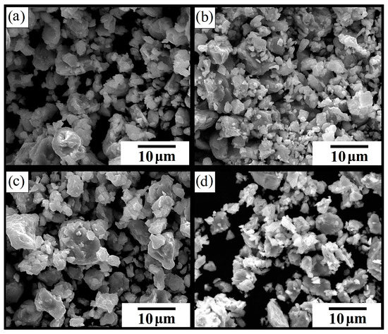
Figure 1.
Scanning electron microscopy (SEM) images of the local microstructures of the following samples: (a) (Fe0.65Co0.35)0.95(La2O3)0.05, (b) (Fe0.65Co0.35)0.95(Nd2O3)0.05, (c) (Fe0.65Co0.35)0.95(Sm2O3)0.05, and (d) Fe0.65Co0.35 (3 h).
It can be observed that the particles of pure and RE-substituted Fe0.65Co0.35 (3 h) samples have an irregular shape. A large number of agglomerates and clusters was noticed. This was explained by the presence of strong magnetic interactions in the Fe–Co based alloys and by the high surface energy in the grain boundaries of powders produced during effective high-energy ball milling [25]. The average particle size distribution of all compositions is presented in Figure 2.
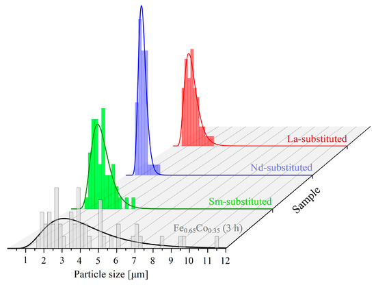
Figure 2.
Average particle size distribution of Fe0.65Co0.35 and (Fe0.65Co0.35)0.95(RE2O3)0.05 (RE = La, Nd, and Sm) formed during effective high-energy ball milling for 3 h.
It can be observed that the average particle size distribution of RE-substituted samples is smaller than that for the pure Fe0.65Co0.35 (3 h) sample. This was explained by the increase in the hardness and brittleness of Fe-Co ductile powders after RE2O3 substitution [25].
The addition of RE2O3 leads to an increase in the grain fragmentation of Fe-Co powders, and to a decrease in the particle size. The decrement in the particle size after the RE-substitution can also be explained by the presence of secondary phases (NdFeO3 and LaFeO3) located at the grain boundaries, which can hinder the particles’ growth [26].
EDS analysis revealed elemental abundances, which are summarized in Table 1. It is thought that the samples show the appropriate stoichiometry. The short duration of high-energy milling prevented contamination from the milling vial and balls (tungsten carbide).

Table 1.
Results of X-ray microprobe analysis (EDS) recorded for the obtained powders.
Figure 3 shows the chemical distribution of the (Fe0.65Co0.35)0.95(La2O3)0.05 sample. All constituents are homogenously dispersed in ferrite particles after 3 h of milling. This indicates that the elements are completely incorporated into the Fe structure. No traces of grains of RE2O3 oxides were detected.
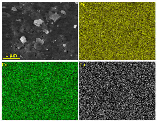
Figure 3.
Chemical distribution of (Fe0.65Co0.35)0.95(La2O3)0.05 sample.
3.2. X-ray Diffraction (XRD) Analysis
The structural and microstructural properties of the lanthanide-substituted Fe0.65Co0.35 (3 h) nanoparticles were investigated using the X-ray diffraction (XRD) technique. The XRD patterns of the prepared nanoparticles are shown in Figure 4.
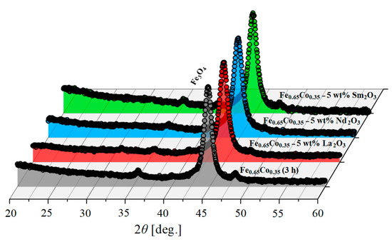
Figure 4.
X-ray diffraction (XRD) patterns of Fe0.65Co0.35 (3 h) alloy and (Fe0.65Co0.35)0.95(RE2O3)0.05 samples with RE = La, Nd, and Sm. The Y-axis was square-rooted to magnify small peaks.
Figure 4 shows the XRD spectrums of the Fe0.65Co0.35 (3 h) alloy and (Fe0.65Co0.35)0.95(RE2O3)0.05 samples with RE = La, Nd, and Sm. The characteristic peaks of Co, La2O3, Nd2O3, and Sm2O3 are no longer visible after 3 h of milling. The XRD patterns show the peaks characteristic for the body-centered cubic (bcc) iron structure (Im3m, COD 04-004-2474) for all investigated samples. These results confirm that Co, La3+, Nd3+, Sm3+, and O2– were dissolved in the bcc-Fe structure. Traces of Fe3O4 are observed for all investigated specimens due to the initial impurity of Fe powder. Furthermore, extremely small peaks seem to originate from traces of the REFeO3 phase, as presented by Suo et al. [27] and Kanna et al. [28]. The presence of these secondary phases suggests that the solubility of La3+ and Nd3+ inside the bcc-Fe structure is not complete due to their large radius (1.15 Å and 0.983 Å, respectively) [27]. However, the Sm3+ ions with a smaller radius are completely incorporated into the Fe0.65Co0.35 nanoparticles, as no additional peaks of any impurity phase were detected (Figure 4). The difference between the Fe and Co atomic radii is less than 15% and they also have the same valence (+3), which is necessary to reach a maximum solubility between atoms [29]. Moreover, the electro-negativity values of Fe and Co are almost the same, 1.83 and 1.88, respectively, which leads to a high solubility between them according to the Hume-Rothery rules [30,31]. The crystallite size, microstrain and lattice parameter refined by the Rietveld analysis, for the Fe0.65Co0.35 alloy and (Fe0.65Co0.35)0.95(RE2O3)0.05 samples with RE = La, Nd, and Sm, are shown in the Figure 5. The lattice parameters are the same for all the samples within the experimental error.
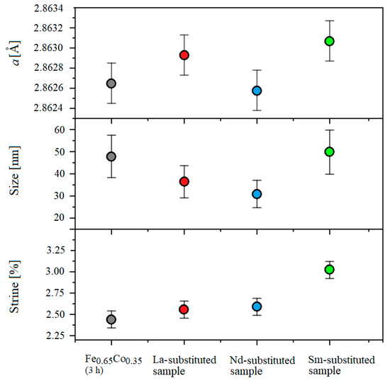
Figure 5.
The crystallite size, microstrain and lattice parameter refined by the Rietveld analysis for pure Fe0.65Co0.35 alloy and for (Fe0.65Co0.35)0.95(RE2O3)0.05 samples with RE = La, Nd, and Sm.
As shown in Figure 5, the average crystallite size in RE-substituted Fe0.65Co0.35 (3 h) samples is slightly smaller than that for the pure Fe0.65Co0.35 (3 h) alloy, except for the Sm-substituted sample. On the other hand, the microstrain values are larger in the (Fe0.65Co0.35)0.95(RE2O3)0.05 samples with RE = La, Nd, and Sm than in the Fe0.65Co0.35 alloy. This behavior could be explained by the difference between the mechanical alloying process of ductile–ductile powders (Fe–Co) and ductile–fragile ones (Fe, Co-RE2O3). In the first stage of ductile–fragile powders milling, the ductile particles (Fe, Co) exhibited plastic deformation, while the brittle particles (RE2O3) exhibited fragmentation. After the welding of the ductile particles, the fragile particles are placed between ductile particles at the collision time [32]. The fragmented particles are placed in the interfacial boundaries of the welded particles during effective high-energy ball milling. These successive phenomena, severe deformation, cold welding and solid dispersion, generate various defects (mainly dislocations) that lead to the increase in microstrain, material hardening and the enhancement of fragmentation. At the final stage, the equilibrium between the welding and fracture mechanisms is observed, leading to the formation of composite particles with a refined microstructure. In addition, the inclusions of the secondary phases at the grain boundaries inhibit the diffusion and hinder the growth of grains. The difference between Fe, Co metallic radii and RE covalent radii inside the lattice generates local disturbance and creates a strain in the crystal, which, in general, affects the nucleation rate and the crystallite sizes.
3.3. Mössbauer Spectrometry
The Mössbauer spectra measured at room temperature for the base sample of Fe0.65Co0.35 (0 h) and Fe0.65Co0.35 (3 h) alloy, (Fe0.65Co0.35)0.95(RE2O3)0.05 samples with RE = La, Nd, and Sm milled for 3 h, are shown in the Figure 6, together with the calculated data.
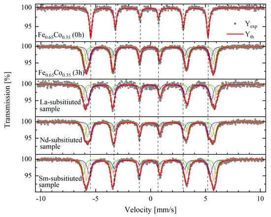
Figure 6.
Mössbauer spectra of the base sample of Fe0.65Co0.35 (0 h), Fe0.65Co0.35 (3 h) alloy, and (Fe0.65Co0.35)0.95(RE2O3)0.05 samples with RE = La, Nd, and Sm milled for 3 h. Full circles - measured data; blue, orange and green - three fit components; red - resulting fit.
For the raw sample (mixture of initial powders), one sextet component was sufficient to satisfactorily fit the experimental data. The spectra for the Fe0.65Co0.35 alloy and composites with RE2O3 were fitted with three magnetically split (sextet) components. Hyperfine interaction parameters (isomer shift and hyperfine magnetic field) for each component (denoted as S1, S2, and S3), together with its relative contribution, are listed in the Table 2. In addition, the mean value of the hyperfine magnetic field, <H> and the isomer shift, with respect to α-Fe calibration <IS> for each sample, are presented in Table 2.
Table 2.
Hyperfine interactions parameters for each sextet (S) (relative contribution and isomer shift with respect to Fe – IS; hyperfine magnetic field – H) for each component and their respective mean values (<IS>, <H>) obtained from the fits of measured Mössbauer spectra.
The Mössbauer spectrum of the raw Fe0.65Co0.35 (0 h) shows a sextet typical for magnetic behavior with a mean hyperfine magnetic field <H> ~ 33.0 T and an average isomer shift <IS> = 0.0021(10) mm/s. This behavior corresponds to the pure bcc-Fe, i.e., Fe has only Fe atoms in the neighbor’s shells, as no cobalt substitution takes place. This result shows that for a raw sample there is no Fe–Co interaction. After 3 h of milling, a broadening of the external lines is observed in the Fe0.65Co0.35 (3 h) Mössbauer spectra. This effect is attributed to the substitution of the Fe atoms by Co atoms in Fe–Co systems. The increase in the average hyperfine magnetic field to 35.7 T and the disappearance of the component with a magnetic field of 33.3 T is due to the formation of the Fe0.65Co0.35 (3 h) alloy, where Fe atoms coexist with randomly distributed Co atoms occupying bcc-Fe sites. This is further confirmed by an increase in the isomer shift up to 0.04 mm/s. These values are similar to those reported in previous works for the Fe–Co alloys [14,33].
The spectra of RE-substituted Fe0.65Co0.35 (3 h) samples show three sextets, indicating the coexistence of different magnetic environments of Fe atoms. The La-substituted Fe0.65Co0.35 (3 h) presents the highest average hyperfine field (36.1 T), which is in close agreement with the values found by Zelenakova et al. (36.16 T) [34]. For Nd- and Sm-substituted compositions, the average hyperfine field is about 35.0 T, which is in agreement with previous works for Fe–Co milled alloys [33,35,36]. The presence of RE elements in the Fe–Co structure may generate additional sextets in the Mössbauer spectra. The magnetic patterns were detected in the Mössbauer spectra of the RE-substituted Fe0.65Co0.35 (3 h) due to a small RE concentration.
3.4. The Magnetic Properties
The effect of RE2O3 (RE = La, Nd, and Sm) addition on the magnetic properties of Fe0.65Co0.35 (3 h) nanoparticles was investigated, based on the M vs. H dependencies, recorded at 10 K and 300 K. Figure 7 presents the hysteresis loops for the Fe0.65Co0.35 (3 h) alloy and (Fe0.65Co0.35)0.95(RE2O3)0.05 samples with RE = La, Nd, and Sm showing soft ferromagnetic behavior.
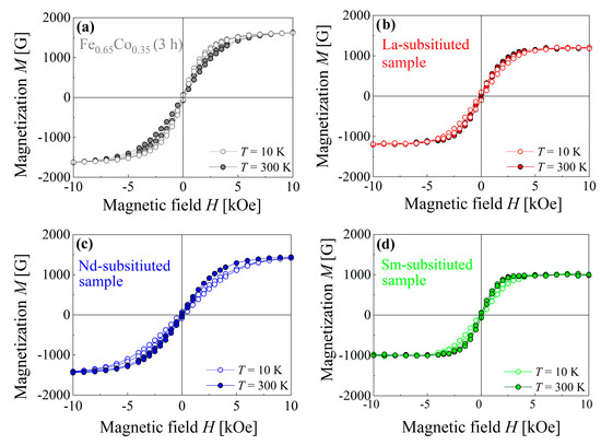
Figure 7.
Magnetization M as a function of the magnetic field H for (a) Fe0.65Co0.35 (3 h) alloy and (Fe0.65Co0.35)0.95(RE2O3)0.05 samples with RE = (b) La, (c) Nd, and (d) Sm, recorded at 10 K and 300 K.
Table 3 summarizes the magnetic properties of saturation magnetization (Ms), coercive field (Hc), remnant magnetization (Mr) and magnetic energy product (EM) derived from these hysteresis loops for all investigated compositions.
Table 3.
The magnetic properties of RE-substituted Fe0.65Co0.35 (3 h).
For T = 300 K, the saturation magnetization value of Fe0.65Co0.35 (3 h) equals 1660(10) G, whereas the saturation magnetization values of the RE-substituted Fe0.65Co0.35 nanoparticles with La3+, Nd3+, and Sm3+ are equal to 1190(10) G, 1490(10) G, and 1000(10) G, respectively. The saturation magnetization value of the RE-substituted Fe0.65Co0.35 (3 h) nanoparticles decreased by 40% when compared with the pure one. The magnetic exchange interactions play a key role in the magnetization process in the nanoparticles. The RE (La3+, Nd3+, and Sm3+) ions partially replace Fe atoms. The ionic radii of La3+, Nd3+, and Sm3+ ions are higher than the atomic radii of Fe atoms, which results in the weakening of the exchange interactions. This causes a decrease in Ms for the substituted compounds; thus, the saturation magnetization of the Fe0.65Co0.35 (3 h) system is reduced [37,38].
The decrease in Ms for RE-substituted samples is caused by the decrease in the Fe–Fe and Fe–Co interactions (3d–3d coupling), due to the reduction in the concentration of Fe and Co ferromagnetic atoms, together with the presence of very weak RE–Fe (4f–3d coupling) and RE–RE (4f–4f coupling) interactions, when compared to 3d–3d ones [39,40]. The saturation magnetization value of RE-substituted Fe0.65Co0.35 nanoparticles depends mainly on their magnetic moments. The Ms value for Nd-substituted Fe0.65Co0.35 (3 h) nanoparticles is higher than for the other samples. This is explained by the fact that the Nd3+ ions have a higher magnetic moment (J = 3.6µB), when compared with La3+ and Sm3+ ions (J = 0 and J = 1.38µB) [41]. The coercivity values of RE-substituted Fe0.65Co0.35 (3 h) nanoparticles are higher than those for the pure Fe0.65Co0.35 nanoparticles. It is well known that the coercivity is strongly influenced by the microstructure and heavy plastic deformation during the MA process, which leads to the formation of defects and generation of internal strain inside the material [42,43].
The maximum coercivity values were reported for the La-, Nd-substituted Fe0.65Co0.35 (3 h) samples. As pointed out by XRD characterization, the solubility of La3+ and Nd3+ ions in the Fe0.65Co0.35 (3 h) structure is limited and some ions do not enter the Fe lattice structure, but precipitate as secondary phases at the grain boundaries. The antiferromagnetic behavior of these secondary phases significantly alters the magnetic response of the samples. Moreover, the presence of inclusions hinders the domain walls’ motion. As a result, the coercive field of the La- and Nd-substituted samples is higher (100(5) Oe for the La-substituted sample and 100(10) Oe for the Nd-substituted, respectively) than for other samples (85(5) Oe for the Sm-substituted sample and 75(5) Oe for the Fe0.65Co0.35 (3 h) alloy). However, for the La-substituted material, the magnetic energy product (EM) is the smallest (at 300 K), and is equal to 0.450 MG·Oe.
For T = 10 K, an enhancement of both Ms and Hc values of all prepared nanoparticles was noticed. The highest Ms value of 1515(15) G was observed for the Nd-substituted sample, which was ~2% higher than that observed at 300 K. In the La-substituted sample, we observed a lower value of saturation magnetization. The Sm-substituted nanoparticles demonstrate the lowest value of Ms, 1025(10) G. The magnetization is mainly governed by the spin state and the magnetic moments of atoms; thus, the increase in Ms was principally due to the reduction in thermal fluctuation of the magnetic moments, and therefore the increase in magnetic ordering [44,45,46,47]. The coercivity has shown a substantial increase at 10 K, for all the investigated nanoparticles. For La- and Nd-substituted samples, the increase in Hc ranged from 100(5) Oe for 300 K to 290 K and 260(5) Oe for 10 K (by ∼66% and ∼62%, respectively). We noted a higher value for the Sm-substituted sample with an increase from 85(5) Oe for 300 K to 330(5) Oe for 10 K (by ~75%). It is apparent that Hc is strongly dependent on temperature. For a particle, thermal energy is essential to reverse its spin and to overcome the energy barrier. For T = 10 K, the particles did not have sufficient thermal energy; therefore, they required a stronger field to reverse the magnetization [48].
3.5. Differential Scanning Calorimetry (DSC) Analysis
The structural stability was studied using the differential scanning calorimetry (DSC) method. Figure 8a presents the DSC curves of the Fe0.65Co0.35 (3 h) alloy, and (Fe0.65Co0.35)0.95(RE2O3)0.05 samples with RE = La, Nd, and Sm, mechanically alloyed for 3 h in the mechanosynthesis process during effective high-energy ball milling. The results confirm the formation of solid solutions.
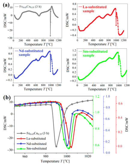
Figure 8.
(a) The DSC curves for Fe0.65Co0.35 (3 h) alloy, and (Fe0.65Co0.35)0.95(RE2O3)0.05 samples with RE = La, Nd, and Sm, mechanically alloyed for 3 h in the mechanosynthesis process during effective high-energy ball milling. (b) The sharp peaks in all alloys related to the transition from body-centered cubic (bcc) ferromagnetic to the face-centered cubic (fcc) paramagnetic structure (range 950 – 1040 °C).
A broad exothermic peak occurs at the temperature range 110 – 120 °C for all investigated compounds. This peak originates from the recovery, strain relaxation, grain growth and recrystallization of the nanocrystalline compositions [48]. The DSC scans show the presence of two main exothermic peaks. The first one is broad with the onset temperature of 670–680 °C, which can be attributed to the disordered (bcc) – ordered B2 (bcc) structural transformation. This is in good agreement with the Fe–Co phase diagram [49,50]. The second sharp peak observed for all investigated samples is related to the transition from body-centered cubic ferromagnetic to the face-centered cubic paramagnetic structure [50].
Note that the onset temperatures of the peaks are 987 °C, 994.8 °C, 995.6 °C, and 996.4 °C for Fe0.65Co0.35 (3 h) alloy, Sm-substituted sample – (Fe0.65Co0.35)0.95(Sm2O3)0.05, Nd-substituted sample – (Fe0.65Co0.35)0.95(Nd2O3)0.05, and La-substituted sample – (Fe0.65Co0.35)0.95(La2O3)0.05, respectively (Figure 8b). It seems that the RE2O3 addition stabilizes the bcc structure at high temperatures and increases the magnetic order temperature of the Fe0.65Co0.35 alloy.
4. Conclusions
The pure Fe0.65Co0.35 (3 h) alloy and (Fe0.65Co0.35)0.95(RE2O3)0.05 samples with RE = La, Nd, and Sm SMNs were successfully prepared with the mechanical alloying method. The research reported particles (agglomerates) of irregular shape that were 0.2 ̶ 12 µm in size. According to EDS analysis, uniform distribution of the elements was achieved. The homogeneous phase formation in the investigated samples was confirmed using the XRD technique.
The X-ray diffraction patterns of the substituted Fe0.65Co0.35 (3 h) alloy demonstrated the bcc-Fe structure with traces of Fe3O4 that originated from the initial impurity of the Fe powder used. Rietveld refinement was used to obtain the lattice parameter, crystallite size and microstrain values. The RE-substituted Fe0.65Co0.35 nanoparticles showed a similar crystallite size (30–50 nm) and higher microstrain, when compared to the pure Fe0.65Co0.35 (3 h). The La-substituted sample seemed to behave differently from the other samples, presumably due to the larger La radius, compared to the other rare-earth metals. This was reflected in the hyperfine interactions, as it exhibited the largest mean isomeric shift and hyperfine magnetic field values (larger than for the parent alloy).
Magnetic measurements performed at 10 K and 300 K have shown the soft ferromagnetic nature of the (Fe0.65Co0.35)0.95(RE2O3)0.05 nanocomposites. The magnetization saturation and coercivity were found to be strongly dependent on RE-substitution and temperature. RE-substitution increased the magnitude of Hc and decreased the Ms. At 300 K, the La-substituted sample was softer (0.450 MG.Oe) than the parent alloy (0.608 MG.Oe), requiring lower energy to reverse magnetization. On the other hand, for the Sm-substituted sample, higher energy was required to flip magnetization (0.710 MG.Oe). Substitution stabilizes the bcc structure at high temperatures, which is associated with an increase in the magnetic ordering temperature of the (Fe0.65Co0.35)0.95(RE2O3)0.05 samples, with respect to the parent alloy. For La-substituted sample, the highest ordering temperature of 1006 °C was reported. Low temperature behavior is also strongly modified by substitution with rare-earth metals. The coercive field increases at least by a factor of 2, while the remanence only slightly increases. The reported research shows a simple and effective route to produce novel materials with desired magnetic properties.
Author Contributions
Conceptualization, P.P. and D.E.M.; methodology, N.D., P.P., E.N. and T.T.; validation, P.P., D.E.M., P.M. and Ł.G.; formal analysis, N.D., P.P., E.N., T.T., J.P., J.M.M. and P.M.; investigation, N.D., P.P., T.T., J.P., J.M.M. and Ł.G.; data curation, N.D., P.P., E.N., J.P., J.M.M. and Ł.G.; resources, N.D.; writing - original draft preparation, N.D., P.P., T.T. and J.M.M.; writing - review and editing, N.D., P.P., E.N., T.T., J.P., J.M.M., P.M. and Ł.G.; visualization, P.P., T.T., J.M.M. and Ł.G.; supervision, Ł.G.; project administration, P.P. and T.T.; funding acquisition, P.P., J.P., J.M.M. and Ł.G. All authors have read and agreed to the published version of the manuscript.
Funding
This research received no external funding.
Institutional Review Board Statement
Not applicable.
Informed Consent Statement
Not applicable.
Data Availability Statement
The data presented in this study are available on request from the corresponding author.
Acknowledgments
This work was partly supported by the subsidies budget of the Faculty of Mathematics and Natural Sciences of Cardinal Stefan Wyszyński University (Warsaw, Poland), the Institute of Physics of the Polish Academy of Sciences (Warsaw, Poland), and the Faculty of Physics and Applied Computer Science of AGH University Science and Technology (Kraków, Poland) via international cooperation.
Conflicts of Interest
The authors declare no conflict of interest. The funders had no role in the research design, data collection, analyses, interpretation, writing the manuscript, or publishing the results.
References
- Hasegawa, T.; Kanatani, S.; Kazaana, M.; Takahashi, K.; Kumagai, K.; Hirao, M.; Ishio, S. Conversion of FeCo from soft to hard magnetic material by lattice engineering and nanopatterning. Sci. Rep. 2017, 7, 13215. [Google Scholar] [CrossRef] [PubMed]
- Alonso, J.; Khurshid, H.; Sankar, V.; Nemati, Z.; Phan, M.H.; Garayo, E.; García, J.A.; Srikanth, H. FeCo nanowires with enhanced heating powers and controllable dimensions for magnetic hyperthermia. J. Appl. Phys. 2015, 117, 17D113. [Google Scholar] [CrossRef]
- Habib, A.H.; Ondeck, C.L.; Chaudhary, P.; Bockstaller, M.R.; McHenry, M.E. Evaluation of iron-cobalt/ferrite core-shell nanoparticles for cancer thermotherapy. J. Appl. Phys. 2008, 103, 2012–2015. [Google Scholar] [CrossRef]
- Lee, J.L.; Sherlock, S.P.; Terashima, M.; Kosuge, H.; Suzuki, Y.; Goodwin, A.P.; Robinson, J.; Seo, W.S.; Liu, Z.; Loung, R.; et al. High-contrast in vivo visualization of microvessels using novel FeCo/GC magnetic nanocrystals. Mag. Res. Med. 2009, 62, 1497–1509. [Google Scholar] [CrossRef] [PubMed]
- Seo, W.S.; Lee, J.H.; Sun, X.; Suzuki, Y.; Mann, D.; Liu, Z.; Terashima, M.; Yang, P.C.; McConnell, M.V.; Nishimura, D.G.; et al. FeCo/graphitic-shell nanocrystals as advanced magnetic-resonance-imaging and near-infrared agents. Nat. Mater. 2006, 5, 971–976. [Google Scholar] [CrossRef] [PubMed]
- Wen, X.; Jackson, E.F.; Price, R.E.; Kim, E.E.; Wu, Q.; Wallace, S.; Charnsangavej, C.; Gelovani, J.G.; Li, C. Synthesis and characterization of poly(L-glutamic acid) gadolinium chelate: A new biodegradable MRI contrast agent. Bioconjug. Chem. 2004, 15, 1408–1415. [Google Scholar] [CrossRef] [PubMed]
- Das, B.K.; Rastogi, A.C. Thin films for secondary data storage. IETE J. Res. 1997, 43, 221–232. [Google Scholar] [CrossRef]
- Zare, Y.; Shams, M.H.; Jazirehpour, M. Tuning microwave permittivity coefficients for enhancing electromagnetic wave absorption properties of FeCo alloy particles by means of sodium stearate surfactant. J. Alloys Compd. 2017, 717, 294–302. [Google Scholar] [CrossRef]
- Chrobak, A. High and ultra-high coercive materials in spring-exchange systems—Review, simulations and perspective. Materials 2022, 15, 6506. [Google Scholar] [CrossRef]
- Berasategi, J.; Gomez, A.; Bou-Ali, M.M.; Gutiérrez, J.; Barandiarán, J.M.; Beketov, I.V.; Safronov, A.P.; Kurlyandskaya, G.V. Fe nanoparticles produced by electric explosion of wire for new generation of magneto-rheological fluids. Smart Mater. Struct. 2018, 27, 045011. [Google Scholar] [CrossRef]
- Hou, C.; Shan, Y.; Wu, H.; Bi, X. Effect of a small addition of Cr on soft magnetic and mechanical properties of Fe-49Co-2V alloy. J. Alloys Compd. 2013, 556, 51–55. [Google Scholar] [CrossRef]
- Khosravi, S.; Alizadeh, M.; Sharafi, S.; Karimi-Maleh, H.; Atar, N. Structural, magnetic and electron transfer effect of Cr additive on Fe65Co35 nanopowder fabricated mechanical alloying. Powder Technol. 2015, 279, 262–268. [Google Scholar] [CrossRef]
- Hocine, M.; Guittoum, A.; Hemmous, M.; Martínez-Blanco, D.; Gorria, P.; Rahal, B.; Blanco, J.A.; Sunol, J.J.; Laggoun, A. The role of silicon on the microstructure and magnetic behaviour of nanostructured (Fe0.7Co0.3)100−xSix powders. J. Magn. Magn. Mater. 2017, 422, 149–156. [Google Scholar] [CrossRef]
- Yousefi, M.; Sharafi, S. The effect of simultaneous addition of Si and Co on microstructure and magnetic properties of nanostructured iron prepared by mechanical alloying. Mat. Design 2012, 37, 325–333. [Google Scholar] [CrossRef]
- Chaudhary, V.; Ping Tan, L.; Sharma, V.K.; Ramanujan, R.V. Accelerated study of magnetic Fe ̶ Co ̶ Ni alloys through compositionally graded spark plasma sintered samples. J. Alloys Compd. 2021, 869, 159318. [Google Scholar] [CrossRef]
- Jain, V.; Chandra, A.R.; Lakshmi, N.; Reddy, V.R.; Jani, S. Magnetic behaviour of 57Fe/Co/Al multilayers deposited on a glass substrate. Bull. Mater. Sci. 2019, 42, 161. [Google Scholar] [CrossRef]
- Sarkar, A.; Hembram, S.; Chatterjee, S.; Basu Mallick, A. Effect of annealing treatments on the magnetic properties of FeCo/Cu core shell nanostructures. Mater. Today Proc. 2018, 5, 745–751. [Google Scholar] [CrossRef]
- Vadillo, V.; Gutiérrez, J.; Insausti, M.; Garitaonandia, J.S.; de Muro, I.G.; Quintana, I.; Barandiaran, J.M. Synthesis and characterization of Fe-Co-V high magnetization nanoparticles obtained by physical routes. IEEE Magn. Lett. 2019, 10, 6104805. [Google Scholar] [CrossRef]
- Xu, F.; Xu, Z.; Yin, Y. Tuning of the microwave magnetization dynamics in Dy-doped Fe65Co35-based thin films. IEEE Trans. Magn. 2015, 51, 2800904. [Google Scholar] [CrossRef]
- Younsi, K.; Russier, V.; Bessais, L. Structure and magnetic properties of nanocrystalline PrCo3. J. Appl. Phys. 2010, 107, 083916. [Google Scholar] [CrossRef]
- Younsi, K.; Crivello, J.C.; Paul-Boncour, V.; Bessais, L.; Porcher, F.; André, G. Study of the magnetic and electronic properties of nanocrystalline PrCo3 by neutron powder diffraction and density functional theory. J. Phys. Condens. Matter. 2013, 25, 116001. [Google Scholar] [CrossRef] [PubMed]
- Sirvent, P.; Berganza, E.; Aragón, A.M.; Bollero, A.; Moure Arroyo, A.; García-Hernández, M.; Marín, P.; Fernández, J.; Quesada, A. Effective high-energy ball milling in air of Fe65Co35 alloys. J. Alloys Compd. 2014, 115, 17B505. [Google Scholar] [CrossRef]
- Chermahini, M.D.; Zandrahimi, M.; Shokrollahi, H.; Sharafi, S. The effect of milling time and composition on microstructural and magnetic properties of nanostructured Fe-Co alloys. J. Alloys Compd. 2009, 477, 45–50. [Google Scholar] [CrossRef]
- Manh, D.H.; Tung, D.K.; Phong, L.T.H.; Phuc, N.X.; Jutimoosik, J.; Yimnirun, R. Complementary studies of phase formation during fabrication of Fe0.65Co0.35 nanoparticles by mechanical alloying. J. Elec. Mat. 2016, 45, 2501–2507. [Google Scholar]
- Hosseini, H.R.M.; Bahrami, A. Preparation of nanocrystalline Fe-Si-Ni soft magnetic powders by mechanical alloying. Mater. Sci. Eng. B Sol. Stat. Mater. Adv. Technol. 2005, 123, 74–79. [Google Scholar] [CrossRef]
- Gadkari, A.; Shinde, T.; Vasambekar, P. Influence of rare-earth ions on structural and magnetic properties of CdFe2O4 ferrites. Rare Met. 2010, 29, 168–173. [Google Scholar] [CrossRef]
- Suo, N.; Sun, A.; Yu, L.; Zuo, Z.; Pan, X.; Zhang, W.; Zhao, X.; Zhang, Y.; Shao, L. Effect of different rare earth (RE = Y3+, Sm3+, La3+, and Yb3+) ions doped on the magnetic properties of Ni–Cu–Co ferrite nanomagnetic materials. J. Mater. Sci. Mater. Electron. 2021, 32, 246–264. [Google Scholar] [CrossRef]
- Kanna, R.R.; Sakthipandi, K.; Seeni Mohamed Aliar Maraikkayar, S.M.; Lenin, N.; Sivabharathy, M. Doping effect of rare-earth (lanthanum, neodymium and gadolinium) ions on structural, optical, dielectric and magnetic properties of copper nanoferrites. J. Rare Earths 2018, 36, 1299–1309. [Google Scholar] [CrossRef]
- Shannon, R.D.; Prewitt, C.T. Effective ionic radii in oxides and fluorides. Acta Crystallogr. B 1969, 25, 925–945. [Google Scholar] [CrossRef]
- Chaubey, G.S.; Barcena, C.; Poudyal, N.; Rong, C.; Gao, J.; Sun, S.; Liu, J.P. Synthesis and stabilization of FeCo nanoparticles. J. Am. Chem. Soc. 2007, 129, 7214–7215. [Google Scholar] [CrossRef] [PubMed]
- Klencsár, Z.; Németh, P.; Sándor, Z.; Horváth, T.; Sajó, I.E.; Mészáros, S.; Mantilla, J.; Coaquira, J.A.H.; Garg, V.K.; Kuzmann, E.; et al. Structure and magnetism of Fe-Co alloy nanoparticles. J. Alloys Comp. 2016, 674, 153–161. [Google Scholar] [CrossRef]
- Zhang, X.; Yan, Q.; Yang, C.; Wang, T.; Ge, C. Microstructure, mechanical properties and bonding characteristic of deformed tungsten. Int. J. Refract. Met. Hard Mater. 2014, 43, 302–308. [Google Scholar] [CrossRef]
- Moumeni, H.; Alleg, S.; Djebbari, C.; Bentayeb, F.Z.; Grenèche, J.M. Synthesis and characterisation of nanostructured FeCo alloys. J. Mater. Sci. 2004, 39, 5441–5443. [Google Scholar] [CrossRef]
- Zeleňáková, A.; Olekšáková, D.; Degmová, J.; Kováč, J.; Kollár, P.; Kusý, M.; Sovák, P. Structural and magnetic properties of mechanically alloyed FeCo powders. J. Magn. Magn. Mater. 2007, 316, e519–e522. [Google Scholar] [CrossRef]
- Moumeni, H.; Alleg, S.; Greneche, J.M. Structural properties of Fe50Co50 nanostructured powder prepared by mechanical alloying. J. Alloys Compd. 2005, 386, 12–19. [Google Scholar] [CrossRef]
- Rincón Soler, A.I.; Rodríguez Jacobo, R.R.; Medina Barreto, M.H.; Cruz-Muñoz, B. Structural and magnetic properties of FeCoC system obtained by mechanical alloying. Hyperfine Interact. 2017, 238, 48. [Google Scholar] [CrossRef]
- Majeed, A.; Khan, M.A.; Raheem, F.; Hussain, A.; Iqbal, F.; Murtaza, G.; Akhtar, M.N.; Shakir, I.; Warsi, M.F. Structural elucidation and magnetic behavior evaluation of rare earth (La, Nd, Gd, Tb, Dy)-doped BaCoNi ̶ X hexagonal nano-sized ferrites. J. Magn. Magn. Mater. 2016, 408, 147–151. [Google Scholar] [CrossRef]
- Kanna, R.R.; Lenin, N.; Sakthipandi, K.; Kumar, A.S. Structural, optical, dielectric, and magnetic studies of gadolinium-added Mn-Cu nanoferrites. J. Magn. Magn. Mater. 2018, 453, 78–90. [Google Scholar] [CrossRef]
- Padalia, D.; Johri, U.C.; Zaidi, M.G.H. Effect of cerium substitution on structural and magnetic properties of magnetite nanoparticles. Mater. Chem. Phys. 2016, 169, 89–95. [Google Scholar] [CrossRef]
- Prozorov, T.; Bazylinski, D.A.; Mallapragada, S.K. Novel magnetic nanomaterials inspired by magnetotactic bacteria: Topical review. Mat. Sci. Eng. 2013, R.74, 133–172. [Google Scholar] [CrossRef]
- Dalal, M. A Textbook of Physical Chemistry, 1st ed.; Dalal Institute: Rohtak, India, 2017; Volume 1. [Google Scholar]
- Lu, G.D.; Miao, X.S.; Cheng, W.M.; Huang, X.F.; Yang, L.; Pan, L.Q. Influence of Cu underlayer on the high-frequency magnetic properties of FeCoSiO thin films. IEEE Trans. Magn. 2015, 51, 2801504. [Google Scholar] [CrossRef]
- Yang, B.; Wu, Y.; Li, X.; Yu, R. Chemical synthesis of high-stable amorphous FeCo nanoalloys with good magnetic properties. Nanomaterials 2018, 8, 154. [Google Scholar] [CrossRef] [PubMed]
- Almessiere, M.A.; Slimani, Y.; Baykal, A. Structural and magnetic properties of Ce-doped strontium hexaferrite. Ceram. Int. 2018, 44, 9000–9008. [Google Scholar] [CrossRef]
- Almessiere, M.A.; Slimani, Y.; Sertkol, M.; Gungunes, H.; Wudil, Y.S.; Demir Korkmaz, A.; Baykal, A. Impact of Gd substitution on the structure, hyperfine interactions, and magnetic properties of Sr hexaferrites. Ceram. Int. 2021, 47, 33853–33864. [Google Scholar] [CrossRef]
- Slimani, Y.; Güngüneş, H.; Nawaz, M.; Manikandan, A.; El Sayed, H.S.; Almessiere, M.A.; Sözeri, H.; Shirsath, S.E.; Ercan, I.; Baykal, A. Magneto-optical and microstructural properties of spinel cubic copper ferrites with Li ̶ Al co-substitution. Ceram. Int. 2018, 44, 14242–14250. [Google Scholar] [CrossRef]
- Kumar, D.; Sarin, A.; Verma, V.; Venkatraman, R. Pulsed laser deposition assisted fabrication and characterization of Fe-Co nanoparticles embedded in TiN thin film matrix. Thin Solid Film. 2013, 534, 561–565. [Google Scholar] [CrossRef]
- Poudyal, N.; Rong, C.; Zhang, Y.; Wang, D.; Kramer, M.J.; Hebert, R.J.; Ping Liu, J. Self-nanoscaling in FeCo alloys prepared via severe plastic deformation. J. Alloys Compd. 2012, 521, 55–59. [Google Scholar] [CrossRef]
- Chitsazan, B.; Shokrollahi, H.; Behvandi, A.; Mirzaee, O. Characterization and magnetic coercivity of nanostructured (Fe50Co50)100-XVX=0,2,4 powders containing a small amount of Co 3V intermetallic obtained by mechanical alloying. Powder Technol. 2011, 214, 105–110. [Google Scholar] [CrossRef]
- Sourmail, T. Near equiatomic FeCo alloys: Constitution, mechanical and magnetic properties. Prog. Mater. Sci. 2005, 50, 816–880. [Google Scholar] [CrossRef]
Publisher’s Note: MDPI stays neutral with regard to jurisdictional claims in published maps and institutional affiliations. |
© 2022 by the authors. Licensee MDPI, Basel, Switzerland. This article is an open access article distributed under the terms and conditions of the Creative Commons Attribution (CC BY) license (https://creativecommons.org/licenses/by/4.0/).