Article

A Theoretical Investigation into the Homo- and Hetero-leptic Cu(I) Phosphorescent Complexes Bearing 2,9-dimethyl-1,10-phenanthroline and bis [2-(diphenylphosphino)phenyl]ether Ligand

1
Department of Science, Jilin Jianzhu University, Changchun 130118, China
2
Laboratory of Theoretical and Computational Chemistry, Institute of Theoretical Chemistry, College of Chemistry, Jilin University, Changchun 130023, China
3
School of Physics, Northeast Normal University, Changchun 130024, China
*
Authors to whom correspondence should be addressed.
These authors contributed equally to this work.
Materials 2022, 15(20), 7253; https://doi.org/10.3390/ma15207253
Submission received: 30 September 2022 / Revised: 12 October 2022 / Accepted: 13 October 2022 / Published: 17 October 2022

Abstract

:
Cu(I) complexes have received widespread attention as a promising alternative to traditional noble-metal complexes. Herein, we systematically study the properties of Cu(I) complexes from homo- to hetero-ligands, and found the following: (1) hetero-ligands are beneficial to regulate phosphorescent efficiency; (2) when the hetero-ligands in a tetracoordinated Cu(I) complex are 1:1, the ligands coordinate along the dx2-y2 direction of Cu(I) ion, which can observably suppress structural deformation; (3) unlike the P^P ligand, the N^N ligand can enhance the participation of Cu(I) during the transition process; (4) the addition of an appropriate amount of P^P ligand can effectively raise the energy level of HOMO (highest occupied molecular orbital), enhance the proportion of LLCT (ligand–ligand charge transfer), and thereby increase the available singlet emission transition moments which can be borrowed, thus promoting the radiative decay process. As a result, this work provides a detailed understanding of the effects of different ligands in Cu(I) complexes, and provides a valuable reference and theoretical basis for regulating and designing the phosphorescent properties of Cu(I) complexes in the future.

1. Introduction

Transition-metal complexes, such as Ir [1], Pt [2], Os [3], Au [4] and other complexes are widely used in solar energy conversion [5], chemical sensing [6], photocatalysis [7] and displays, as well as biological molecular probes and materials in the science industry [8]. These complexes are especially attractive as candidates for efficient OLEDs, as they can both harvest singlet and triplet excitons and show higher phosphorescence quantum yields [9]. However, there are some potential drawbacks of transition-metal complexes, including high costs and environmental concerns. Cu(I) complexes are ideal candidates for metal complex luminescent materials because of their low cost, environmental friendliness, lack of MC (metal-centered excited state) transitions [10], and diverse coordination structures [11,12,13,14]. The coordination behavior depends on the electron structure of Cu(I). Because of the fulfilling of the d-orbital (d10 configuration), the ligand coordination sites around the metal center have the least electrostatic repulsion from each other and have tetrahedral structures due to the symmetrical distribution of their charges. It is clear that d10 configuration prevents the d–d centered metallic electron transition in Cu(I) compounds [10].
The luminescence performance of Cu(I) complexes have been extensively investigated. Initially, the four coordination Cu(I) complexes [Cu(N^N)2]+ and [Cu(P^P)2]+ were studied [15,16,17]. There have been many reports about homogeneous complexes in the form of [Cu(N^N)2]+ (N^N = phenanthroline). For example, the introduction of larger ligands at the 2 and 9 positions of [Cu(N^N)2]+ can inhibit the quenching of the complexes, the effective size increases in the following order: methyl< n-butyl≈n-octyl< neopentyl< sec-butyl [15]. For the past few years, the Cu(I) complex [Cu(N^N) (P^P)]+ (POP = bis [2-(11)(diphenylphosphino)phenyl]ether, N^N = 1,10-phenanthroline]) has attracted more attention [18,19]. One of the most important reasons is that N^N ligands (such as phenanthroline) are strong electron-acceptors due to their large π characteristics. It is regarded as the target of metal-to-ligand charge transfer, which affects the LUMOs (lowest unoccupied molecular orbital) of the complexes [20]. The other reason is that, as an electron-donor, the phosphine ligands can stabilize the HOMOs (highest occupied molecular orbital) and can also enhance the luminous quantum yield of Cu(I) complexes, so these complexes have a longer excited state life and color adjustability [21]. The luminescence performance of [Cu(N^N) (P^P)]+ complexes was improved by modifying the ligand species, such as POP or dppb (1,2-bis-(diphenylphosphino)-benzene) ligands [14,22]. These hybrid ligand complexes improve luminescence performance by reducing the flattening of the Cu(I) center, so as to obtain a lower non-radiative decay [23].
Therefore, the study of hetero-leptic Cu(I) complexes, with an evolution from the N^N ligand to the P^P ligand, is a very critical step towards finding an alternative to the traditional precious metal complexes in electroluminescent devices. However, the current research in this area mainly focuses on the properties of delayed fluorescence and the mechanism of light emission [24]. Some scattered experimental and theoretical studies of nitrogen–phosphorus hetero-ligands have mainly focused on the properties and performance issues of nitrogen–phosphorus hetero-ligands in dinuclear or trinuclear complexes. There are relatively few studies devoted to how does the cooperation between different ligands effect the luminescence properties. Therefore, in this study, four Cu(I) complexes (shown in Figure 1) with various ligands (gradually change from N^N ligands to P^P ligands) are investigated, to gain a better understanding of the effects of different N^N and P^P homo- and hetero-ligands on the phosphorescent quantum efficiency. We expect that this work can contribute to a detailed understanding of the effects of N^N and P^P homogeneous and heterogeneous ligands in Cu(I) complexes on the electronic structure and light emission properties, to provide a reliable theoretical basis for regulating and rationally designing the luminescence properties of Cu(I) complexes.

2. Computation Methods

All the calculations in this paper are completed by Gaussian 09 package [25]. Density functional theory (DFT) [26] and the unrestricted DFT method were used to optimize the S0 state and T1 state geometry of the complexes. The vibration frequencies were calculated to make sure all the structures are stable and have no imaginary frequency. The LANL2DZ basis set was used for Cu(I) atom [27], and the 6–31G(d) basis set was employed for all nonmetallic atoms [28]. The transition properties of Cu(I) complexes were calculated using a polarizable continuum model [29] (CH2Cl2) by time-dependent density functional (TD–DFT) [30].
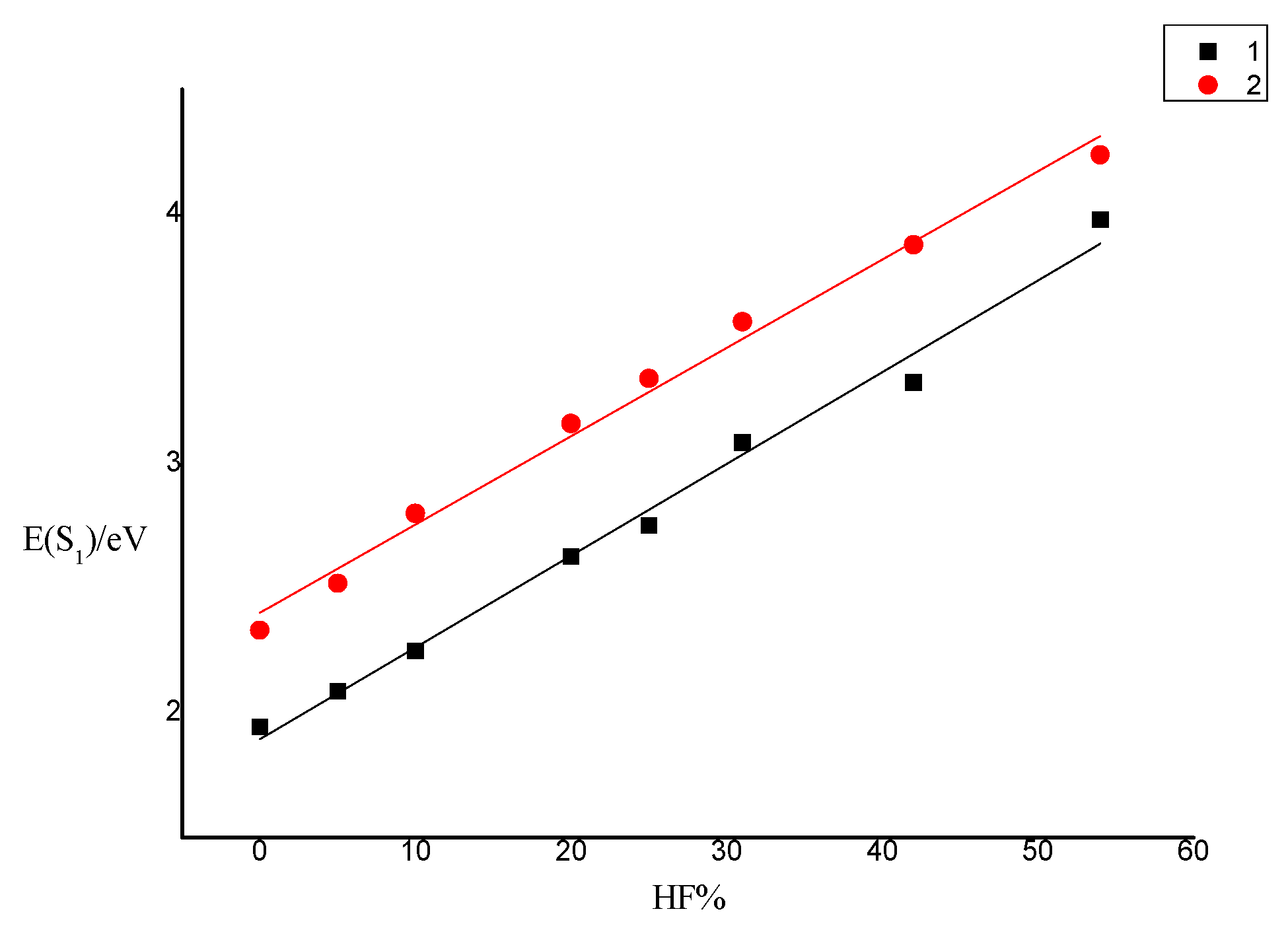
It is well known that the optical properties of complexes are very sensitive to the selection of hybrid functionals. It has been reported that the calculation of the vertical excitation energy E(S1) by the TD–DFT method has an important dependence on the Hartree–Fock percentage (HF%) contained in functionals [31]. Taking complexes 1 and 2 as examples, functionals with different HF% were simulated: BLYP [32,33], MPWLYP1M [34], TPSSH [35], B3LYP [36], PBE0 [37,38], MPW1B95 [38], BMK [39], M06-2x [40], the vertical excitation energy E(S1) data in CH2Cl2 solvent is shown in Table 1. The fit curve of E(S1) as a function of HF% is shown in Figure 2. The results show that E(S1) is positively correlated with HF%. When HF% < 54%, a good linear relationship (R12 = 0.99521, R22 = 0.99588) between E(S1) and HF% is observed (Figure 2).
1: E1(S1) = 1.89465 + 0.03879HF%
2: E2(S1) = 2.40138 + 0.03541HF%
In the experiment, the E(S1) of complex 1 was equal to 2.71 eV, according to Equation (1), the best HF% is 21.02%. According to Equation (2), the best HF% is 23.60% with the value of experiment (E(S1) = 3.24 eV). Therefore, B3LYP and PBE0 are considered as a suitable functionals. Using B3LYP and PBE0 functionals to calculate the absorption spectrum and emission spectrum of complexes 13, the calculated data are listed in Tables S1 and S2. The calculated results of PBE0 are very consistent with the experimental values, so PBE0 can be used to accurately describe the properties of this series of complexes.

3. Results and Discussion

3.1. Geometric and Electronic Structures

The structure diagrams of the studied Cu(I) complexes are shown in Figure 2. The important bond length and bond angle of the complexes are listed in Table S3. The isolated single complex that extracts from the single crystal structure is used as the initial structure of the optimization, so the effect of intermolecular interaction is lacking, the bond lengths of Cu–N and Cu–P in the optimized ground state geometry are evidently about 3–5% longer than those in the crystal structure. Compared with the experiment, the bond angle and dihedral angle have smaller errors (1–3%).
It can be seen from Table S3, in the S0 state, from the homo-coordination complexes composed of N^N ligands, to hetero-coordination complexes of N^N ligands and P^P ligands, and to homo-coordination complexes of P^P ligands (the complexes are from 1 to 4), the bond length of Cu–N and Cu–P gradually increases. This means that the Cu(I) move away from the ligands (N^N ligands and P^P ligands) gradually due to the change of the ligand. This can weaken the coordination between the metal and the ligands, which is not conducive to the MLCT process. For complex 4, the bond length of Cu–P4 is 4.1866 Å, which is far beyond the range of the bond length of a single bond (the van der Waals radius of Cu and P are 1.86 Å and 1.96 Å, respectively) [41]. It means that the metal center does not form a bond with the P4 atom. In addition, the bond angle of complex 4 is P1–Cu–P2: 113.14°; P2–Cu–P3: 120.05°; P1–Cu–P3: 124.38°; the sum is 357.57°, which is very close to 360°. The geometric structure of the three coordinated P atoms is a plane-like triangle instead of a tetrahedron. The reason why the fourth P atom does not bond with Cu(I) is probably because of the steric hindrance of the diphenyl ether skeleton and POP ligand [42].
Comparing the structural changes from the S0 to the T1 state of each complex, we found that the bond length of Cu–N gradually decreases, while the Cu–P bond length becomes longer, that is, Cu(I) is closer to the N^N ligand and away from the P^P ligand during the change from the S0 state to T1 state. It is found that the change in DHA1 (the dihedral angle between the ligands) has been suppressed by the addition of the P^P ligand (1: 89.64°→60.56°, 2: 82.36°→68.52°, 3: 83.35°→83.44°, 4: 82.33°→80.01°), which may bring a smaller structure distortion between the S0 and T1 states. Pymol software was used to superimpose the molecular structure of two states and calculate the RMSD (root mean square deviation) [43]. The data are shown in Figure S1. The results indicate that the structural distortions of complexes 1, 2, 4 decrease with the addition of the P^P ligand. Although the DHA1 of complex 3 is closest to 90° and the change from the S0 to T1 state is the smallest (Δ = 0.09°) one, complex 3 has the largest RMSD. This can be attributed to the asymmetric coordination of N^P ligand, which lead to an imbalance on the donating and accepting electrons, then result in a large distortion between the S0 and T1 state.

3.2. Natural Population Analysis

In order to deeply investigate the relationship between the diverse ligands, the Natural Population Analysis is applied. The data are listed in Table 2. During the change of the complexes from 1 to 4, the net charge of Cu(I) ion also decreases and then increases (1: 0.5615e, 2: 0.3633e, 3:0.2257e, 4: 0.5070e). These mean that the Cu(I) ion tends to lose charge from the homo-coordination (complexes 1 and 4) to the hetero-coordination (complexes 2 and 3). It is also found that the tends of lose charge decreases with the increasing of the number of P atoms.
As shown in Table S4, the net charges of the N atoms are negative, while the net charge of P atoms are positive. This means that the net charge of the central Cu(I), N and P atoms can be strongly affected by the change of the ligands. The charge distributions of the two ligands are similar due to having a homo-coordination.
From the S0 state to the T1 state, the change of net charge of Cu(I) ion in complex 1 is smaller than that of other complexes (1: Δ = 0.0002e). According to the result, complex 3 may have the highest metal participation in the charge transition process, while the complexes with hetero-coordination (complexes 2 and 3) may have stronger charge transitions between the two ligands (LLCT) and between the intra-ligands (ILCT).

3.3. Ionization Energy (IP) and Electron Affinity (EA) and Reorganization Energy

It is critical to study whether the redox potential of the material complexes in a multilayer device can constitute a suitable barrier gradient, and thus allow different carriers to pass through smoothly. For the material of the OLED emission layer, low injection energy barrier and balanced charge transport ability are the goals pursued by design materials. Ionization energy (IP) and electron affinity (EA) are used to assess the ability of material complexes to lose and accept an electron. We calculated the complex’s IP and EA using PBE0/6-31G (d), and the results are listed in Table 3. As the addition of the P^P ligand, the IP and EA values of the complex gradually decrease, which means that the hole injection barrier of the complex is gradually reduced, and the electron injection barrier is heightened. That is the possibility of becoming a hole transport material increase.
The reorganization energy (λ) represents the geometrical relaxation energy experienced by the material complexes when they are injected and transferred a hole/electron. According to the Marcus formula, the reorganization energy is located in the exponential term of transfer rate formula, so it is one of the important factors that limit the size of the transmission rate. From Table 3, λelectron of all complexes are smaller than λhole, indicating that the electron transport rate of these complexes will be faster than the hole transport rate. The main reason is that the energy required to extract holes (HEP value) is much higher than the electron extraction energy (EEP value), that is, the holes have deeper traps. Compared with the hetero-leptic complexes 2 and 3, the electron-reorganization energies of the homo-leptic compounds 1 and 4 are both smaller, while the hole reorganization energy values are larger. The electron and hole reorganization energy values of complex 2 are closer to each other (the difference between them: 1: 0.51, 2: 0.15, 3: 0.27, 4: 0.75), so complex 2 as OLED light-emitting layer material will exhibit better electron/hole transport balanced performance and higher luminescent efficiency. Compared with the excellent electron transport material Alq3 the EA value of Alq3 is 3.0 eV) [44], it can be seen that the EA values of the complexes 1, 2, 3 are close to it or larger than it, indicating as an OLED material they have a more suitable electron affinity potential.

3.4. Frontier Molecular Orbitals

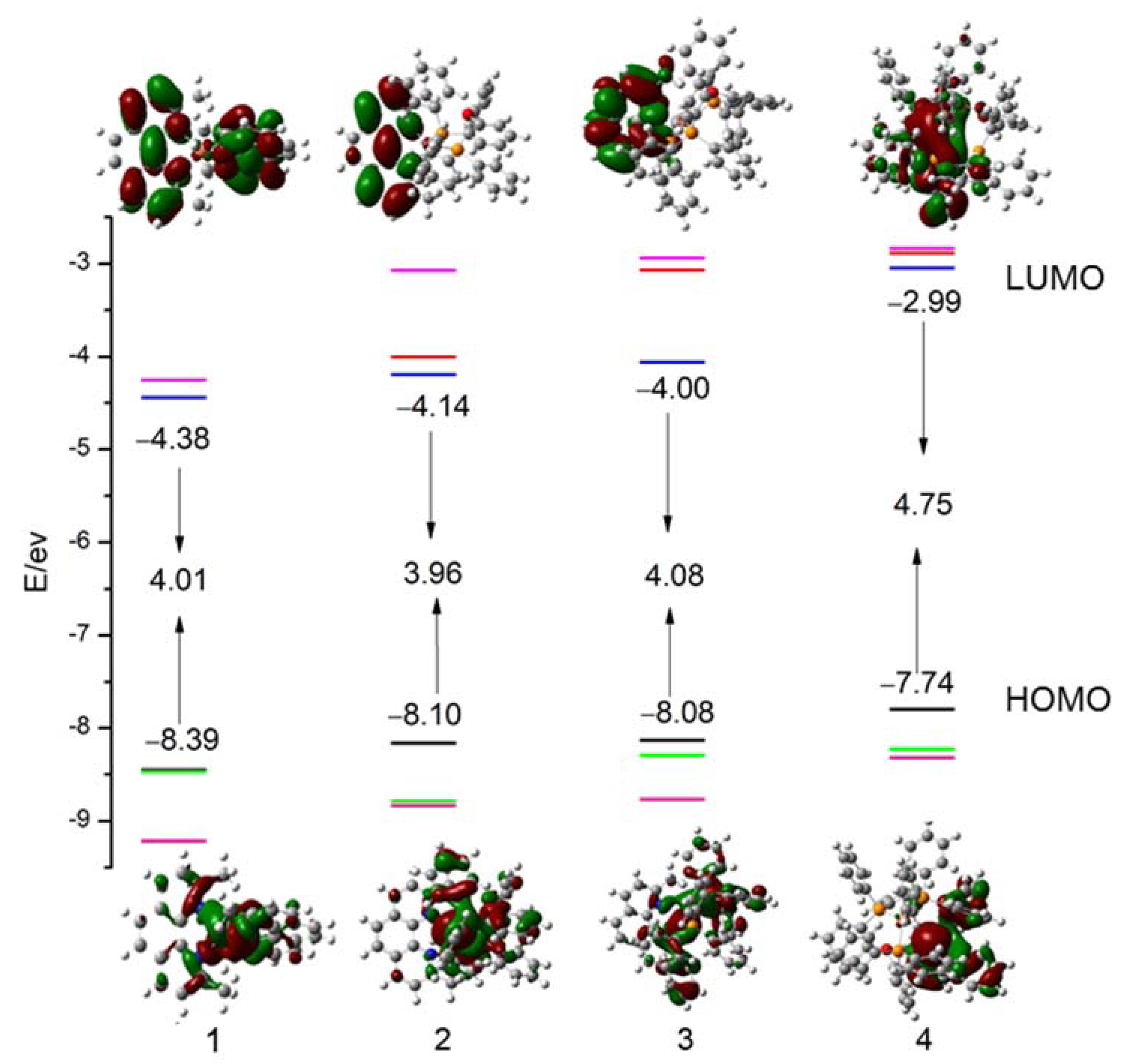
It is important to analyze the highest occupied molecular orbital (HOMO) and lowest unoccupied molecular orbital (LUMO) of the complexes. In order to more intuitively observe the effect of structural changes on the electronic properties, the energy levels of HOMOs, LUMOs, and HOMO-LUMO energy gaps (∆EH-L) were obtained by using the DFT method as shown in Figure 3.
Firstly, the electron cloud distribution of Cu(I) should be taken into consideration. For complex 1, which has two homo-coordination N^N ligands, Cu(I) has the highest proportion (70.5%) in the HOMOs, and the proportion of two N^N ligands are same (14.8%). Because of the existence of the non-bonding P4 atom, complex 4 has a different proportion of the P^P ligands, and the lowest proportion of Cu(I) in the HOMO. When Cu(I) ion is bonded to the hetero-coordination ligands (complexes 2 and 3), the proportion of Cu(I) in the HOMO decreases by about 30% compared to complex 1, and the HOMO is mainly localized on the P^P ligands.
Secondly, the changes in the molecular orbital in the two ligands are discussed. It is found that with the addition of the P^P ligand, the electron cloud distribution of the HOMO changes significantly. The electron cloud moves from the N^N ligand to the P^P ligand gradually (the proportion of P^P ligand in the HOMO: 1: 0%, 2: 57.5%, 3: 64.3%, 4: 100%). However, the LUMOs mainly locate on the N^N ligands (the proportion of N^N ligand in the LUMO: 1: 96.6%, 2: 92.3%, 3: 98.5%%, 4: 0%). For complex 3, it is clear that the LUMOs mainly locate on the quinoline group in N^P ligand. The results indicate that P^P ligands have stronger electron donor properties than N^N ligands among complexes with hetero-coordination. However, in the complexes with homo-coordination, the N^N ligands are both the electron-donors and electron-acceptors. As a result, the advantage of N^N ligand as an electron-acceptor is not significant, which may not be beneficial to the charge transition. From the above discussions, we know that the location of Frontier molecular orbitals (MOs) changed with the addition of P^P ligands, which led to a significant variation of the energy levels of HOMOs and LUMOs. The energy level of the HOMOs and LUMOs rise observably with the addition of the P^P ligands. In the meantime, the energy gaps display little change (1: ΔEH-L = 4.01 eV, 2: ΔEH-L = 3.96 eV, 3: ΔEH-L = 4.08 eV). For complex 4, the energy of the LUMO rises to 2.99 eV due to the number of P^P ligands changed from 3 to 4. This means that the energy of the LUMO is increased by the electron cloud distribution of the P^P ligands. This also indicates that the photodynamic stability increased with the addition of the P^P ligands. In addition, complex 4 has a strong reducibility, which can be attributed to the uncoordinated P atom and large steric hindrance.

3.5. Absorption and Emission Spectra in PCM Solvent

3.5.1. Absorption Spectra

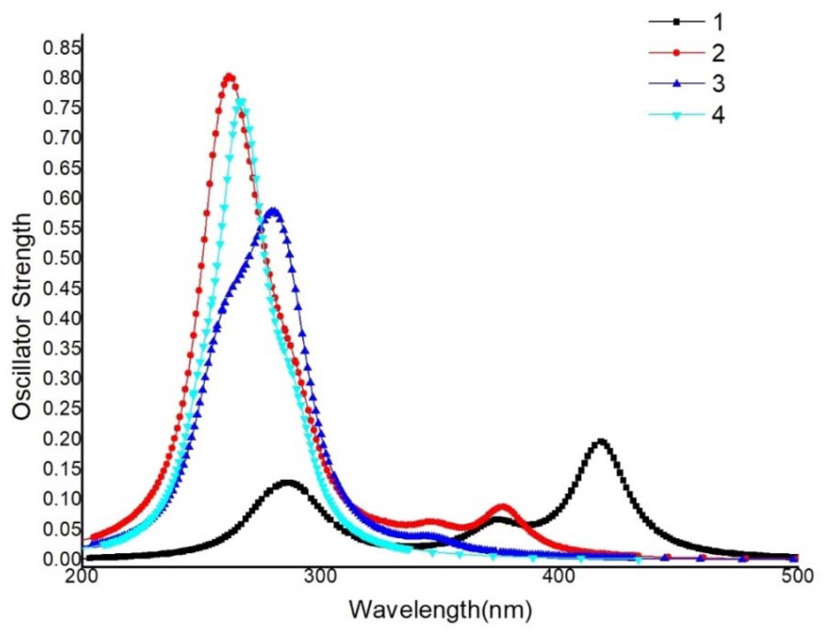
Based on the optimized structure of the S0 state and considering the solvent effect of CH2Cl2, the simulated absorption spectrums are shown in Figure 4. The calculated wavelength, oscillator strength, transition composition and the experimental values are listed in Table S6. From Table S6, it is clear that the calculated values agree well with the experiment (2: λmaxabs=376.5 nm, λexpt=383 nm, 3: λmaxabs =347.2 nm, λexpt =350 nm,) [45], which indicates that the PBE0 functional can simulate the electronic properties well. Notably, compared to complex 3, complex 2 has the larger oscillator strength, which may be conducive to the improvement of phosphorescence efficiency.
For the absorption beyond 300nm, the maximum absorption peak of complex 1 is at 417.2 nm, corresponding to the mixed transition of H−1→L+1 (56.2%) and H→L (44.2%), which is regarded as MLNLNCT from the metal (Cu, dxz, dxy) to the two ligands. There are no other absorption peaks for other three complexes in this region, indicating that the addition of the P^P ligands will blue-shift the absorption spectrum. Complex 2 has a relatively weak absorption peak at 376.5 nm, which is mainly derived from the transition of H→L (92.5%). That is a mixture of metal (Cu, dx2-y2) to ligand (N^N) charge transfer (MLNCT) and P^P ligand to N^N ligand charge transfer (LPLNCT). Complex 3 has a weak absorption at 347.2 nm, corresponding to H−1→L (87.1%) and H→L (10.6%), which is also a mixture of MLNCT and LPLNCT, but Cu(dxy, dx2-y2) participates in charge transfer. There is no absorption peak in the long wavelength region for complex 4. The above facts indicate that the addition of the P^P ligand causes a blue shift in the absorption spectrum.
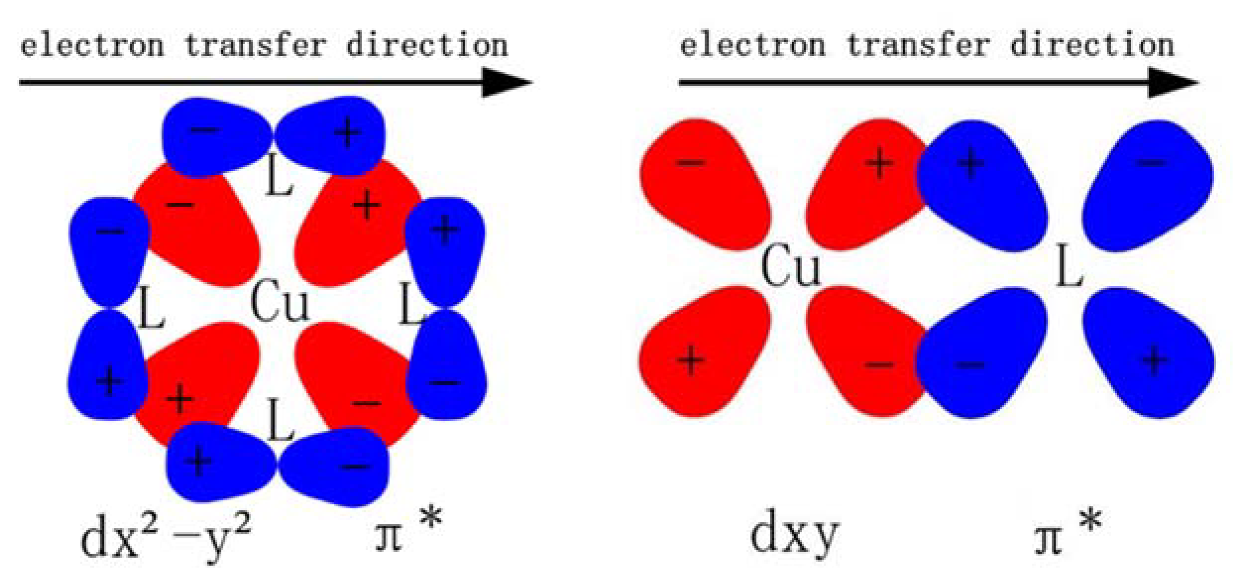
In short, in complex 2, the N^N ligand coordinates with Cu(I) along the orbital direction of dx2-y2, then form a charge transfer transition from metal (Cu, dx2-y2) to ligand (N^N, π*). The other complexes, such as the homo-coordinated complex 1 and 4, the hetero-coordinated complex 3, all coordinate along the orbital direction of (dxz, dxy) and dyz, and therefore have a similar CT transition from metal (Cu, dxz/dxy/dyz) to the ligand (N^N and P^P, π*). This illustrates that different numbers of phosphorus atoms in ligands lead to different ligand fields with different metal d-orbitals (Figure 5), resulting in different degrees of structural deformation by way of the excited process, and finally affecting the luminescent efficiency. A detailed analysis of the effect on phosphorescent quantum efficiency will be further discussed below.

3.5.2. Phosphorescent Spectrum

Based on the structure of the T1 state optimized by the UPBE0/6-31G (d) and LAN2DZ method, the emission spectra of all complexes in dichloromethane solution are listed in Table 4. It is found that the phosphorescent transitions of the four complexes mainly occurred from the HOMO to LUMO, so the properties of the transition can be replaced by the discussion of the HOMO and LUMO. The orbital compositions of the T1 state are listed in Table S7. Complex 1 has the highest 3MLCT ratio because of the highest proportion of Cu(I) in the HOMO. The ratios of complexes 2 and 3 are lower than complex 1. Complex 4 has the lowest 3MLCT proportion owing to the lowest composition of Cu(I) in the HOMO (only 0.4%). Further analyzing the contribution of each fragment to the HOMOs and LUMOs, N^N ligands exhibit strong acceptor properties in the T1 state. When the P^P ligand is coordinated with Cu(I), the P^P ligands exhibit strong donor properties, that is, the contribution of the P^P ligand in the HOMO reaches 47.3%, and at the same time, the contribution of Cu(I) in the HOMO decreases to 39.4%. When the large π-conjugation in the N^N ligand is destroyed and replaced with a diphenylphosphine ligand (complex 3), the contribution of the N^P fragment to the HOMO is significantly increased. When all the ligands are replaced by the P^P ligands, the proportion of Cu(I) in the HOMO reduces to 0.4% due to the strong donor properties, while the proportion of P^P (independent P) ligands reaches 98.3%. It should be pointed out that the LUMOs are not sensitive to the structure changes in P^P ligands, which is different from the HOMOs. The LUMOs of complexes 2 and 3 are mainly located on the N^N ligand. Based on the above discussion, the transition type of each complex can be identified as follows: the transition type of complex 1 is from the metal d-orbital to the π*-orbital of the N^N ligand (MLNLNCT). Complex 2 is a mixture of the transition from the π-orbital of the P^P ligand and the d-orbital of the metal to the π*-orbital of the N^N ligand (LPLNCT and MLNCT). Complex 3 is a mixture of the metal d-orbital and the π-orbital of the P^P ligand to the π*-orbital of the N^N ligand (MLNCT and LPLNCT). Complex 4 is from the π-orbital to the π*-orbital in the P^P ligand (independent P) (LP1LP2CT). From the above transition types, we found that complex 1 has only MLCT transition, so the participation of the N^N ligand is small. However, complex 4 has only ILCT transition in the P^P ligand, and the metal participation is close to zero. This may indicate that the phosphorescent radiative decay rate of complexes 1 and 4 may be unsatisfactory. However, for complexes 2 and 3, they have a suitable proportion of MLCT and LLCT (between the N^N ligand and the P^P ligand) in the transition progress, which may be beneficial to the radiative decay.
In addition, complex 4 is easy to oxidize and is used as a catalyst for the organic reaction due to the non-bonding P atom. Therefore, complex 4 is not suitable to be a stable light-emitting material, and the experiments have confirmed that complex 4 did not emit light [46], which will be discussed in the following section.

3.6. The Quantum Yields ( ϕ p )

The internal quantum yield for studied complexes is an important theoretical parameter to measure whether a complex can become a potential OLED material. The internal quantum yield ( ϕ p ) can be expressed as:
ϕ p = k r k r + k n r
where kr is the radiative decay rate and knr is the non-radiative decay rate.

3.6.1. Radiative Decay

The radiative decay rate is derived from Einstein’s spontaneous emission coefficient and can be expressed as follows [47], the formula details are listed in the Supporting Materials:
k r α ( T 1 S 0 ) = η 3 E ( T 1 ) 3 1.5 { n T 1 α | H ^ S O C | S n E ( S n ) E ( T 1 ) × ( f n E ( S n ) ) 1 / 2 } 2
k r , a v g R T ( T 1 S 0 ) = 1 3 α k r α
Based on Equation (4), the calculated radiative decay rates and experimental values are listed in Table 5. The related spin–orbit coupling (SOC) matrix elements and oscillator strength are listed in Tables S8–S11. It can be seen from Equation (4) that the radiative decay rate is directly related to the excited triplet energy E(T1), the excited singlet energy E(Sn), the oscillator strength of Sn→S0, the singlet–triplet energy difference (E(Sn)−E(T1)) and the SOC matrix elements. The SOC matrix element is calculated by BDF Software [48,49].
From Table 5, it is found that complex 2 has the highest T1 state excited energy (E(T1) = 16,322 cm−1), which led the smallest energy difference between the singlets and triplets (E (Sn)−E (T1)) (1: 3953 cm−1, 2: 2121 cm−1, 3: 4388 cm−1, 4: 4483 cm−1). From Equation (4), we can see that a larger E (T1) and energy gap between E (Sn) and E (T1) may bring a faster radiative decay rate. As a result, complex 2 may have a fast radiative decay rate. Furthermore, complex 1 has a smaller maximum SOC value than the other complexes (1: 83.61 cm−1, 2: 212.51 cm−1, 3: 202.96 cm−1, 4: 169.26 cm−1), while the number of SOC matrix elements with the lager absolute value is also significantly less than other complexes. For complexes 2 and 3, both the absolute value and the number of the SOC matrix elements are larger than complexes 1 and 4, that is, complexes 2 and 3 have larger SOC, and complexes 1 and 4 have smaller ones. Concerning the calculation process, the radiative decay rate is not only related to the absolute value of SOC, but also closely to the cancelling of the positive and negative SOC values. Only when the singlet states have a large oscillator strength, effective SOC matrix element, and no-cancelling of the positive and negative value in the summation calculation, can the complexes have a large radiative decay rate.
Based on the data above, although the MLCT ratio in complex 1 is the highest, the SOC matrix element is weaker than the other complexes due to the single MLCT transition caused by the homogeneous N^N ligands. In the meantime, complex 1 has the lowest excited energy E (T1). Considering these two points, complex 1 has the lowest radiative decay rate. Complex 2 and complex 3 have similar MLCT ratios, and the mixed transition types (MLCT and LLC), and then have similarly sized SOC matrix elements. However, owing to the high matching of the oscillator strength and the SOC matrix element in complex 2, the sum of the product for these two terms is significantly greater than that of complex 3 (see Supporting Materials). At the same time, complex 2 has the highest triplet energy due to the presence of pure P^P ligands. Finally, complex 2 has the fastest radiative decay rate. Because that one branch of the N^N ligand is replaced by the diphenylphosphine ligand in complex 3, the triplet energy reduces (E (T1) = 13,780cm−1), and then its radiative decay rate becomes lower.

3.6.2. Non-Radiative Decay

The non-radiative decay rate knr can be estimated by Equation (6) [50,51,52], in which the contributions of the low-frequency (ωlf ≤ 1000 cm−1) and high-frequency (1700 > ωhf > 1000 cm−1) vibration mode to knr are discussed, respectively. The related data are shown in Table 6. When the sum of the Huang–Rhys factor in low frequency is greater than 1 (Slf > 1), the strong coupling limit is required. In case that the Huang–Rhys factor is less than 1 (S < 1) or ħωM >> kBT, the weak coupling limit is required. Therefore, knr is estimated based on the strong coupling limit for lf modes and weak coupling limit for hf modes in the studied complexes. The details of the formula are listed in the Supporting Materials.
k n r ( T 1 S 0 ) = 2 π T 1 | H S O C | S m 2 2 [ 2 π 2 ( D 1 2 + P 2 ) ] 1 2 e x p [ ( Δ E 00 n M ω M λ 1 μ ) 2 π 2 ( D 1 2 + P 2 ) ] e x p ( S M S M n M n M ! )    
As shown in Table 6, the spin–orbit coupling (SOC) effect between the T1 and S0 states of complex 1 is significantly weaker than that of other complexes, with a value of only 0.014 cm−1. For phosphorescent complexes, besides the abundant aggregation of the triplet excitons, allowing the transition between the T1 and S0 state is necessary. That is, an appropriate spin–orbit coupling between the T1 and S0 state can successfully promote the transition of excitons gathered in the triplet states. Therefore, although complex 1 has a strong SOC effect between the T1 and S1 state, and a certain aggregation of the T1 state excitons, owing to the excessively small SOC effect between the T1 and S0 state, the phosphorescence emitting channel from the triplet state is blocked. This is consistent with the fact that complex 1 appears as a fluorescent emission in the experiment [15].
Reorganization energy is an important factor which can display the degree of electron–vibration coupling between two electronic states. Complex 1 has the lowest reorganization energy from the excited state to the ground state because of the high π-conjugation properties of the homogeneous N^N ligands. With the addition of the P^P ligand, large-sized phenyl-phosphine structures were introduced, and there is no obvious π-conjugated connection between the benzene rings comparing with the phenanthroline structure of 1, resulting in a greater structural distortion between the S0 state and the T1 state, that is, the reorganization energy increases significantly. In particular, complex 3 has a significant structural distortion due to one branch of the N^N ligand is replaced with the diphenylphosphine group, then the reorganization energy of complex 3 reaches 15,713.9cm−1.
In order to deeply investigate the origin of the reorganization energy, we divide the reorganization energy into a low-frequency vibration region (ω ≤ 1000cm−1) and a high-frequency vibration region (ω > 1000cm−1), respectively. It is verified that the increased reorganization energy mainly comes from low-frequency vibrations. The three larger vibrational modes of complexes 1, 3 and 4 that contribute largely to reorganization energy are derived from the low-frequency vibrational modes, and especially for complexes 2 and 3, the vibrational modes of largest reorganization energies mainly came from the low-frequency modes below 200 cm−1 (see Figures S3–S6).
According to Figures S3–S6, the vibration modes that have the largest contribution to the reorganization energy are analyzed. Evidently, the vibration mode 273 cm−1 of complex 1 mainly comes from the out-plane bending vibration of the N^N ligand, especially the atoms and methyl near the Cu(I) ion. However, when the N^N ligand is replaced by the P^P ligand (complex 2: 179cm−1), the vibrations of the P^P ligand and the N^N ligand are suppressed due to large steric effects, only the methyl groups on the N^N ligand have rocking vibration. For complex 3, the vibration mode 87 cm−1 has the largest contribution to the reorganization energy, which is attributed to the rocking vibrations of the two benzene rings connected to the P atom in the N^P ligand. In addition, the reorganization energy of the largest vibration modes already reaches 1347 cm−1, and several of the reorganization energy levels of the vibration mode exceed 1000 cm−1, these are derived from the rocking vibration of the benzene ring directly connected to the P atom in the N^P ligand. These results mean that the addition of the diphenylphosphine structure in the N^N ligand breaking of the original π conjugation, which led to a strengthening of vibrations from the benzene ring, finally result in a larger reorganization energy. The vibration with the largest contribution to the reorganization energy in complex 4 (163 cm−1) is mainly derived from the rocking vibration of the benzene ring connected to the O atom in the P^P ligand (independent P), and the reorganization energy also reaches 1333 cm−1.
As shown in Table 6, the values of the temperature-dependent term μ1 of complex 1 and 2 below 200 cm−1(1: μ1 = −2011 cm−1, 2: μ1 = −712 cm−1) are negative and can increase the effective energy gap. To complexes 3 and 4, the values of μ1 are positive, and the values of the temperature-dependent term μ2 of the high energy region are also positive, both of them reduce the effective energy gap, and as a result, bring about a sharp increase in knr.
In summary, for complex 1, although the phosphorescent radiative decay rate is calculated by Equation (4), considering the weak spin–orbit coupling between the T1 and S0 states, the excitons of the T1 state cannot accomplish phosphorescence emission. In spite of the lower proportion of MLCT in complex 2, effective spin–orbit coupling matrix element, large singlet transition moments that can be borrowed, as well as the highest triplet excitation energy, result in the fastest radiative decay rate. The addition of the P^P ligand can effectively suppress the out-plane bending vibration of the N^N ligand, reduce the structural distortion between the excited state of the ground state, hence a smaller non-radiative decay rate was obtained, and finally, complex 2 has the highest quantum yield. Although complex 3 has a similar MLCT proportion to complex 2, it has a lower kr ascribing to the lower matching between the effective spin–orbit coupling and the larger oscillator strength (i.e., the small singlet transition moments that can be borrowed). At the same time, because of the addition of diphenylphosphine in the N^P ligand, the π conjugation of the original N^N ligand is broken, which results in a significant vibration of the benzene ring connected to the P atom in the N^P ligand. The knr of complex 3 is faster with the increase in reorganization energy, so complex 3 has a lower quantum yield than complex 2. For complex 4, with the presence of an un-chelated P atom, the contribution of the benzene ring, which is connected to the dangling P atom, to the reorganization energy is increased, so the quantum yield of complex 4 is lower than that of complex 2.

4. Conclusions

In this paper, DFT/TDDFT methods are used to study the ground- and excited-state structures of a series of Cu(I) complexes bearing the 2,9-dimethyl-1,10-phenanthroline and bis [2-(diphenylphosphino)phenyl]ether ligand. The radiative decay rate, the factors affecting the non-radiative decay rate and the effects of different ligand structures from N^N to P^P ligand, including of mixed ligands, on the quantum efficiency are illustrated from the perspective of theoretical calculation. The results confirm that although the N^N homo-coordination complex has the highest metal participation in the transition process, the borrowable singlet transition moment is small, ascribing to the single MLCT transition, resulting in an unsatisfactory phosphorescent radiative decay. The addition of the P^P ligand can effectively raise the HOMO energy level, reduce the participation of Cu(I) ion and heighten the proportion of LLCT in the transition process, thus enhancing the probability of borrowable singlet transition moments, which is beneficial to the radiative decay. Complex 2, because of the fastest radiative decay rate and effective inhibition of the bending vibration of the N^N ligand, has the highest quantum yield. For complex 3, due to the addition of the diphenylphosphine structure in the N^N ligand, the conjugated nature of the original N^N ligand is destroyed to some extent, resulting in an imbalance of the electron-donating and -accepting in the N^P ligand, and moreover, there is no large steric hindrance, allowing the rocking vibration of the benzene ring connected to the P atom in the N^P ligand to increase, resulting in the largest reorganization energy and the largest non-radiative decay rate.
In summary, due to the high degree of coordination of electron-donating and electron-accepting between the N^N and P^P ligands and the perfect spatial chelation structure, complex 2 has a higher radiative decay rate and a lower non-radiative transition rate, that is, the hetero-coordinated structure with the N^N ligand and the P^P ligand has better applicable prospects for phosphorescent materials. In this paper, through the study of this series of Cu(I) complexes, the effects of the N^N and P^P ligands in Cu(I) complexes on the electronic structure and light-emitting properties are explained in depth, and the regulating effect of homo-leptic and hetero-leptic ligands on the phosphorescent emitting properties of Cu(I) complexes are scrutinized, which provides a valuable theoretical basis and reference.

Supplementary Materials

The following supporting information can be downloaded at: https://www.mdpi.com/article/10.3390/ma15207253/s1. Table S1: absorption spectra and emission wavelengths at the optimized S0 geometries for complexes 1, 2, 3 in PCM solution (CH2Cl2) together with the experimental values; Table S2: selected bond lengths (Å), bond angles (°) at the optimized S0 geometries for complexes 13 by PBE0/LANL2DZ&6-31G(d); Table S3: selected bond lengths (Å), bond angles (°) and dihedral angles (°) at optimized S0 and T1 geometries for complexes by PBE0/LANL2DZ&6-31G(d); Table S4: Natural charge population in S0 and T1 on the N and the P atoms of the complexes by using NBO method at the PBE0/LANL2DZ and 6-31 G(d) level; Table S5: Frontier molecular orbital (FMO) energies (eV) and compositions (%) of different fragments in the ground state for the complex. under considering solvent effect with DCM (Only FMO components contributing more than 10% are counted); Table S6: absorption spectra obtained by the TDDFT method in the CH2Cl2 for these complexes, together with experimental values; Table S7: Frontier molecular orbital energies (eV) and compositions (%) of different fragments in the T1 state for the complexes. Table S8: the SOC matrix elements and oscillator strength of complex 1 at the optimized T1 state geometry by BDF package; Table S9: the SOC matrix elements and oscillator strength of complex 2 at the optimized T1 state geometry by BDF package; Table S10: the SOC matrix elements and oscillator strength of complex 3 at the optimized T1 state geometry by BDF package; Table S11: the SOC matrix elements and oscillator strength of complex 4 at the optimized T1 state geometry by BDF package; Figure S1: superimposed structures of S0 (green) and T1 (red) states of studied complexes; Figure S2: electronic density contours of the frontier orbitals in the T1 state for the complexes studied; Figure S3: diagrammatic illustration of the displacement vectors of the selected vibrational normal modes with the largest reorganization energies of complex 1; Figure S4: diagrammatic illustration of the displacement vectors of the selected vibrational normal modes with the largest reorganization energies of complex 2; Figure S5: diagrammatic illustration of the displacement vectors of the selected vibrational normal modes with the largest reorganization energies of complex 3; Figure S6: siagrammatic illustration of the displacement vectors of the selected vibrational normal modes with the largest reorganization energies of complex 4.

Author Contributions

Data curation, L.S. and Y.-Y.W.; Investigation, L.S. and Y.-Y.W.; methodology, L.-Y.Z. and T.-F.H.; software, J.-F.G.; formal analysis, A.-M.R.; writing—original and draft, L.S. and Y.-Y.W.; writing—review and editing, A.-M.R. and J.-F.G.; funding acquisition, A.-M.R., J.-F.G. and L.S. All authors have read and agreed to the published version of the manuscript.

Funding

This research was funded by the Science and Technology Project of the 14th Five-year Plan of Jilin Provincial Department of Education (No. JJKH20220258KJ), the National Key R&D Program of China (Grant No. 2018YFB1501903); the Natural Science Foundation of Science and Technology Department of Jilin Province, China (no. 20190201228JC); The National Natural Science Foundation of China (No. 21473071, 21173099, 20973078, 20673045 and 51673). The 13th Five-year Plan of Jilin Provincial Department of Education (no. JJKH20211041KJ).

Institutional Review Board Statement

Not applicable.

Informed Consent Statement

Not applicable.

Data Availability Statement

The data presented in this study are available on request from the corresponding author.

Conflicts of Interest

The authors declare no conflict of interest.

References

  1. Chi, Y.; Chou, P.T. Transition-metal phosphors with cyclometalating ligands: Fundamentals and applications. Chem. Soc. Rev. 2010, 39, 638–655. [Google Scholar] [CrossRef]
  2. Arena, G.; Calogero, G.; Campagna, S.; Monsù Scolaro, L.; Ricevuto, V.; Romeo, R. Synthesis, Characterization, Absorption Spectra, and Luminescence Properties of Organometallic Platinum(II) Terpyridine Complexes. Inorg. Chem. 1998, 37, 2763–2769. [Google Scholar] [CrossRef] [PubMed]
  3. Tung, Y.-L.; Wu, P.-C.; Liu, C.-S.; Chi, Y.; Yu, J.-K.; Hu, Y.-H.; Chou, P.-T.; Peng, S.-M.; Lee, G.-H.; Tao, Y.; et al. Highly Efficient Red Phosphorescent Osmium(II) Complexes for OLED Applications. Organometallics 2004, 23, 3745–3748. [Google Scholar] [CrossRef]
  4. Osawa, M.; Hoshino, M.; Akita, M.; Wada, T. Synthesis and Characterization of Phenanthrylphosphine Gold Complex:  Observation of Au-Induced Blue-Green Phosphorescence at Room Temperature. Inorg. Chem. 2005, 44, 1157–1159. [Google Scholar] [CrossRef] [PubMed]
  5. Zhang, X.; Thavasi, V.; Mhaisalkar, S.G.; Ramakrishna, S. Novel hollow mesoporous 1D TiO2 nanofibers as photovoltaic and photocatalytic materials. Nanoscale 2012, 4, 1707–1716. [Google Scholar] [CrossRef]
  6. Xu, W.; Schmidt, R.; Whaley, M.; Demas, J.N.; DeGraff, B.A.; Karikari, E.K.; Famer, B.A. Oxygen sensors based on luminescence quenching: Interactions of pyrene with the polymer supports. Anal. Chem. 1995, 67, 3172–3180. [Google Scholar] [CrossRef]
  7. Kim, Y.I.; Salim, S.; Huq, M.J.; Mallouk, T.E. Visible-light photolysis of hydrogen iodide using sensitized layered semiconductor particles. J. Am. Chem. Soc. 1991, 113, 9561–9563. [Google Scholar] [CrossRef]
  8. Gafney, H.D. Spectral, photophysical and photochemical properties of Ru(bpy)32+ on porous vycor glass. Coord. Chem. Rev. 1990, 104, 113–141. [Google Scholar] [CrossRef]
  9. Yersin, H.; Rausch, A.F.; Czerwieniec, R.; Hofbeck, T.; Fischer, T. The triplet state of organo-transition metal compounds. Triplet harvesting and singlet harvesting for efficient OLEDs. Coord. Chem. Rev. 2011, 255, 2622–2652. [Google Scholar] [CrossRef]
  10. Barbieri, A.; Accorsi, G.; Armaroli, N. Luminescent complexes beyond the platinum group: The d10 avenue. Chem. Commun. 2008, 19, 2185–2193. [Google Scholar] [CrossRef] [PubMed]
  11. Osawa, M.; Hoshino, M.; Hashimoto, M.; Kawata, I.; Igawa, S.; Yashima, M. Application of three-coordinate copper(I) complexes with halide ligands in organic light-emitting diodes that exhibit delayed fluorescence. Dalton Trans. 2015, 44, 8369–8378. [Google Scholar] [CrossRef]
  12. Chakkaradhari, G.; Belyaev, A.A.; Karttunen, A.J.; Sivchik, V.; Tunik, S.P.; Koshevoy, I.O. Alkynyl triphosphine copper complexes: Synthesis and photophysical studies. Dalton Trans. 2015, 44, 13294–13304. [Google Scholar] [CrossRef] [Green Version]
  13. Gneuss, T.; Leitl, M.J.; Finger, L.H.; Rau, N.; Yersin, H.; Sundermeyer, J. A new class of luminescent Cu(I) complexes with tripodal ligands-TADF emitters for the yellow to red color range. Dalton Trans. 2015, 44, 8506–8520. [Google Scholar] [CrossRef] [PubMed] [Green Version]
  14. Sandroni, M.; Kayanuma, M.; Rebarz, M.; Akdas-Kilig, H.; Pellegrin, Y.; Blart, E.; Le Bozec, H.; Daniel, C.; Odobel, F. Heteroleptic diimine copper(I) complexes with large extinction coefficients: Synthesis, quantum chemistry calculations and physico-chemical properties. Dalton Trans. 2013, 42, 14628–14638. [Google Scholar] [CrossRef] [PubMed] [Green Version]
  15. Penfold, T.J.; Karlsson, S.; Capano, G.; Lima, F.A.; Rittmann, J.; Reinhard, M.; Rittmann-Frank, M.H.; Braem, O.; Baranoff, E.; Abela, R.; et al. Solvent-Induced Luminescence Quenching: Static and Time-Resolved X-Ray Absorption Spectroscopy of a Copper(I) Phenanthroline Complex. J. Phys. Chem. A 2013, 117, 4591–4601. [Google Scholar] [CrossRef]
  16. Moudam, O.; Kaeser, A.; Delavaux-Nicot, B.; Duhayon, C.; Holler, M.; Accorsi, G.; Armaroli, N.; Séguy, I.; Navarro, J.; Destruel, P.; et al. Electrophosphorescent homo- and heteroleptic copper(i) complexes prepared from various bis-phosphine ligands. Chem. Commun. 2007, 29, 3077–3079. [Google Scholar] [CrossRef]
  17. Kaeser, A.; Mohankumar, M.; Mohanraj, J.; Monti, F.; Holler, M.; Cid, J.-J.; Moudam, O.; Nierengarten, I.; Karmazin-Brelot, L.; Duhayon, C.; et al. Heteroleptic Copper(I) Complexes Prepared from Phenanthroline and Bis-Phosphine Ligands. Inorg. Chem. 2013, 52, 12140–12151. [Google Scholar] [CrossRef] [PubMed]
  18. Cuttell, D.G.; Kuang, S.-M.; Fanwick, P.E.; McMillin, D.R.; Walton, R.A. Simple Cu(I) Complexes with Unprecedented Excited-State Lifetimes. J. Am. Chem. Soc. 2002, 124, 6–7. [Google Scholar] [CrossRef] [PubMed]
  19. Evans, R.C.; Douglas, P.; Winscom, C.J. Coordination complexes exhibiting room-temperature phosphorescence: Evaluation of their suitability as triplet emitters in organic light emitting diodes. Coord. Chem. Rev. 2006, 250, 2093–2126. [Google Scholar] [CrossRef]
  20. Bergmann, L.; Braun, C.; Nieger, M.; Bräse, S. The coordination- and photochemistry of copper(i) complexes: Variation of N^N ligands from imidazole to tetrazole. Dalton Trans. 2018, 47, 608–621. [Google Scholar] [CrossRef] [PubMed]
  21. Femoni, C.; Muzzioli, S.; Palazzi, A.; Stagni, S.; Zacchini, S.; Monti, F.; Accorsi, G.; Bolognesi, M.; Armaroli, N.; Massi, M.; et al. New tetrazole-based Cu(i) homo- and heteroleptic complexes with various P^P ligands: Synthesis, characterization, redox and photophysical properties. Dalton Trans. 2013, 42, 997–1010. [Google Scholar] [CrossRef] [PubMed]
  22. Saito, K.; Arai, T.; Takahashi, N.; Tsukuda, T.; Tsubomura, T. A series of luminescent Cu(i) mixed-ligand complexes containing 2,9-dimethyl-1,10-phenanthroline and simple diphosphine ligands. Dalton Trans. 2006, 37, 4444–4448. [Google Scholar] [CrossRef]
  23. Iwamura, M.; Takeuchi, S.; Tahara, T. Substituent effect on the photoinduced structural change of Cu(i) complexes observed by femtosecond emission spectroscopy. Phys. Chem. Chem. Phys. 2014, 16, 4143–4154. [Google Scholar] [CrossRef] [PubMed]
  24. Mohankumar, M.; Holler, M.; Meichsner, E.; Nierengarten, J.-F.; Niess, F.; Sauvage, J.-P.; Delavaux-Nicot, B.; Leoni, E.; Monti, F.; Malicka, J.M.; et al. Heteroleptic Copper(I) Pseudorotaxanes Incorporating Macrocyclic Phenanthroline Ligands of Different Sizes. J. Am. Chem. Soc. 2018, 140, 2336–2347. [Google Scholar] [CrossRef] [PubMed]
  25. Frisch, M.J.; Trucks, G.W.; Schlegel, H.B.; Scuseria, G.E.; Robb, M.A.; Cheeseman, J.R.; Scalmani, G.; Barone, V.; Mennucci, B.; Petersson, G.A.; et al. Fox, Gaussian 09, Revision A.02; Gaussian Inc.: Wallingford, CT, USA, 2009. [Google Scholar]
  26. Runge, E.; Gross, E.K.U. Density-Functional Theory for Time-Dependent Systems. Phys. Rev. Lett. 1984, 52, 997–1000. [Google Scholar] [CrossRef]
  27. Ross, R.B.; Ermler, W.C. Ab initio calculations including relativistic effects for diatomic silver, diatomic gold, silver-gold (AgAu), silver hydride (AgH), and gold hydride (AuH). J. Phys. Chem. 1985, 89, 5202–5206. [Google Scholar] [CrossRef]
  28. Hariharan, P.C.; Pople, J.A. Accuracy of AH n equilibrium geometries by single determinant molecular orbital theory. Mol. Phys. 1974, 27, 209–214. [Google Scholar] [CrossRef]
  29. Tomasi, J.; Mennucci, B.; Cammi, R. Quantum Mechanical Continuum Solvation Models. Chem. Rev. 2005, 105, 2999–3093. [Google Scholar] [CrossRef] [PubMed]
  30. Autschbach, J.; Patchkovskii, S.; Ziegler, T.; van Gisbergen, S.J.A.; Jan Baerends, E. Chiroptical properties from time-dependent density functional theory. II. Optical rotations of small to medium sized organic molecules. J. Chem. Phys. 2002, 117, 581–592. [Google Scholar] [CrossRef]
  31. Huang, S.; Zhang, Q.; Shiota, Y.; Nakagawa, T.; Kuwabara, K.; Yoshizawa, K.; Adachi, C. Computational Prediction for Singlet- and Triplet-Transition Energies of Charge-Transfer Compounds. J. Chem. Theory Comput. 2013, 9, 3872–3877. [Google Scholar] [CrossRef] [PubMed]
  32. Becke, A.D. Density-functional exchange-energy approximation with correct asymptotic behavior. Phys. Rev. A 1988, 38, 3098–3100. [Google Scholar] [CrossRef] [PubMed]
  33. Lee, C.; Yang, W.; Parr, R.G. Development of the Colle-Salvetti correlation-energy formula into a functional of the electron density. Phys. Rev. B 1988, 37, 785–789. [Google Scholar] [CrossRef] [PubMed] [Green Version]
  34. Schultz, N.E.; Zhao, Y.; Truhlar, D.G. Density Functionals for Inorganometallic and Organometallic Chemistry. J. Phys. Chem. A 2005, 109, 11127–11143. [Google Scholar] [CrossRef] [PubMed]
  35. Staroverov, V.N.; Scuseria, G.E.; Tao, J.; Perdew, J.P. Comparative assessment of a new nonempirical density functional: Molecules and hydrogen-bonded complexes. J. Chem. Phys. 2003, 119, 12129–12137. [Google Scholar] [CrossRef]
  36. Reiher, M.; Salomon, O.; Artur Hess, B. Reparameterization of hybrid functionals based on energy differences of states of different multiplicity. Theor. Chem. Acc. 2001, 107, 48–55. [Google Scholar] [CrossRef]
  37. Perdew, J.P.; Burke, K.; Ernzerhof, M. Generalized Gradient Approximation Made Simple. Phys. Rev. Lett. 1996, 77, 3865–3868. [Google Scholar] [CrossRef] [Green Version]
  38. Adamo, C.; Barone, V. Accurate excitation energies from time-dependent density functional theory: Assessing the PBE0 model for organic free radicals. Chem. Phys. Lett. 1999, 314, 152–157. [Google Scholar] [CrossRef]
  39. Boese, A.D.; Martin, J.M.L. Development of density functionals for thermochemical kinetics. J. Chem. Phys. 2004, 121, 3405–3416. [Google Scholar] [CrossRef] [PubMed] [Green Version]
  40. Zhao, Y.; Truhlar, D.G. The M06 suite of density functionals for main group thermochemistry, thermochemical kinetics, noncovalent interactions, excited states, and transition elements: Two new functionals and systematic testing of four M06 functionals and 12 other functionals. Theor. Chem. Acc. 2008, 119, 525. [Google Scholar] [CrossRef] [Green Version]
  41. Sheng-Zhi, H.; Zhao-Hui, Z.; Khi-Rui, T. Average van der Waals Radii of Atoms in Crystals. Acta Phys. Chim. Sin. 2003, 19, 1073–1077. [Google Scholar] [CrossRef]
  42. Venkateswaran, R.; Balakrishna, M.S.; Mobin, S.M.; Tuononen, H.M. Copper(I) Complexes of Bis(2-(diphenylphosphino)phenyl) Ether:  Synthesis, Reactivity, and Theoretical Calculations. Inorg. Chem. 2007, 46, 6535–6541. [Google Scholar] [CrossRef] [PubMed]
  43. The PyMOL Molecular Graphics System, Version 1.6.0.0; Schrodinger LLC: New York, NY, USA, 2010.
  44. Tang, C.W.; VanSlyke, S.A. Organic electroluminescent diodes. Appl. Phys. Lett. 1987, 51, 913–915. [Google Scholar] [CrossRef]
  45. Qin, L.; Zhang, Q.; Sun, W.; Wang, J.; Lu, C.; Cheng, Y.; Wang, L. Novel luminescent iminephosphine complex of copper(i) with high photochemical and electrochemical stability. Dalton Trans. 2009, 43, 9388–9391. [Google Scholar] [CrossRef]
  46. Krylova, V.A.; Djurovich, P.I.; Conley, B.L.; Haiges, R.; Whited, M.T.; Williams, T.J.; Thompson, M.E. Control of emission colour with N-heterocyclic carbene (NHC) ligands in phosphorescent three-coordinate Cu(i) complexes. Chem. Commun. 2014, 50, 7176–7179. [Google Scholar] [CrossRef]
  47. Siddique, Z.A.; Yamamoto, Y.; Ohno, T.; Nozaki, K. Structure-Dependent Photophysical Properties of Singlet and Triplet Metal-to-Ligand Charge Transfer States in Copper(I) Bis(diimine) Compounds. Inorg. Chem. 2003, 42, 6366–6378. [Google Scholar] [CrossRef] [PubMed]
  48. Liu, W.; Hong, G.; Dai, D.; Li, L.; Dolg, M. The Beijing four-component density functional program package (BDF) and its application to EuO, EuS, YbO and YbS. Theor. Chem. Acc. 1997, 96, 75–83. [Google Scholar] [CrossRef]
  49. Liu, W.; Wang, F.; Li, L. The Beijing Density Functional (BDF) Program Package: Methodologies and Applications. J. Theor. Comput. Chem. 2003, 2, 257–272. [Google Scholar] [CrossRef]
  50. Freed, K.F. Influence of Frequency Shifts on Electron Transfer Processes. J. Phys. Chem. B 2003, 107, 10341–10343. [Google Scholar] [CrossRef]
  51. Tong, G.S.M.; Chow, P.K.; To, W.-P.; Kwok, W.-M.; Che, C.-M. A Theoretical Investigation into the Luminescent Properties of d8-Transition-Metal Complexes with Tetradentate Schiff Base Ligands. Chem. A Eur. J. 2014, 20, 6433–6443. [Google Scholar] [CrossRef]
  52. Brunschwig, B.S.; Sutin, N. Rate-Constant Expressions for Nonadiabatic Electron-Transfer Reactions. Comments Inorg. Chem. 1987, 6, 209–235. [Google Scholar] [CrossRef]
Figure 1. Chemical structures of the studied complexes.
Materials 15 07253 g001
Figure 2. Dependence of E(S1) of complex 1 on the HF% in TD–DFT functionals.
Materials 15 07253 g002
Figure 3. Frontier molecular orbital energy levels, the energy gaps ∆EH-L and the electronic density contours of the frontier molecular orbital in the S0 state for studied complexes.
Materials 15 07253 g003
Figure 4. Simulated absorption spectrum of these complexes in PCM solution (CH2Cl2).
Materials 15 07253 g004
Figure 5. A schematic illustration of the symmetry of the ligand’s π-orbital with the d-orbital of Cu(I) and the charge transfer of the formed metal→ligand.
Materials 15 07253 g005
Table 1. Calculated E(S1) using various functionals and LANL2DZ/6–31G(d) basis set in CH2Cl2 media for complex 1 and 2.
BLYP
(0%)
MPWLY
P1M (5%)
TPSSH
(10%)
B3LYP
(20%)
PBE0
(25%)
MPW1
B95 (31%)
BMK
(42%)
M06-2x
(54%)
HF%05102025314254
1 E(S1)/eV1.94322.08632.24682.62592.75083.08253.32423.9777
2 E(S1)/eV2.33082.52022.79973.16053.34003.56793.87704.2369
Note: Hartree–Fock percentage is listed in the parentheses.
Table 2. Calculated natural population analysis in S0 and T1 states (unit in e) by PBE0/[LANL2DZ-ECP/6-31G(d)].
ComplexS0T1S0-T1
CuLigand 1Ligand 2CuLigand 1Ligand 2CuLigand 1Ligand 2
10.56150.21930.21930.56170.21920.21920.00020.00010.0001
20.36330.22620.41050.36770.19730.43500.00440.0289−0.0245
30.22570.32170.45260.30760.28090.41160.08190.04080.0410
40.50700.29190.20110.51490.29390.19180.0079−0.00200.0093
Table 3. IPs, EAs, extraction potentials, and reorganization energies for complexes (unit in eV).
IP (v)IP (a)HEPEA (v)EA (a)EEPλholeλelectron
19.559.168.883.283.373.450.670.16
29.038.708.422.943.183.410.610.46
38.948.558.162.833.083.330.770.50
48.818.347.722.182.332.511.090.33
Table 4. Emission spectra obtained by the TDDFT method in CH2Cl2 for studied complexes, together with experimental values.
Electronic Transition λ m a x e m m   ( nm ) Configuration (%)Assignment
1T1→S0782.9/730exptH→L (98.0)MLNLNCT
H→L+4 (2.0)MLNLNCT
2T1→S0612.7/570exptH→L (92.5)MLNCT/LPLNCT
H−1→L (2.9)MLNCT/LPLNCT
3T1→S0725.7/608exptH→L (72.0)MLNCT/LPLNCT
H−4→L (19.5)MLNCT/LPLNCT
H−3→L (4.5)MLNCT/LPLNCT
4T1→S0647.3H→L (89.8)LP1LP2CT
Table 5. Calculated excited triplet energy E(T1) and kr based on the T1 geometry by TD PBE0//UPBE0/6-31G(d)&LAN2DZ.
E(T1) (cm−1)krx (s−1)kry (s−1)krz (s−1)kravg (s−1)krexp (s−1)
112,7732074142207514304444
216,32212,41316,30012,41313,70810,490
313,780330210,2393302561417,143
415,45010,5064710,50670200
Table 6. Calculated spin–orbit coupling matrix, emission energy ΔE00, reorganization energy λ, Huang–Rhys factor (S) and the parameters related to knr. (Energy unit in cm−1, rate unit in s−1).
<T1|SOC|S0>2ΔE00λ1μ1μ2nM1nM2λMλLλSlfSMSknrknr (expt)
10.01417,7812141−201118512117832735351841.140.530.41762.8 × 10−5
2477720,9972977−712567121113783073445133.960.9334.899469.65.94 × 104
3160319,24313,76944285333226713,44715,714254.281.55255.821.2 × 10111.41 × 106
449725,91812,409416111998137112,18213,55366.620.9769.598.8 × 105
Publisher’s Note: MDPI stays neutral with regard to jurisdictional claims in published maps and institutional affiliations.