

Molecular Cocrystals with Hydrogen-Bonded Polymeric Structures and Polarized Luminescence
 , , and
Abstract
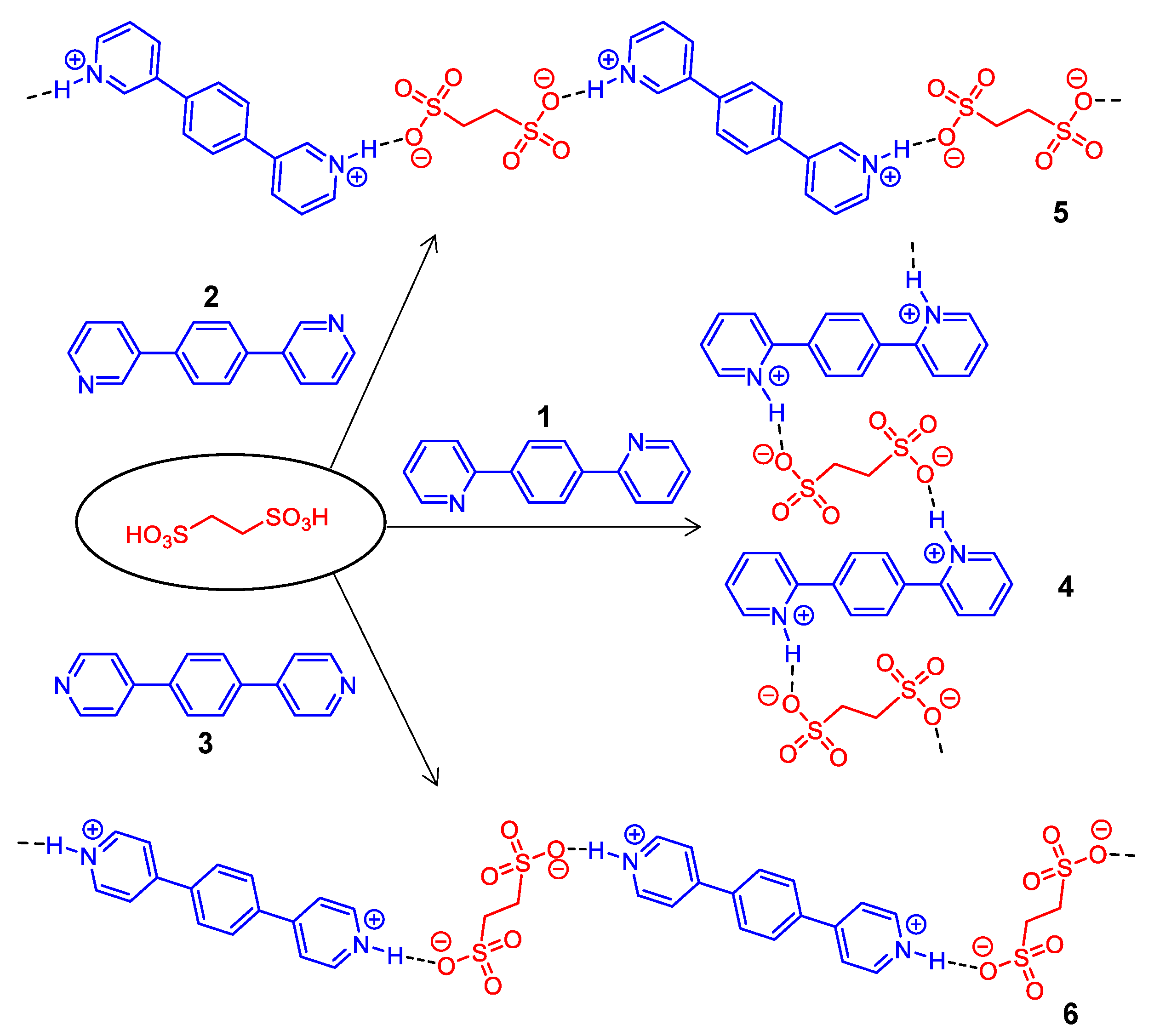
:1. Introduction
2. Materials and Methods
2.1. Materials
2.2. Sample Preparation
2.3. Characterization
2.4. Equipment Information of Physical Measurements
2.5. Methods of Calculation
3. Results
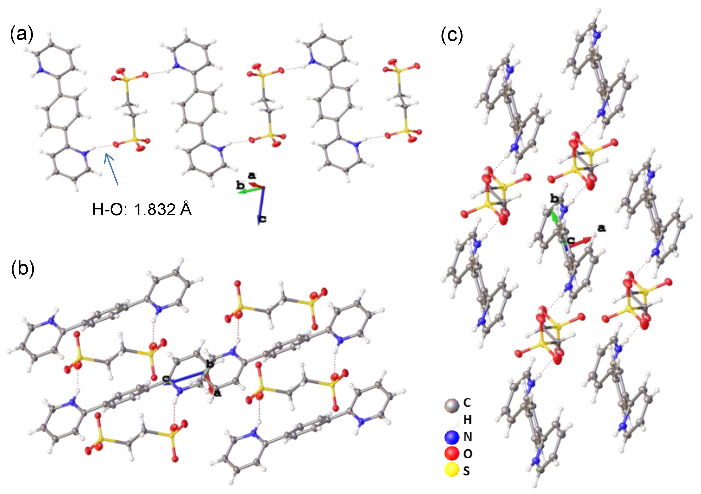
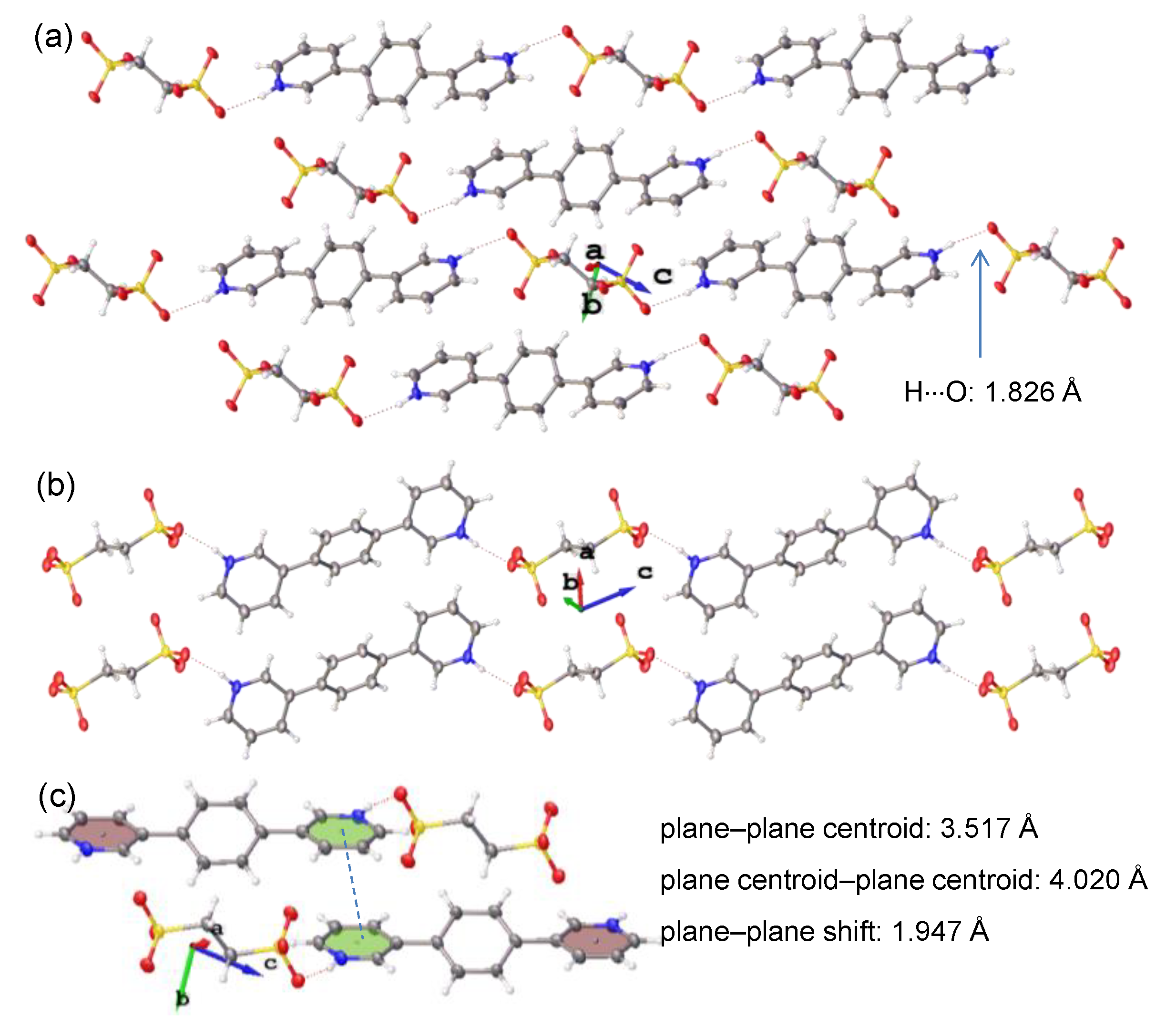
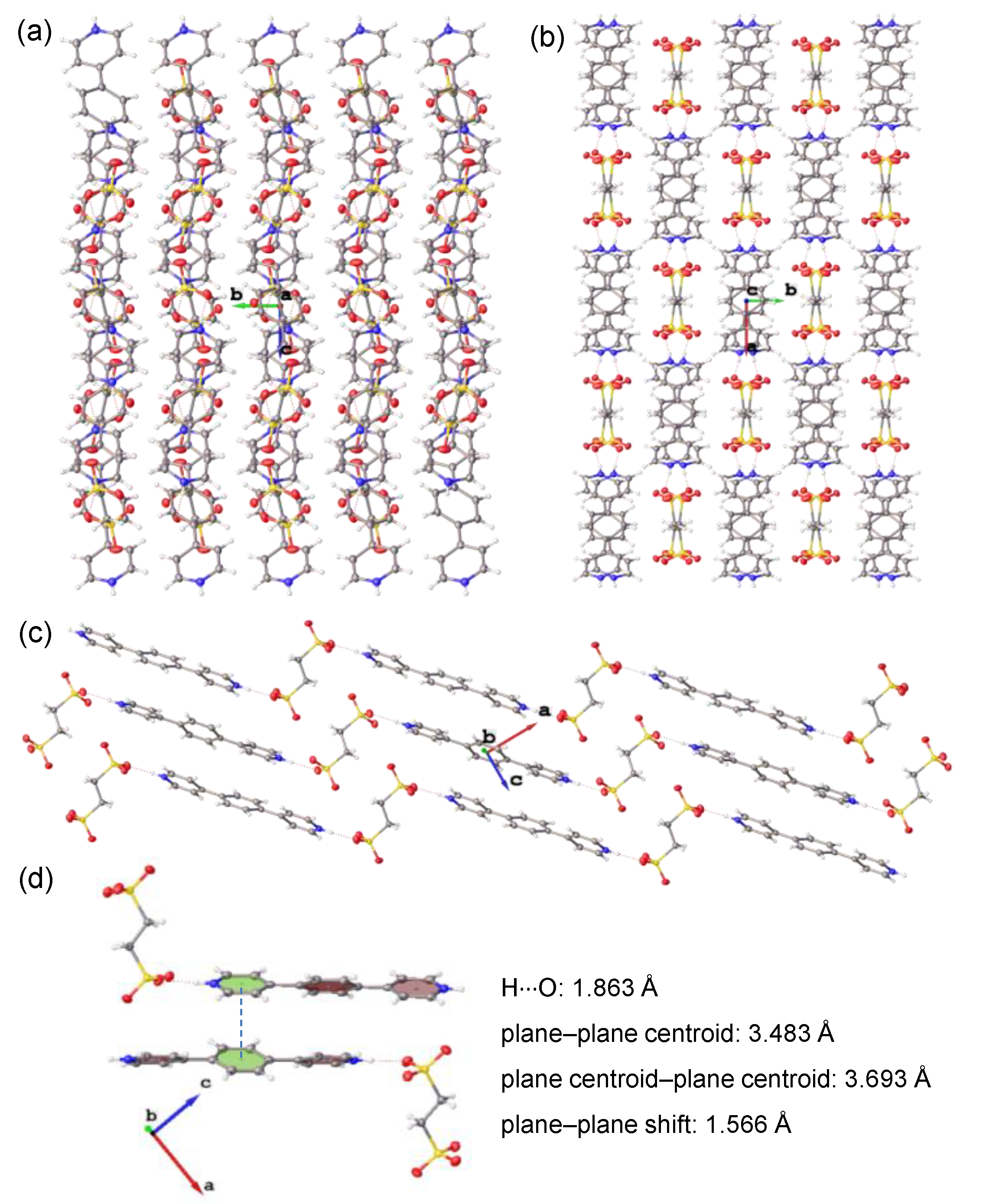
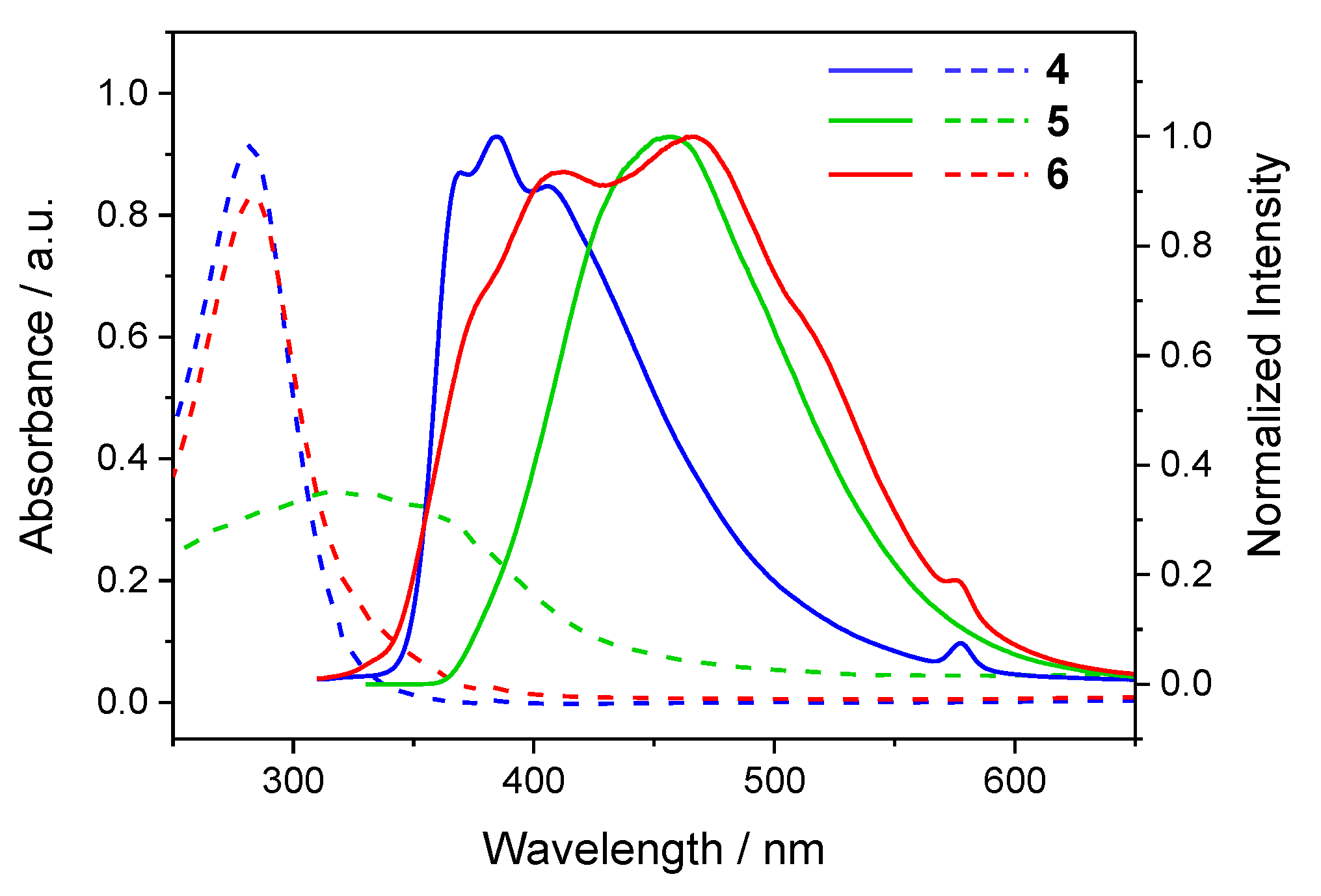
3.1. Studies on Single Crystals
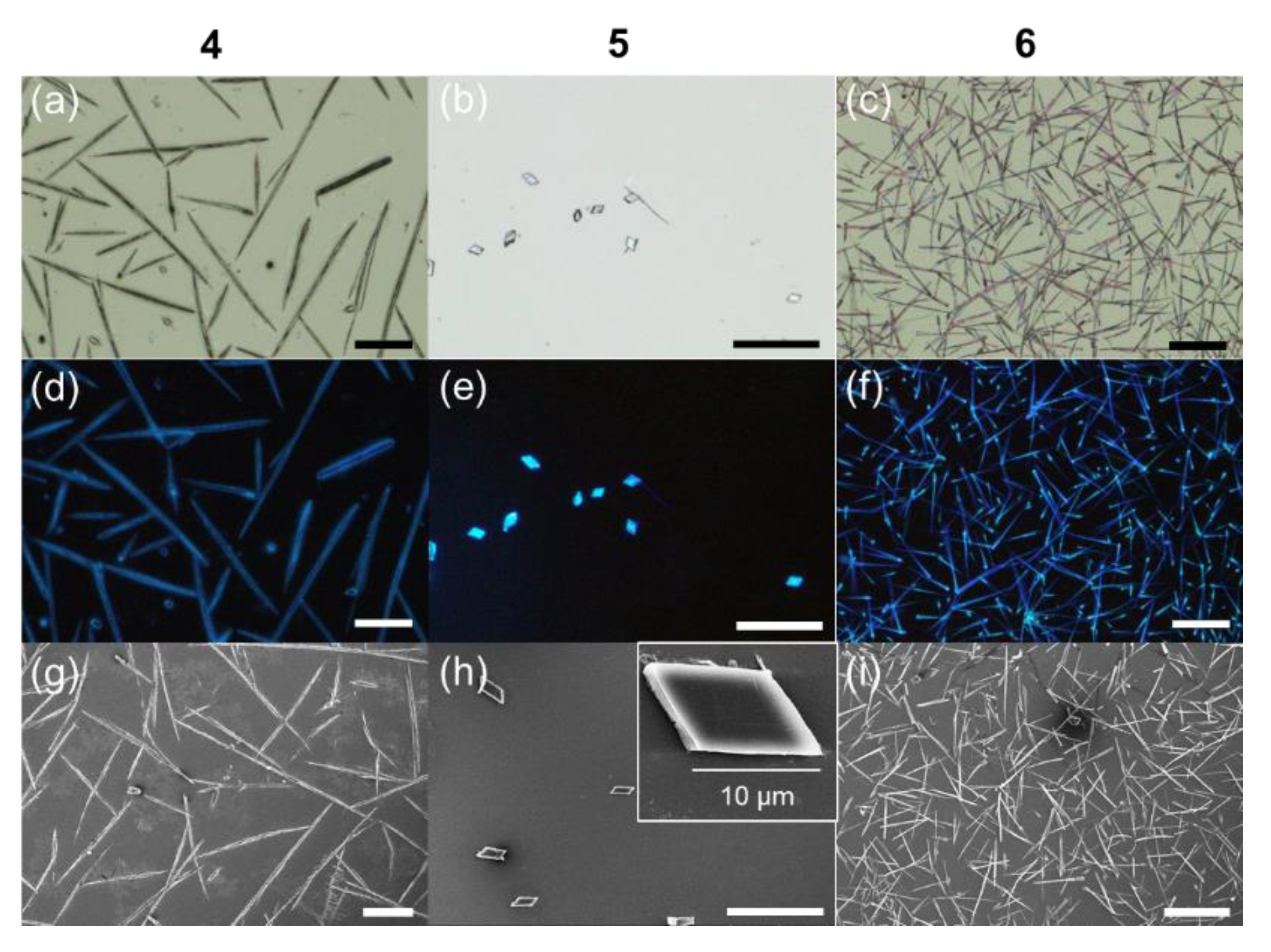
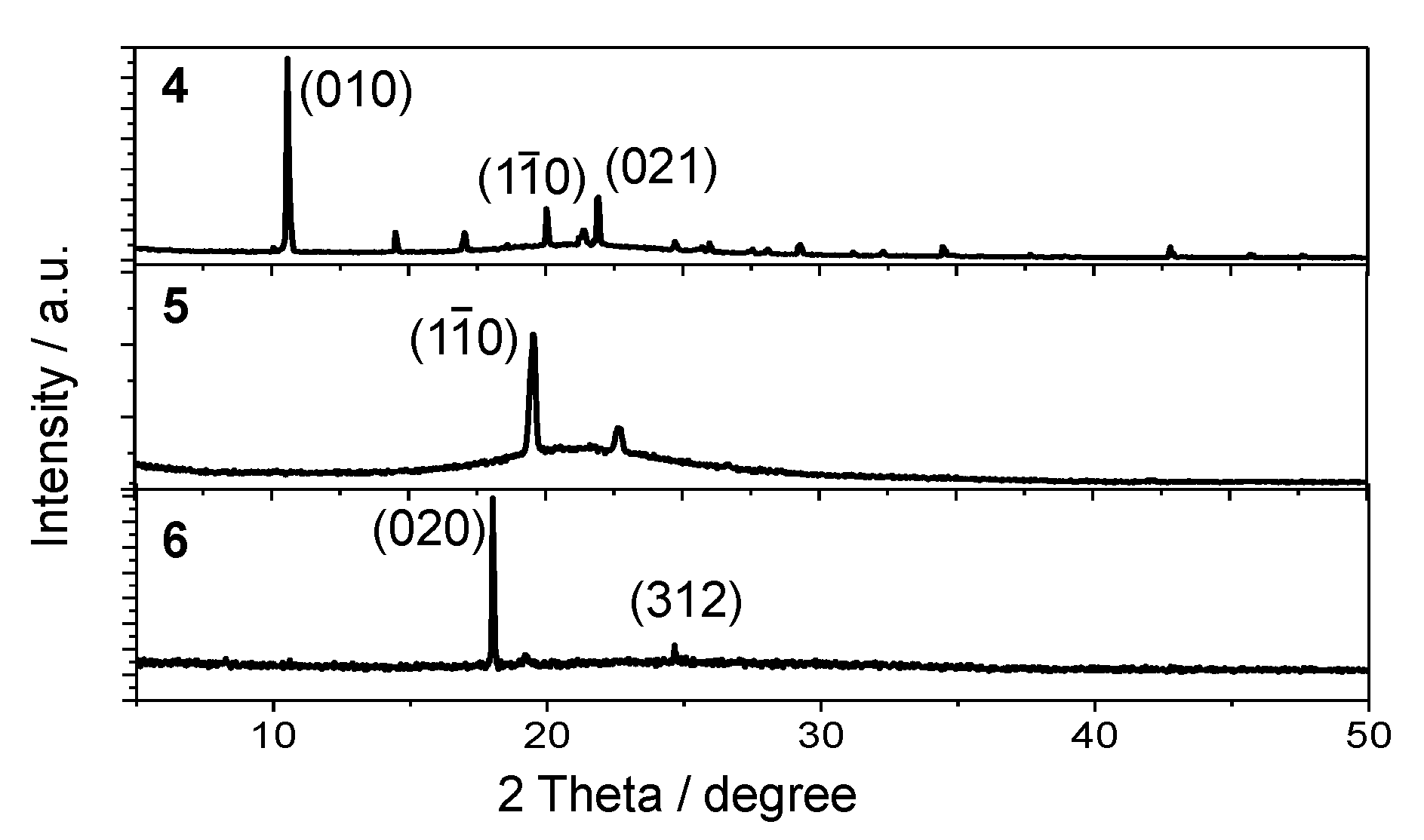
3.2. Studies of Microcrystals
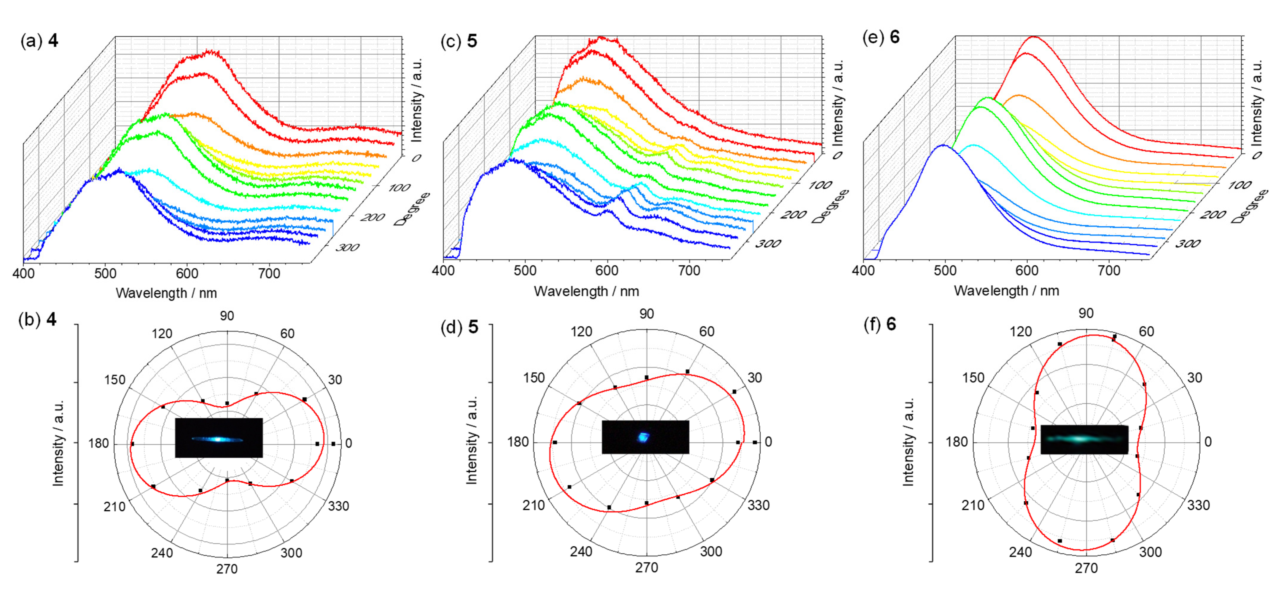
3.3. Polarized Luminescence
4. Conclusions
Supplementary Materials
Author Contributions
Funding
Data Availability Statement
Acknowledgments
Conflicts of Interest
References
- Li, S.; Hao, Y.; Guo, S.; Ding, C.; Ma, Y.; Liu, R.; Yan, D. Three-primary-color molecular cocrystals showing white-light luminescence, tunable optical waveguide and ultrahigh polarized emission. Sci. China Chem. 2022, 65, 408–417. [Google Scholar] [CrossRef]
- Ma, X.; Diroll, B.T.; Cho, W.; Fedin, I.; Schaller, R.D.; Talapin, D.V.; Wiederrecht, G.P. Anisotropic Photoluminescence from Isotropic Optical Transition Dipoles in Semiconductor Nanoplatelets. Nano Lett. 2018, 18, 4647–4652. [Google Scholar] [CrossRef]
- Sun, M.-J.; Liu, Y.; Zeng, W.; Zhao, Y.S.; Zhong, Y.-W.; Yao, J. Photoluminescent Anisotropy Amplification in Polymorphic Organic Nanocrystals by Light-Harvesting Energy Transfer. J. Am. Chem. Soc. 2019, 141, 6157–6161. [Google Scholar] [CrossRef] [PubMed]
- Li, Z.-Q.; Ma, D.-X.; Xu, F.-F.; Dan, T.-X.; Gong, Z.-L.; Shao, J.-Y.; Zhao, Y.S.; Yao, J.; Zhong, Y.-W. Selective, Anisotropic, or Consistent Polarized-Photon Out-Coupling of 2D Organic Microcrystals. Angew. Chem. Int. Ed. 2022, 61, e202205033. [Google Scholar]
- Gong, Z.-L.; Zhu, X.; Zhou, Z.; Zhang, S.-W.; Yang, D.; Zhao, B.; Zhang, Y.-P.; Deng, J.; Cheng, Y.; Zheng, Y.-X.; et al. Frontiers in Circularly Polarized Luminescence: Molecular Design, Self-Assembly, Nanomaterials, and Applications. Sci. China Chem. 2021, 64, 2060–2104. [Google Scholar] [CrossRef]
- Sang, Y.; Han, J.; Zhao, T.; Duan, P.; Liu, M. Circularly Polarized Luminescence in Nanoassemblies: Generation, Amplification, and Application. Adv. Mater. 2020, 32, 1900110. [Google Scholar] [CrossRef] [PubMed]
- Zhao, J.; Zhang, T.; Dong, X.-Y.; Sun, M.-E.; Zhang, Z.; Li, X.; Zhao, Y.S.; Zang, S.-Q. Circularly Polarized Luminescence from Achiral Single Crystals of Hybrid Manganese Halides. J. Am. Chem. Soc. 2019, 141, 15755–15760. [Google Scholar] [CrossRef]
- Qiao, C.; Zhang, C.; Zhou, Z.; Yao, J.; Zhao, Y.S. An Optically Reconfigurable Förster Resonance Energy Transfer Process for Broadband Switchable Organic Single-Mode Microlasers. CCS Chem. 2022, 4, 250–258. [Google Scholar] [CrossRef]
- Zhang, W.; Yao, J.; Zhao, Y.S. Organic Micro/Nanoscale Lasers. Acc. Chem. Res. 2016, 49, 1691–1700. [Google Scholar] [CrossRef]
- Li, R.; Gong, Z.-L.; Zhu, Q.; Sun, M.-J.; Che, Y.; Yao, J.; Zhong, Y.-W. A Pre-Organized Monomer-Reservoir Strategy to Prepare Multidimensional Phosphorescent Organoplatinum Nanocrystals and Suprastructures. Sci. China Chem. 2022, 65, 328–338. [Google Scholar] [CrossRef]
- Yan, Y.; Zhao, Y.S. Organic Nanophotonics: From Controllable Assembly of Functional Molecules to Low-Dimensional Materials with Desired Photonic Properties. Chem. Soc. Rev. 2014, 43, 4325–4340. [Google Scholar] [CrossRef] [PubMed]
- Chen, S.; Wang, X.-D.; Zhuo, M.-P.; Lv, Q.; Liu, J.-F.; Liao, L.-S. Organic white-light sources: Multiscale construction of organic luminescent materials from molecular to macroscopic level. Sci. China Chem. 2022, 65, 740–745. [Google Scholar] [CrossRef]
- Wang, B.; Lin, R.-B.; Zhang, Z.; Xiang, S.; Chen, B. Hydrogen-Bonded Organic Frameworks as a Tunable Platform for Functional Materials. J. Am. Chem. Soc. 2020, 142, 14399–14416. [Google Scholar] [CrossRef]
- Chen, S.; Yin, H.; Wu, J.-J.; Lin, H.; Wang, X.-D. Organic Halogen-Bonded Co-crystals for Optoelectronic Applications. Sci. China Mater. 2020, 63, 1613–1630. [Google Scholar] [CrossRef]
- Pop, F.; Zigon, N.; Avarvari, N. Main-Group-Based Electro- and Photoactive Chiral Materials. Chem. Rev. 2019, 119, 8435–8478. [Google Scholar] [CrossRef] [PubMed]
- Li, Z.-Q.; Gong, Z.-L.; Shao, J.-Y.; Yao, J.; Zhong, Y.-W. Full-Color and White Circularly Polarized Luminescence of Hydrogen-Bonded Ionic Organic Microcrystals. Angew. Chem., Int. Ed. 2021, 60, 14595–14600. [Google Scholar] [CrossRef] [PubMed]
- Tydlitat, J.; Achelle, S.; Rodriguez-Lopez, J.; Pytela, O.; Mikysek, T.; Cabon, N.; Guen, F.R.-l.; Miklik, D.; Ruzickova, Z.; Bures, F. Photophysical Properties of Acid-Responsive Triphenylamine Derivatives Bearing Pyridine Fragments: Towards White Light Emission. Dyes Pigment. 2017, 146, 467–478. [Google Scholar] [CrossRef]
- Li, R.; Gong, Z.-L.; Tang, J.-H.; Sun, M.-J.; Shao, J.-Y.; Zhong, Y.-W.; Yao, J. Triarylamines with Branched Multi-Pyridine Groups: Modulation of Emission Properties by Structural Variation, Solvents, and Tris(pentafluorophenyl)borane. Sci. China Chem. 2018, 61, 545–556. [Google Scholar] [CrossRef]
- Ma, C.; Xu, B.; Xie, G.; He, J.; Zhou, X.; Peng, B.; Jiang, L.; Xu, B.; Tian, W.; Chi, Z.; et al. An AIE-active Luminophore with Tunable and Remarkable Fluorescence Switching Based on the Piezo and Protonation-Deprotonation Control. Chem. Commun. 2014, 50, 7374–7377. [Google Scholar] [CrossRef] [PubMed]
- Zhou, T.; Jia, T.; Zhao, S.; Guo, J.; Zhang, H.; Wang, Y. Acid-Stimuli-Luminescence and Carbonyl-Proton Interaction Dependent Emission Properties of 2,6-Biphenyl-4-pyrone Crystals. Cryst. Growth Des. 2012, 12, 179–184. [Google Scholar] [CrossRef]
- Zhang, J.; Chen, J.; Xu, B.; Wang, L.; Ma, S.; Dong, Y.; Li, B.; Ye, L.; Tian, W. Remarkable Fluorescence Change Based on the Protonation-Deprotonation Control in Organic Crystals. Chem. Commun. 2013, 49, 3878–3880. [Google Scholar] [CrossRef] [PubMed]
- Ma, S.; Zhang, J.; Liu, Y.; Qian, J.; Xu, B.; Tian, W. Direct Observation of the Symmetrical and Asymmetrical Protonation States in Molecular Crystals. J. Phys. Chem. Lett. 2017, 8, 3068–3072. [Google Scholar] [CrossRef] [PubMed]
- Dong, Y.; Ma, S.; Zhang, X.; Qian, J.; Zhu, N.; Xu, B.; Ho, C.-L.; Tian, W.; Wong, W.-Y. A Cyanostilbene-Based Molecule with Aggregation-Induced Emission Properties: Amplified Spontaneous Emission, Protonation Effect and Electroluminescence. Sci. China Chem. 2019, 62, 212–219. [Google Scholar] [CrossRef]
- Yano, Y.; Ono, T.; Hatanaka, S.; Gryko, D.T.; Hisaeda, Y. Salt-Cocrystal Continuum for Photofunction Modulation: Stimuli-Responsive Fluorescence Color-Tuning of Pyridine-Modified Intramolecular Charge-Transfer Dyes and Acid Complexes. J. Mater. Chem. C 2019, 7, 8847–8854. [Google Scholar] [CrossRef]
- Bai, L.; Bose, P.; Gao, Q.; Li, Y.; Ganguly, R.; Zhao, Y. Halogen-Assisted Piezochromic Supramolecular Assemblies for Versatile Haptic Memory. J. Am. Chem. Soc. 2017, 139, 436–441. [Google Scholar] [CrossRef]
- Xing, P.; Li, Y.; Xue, S.; Fiona Phua, S.Z.; Ding, C.; Chen, H.; Zhao, Y. Occurrence of Chiral Nanostructures Induced by Multiple Hydrogen Bonds. J. Am. Chem. Soc. 2019, 141, 9946–9954. [Google Scholar] [CrossRef]
- Sun, L.; Zhu, W.; Wang, W.; Yang, F.; Zhang, C.; Wang, S.; Zhang, X.; Li, R.; Dong, H.; Hu, W. Intermolecular Charge-Transfer Interactions Facilitate Two-Photon Absorption in Styrylpyridine–Tetracyanobenzene Cocrystals. Angew. Chem. Int. Ed. 2017, 56, 7831–7835. [Google Scholar] [CrossRef]
- Li, S.; Lu, B.; Fang, X.; Yan, D. Manipulating Light-Induced Dynamic Macro-Movement and Static Photonic Properties within 1D Isostructural Hydrogen-Bonded Molecular Cocrystals. Angew. Chem. Int. Ed. 2020, 59, 22623–22630. [Google Scholar] [CrossRef]
- Gadade, D.D.; Kulkarni, D.A.; Rathi, P.B.; Pekamwar, S.S.; Joshi, S.S. Solubility Enhancement of Lornoxicam by Crystal Engineering. Indian J. Pharm. Sci. 2017, 79, 277–286. [Google Scholar] [CrossRef]
- Pekamwar, S.; Kulkarni, D.; Gadade, D. Accidental Formation of Eutectics during Crystal Engineering of Lamotrigine with Solubility Advantage and Drug Release Efficiency. Asian J. Pharm. 2021, 15, 60–67. [Google Scholar]
- Pekamwar, S.S.; Kulkarni, D.A. Development and Evaluation of Bicomponent Cocrystals of Aceclofenac for Efficient Drug Delivery with Enhanced Solubility and Improved Dissolution. Indian Drugs 2021, 58, 54–60. [Google Scholar] [CrossRef]
- Vishweshwar, P.; Nangia, A.; Lynch, V.M. Recurrence of Carboxylic Acid-Pyridine Supramolecular Synthon in the Crystal Structures of Some Pyrazinecarboxylic Acids. J. Org. Chem. 2002, 67, 556–565. [Google Scholar] [CrossRef] [PubMed]
- Bhogala, B.R.; Nangia, A. Cocrystals of 1,3,5-Cyclohexanetricaboxylic Acid with 4,4’-Bipyridine Homologues: Acid∙Pyridine Hydrogen Bonding in Neutral and Ionic Complexes. Cryst. Growth Des. 2003, 3, 547–554. [Google Scholar] [CrossRef]
- Lemmerer, A.; Govindraju, S.; Johnston, M.; Motloung, X.; Savig, K.L. Co-crystals and molecular salts of carboxylic acid/pyridine complexes: Can calculated pKa’s predict proton transfer? A case study of nine complexes. Cryst. Eng. Comm. 2015, 17, 3591–3595. [Google Scholar] [CrossRef] [Green Version]
- Mohamed, S.; Tocher, D.A.; Vickers, M.; Karamertzanis, P.G.; Price, S.L. Salt or Cocrystal? A New Series of Crystal Structures Formed from Simple Pyridines and Carboxylic Acids. Cryst. Growth Des. 2009, 9, 2881–2889. [Google Scholar] [CrossRef]
- Feng, H.-T.; Xiong, J.-B.; Luo, J.; Feng, W.-F.; Yang, D.; Zheng, Y.-S. Selective Host-Guest Co-crystallization of Pyridine-Functionalized Tetraphenylethylenes with Phthalic Acids and Multicolor Emission of the Co-Crystals. Chem. Eur. J. 2017, 23, 644–651. [Google Scholar] [CrossRef] [PubMed]
- Sui, L.-Z.; Yang, W.-W.; Yao, C.-J.; Xie, H.-Y.; Zhong, Y.-W. Charge Delocalization of 1,4-Benzenedicyclometalated Ruthenium: A Comparison between Tris-bidentate and Bis-tridentate Complexes. Inorg. Chem. 2012, 51, 1590–1598. [Google Scholar] [CrossRef]
- Sudan, S.; Fadaei-Tirani, F.; Scopelliti, R.; Ebbert, K.E.; Clever, G.H.; Severin, K. LiBF4-Induced Rearrangement and Desymmetrization of a Palladium-Ligand Assembly. Angew. Chem. Int. Ed. 2022, 61, e202201823. [Google Scholar] [CrossRef]
- Liu, Y.; Hu, H.; Xu, L.; Qiu, B.; Liang, J.; Ding, F.; Wang, K.; Chu, M.; Zhang, W.; Ma, M.; et al. Orientation-Controlled 2D Anisotropic and Isotropic Photon Transport in Co-crystal Polymorph Microplates. Angew. Chem. Int. Ed. 2020, 59, 4456–4463. [Google Scholar] [CrossRef]
- Zhuo, M.-P.; Tao, Y.-C.; Wang, X.-D.; Wu, Y.; Chen, S.; Liao, L.-S.; Jiang, L. 2D Organic Photonics: An Asymmetric Optical Waveguide in Self-Assembled Halogen-Bonded Cocrystals. Angew. Chem. Int. Ed. 2018, 57, 11300–11304. [Google Scholar] [CrossRef]
- Xu, F.-F.; Zeng, W.; Sun, M.-J.; Gong, Z.-L.; Li, Z.-Q.; Zhao, Y.S.; Yao, J.; Zhong, Y.-W. Organoplatinum(II) Cruciform: A Versatile Building Block to Fabricate 2D Microcrystals with Full-Color and White Phosphorescence and Anisotropic Photon Transport. Angew. Chem. Int. Ed. 2022, 61, e202116603. [Google Scholar]
- Lyu, Z.-Y.; Dong, H.; Yang, X.-F.; Sun, L.-D.; Yan, C.-H. Highly Polarized Upconversion Emissions from Lanthanide-Doped LiYF4 Crystals as Spatial Orientation Indicators. J. Phys. Chem. Lett. 2021, 12, 11288–11294. [Google Scholar] [CrossRef] [PubMed]
- Lee, C.; Yang, W.; Parr, R.G. Development of the Colle-Salvetti correlation-energy formula into a functional of the electron density. Phys. Rev. B Condens. Matter Mater. Phys. 1988, 37, 785–789. [Google Scholar] [CrossRef] [PubMed]
- Li, Z.-Z.; Tao, Y.-C.; Wang, X.-D.; Liao, L.-S. Organic Nanophotonics: Self-Assembled Single-Crystalline Homo-/Heterostructures for Optical Waveguides. ACS Photonics 2018, 5, 3763–3771. [Google Scholar] [CrossRef]


| Compound | 4 | 5 | 6 |
|---|---|---|---|
| CCDC number | 2181166 | 2208263 | 2181168 |
| empirical formula | C18H18N2O6S2 | C18H18N2O6S2 | C18H18N2O6S2 |
| formula weight | 422.46 | 422.46 | 422.46 |
| temperature (K) | 170.00(10) | 170.00(10) | 169.99(10) |
| crystal system | Triclinic | Triclinic | Monoclinic |
| space group | P-1 | P-1 | P2/c |
| a (Å) | 5.18630(10) | 5.4682(3) | 16.0369(2) |
| b (Å) | 8.3578(2) | 8.5731(4) | 9.90610(10) |
| c (Å) | 10.4965(2) | 9.9083(5) | 11.22670(10) |
| α (̊) | 82.122(2) | 81.978(4) | 90 |
| β (̊) | 86.845(2) | 75.842(5) | 90.3780(10) |
| γ (̊) | 88.969(2) | 87.649(4) | 90 |
| V (Å3) | 449.980(16) | 445.97(4) | 1783.47(3) |
| Z value | 1 | 1 | 4 |
| density (g/cm3) | 1.559 | 1.573 | 1.573 |
| R1 (final) | 0.0326 | 0.0421 | 0.0310 |
| wR2 (final) | 0.0889 | 0.1177 | 0.0815 |
| R1 (all) | 0.0329 | 0.0438 | 0.0325 |
| wR2 (all) | 0.0892 | 0.1192 | 0.0826 |
Publisher’s Note: MDPI stays neutral with regard to jurisdictional claims in published maps and institutional affiliations. |
© 2022 by the authors. Licensee MDPI, Basel, Switzerland. This article is an open access article distributed under the terms and conditions of the Creative Commons Attribution (CC BY) license (https://creativecommons.org/licenses/by/4.0/).
Share and Cite
Zhao, J.-Y.; Xu, F.-F.; Li, Z.-Q.; Gong, Z.-L.; Zhong, Y.-W.; Yao, J. Molecular Cocrystals with Hydrogen-Bonded Polymeric Structures and Polarized Luminescence. Materials 2022, 15, 7247. https://doi.org/10.3390/ma15207247
Zhao J-Y, Xu F-F, Li Z-Q, Gong Z-L, Zhong Y-W, Yao J. Molecular Cocrystals with Hydrogen-Bonded Polymeric Structures and Polarized Luminescence. Materials. 2022; 15(20):7247. https://doi.org/10.3390/ma15207247
Chicago/Turabian StyleZhao, Jing-Yi, Fa-Feng Xu, Zhong-Qiu Li, Zhong-Liang Gong, Yu-Wu Zhong, and Jiannian Yao. 2022. "Molecular Cocrystals with Hydrogen-Bonded Polymeric Structures and Polarized Luminescence" Materials 15, no. 20: 7247. https://doi.org/10.3390/ma15207247
APA StyleZhao, J.-Y., Xu, F.-F., Li, Z.-Q., Gong, Z.-L., Zhong, Y.-W., & Yao, J. (2022). Molecular Cocrystals with Hydrogen-Bonded Polymeric Structures and Polarized Luminescence. Materials, 15(20), 7247. https://doi.org/10.3390/ma15207247