Article

Enhanced Photocatalytic Activity of Spherical Nd3+ Substituted ZnFe2O4 Nanoparticles

1
Faculty of Chemistry, Thai Nguyen University of Education, Thai Nguyen 24000, Vietnam
2
Faculty of Fundamental Sciences, Thai Nguyen University of Technology, Thai Nguyen 24000, Vietnam
3
NTT Hi-Tech Institute, Nguyen Tat Thanh University, Ho Chi Minh 700000, Vietnam
4
Center of Excellence for Green Energy and Environmental Nanomaterials, Nguyen Tat Thanh University, Ho Chi Minh 700000, Vietnam
*
Authors to whom correspondence should be addressed.
Materials 2021, 14(8), 2054; https://doi.org/10.3390/ma14082054
Submission received: 4 March 2021 / Revised: 15 April 2021 / Accepted: 16 April 2021 / Published: 19 April 2021

Abstract

:
In this study, nanocrystalline ZnNdxFe2−xO4 ferrites with x = 0.0, 0.01, 0.03 and 0.05 were fabricated and used as a catalyst for dye removal potential. The effect of Nd3+ ions substitution on the structural, optical and photo-Fenton activity of ZnNdxFe2−xO4 has been investigated. The addition of Nd3+ ions caused a decrease in the grain size of ferrites, the reduction of the optical bandgap energies and thus could be well exploited for the catalytic study. The photocatalytic activity of the ferrite samples was evaluated by the degradation of Rhodamine B (RhB) in the presence of H2O2 under visible light radiation. The results indicated that the ZnNdxFe2−xO4 samples exhibited higher removal efficiencies than the pure ZnFe2O4 ferrites. The highest degradation efficiency was 98.00%, attained after 210 min using the ZnNd0.03Fe1.97O4 sample. The enhanced photocatalytic activity of the ZnFe2O4 doped with Nd3+ is explained due to the efficient separation mechanism of photoinduced electron and holes. The effect of various factors (H2O2 oxidant concentration and catalyst loading) on the degradation of RhB dye was clarified.

Graphical Abstract

1. Introduction

It has been proved that the discharge of organic compounds, including dyes, from manufacturing plants led to growing contamination in the aquatic ecosystem [1,2]. There are numerous impacts of color pollution, so more and more techniques have been found to address this environmental problem [3]. Among them, photocatalysis, which relies on semiconductors and irradiation-based degradation of organic substances, is an effective method [4,5]. The benefits of this approach include environmental friendliness, the potential to entirely decompose organic pollutants into inorganic molecules, i.e., CO2 and H2O. TiO2 [6,7,8], WO3 [9,10,11,12,13], and BiVO4 [14,15] are typical photocatalysts that have been well studied for dyes degradation. Nano ferrites, another material, have recently received a great deal of interest due to their high stability, strong magnetic properties, and high catalytic performance. In particular, such nanoparticles may be used as a photocatalyst under visible light in wastewater treatment thanks to their narrow band gap [16,17]. In addition, the method used for ferrite synthesis may differ depending on the desired characteristics, and a number of synthesis routes have been investigated so far, like solvothermal [18], sol-gel [19], coprecipitation [20], and combustion method [21]. For example, Xiaojun Guo et al. [17] reported that the NiFe2O4 hollow nanospheres synthesized by solvothermal method had a high photoactivity for methylene blue (MB) degradation, which achieved the removal efficiency of approximately 98.5% only within 50 min in the presence of 5 mM H2O2 and 0.06 g·L−1 H2C2O4. In other extensive studies on ferrite, ZnFe2O4 nanomaterial has been documented to be effective in removing a wide range of organic compounds, such as Orange II [22], Red 88, Acid Orange 8, Malachite Green [23], Congo red [24], methylene blue [25] and tetracycline [26]. Besides, MnFe2O4, CoFe2O4, CuFe2O4 and MgFe2O4 nanospinels have been reported to show effective photocatalytic activity to eliminate distinct categories of dyes [19,20,27,28]. Because of the fact that ferrite catalysts can be easily recoverable using an external magnetic field, these promising potentials have rendered any successful effort to improve their photocatalytic efficiency in substantiating their practical system uses [29].
It was previously noted that the structural features and magnetic, electrical characteristics and catalytic activity of ferrites could be dependent on the metals in the ferrite lattice structure [30]. In addition, several studies have reported the enhancement in the catalytic activity of ferrites with different metal substitution and change in cation distribution. For instance, the photocatalytic activity of cobalt zinc ferrite systems on Mn substitution has been documented by Santosh Bhukal et al. [31], showing that the decolorization ratio of methyl orange is enhanced along with increasing Mn3+ ions content. In another study, MgFe2O4 doped with Co2+ ions by modified sol-gel combustion method exhibited higher degradation efficiency for methylene blue in comparison with that of pure MgFe2O4 sample [32]. Similar results were obtained with nickel ferrite when substituted with Zn [33].
On the other hand, the substitution that gained attention in doping ferrite iron was rare-earth ions [34,35,36,37,38]. Since these metals have a strong spin-orbit coupling of angular momentum due to the presence of unpaired electrons in the 4f orbitals, their interaction with ferrites occurs in 3d-4f coupling, resulting in magneto-crystalline anisotropy and thus influencing magnetic, electrical and catalytic features of substituted ferrites [39]. Mariosi et al. found that cobalt ferrite nanoparticles substituted by La3+ ions exhibited structural changes in terms of cationic arrangement of the spinel structure [38]. This change resulted in a decrease in coercivity values and an increase in the surface area. The substitution of other rare-earth ions such as La3+, Nd3+, Gd3+ and Dy3+ into the [B] sites containing iron has been shown to displace Fe3+ into (A) sites, thus altering the structure and electrical and magnetic characteristics the ferrites [34,35,37,39]. Sharma et al. have carried out one prominent study showing the ability of rare-earth doping to boost catalytic activity [40]. Specifically, rare-earth (La3+, Ce3+) substituted CoFe2O4 exhibited higher efficiencies in the elimination of five model pollutants, possibly due to the presence of Ce3+/Ce4+ redox pair. The synthesis of samarium (Sm3+) substituted manganese ferrite nanoparticles (MnFe2-xSmxO4) using oleic acid as a surfactant was reported by Rashmi et al. [41]. Such synthesized nanomaterials were tested for photocatalytic degradation of colors under visible light irradiation. Such synthesized nanomaterials were tested for photocatalytic degradation of colors under visible light irradiation. The result indicated that samarium replacement significantly increased the photocatalytic activity of MnFe2O4 nanoparticles. The value of x varied from 0, 0.5, 1.0, 1.5 and 2.0, and the best results were obtained at x = 1.5. The higher activity of x = 1.5 was related to its minimum band gap energy value (1.64 eV). After that, Patil et al. [42] synthesized Gd3+ doped ZnFe2O4 (ZnFe2−xGdxO4) nanoparticles via coprecipitation method. Synthesized photocatalysts were checked for MB photo-degradation, resulting in enhanced degradation of MB, from about 95 to 99% in the presence of photocatalysts ZnFe2−xGdxO4 (x = 0, 0.3, 0.5 and 0.7) along with 8 ppm of H2O2. The efficiency level was found higher than that of the pristine ZnFe2O4 and could be attributable to the fact that Fermi energy levels of substituted catalyst were just below the conduction band within the energy band gap. In addition, formation of lattice strains due to the difference between ionic radii of Gd3+ (0.94 Å) and that of Fe3+ (0.78 Å) is also partially responsible for the enhancement. However, the impact of rare earth substitution (e.g., Nd) on the photo-Fenton activity of ferrites for dye degradation is still a gap in the literature.
The present study aims to investigate the structural and catalytic properties of ferrites by the doping substitution of Nd3+ ions. ZnFe2O4 was incorporated with various Nd3+ molar ratio (0–0.05 mol%) using urea as a fuel additive. The as-synthesized ferrite was then characterized using several techniques (XRD, SEM, TEM, EDX, and FT-IR before being tested for photocatalytic activities toward Rhodamine B. The effect of catalyst loading, H2O2 concentration, and contact time on the photo catalytic activity of ZnFe2O4 nanoparticles was surveyed meticulously.

2. Materials and Methods

2.1. Synthesis of ZnNdxFe2−xO4 Nanoparticles

Firstly, urea coprecipitation method was adopted to fabricate nanocrystalline ZnNdxFe2−xO4 (x = 0, 0.01, 0.03, 0.05) ferrites according to a previous publication with a moderate modification [34]. Analytical grade zinc nitrate tetrahydrate [Zn(NO3)2·4H2O, 98% pure, Sigma-Aldrich, Darmstadt, Germany], iron(II) nitrate nonahydrate [Fe(NO3)3·9H2O, 99.9% pure, Sigma-Aldrich], and neodymium(III) nitrate hexahydrate (Nd(NO3)3·6H2O, 99.9% pure, Sigma-Aldrich) were used as oxidizer and urea (CH4N2O, >99% pure, Sigma-Aldrich) was used as a fuel additive. A number of proper ions: Zn2+ (1 mmol) combined with Fe3+ [(2−x) mol] and Nd3+ (x mol) (x = 0, 0.01, 0.03, 0.05) were dissolved in distilled water. The final pH of the solution was adjusted at 5.0, and then was heated to 100 °C for 1 h. The precipitation was collected, washed with distilled water (3 × 20 mL) to eliminate metal ion and anion traces and then calcinated up to 500 °C for 2 h (ramping rate of 10 °C/min). The product was ground and stored at a vacuum container.

2.2. Characterization

The as-synthesized particles were characterized using a number of techniques including X-ray diffraction (XRD), Fourier-transform infrared spectroscopy (FT-IR), scanning electron microscope (SEM), transmission electron microscopy (TEM), energy dispersive X-ray spectroscopy (EDX) and UV-Vis absorption spectroscopy. Respective instruments for those analyses include D8 Advance diffractometer (Brucker, Madison, WI, USA) with CuKα radiation (λ = 1.5406 Å) in a 2θ angle ranging from 20° to 70° with a step of 0.03° source, FTIR Affinity-1S (Shimadzu, Kyoto, Japan), Hitachi S-4800 (Tokyo, Japan), JEOL-JEM-1010 (Tokyo, Japan), JEOL JED 2300 Analysis Station (Tokyo, Japan) and U-4100 (Hitachi, Tokyo, Japan) operating in the wavelength range of 200–800 nm.

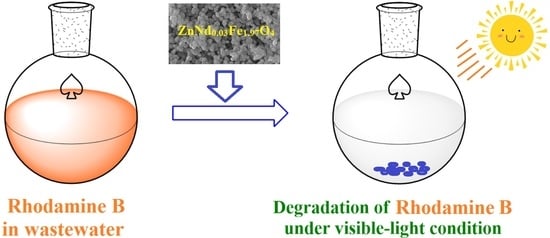
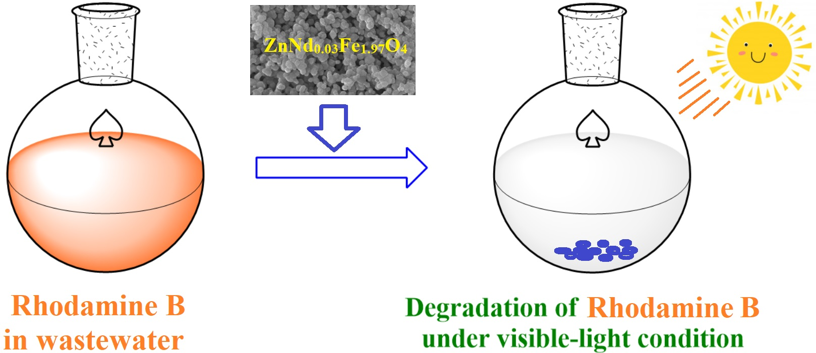
2.3. Photocatalytic Degradation of Rhodamine B

In this study, Rhodamine B (RhB) was used as target pollutant to assess the photocatalytic potential of as-synthesized ferrite samples. Accordingly, the RhB degradation reaction was carried out in a reactor containing the ZnNdxFe2−xO4 (x = 0, 0.01, 0.03, 0.05) nanoparticles and RhB dye under visible light irradiation (using 30 W Led lamps, Philips, Amsterdam, Netherlands). In a typical experiment, 0.1 g of catalyst was introduced into 200 mL of RhB aqueous solution (10 mg·L−1) and suspended on a shaker table at 200 rpm. The suspension was first stirred in the dark for 30 min to attain the adsorption-desorption equilibrium state between the catalyst and RhB. Afterwards, the reaction was stirred and H2O2 30% (w/w) in H2O (Sigma-Aldrich) was added to the mixture, which was then irradiated under visible light for 210 min. To determine the RhB concentration in the mixture, 5 mL of each aliquot was taken out periodically, then centrifuged to remove the solid catalyst. The effect of two factors including H2O2 concentrations (0.02 M, 0.04 M and 0.06 M) and the catalyst dosages (0.5, 0.75 and 1.0 g/L) on the photo-degradation efficiency was studied.
The efficient degradation of RhB (H) was calculated according to the formula Equation (1).
H = C o C t C o × 100
where Co and Ct are the concentration of RhB (mg·L−1) at the time 0 and t. The samples were measured by scanning at the maximum wavelength λ = 553 nm.

3. Results

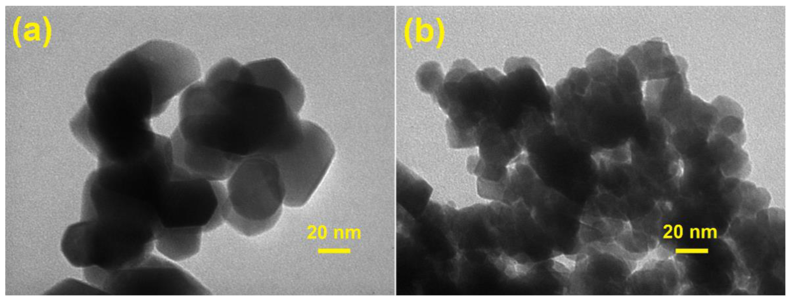
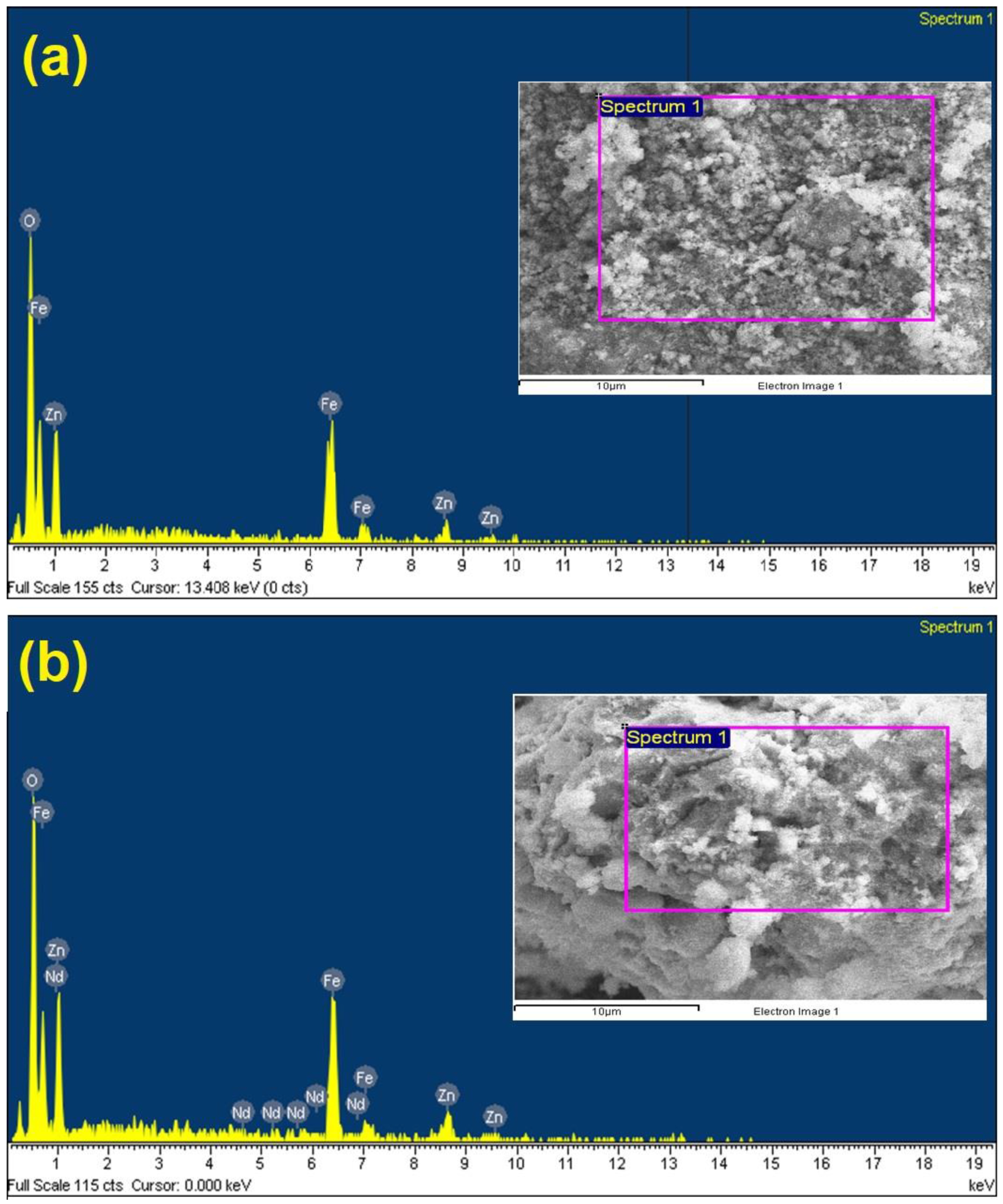
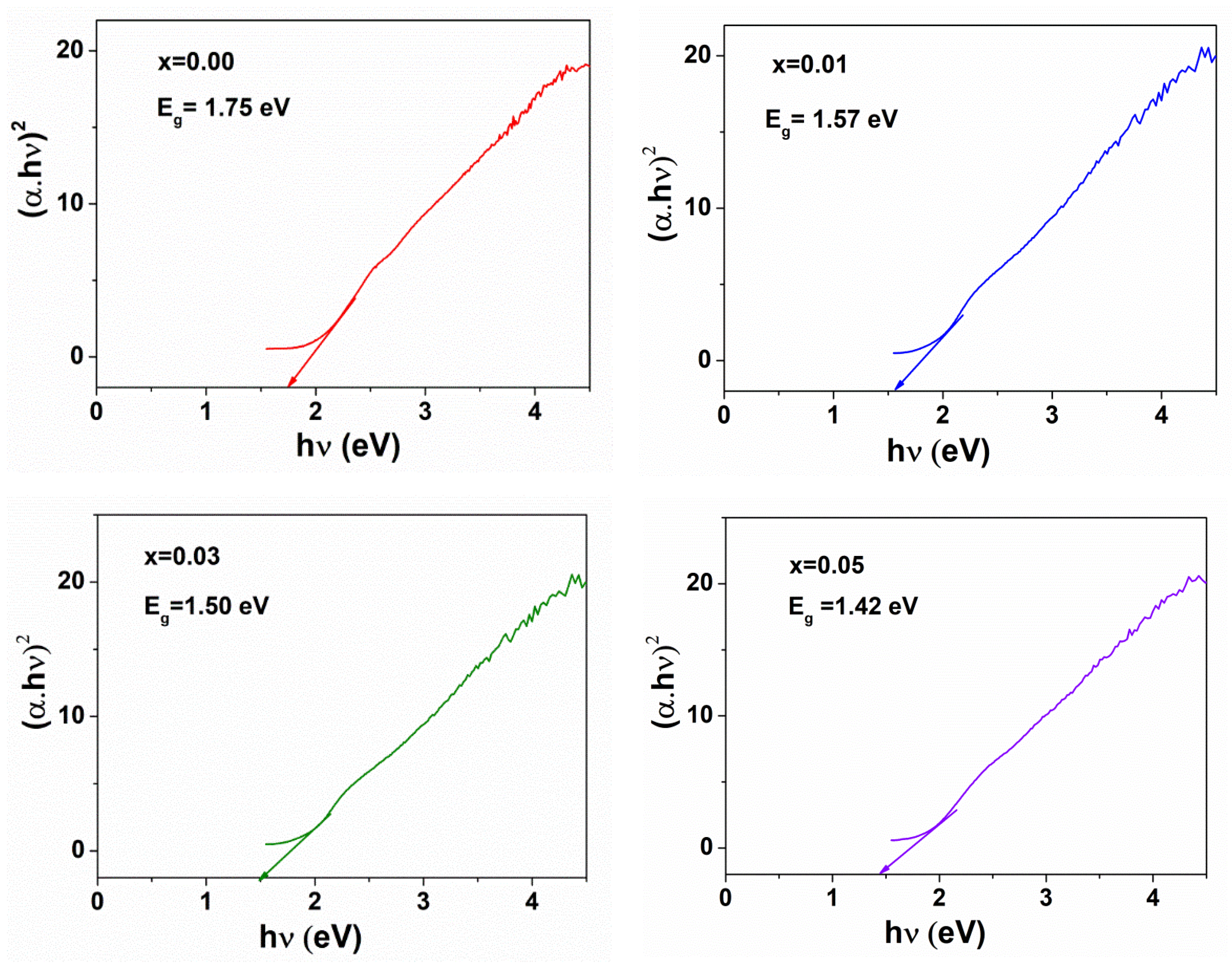
3.1. Characterization

Figure 1 illustrates XRD patterns of ZnFe2O4 and different ZnNdxFe2−xO4 (x = 0.01, 0.03, 0.05) samples synthesized at 500 °C. The formation of zinc ferrite (JCPDS number 022-1012) was evidenced by reflection peaks corresponding to the characteristic spacing between (220), (311), (400), (422), (511) and (440) planes of a cubic spinel structure. Employing Scherrer’s equation, average crystallite size of the samples could be calculated as follows:
D XRD = k λ β cos θ
where λ, k, β and θ wavelength of the X-ray (0.1504 nm), the Scherrer’s constant (k = 0.89), the full width at half maximum observed in radians and the angle of diffraction of the (311) peak with the highest intensity, respectively.
To determine the lattice constant (a) at the most intense peak (311), following formula was used
a = d hkl h 2 + k 2 + l 2
where d is interplanar distance and h, k, l are Miller indices. The change in crystalline structure with doping of Nd3+ ions could be observed from the XRD data given in Table 1. It clearly indicates that the average crystallite sizes significantly decrease from 22 mm to 12 nm with increasing the content of Nd3+ from 0 to 0.05 mol, which was in good agreement with a previous publication [36]. Moreover, the lattice constants for the samples of zinc ferrites nanoparticles increase slightly from 8.43 to 8.45 Å as the amount of Nd3+ added increases. This outcome can be due to the difference of the radius of ferrites, or more specifically, metal ions radius (Nd3+, Zn2+, Fe3+). Indeed, the ionic radius of Nd3+ ion (0.98 Å) is larger than the ionic radius of Zn2+ (0.74 Å) and Fe3+ (0.67 Å); hence Nd3+ ions prefer to occupy more octahedral sites (B-sites) than Fe3+ ions [36]. It is likely for Nd3+ ions to be distributed in the grain boundaries, thus contributing to the improvement of energy barrier of Zn2+ or Fe3+ movement [38]. As a result, the growth of ferrites nanoparticles grains and the crystallite size of zinc ferrites tends to decrease while their crystal lattice constant increases. The same phenomena in decreasing crystallite size due to increasing rare-earth ions content have been observed previously in cobalt ferrites [38,39], nickel ferrites [37] and zinc ferrites [36]. To sum up, the dope of Nd3+ showed a significant effect on the crystalline nanostructure of origin zinc ferrites.
Chemical bonds diagnosed from the FT-IR spectra in Figure 2 can suggest two most characteristic bands for the as-synthesized ZnNdxFe2−xO4 nanoparticles. The first band is located at 522.7−528.6 cm−1 (Table 1), which corresponds to the stretching vibration in the tetrahedral bonding of Zn-O [26,37]. The other band appeared at 418.5–451.3 cm−1, which is attributable to the stretching frequency of the octahedral bonding of Fe−O and Nd−O. The change in the lattice parameters can reflect the shift of the band vibrations. The position and intensity of ν1 and ν2 tend to change with increasing Nd3+ ions content. Finally, the frequency change confirms the presence of the Nd3+ ions occupying the octahedral sites in the ferrites lattice.
To better understand the structure of samples, the morphology of ZnNdxFe2−xO4 nanoparticles is observed by SEM technique. The samples including pure ZnFe2O4 and synthesized ZnNdxFe2−xO4 nanoparticles (x = 0.01; 0.03 and 0.05) all display a type of uniform sphere (Figure 3). The crystallite size of the zinc ferrites decreases with increasing Nd content, which is consistent with the result of XRD analysis.
Figure 4 displays the TEM photomicrography of the pure ZnFe2O4 and ZnNd0.03Fe1.97O4 annealed at 500 °C. Both samples ZnNd0.03Fe1.97O4 and ZnFe2O4 exhibit mostly homogeneous microspheres. In particular, the agglomeration or clustering of these microspheres is rarely observed. Although the effect of Nd3+ ions on the morphology is almost inconsiderable, the particle size of ZnNd0.03Fe1.97O4 sample is smaller than that of the ZnFe2O4 sample. The grain size from TEM studied is the close agreement with the XRD data and SEM photomicrography. Moreover, the chemical composition of samples was confirmed by EDX spectra. The presence of all elements in the XRD profile indicates that synthesized material was of high purity (Figure 5).
The band gap of the spinel nanoparticles was determined by DRS. Kubelka-Munk model was used to calculate band gaps (Eg) of zinc ferrites nanoparticles with the absorption coefficient (α) obtainable from DRS spectra as Equation (4).
F ( R ) = α = ( 1 R ) 2 2 R
where, F(R) represents the Kubelka-Munk function, α is the absorption coefficient and R is the reflectance. The following relationship could be used to determine the band gap energy (Eg) as shown in Equation (5).
α h ν = A ( h ν E g ) n
where, hν: energy of the photon, α: the the absorption coefficient, A: material parameter and n: transition parameter, n = 2 represent indirect transitions. The slope of plotting (αhν)2 against hν could be used to measure the band gap energy for the absorption peak, as shown in Figure 6. The band gap values of ZnNdxFe2−xO4 (x = 0, 0.01, 0.03, 0.05) nanoparticles are found to be 1.75, 1.57, 1.50 and 1.42 eV, respectively. This indicates that the Nd3+ ions concentration affected the band gap energy of zinc ferrites nanoparticles. The band gap energy value decreased with increasing the Nd3+ ions concentration. Due to the larger ionic radius, the crystal lattice is bound to distort leading to generation of interface defects [38]. In zinc ferrites nanoparticles, the orbital overlapping between O-2p and Fe-3d energy levels caused the formation of the energy band gap. There is the 4f Fermi energy level of Nd in ZnNdxFe2−xO4 samples, thus resulting in decreased band gap energy value [42,43]. The optical band gap of the CoFe2O4 samples doped with La decreases from 1.35 to 1.1 eV [38].

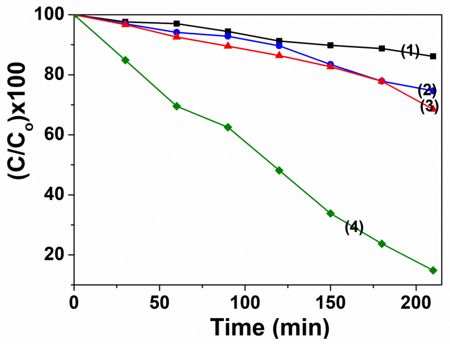
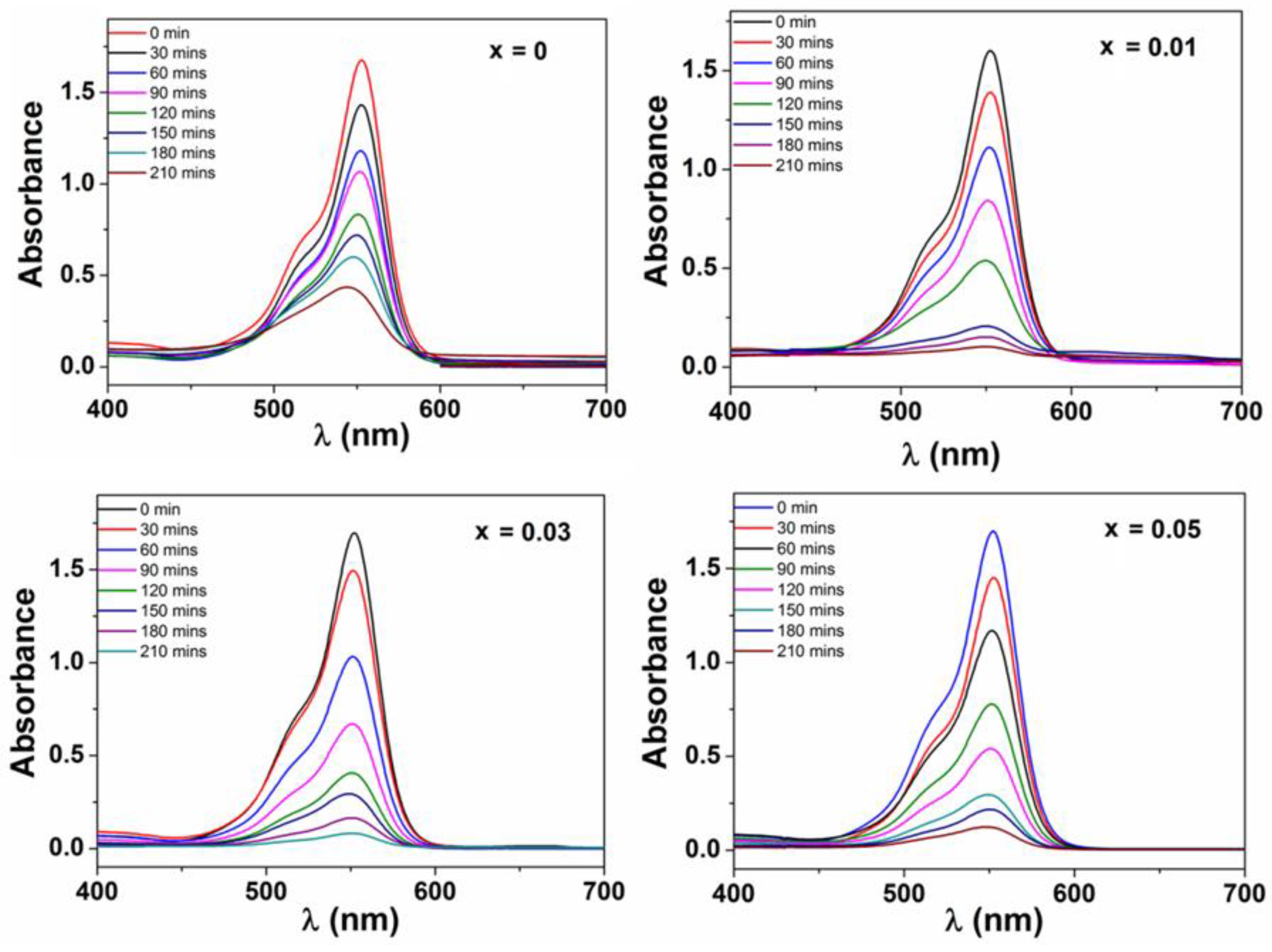
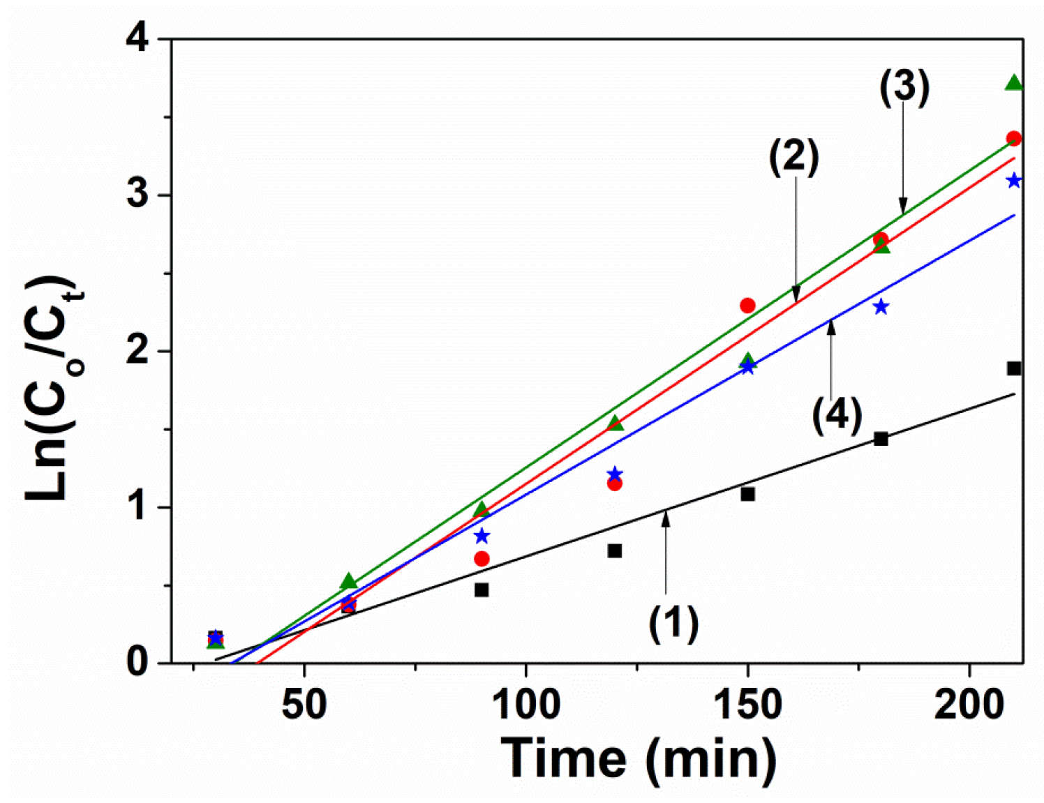
3.2. Photocatalytic Activity

3.2.1. Influence of Experimental Conditions

The photo-Fenton catalytic degradation activities of ZnFe2O4 catalyst occurring at different experimental parameters are illustrated in Figure 7. The lowest RhB removal efficiency is 13.87%, reached only when there is only H2O2 existed in the solution. Under ZnFe2O4/Visible-light system, the decolorization ratio achieved 25.35%. This figure was enhanced to 31.51% when ZnFe2O4 combined with H2O2. However, the removal rate of RhB reaches to 85.14% under irradiation and in the presence of ZnFe2O2 and H2O2. The high removal rate could be explained by the h+ in the valence of ZnFe2O4 and photodecomposition of H2O2 that produce more •OH. Both of which contributed to the improved oxidation of dyes [22]. On the other hand, the production of •OH could also be promoted by decreased recombination of electrons and holes, caused by the participation of photo-induced electrons in the Fe3+/Fe2+ cycle reaction [26].
When the ZnFe2O4 crytals are doped with Nd3+ ions, their photocatalytic degradation of RhB are enhanced. The higher photocatalytic performance at higher Nd3+ introduced may be due to smaller crystallite sizes from 22 to 12 nm with increasing Nd3+ ions content. This may be leading to the larger surface area and higher amount of active photocatalytic sites. Moreover, another effect is band gap energy value decreasing from 1.75 to 1.42 eV with increasing Nd3+ ions concentration, which aids the formation of ∙OH active species and stimulates oxidative degradation of dye molecules. UV–vis absorption spectra of RhB during the degradation by ZnNdxFe2−xO4 (x = 0–0.05) at the different irradiation time as shown in Figure 8. The photocatalytic degradation efficiency of RhB and kinetic constant after 210 min irradiation are 96.53% and 0.0095 min−1, 98.00% and 0.0189 min−1, 95.46% and 0.0163 min−1 in the presence of H2O2 and ZnNdxFe2−xO4 with x = 0.01, 0.03 and 0.05, respectively (Figure 9 and Table 2). This phenomenon can rely on the combination of rare earth ions and the ions in the crystal lattice of ferrite to generate the energy levels and the defects, which has been confirmed by XRD and DRS measurements [38,39].

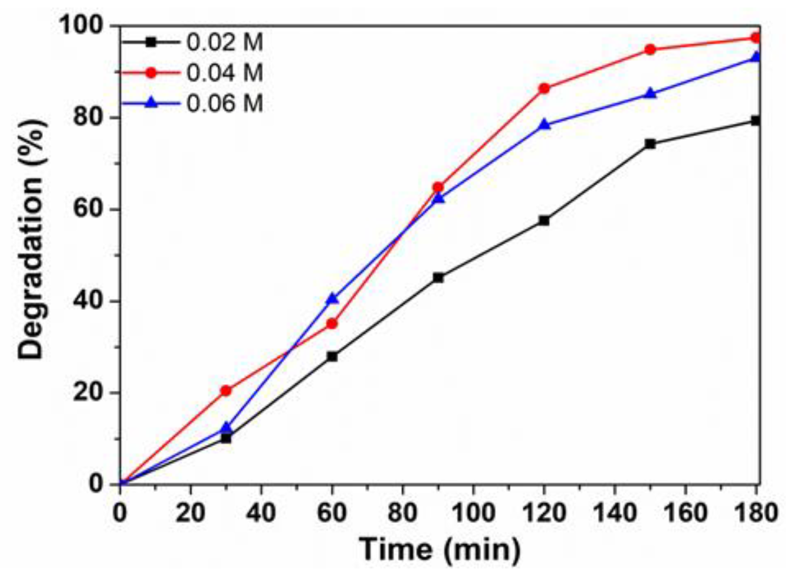
3.2.2. Influence of H2O2 Concentration

Figure 10 shows the removal efficiency of RhB under different concentrations of H2O2. When initial H2O2 concentration increased from 0.02 M to 0.04 M, the degradation efficiency increased from 79.4% to 97.42%. However, the degradation efficiency decreased to 93.02% when H2O2 concentration increased to 0.06 M. The initial increase in the degradation could be explained due to the generation of the higher number of OH active species which are mainly responsible for the oxidative degradation of dye molecules, Equation (3). When hydrogen peroxide presents in high concentration, OH could be scavenged (Equations (4)–(6)) and reduced [22,26]. Therefore, the degradation efficiency of RhB dye is greatly reduced. The optimal initial H2O2 content was 0.04 M.
H2O2 → 2OH
OH + H2O2OOH + H2O
OH + OH → H2O2
OH + OOH → H2O + O2

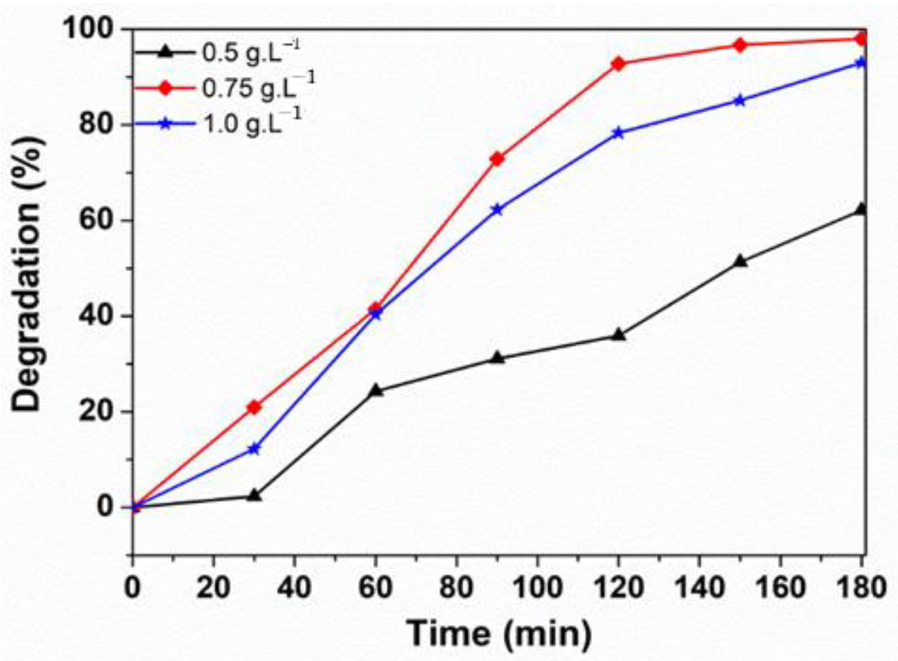
3.2.3. Influence of the Catalyst Loading

The effect of the ferrite sample amount on the RhB removal rate is shown in Figure 11. When the ZnNd0.03Fe1.97O4 dosage increases from 0.5 g/L to 0.75 g/L, the efficient degradation of RhB increases from 62.13% to 98.01% at 180 min. However, the removal rate of RhB decreased to 93.02% when increasing the ZnNd0.03Fe1.97O4 dosage to 1.0 g/L. This outcome is because when increasing catalyst dosage, OH radical amount increases due to the reaction of h+ in the valence of ferrite sample [22]. However, with a high catalyst dosage, the degradation efficiency of RhB dye decreases due to increased solution turbidity, in turn obstructing light irradiation and activating the totality of the catalyst suspension [43]. Therefore, the optimal catalyst dosage was 0.75 g/L.

4. Conclusions

Nd3+ substituted zinc ferrite nanoparticles were successfully synthesized via solution combustion technique. The physical and chemical characteristic of the ZnNdxFe2−xO4 samples were investigated by XRD, EDX, FT-IR, SEM and TEM. The average crystallite size and the optical band gap values reduced from 22 to 12 nm and from 1.75 to 1.42 eV, respectively, with increasing Nd3+ ions content. The substitution of Nd3+ ions on octahedral sites was confirmed by the change of ν1 and ν2 frequency. The enhanced photocatalytic activity of the zinc ferrite samples was observed with increasing Nd3+ ions concentration. The ZnNd0.03Fe1.97O4 nanoparticles have the highest efficient degradation for Rhodamine B. The removal efficiency of Rhodamine B dye was affected by the concentration of H2O2, catalyst amount. The optimal initial H2O2 content was 0.04 M and the optimal dosage of the catalyst was 0.75 g/L.

Author Contributions

Conceptualization, L.T.T.N.; methodology, H.T.T.N.; software, T.H.L.; validation, L.T.H.N.; formal analysis, H.Q.N.; investigation, L.T.T.N.; software, T.T.H.P.; resources, N.D.B.; data curation, N.T.K.T.; writing—review and editing, D.T.C.N.; writing—original draft preparation, L.T.T.N.; writing—review and editing, T.V.L.; visualization, T.V.L.; supervision, T.V.L., T.V.T.; project administration, T.V.T.; funding acquisition, T.V.T. All authors have read and agreed to the published version of the manuscript.

Funding

This work was supported by Thai Nguyen University of Education of Vietnam (No. CS2020-02).

Institutional Review Board Statement

Not applicable.

Informed Consent Statement

Not applicable.

Data Availability Statement

The data presented in this study are available on request from the corresponding author.

Conflicts of Interest

The authors declare no conflict of interest.

References

  1. Van Tran, T.; Phan, T.-Q.T.; Nguyen, D.T.C.; Nguyen, T.T.; Nguyen, D.H.; Vo, D.-V.N.; Bach, L.G.; Nguyen, T.D. Recyclable Fe3O4@C nanocomposite as potential adsorbent for a wide range of organic dyes and simulated hospital effluents. Environ. Technol. Innov. 2020, 20, 101122. [Google Scholar] [CrossRef]
  2. Nguyen, D.T.C.; Dang, H.H.; Vo, D.-V.N.; Bach, L.G.; Nguyen, T.D.; Van Tran, T. Biogenic synthesis of MgO nanoparticles from different extracts (flower, bark, leaf) of Tecoma stans (L.) and their utilization in selected organic dyes treatment. J. Hazard. Mater. 2021, 404, 124146. [Google Scholar] [CrossRef]
  3. Van Tran, T.; Nguyen, H.-T.T.; Dang, H.H.; Nguyen, D.T.C.; Nguyen, D.H.; Van Pham, T.; Van Tan, L. Central composite design for optimizing the organic dyes remediation utilizing novel graphene oxide@CoFe2O4 nanocomposite. Surf. Interfaces 2020, 21, 100687. [Google Scholar] [CrossRef]
  4. Hsiao, P.-H.; Li, T.-C.; Chen, C.-Y. ZnO/Cu 2 O/Si Nanowire Arrays as Ternary Heterostructure-Based Photocatalysts with Enhanced Photodegradation Performances. Nanoscale Res. Lett. 2019, 14, 244. [Google Scholar] [CrossRef] [PubMed] [Green Version]
  5. Hsiao, P.-H.; Timjan, S.; Kuo, K.-Y.; Juan, J.-C.; Chen, C.-Y. Optical Management of CQD/AgNP@ SiNW Arrays with Highly Efficient Capability of Dye Degradation. Catalysts 2021, 11, 399. [Google Scholar] [CrossRef]
  6. Khalid, N.R.; Ahmed, E.; Niaz, N.A.; Nabi, G.; Ahmad, M.; Tahir, M.B.; Rafique, M.; Rizwan, M.; Khan, Y. Highly visible light responsive metal loaded N/TiO2 nanoparticles for photocatalytic conversion of CO2 into methane. Ceram. Int. 2017, 43, 6771–6777. [Google Scholar] [CrossRef]
  7. Khalid, N.R.; Majid, A.; Tahir, M.B.; Niaz, N.A.; Khalid, S. Carbonaceous-TiO2 nanomaterials for photocatalytic degradation of pollutants: A review. Ceram. Int. 2017, 43, 14552–14571. [Google Scholar] [CrossRef]
  8. Khalid, N.R.; Liaqat, M.; Tahir, M.B.; Nabi, G.; Iqbal, T.; Niaz, N.A. The role of graphene and europium on TiO2 performance for photocatalytic hydrogen evolution. Ceram. Int. 2018, 44, 546–549. [Google Scholar] [CrossRef]
  9. Tahir, M.B.; Nabi, G.; Hassan, A.; Iqbal, T.; Kiran, H.; Majid, A. Morphology tailored synthesis of C-WO3 nanostructures and its photocatalytic application. J. Inorg. Organomet. Polym. Mater. 2018, 28, 738–745. [Google Scholar] [CrossRef]
  10. Tahir, M.B.; Nabi, G.; Iqbal, T.; Sagir, M.; Rafique, M. Role of MoSe2 on nanostructures WO3-CNT performance for photocatalytic hydrogen evolution. Ceram. Int. 2018, 44, 6686–6690. [Google Scholar] [CrossRef]
  11. Tahir, M.; Nabi, G.; Rafique, M.; Khalid, N. Nanostructured-based WO photocatalysts: Recent development, activity enhancement, perspectives and applications for wastewater treatment. Int. J. Environ. Sci. Technol. 2017, 14, 2519–2542. [Google Scholar] [CrossRef]
  12. Tahir, M.B.; Nabi, G.; Khalid, N.R. Enhanced photocatalytic performance of visible-light active graphene-WO3 nanostructures for hydrogen production. Mater. Sci. Semicond. Process. 2018, 84, 36–41. [Google Scholar] [CrossRef]
  13. Tahir, M.B.; Nabi, G.; Khalid, N.R.; Rafique, M. Role of europium on WO3 performance under visible-light for photocatalytic activity. Ceram. Int. 2018, 44, 5705–5709. [Google Scholar] [CrossRef]
  14. Tahir, M.B.; Iqbal, T.; Kiran, H.; Hasan, A. Insighting role of reduced graphene oxide in BiVO4 nanoparticles for improved photocatalytic hydrogen evolution and dyes degradation. Int. J. Energy Res. 2019, 43, 2410–2417. [Google Scholar] [CrossRef]
  15. Nguyen, T.D.; Nguyen, V.-H.; Nanda, S.; Vo, D.-V.N.; Nguyen, V.H.; Van Tran, T.; Nong, L.X.; Nguyen, T.T.; Bach, L.-G.; Abdullah, B. BiVO4 photocatalysis design and applications to oxygen production and degradation of organic compounds: A review. Environ. Chem. Lett. 2020, 18, 1779–1801. [Google Scholar] [CrossRef]
  16. Casbeer, E.; Sharma, V.K.; Li, X.-Z. Synthesis and photocatalytic activity of ferrites under visible light: A review. Sep. Purif. Technol. 2012, 87, 1–14. [Google Scholar] [CrossRef]
  17. Guo, X.; Wang, D. Photo-Fenton degradation of methylene blue by synergistic action of oxalic acid and hydrogen peroxide with NiFe2O4 hollow nanospheres catalyst. J. Environ. Chem. Eng. 2019, 7, 102814. [Google Scholar] [CrossRef]
  18. Feng, J.; Zhang, Z.; Gao, M.; Gu, M.; Wang, J.; Zeng, W.; Lv, Y.; Ren, Y.; Fan, Z. Effect of the solvents on the photocatalytic properties of ZnFe2O4 fabricated by solvothermal method. Mater. Chem. Phys. 2019, 223, 758–761. [Google Scholar] [CrossRef]
  19. Zhao, Y.; Lin, C.; Bi, H.; Liu, Y.; Yan, Q. Magnetically separable CuFe2O4/AgBr composite photocatalysts: Preparation, characterization, photocatalytic activity and photocatalytic mechanism under visible light. Appl. Surf. Sci. 2017, 392, 701–707. [Google Scholar] [CrossRef]
  20. Vinosha, P.A.; Xavier, B.; Anceila, D.; Das, S.J. Nanocrystalline ferrite (MFe2O4, M = Ni, Cu, Mn and Sr) photocatalysts synthesized by homogeneous Co-precipitation technique. Optik 2018, 157, 441–448. [Google Scholar] [CrossRef]
  21. To Loan, N.T.; Hien Lan, N.T.; Thuy Hang, N.T.; Quang Hai, N.; Tu Anh, D.T.; Thi Hau, V.; Van Tan, L.; Van Tran, T. CoFe2O4 nanomaterials: Effect of annealing temperature on characterization, magnetic, photocatalytic, and photo-Fenton properties. Processes 2019, 7, 885. [Google Scholar] [CrossRef] [Green Version]
  22. Cai, C.; Zhang, Z.; Liu, J.; Shan, N.; Zhang, H.; Dionysiou, D.D. Visible light-assisted heterogeneous Fenton with ZnFe2O4 for the degradation of Orange II in water. Appl. Catal. B Environ. 2016, 182, 456–468. [Google Scholar] [CrossRef]
  23. Xiang, Y.; Huang, Y.; Xiao, B.; Wu, X.; Zhang, G. Magnetic yolk-shell structure of ZnFe2O4 nanoparticles for enhanced visible light photo-Fenton degradation towards antibiotics and mechanism study. Appl. Surf. Sci. 2020, 513, 145820. [Google Scholar] [CrossRef]
  24. Li, Y.; Chen, D.; Fan, S.; Yang, T. Enhanced visible light assisted Fenton-like degradation of dye via metal-doped zinc ferrite nanosphere prepared from metal-rich industrial wastewater. J. Taiwan Inst. Chem. Eng. 2019, 96, 185–192. [Google Scholar] [CrossRef]
  25. Cao, Z.; Zhang, J.; Zhou, J.; Ruan, X.; Chen, D.; Liu, J.; Liu, Q.; Qian, G. Electroplating sludge derived zinc-ferrite catalyst for the efficient photo-Fenton degradation of dye. J. Environ. Manag. 2017, 193, 146–153. [Google Scholar] [CrossRef]
  26. Surendra, B.S.; Shekhar, T.R.S.; Veerabhadraswamy, M.; Nagaswarupa, H.P.; Prashantha, S.C.; Geethanjali, G.C.; Likitha, C. Probe sonication synthesis of ZnFe2O4 NPs for the photocatalytic degradation of dyes and effect of treated wastewater on growth of plants. Chem. Phys. Lett. 2020, 745, 137286. [Google Scholar] [CrossRef]
  27. Mohamed, R.M.; Ismail, A.A. Impact of surfactant ratios on mesostructured MnFe2O4 nanocomposites and their photocatalytic performance. Ceram. Int. 2020, 46, 10925–10933. [Google Scholar] [CrossRef]
  28. Kalam, A.; Al-Sehemi, A.G.; Assiri, M.; Du, G.; Ahmad, T.; Ahmad, I.; Pannipara, M. Modified solvothermal synthesis of cobalt ferrite (CoFe2O4) magnetic nanoparticles photocatalysts for degradation of methylene blue with H2O2/visible light. Results Phys. 2018, 8, 1046–1053. [Google Scholar] [CrossRef]
  29. Kefeni, K.K.; Mamba, B.B. Photocatalytic application of spinel ferrite nanoparticles and nanocomposites in wastewater treatment. Sustain. Mater. Technol. 2020, 23, e00140. [Google Scholar] [CrossRef]
  30. Nadumane, A.; Shetty, K.; Anantharaju, K.S.; Nagaswarupa, H.P.; Rangappa, D.; Vidya, Y.S.; Nagabhushana, H.; Prashantha, S.C. Sunlight photocatalytic performance of Mg-doped nickel ferrite synthesized by a green sol-gel route. J. Sci. Adv. Mater. Devices 2019, 4, 89–100. [Google Scholar] [CrossRef]
  31. Bhukal, S.; Bansal, S.; Singhal, S. Magnetic Mn substituted cobalt zinc ferrite systems: Structural, electrical and magnetic properties and their role in photo-catalytic degradation of methyl orange azo dye. Phys. B Condens. Matter 2014, 445, 48–55. [Google Scholar] [CrossRef]
  32. Abraham, A.G.; Manikandan, A.; Manikandan, E.; Vadivel, S.; Jaganathan, S.K.; Baykal, A.; Renganathan, P.S. Enhanced magneto-optical and photo-catalytic properties of transition metal cobalt (Co2+ ions) doped spinel MgFe2O4 ferrite nanocomposites. J. Magn. Magn. Mater. 2018, 452, 380–388. [Google Scholar] [CrossRef]
  33. Sharma, R.; Singhal, S. Structural, magnetic and electrical properties of zinc doped nickel ferrite and their application in photo catalytic degradation of methylene blue. Phys. B Condens. Matter 2013, 414, 83–90. [Google Scholar] [CrossRef]
  34. Joshi, S.; Kumar, M.; Pandey, H.; Singh, M.; Pal, P. Structural, magnetic and dielectric properties of Gd3+ substituted NiFe2O4 nanoparticles. J. Alloys Compd. 2018, 768, 287–297. [Google Scholar] [CrossRef]
  35. Chauhan, L.; Singh, N.; Dhar, A.; Kumar, H.; Kumar, S.; Sreenivas, K. Structural and electrical properties of Dy3+ substituted NiFe2O4 ceramics prepared from powders derived by combustion method. Ceram. Int. 2017, 43, 8378–8390. [Google Scholar] [CrossRef]
  36. Zhang, Y.; Chen, Y.; Kou, Q.; Wang, Z.; Han, D.; Sun, Y.; Yang, J.; Liu, Y.; Yang, L. Effects of Nd concentration on structural and magnetic properties of ZnFe 2 O 4 nanoparticles. J. Mater. Sci. Mater. Electron. 2018, 29, 3665–3671. [Google Scholar] [CrossRef]
  37. Masoudpanah, S.M.; Ebrahimi, S.A.S.; Derakhshani, M.; Mirkazemi, S.M. Structure and magnetic properties of La substituted ZnFe2O4 nanoparticles synthesized by sol–gel autocombustion method. J. Magn. Magn. Mater. 2014, 370, 122–126. [Google Scholar] [CrossRef]
  38. Mariosi, F.R.; Venturini, J.; da Cas Viegas, A.; Bergmann, C.P. Lanthanum-doped spinel cobalt ferrite (CoFe2O4) nanoparticles for environmental applications. Ceram. Int. 2020, 46, 2772–2779. [Google Scholar] [CrossRef]
  39. Almessiere, M.A.; Slimani, Y. Sonochemical synthesis of Eu3+ substituted CoFe2O4 nanoparticles and their structural, optical and magnetic properties. Ultrason. Sonochem. 2019, 58, 104621. [Google Scholar] [CrossRef] [PubMed]
  40. Sharma, R.; Bansal, S.; Singhal, S. Augmenting the catalytic activity of CoFe2O4 by substituting rare earth cations into the spinel structure. RSC Adv. 2016, 6, 71676–71691. [Google Scholar] [CrossRef]
  41. Rashmi, S.K.; Naik, H.S.B.; Jayadevappa, H.; Sudhamani, C.N.; Patil, S.B.; Naik, M.M. Influence of Sm3+ ions on structural, optical and solar light driven photocatalytic activity of spinel MnFe2O4 nanoparticles. J. Solid State Chem. 2017, 255, 178–192. [Google Scholar] [CrossRef]
  42. Patil, S.B.; Naik, H.S.B.; Nagaraju, G.; Viswanath, R.; Rashmi, S.K. Synthesis of visible light active Gd3+-substituted ZnFe2O4 nanoparticles for photocatalytic and antibacterial activities. Eur. Phys. J. Plus 2017, 132, 1–12. [Google Scholar] [CrossRef]
  43. Harish, K.N.; Bhojya Naik, H.S.; Prashanth Kumar, P.N.; Viswanath, R. Optical and photocatalytic properties of solar light active Nd-substituted Ni ferrite catalysts: For environmental protection. ACS Sustain. Chem. Eng. 2013, 1, 1143–1153. [Google Scholar] [CrossRef]
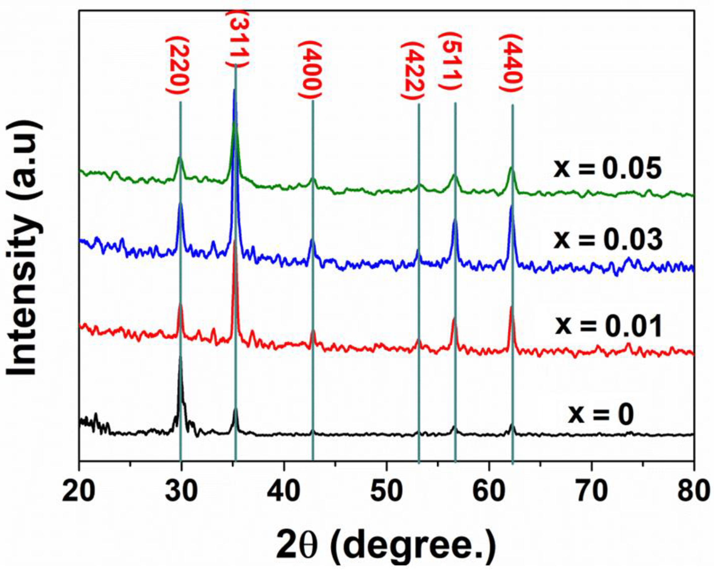
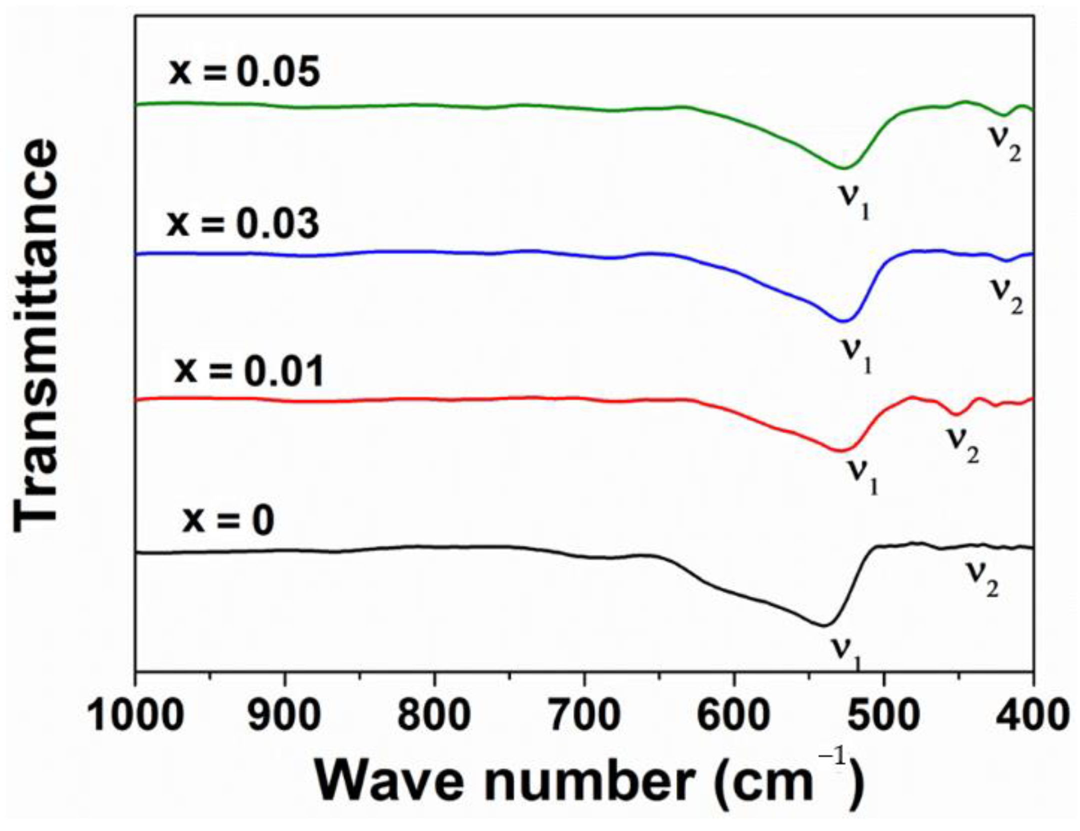
Figure 1. X-ray diffraction patterns of ZnNdxFe2−xO4 nanoparticles (x = 0.0–0.05) annealed at 500 °C.
Materials 14 02054 g001
Figure 2. FT−IR spectrum of ZnNdxFe2−xO4 nanoparticles.
Materials 14 02054 g002
Figure 3. SEM photomicrography of ZnNdxFe2−xO4 nanoparticles: (a) x = 0, (b) x = 0.01, (c) x = 0.03, (d) x = 0.05.
Materials 14 02054 g003
Figure 4. TEM of ZnNdxFe2−xO4 nanoparticles: (a) x = 0; (b) x = 0.03.
Materials 14 02054 g004
Figure 5. EDX spectra of ZnNdxFe2−xO4 nanoparticles: (a) x = 0; (b) x = 0.03.
Materials 14 02054 g005
Figure 6. The band gap energies of ZnNdxFe2−xO4 nanoparticles (x = 0.00–0.05).
Materials 14 02054 g006
Figure 7. The photocatalytic degradation of RhB in different conditions: curve (1), 10.0 mg/L RhB + 0.04 M H2O2 + light; curve (2), 10.0 mg/L RhB + 0.1 g ZnFe2O4 + light; curve (3), 10.0 mg/L RhB + 0.1 g ZnFe2O4+ 0.04 M H2O2 + dark; curve (4), 10.0 mg/L RhB + 0.1 g ZnFe2O4 + 0.04 M H2O2 + light.
Materials 14 02054 g007
Figure 8. The change in absorption of Rhodamine B solution with time in the presence of H2O2 and ZnNdxFe2−xO4 nanoparticles (x = 0–0.05) under irradiation.
Materials 14 02054 g008
Figure 9. The plots of ln(Co/Ct) versus irradiation time (t) in the presence of H2O2 and ZnNdxFe2−xO4 nanoparticles: (1) x = 0, (2) x = 0.01, (3) x = 0.03, (4) x = 0.05.
Materials 14 02054 g009
Figure 10. The influence of H2O2 concentration on the efficient degradation of Rhodamine B using ZnNd0.03Fe1.97O4 as Photo-Fenton.
Materials 14 02054 g010
Figure 11. The effect of ZnNd0.03Fe1.97O4 dosage on the degradation of Rhodamine.
Materials 14 02054 g011
Table 1. Average crystallite size (DXRD), lattice parameter (a), unit cell volume (V) and wave number, ν1 and ν2 for the tetrahedral and octahedral of the ZnNdxFe2−xO4 samples, respectively.
SamplesDXRD
(nm)
a
(Å)
V
3)
ν1
(cm−1)
ν2
(cm−1)
ZnFe2O4228.43599.08522.7447.5
ZnNd0.01Fe1.99O4218.44601.21528.5451.3
ZnNd0.03Fe1.97O4188.45603.35526.0418.5
ZnNd0.05Fe1.95O4128.45603.35526.6420.5
Table 2. The degradation efficiency (H%) and pseudo first-order rate constant (k) for the photocatalytic degradation of RhB in the presence of H2O2 0.04 M using ZnNdxFe2−xO4 nanoparticles.
SamplesH (%)k (min−1)R2
ZnFe2O485.14 ± 0.990.00950.952
ZnNd0.01Fe1.99O496.53 ± 0.950.01890.951
ZnNd0.03Fe1.97O498.00 ± 0.440.01900.964
ZnNd0.05Fe1.95O495.46 ± 0.910.01630.972
Publisher’s Note: MDPI stays neutral with regard to jurisdictional claims in published maps and institutional affiliations.

