Esthetic and Physical Changes of Innovative Titanium Surface Properties Obtained with Laser Technology

 ,
, 
 and
and 
Abstract
1. Introduction
2. Materials and Methods
Surface Analyses
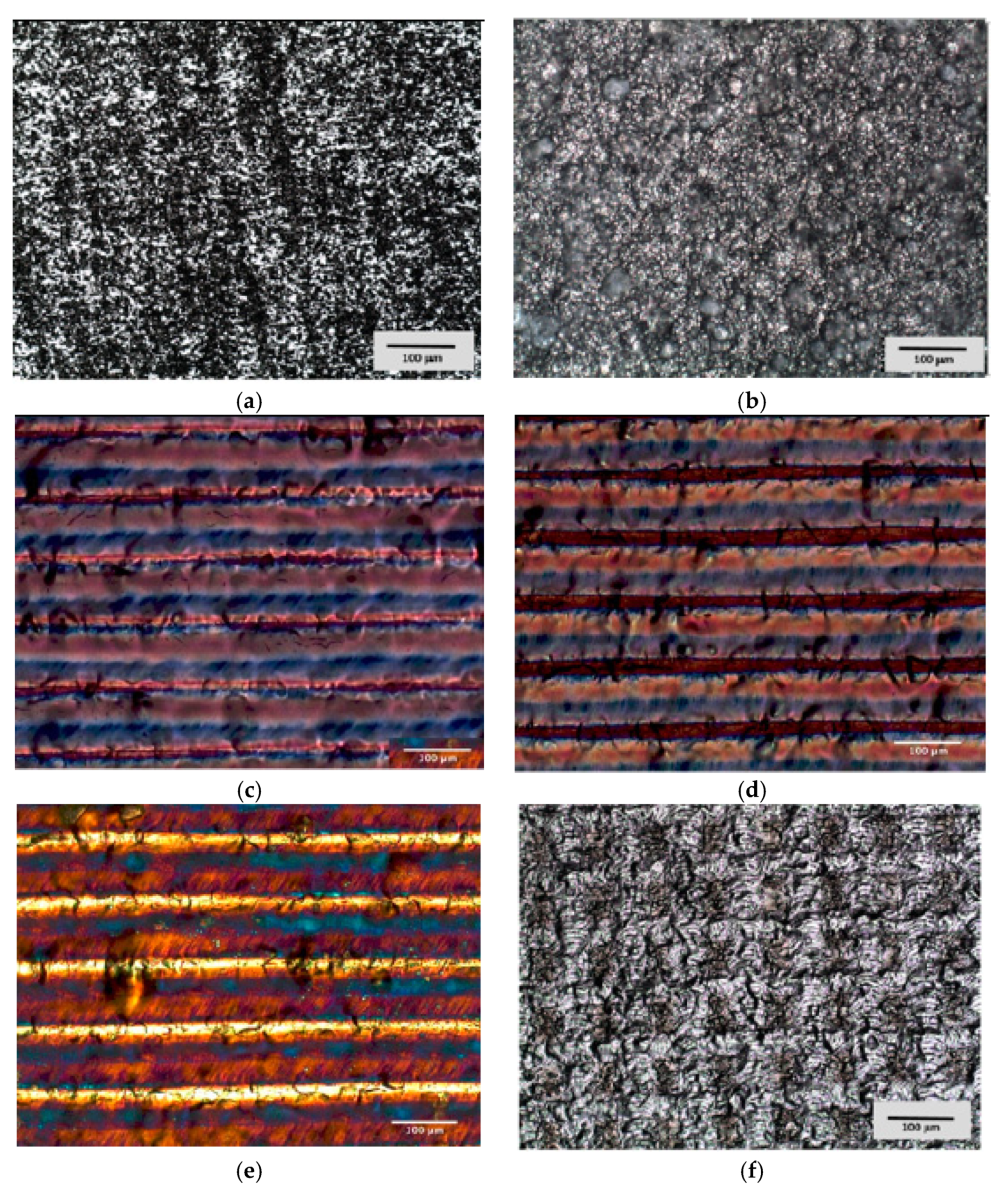
3. Results
4. Discussion
5. Conclusions
Author Contributions
Funding
Conflicts of Interest
References
- GBD 2016 Disease and Injury Incidence and Prevalence Collaborators. Global, regional, and national incidence, prevalence, and years lived with disability for 328 diseases and injuries for 195 countries, 1990–2016: A systematic analysis for the Global Burden of Disease Study 2016. Lancet 2017, 390, 1211–1259.
- Branemark, P.I.; Svensson, B.; van Steenberghe, D. Ten-year survival rates of fixed prostheses on four or six implants ad modum Branemark in full edentulism. Clin. Oral Implant. Res. 1995, 6, 227–231. [Google Scholar] [CrossRef] [PubMed]
- ADA Coucil on Scientific Affairs. Dental endosseous implants: An update. J. Am. Dent. Assoc. 2004, 135, 92–97. [Google Scholar] [CrossRef] [PubMed]
- Sul, Y. The significance of the surface properties of oxidized titanium to the bone response: Special emphasis on potential biochemical bonding of oxidized titanium implant. Biomaterials 2003, 24, 3893–3907. [Google Scholar] [CrossRef]
- Yang, T.-C.; Shu, H.-Y.; Chen, H.-T.; Chung, C.-J.; He, J.-L. Interface between grown osteoblast and micro-arc oxidized bioactive layers. Surf. Coat. Technol. 2014, 259, 185–192. [Google Scholar] [CrossRef]
- El Askary, A.S.; Meffert, R.M.; Griffin, T. Why do dental implants fail? Part I. Implant. Dent. 1999, 8, 173–185. [Google Scholar] [CrossRef]
- Higginbottom, F.L. Implants as an option in the esthetic zone. J. Oral Maxillofac. Surg. 2005, 63, 33–44. [Google Scholar] [CrossRef]
- Paolantoni, G.; Marenzi, G.; Blasi, A.; Mignogna, J.; Sammartino, G. Findings of a Four-Year Randomized Controlled Clinical Trial Comparing Two-Piece and One-Piece Zirconia Abutments Supporting Single Prosthetic Restorations in Maxillary Anterior Region. Biomed. Res. Int. 2016, 2016, 8767845. [Google Scholar] [CrossRef][Green Version]
- Chang, M.; Odman, P.A.; Wennstrom, J.L.; Andersson, B. Esthetic outcome of implant supported single-tooth replacements assessed by the patient and by prosthodontists. Int. J. Prosthodont. 1999, 12, 335–341. [Google Scholar]
- Papaspyridakos, P.; Chen, C.J.; Singh, M.; Weber, H.P.; Gallucci, C.O. Success criteria in implant dentistry: A systematic review. J. Dent. Res. 2012, 91, 242–248. [Google Scholar] [CrossRef]
- Dawson, A.; Chen, S.; Buser, D. The SAC Classification in Implant Dentistry; Quintessence: Berlin, Germany, 2009. [Google Scholar]
- Fuentealba, R.; Jofrè, J. Esthetic failure in implant dentistry. Dent. Clin. N. Am. 2015, 59, 227–246. [Google Scholar] [CrossRef] [PubMed]
- Napoli, G.; Paura, M.; Vela, T.; Di Schino, A. Colouring titanium alloys by anodic oxidation. Metal. Sisak Zagreb 2018, 57, 111. [Google Scholar]
- Oleari, C. Colour in optical coatings. In Optical Thin Films and Coatings: From Materials to Applications; Piegari, A., Flory, F., Eds.; Woodhead Publishing Limited: Cambridge, UK, 2013; pp. 391–424. [Google Scholar]
- Rajab, F.H.; Liauw, C.M.; Benson, P.S.; Li, L.; Whitehead, K.A. Production of hybrid macro/micro/nano surface structures on Ti6Al4V surfaces by picosecond laser surface texturing and their antifouling characteristics. Colloids Surf. B Biointerfaces 2017, 160, 688–696. [Google Scholar] [CrossRef]
- Vanhumbeeck, J.F.; Proost, J. Current understanding of Ti anodization: Functional, morphological, chemical and mechanical aspects. Corros. Rev. 2009, 27, 117–194. [Google Scholar] [CrossRef]
- Diamanti, M.V.; Del Curto, B.; Masconale, V.; Passaro, C.; Pedeferri, M.P. Anodic colouring of titanium and its alloy for jewels production. Colour. Res. Appl. 2012, 37, 384–390. [Google Scholar] [CrossRef]
- Albrektsson, T.; Wennerberg, A. On osseointegration in relation to implant surfaces. Clin. Implant. Dent. Relat. Res. 2019, 21, 4–7. [Google Scholar] [CrossRef]
- Scarano, A.; Piattelli, A.; Quaranta, A.; Lorusso, F. Bone Response to Two Dental Implants with Different Sandblasted/Acid-Etched Implant Surfaces: A Histological and Histomorphometrical Study in Rabbits. Biomed. Res. Int. 2017, 2017, 1–8. [Google Scholar] [CrossRef]
- Rupp, F.; Liang, L.; Geis-Gerstorfer, J.; Scheideler, L.; Hüttig, F. Surface characteristics of dental implants: A review. Dent. Mater. 2018, 34, 40–57. [Google Scholar] [CrossRef]
- Tetè, S.; Mastrangelo, F.; Traini, T.; Vinci, R.; Sammartino, G.; Marenzi, G.; Gherlone, E. A macro- and nanostructure evaluation of a novel dental implant. Implant. Dent. 2008, 17, 309–320. [Google Scholar] [CrossRef]
- Wang, T.; Wang, L.; Lu, Q.; Fan, Z. Changes in the esthetic, physical, and biological properties of a titanium alloy abutment treated by anodic oxidation. J. Prosthet. Dent. 2019, 121, 156–165. [Google Scholar] [CrossRef]
- Chen, J.; Mwenifumbo, S.; Langhammer, C.; McGovern, J.P.; Li, M.; Beye, A.; Soboyejo, W.O. Cell/surface interactions and adhesion on Ti-6Al-4V: Effects of surface texture. J. Biomed. Mater. Res. B Appl. Biomater. 2007, 82, 360–373. [Google Scholar] [CrossRef] [PubMed]
- Tu, Z.M.; Zhu, Y.M.; Li, N.; Hu, H.L.; Cao, L.X. Applications and Advances on surface treatment for titanium and titanium alloy. Surf. Technol. 2009, 38, 76–77. [Google Scholar]
- Liu, J.; Alfantazi, A.; Asselin, E. A new method to improve the corrosion resistance of titanium for hydrometallurgical applications. Appl. Surf. Sci. 2015, 332, 480–487. [Google Scholar] [CrossRef]
- Lim, H.P.; Lee, K.M.; Koh, Y.I.; Park, S.W. Allergic contact stomatitis caused by a titanium nitride-coated implant abutment: A clinical report. J. Prosthetic. Dent. 2012, 108, 209–213. [Google Scholar] [CrossRef]
- Wadhwani, C.; Brindis, M.; Kattadiyil, M.; O’Brien, R.; Chung, K.H. Colourizing titanium-6aluminum-4vanadium alloy using electrochemical anodization: Developing a colour chart. J. Prosthet. Dent. 2018, 119, 26–28. [Google Scholar] [CrossRef]
- Karambakhsh, A.; Afshar, A.; Ghahramani, S.; Malekinejad, P. Pure commercial titanium colour anodizing and corrosion resistance. J. Mater. Eng. Perform. 2011, 20, 1690–1696. [Google Scholar] [CrossRef]
- Al-Nawas, B.; Hangen, U.; Duschner, H.; Krummenauer, F.; Wagner, W. Turned, machined versus double-etched dental implants in vivo. Clin. Implant. Dent. Relat. Res. 2007, 9, 71–78. [Google Scholar] [CrossRef]
- Tetè, S.; Mastrangelo, F.; Quaresima, R.; Vinci, R.; Sammartino, G.; Stuppia, L.; Gherlone, E. Influence of novel nano-titanium implant surface on human osteoblast behavior and growth. Implant. Dent. 2010, 19, 520–531. [Google Scholar] [CrossRef]
- Belser, U.C.; Schmid, B.; Higginbottom, F.; Buser, D. Outcome analysis of implant restorations located in the anterior maxilla: A review of the recent literature. Int. J. Oral Maxillofac. Implant. 2004, 19, 30–42. [Google Scholar]
- Marenzi, G.; Impero, F.; Scherillo, F.; Sammartino, J.C.; Squillace, A.; Spagnuolo, G. Effect of Different Surface Treatments on Titanium Dental Implant Micro-Morphology. Materials 2019, 12, 733. [Google Scholar] [CrossRef]


| Sample | L* | a* | b* | ΔE |
|---|---|---|---|---|
| M | 67.64 | −0.39 | −0.99 | - |
| SBAE | 65.47 | −0.49 | −0.89 | 2.17 |
| P1 | 88.42 | +45.34 | +39.21 | 64.30 |
| P2 | 90.65 | +43.72 | +37.31 | 62.75 |
| I | 34.27 | +44.53 | −21.92 | 59.80 |
| W | 96.31 | +2.45 | −10.91 | 30.69 |
| Sample | Ra | Rq | Rz | Ry | Sm |
|---|---|---|---|---|---|
| M | 0.32 (±0.02) | 0.47 (±0.03) | 2.21 (±0.19) | 2.74 (±0.15) | 53 (±4.86) |
| SBAE | 1.41 (±0.19) | 21.92 ±1.31) | 12.87 (±1.39) | 2.52 (±0.13) | 83 (±8.73) |
| P1 | 0.44 (±0.02) | 0.58 (±0.03) | 3.24 (±0.27) | 1.93 (±0.10) | 54 (±4.99) |
| P2 | 0.54 (±0.03) | 0.72 (±0.05) | 3.86 (±0.36) | 2.61 (±0.14) | 66 (±6.96) |
| I | 0.52 (±0.03) | 0.67 (±0.04) | 3.47 (±0.31) | 2.14 (±0.12) | 66 (±6. 31) |
| W | 0.32 (±0.02) | 0.41 (±0.02) | 2.21 (±0.18) | 1.21 (±0.12) | 37 (±4.01) |
© 2020 by the authors. Licensee MDPI, Basel, Switzerland. This article is an open access article distributed under the terms and conditions of the Creative Commons Attribution (CC BY) license (http://creativecommons.org/licenses/by/4.0/).
Share and Cite
Mastrangelo, F.; Quaresima, R.; Abundo, R.; Spagnuolo, G.; Marenzi, G. Esthetic and Physical Changes of Innovative Titanium Surface Properties Obtained with Laser Technology. Materials 2020, 13, 1066. https://doi.org/10.3390/ma13051066
Mastrangelo F, Quaresima R, Abundo R, Spagnuolo G, Marenzi G. Esthetic and Physical Changes of Innovative Titanium Surface Properties Obtained with Laser Technology. Materials. 2020; 13(5):1066. https://doi.org/10.3390/ma13051066
Chicago/Turabian StyleMastrangelo, Filiberto, Raimondo Quaresima, Roberto Abundo, Gianrico Spagnuolo, and Gaetano Marenzi. 2020. "Esthetic and Physical Changes of Innovative Titanium Surface Properties Obtained with Laser Technology" Materials 13, no. 5: 1066. https://doi.org/10.3390/ma13051066
APA StyleMastrangelo, F., Quaresima, R., Abundo, R., Spagnuolo, G., & Marenzi, G. (2020). Esthetic and Physical Changes of Innovative Titanium Surface Properties Obtained with Laser Technology. Materials, 13(5), 1066. https://doi.org/10.3390/ma13051066