Effect of Yttria Content on the Translucency and Masking Ability of Yttria-Stabilized Tetragonal Zirconia Polycrystal


Abstract
1. Introduction
2. Materials and Methods
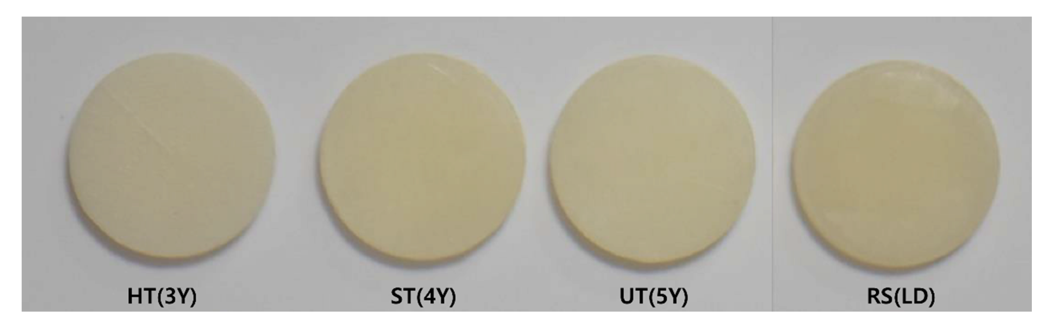
2.1. Preparation of the Zirconia Specimens
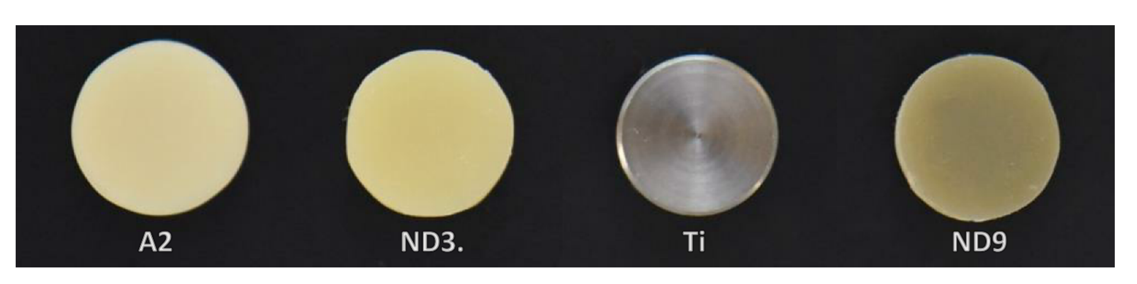
2.2. Preparation of the Background Substrates
2.3. Evaluation of the Translucency Parameter
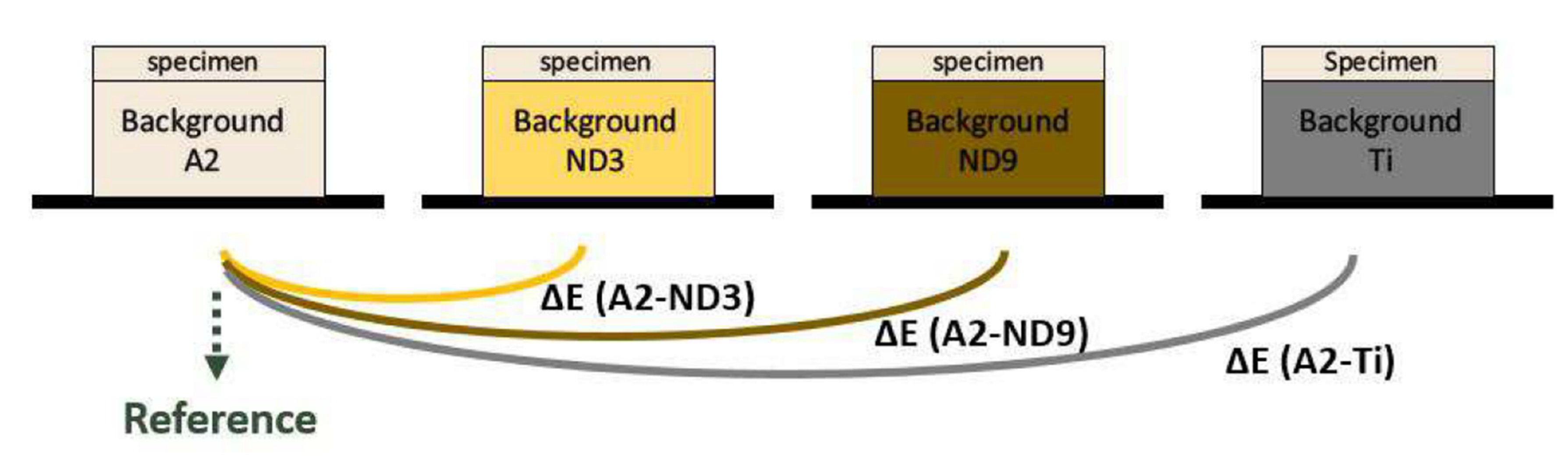
2.4. Evaluation of Masking Ability
2.5. Statistical Analysis
3. Results
3.1. Translucency Parameter
3.2. Masking Ability
4. Discussion
5. Conclusions
- A decrease in zirconia thickness from 1.5 to 0.8 mm significantly increased translucency; however, all specimens showed translucency lower than that of human enamel.
- The TP value increased significantly with an increase in yttria content (mol %) of zirconia.
- 5Y-ZP showed, approximately, 80% translucency at 0.8 mm thickness and 89% at 1.5 mm thickness, compared to that of lithium disilicate.
- Monolithic CAD-CAM ceramics could mask a normal dentin background but could not mask severely discolored dentin at either 0.8 or 1.5 mm thicknesses.
Author Contributions
Funding
Conflicts of Interest
References
- Bonfante, E.A.; Da Silva, N.R.F.A.; Coelho, P.G.; Bayardo-González, D.E.; Thompson, V.P.; Bonfante, G. Effect of framework design on crown failure. Eur. J. Oral Sci. 2009, 117, 194–199. [Google Scholar] [CrossRef]
- Ichikawa, Y.; Akagawa, Y.; Nikai, H.; Tsuru, H. Tissue compatibility and stability of a new zirconia ceramic in vivo. J. Prosthet. Dent. 1992, 68, 322–326. [Google Scholar] [CrossRef]
- Prasad, H.A.; Pasha, N.; Hilal, M.; Amarnath, G.S.; Kundapur, V.; Anand, M.; Singh, S. To evaluate effect of airborne particle abrasion using different abrasives particles and compare two commercial available zirconia on flexural strength on heat treatment. Int. J. Biomed. Sci. IJBS 2017, 13, 93–112. [Google Scholar] [PubMed]
- Júnior, V.V.B.F.; Dantas, D.; Bresciani, E.; Huhtala, M.F.R.L. Evaluation of the bond strength and characteristics of zirconia after different surface treatments. J. Prosthet. Dent. 2018, 120, 955–959. [Google Scholar] [CrossRef] [PubMed]
- Kelly, J.R.; Denry, I. Stabilized zirconia as a structural ceramic: An overview. Dent. Mater. 2008, 24, 289–298. [Google Scholar] [CrossRef] [PubMed]
- Rekow, E.; Silva, N.; Coelho, P.; Zhang, Y.; Guess, P.; Thompson, V. Performance of Dental Ceramics: Challenges for Improvements. J. Dent. Res. 2011, 90, 937–952. [Google Scholar] [CrossRef]
- Zhang, Y.; Lawn, B. Novel zirconia materials in dentistry. J. Dent. Res. 2017, 97, 140–147. [Google Scholar] [CrossRef]
- Tong, H.; Tanaka, C.B.; Kaiser, M.R.; Zhang, Y. Characterization of three commercial Y-TZP ceramics produced for their higher translucency. Ceram. Int. 2016, 42, 1077–1085. [Google Scholar] [CrossRef]
- Kwon, S.J.; Lawson, N.C.; McLaren, E.E.; Nejat, A.H.; Burgess, J.O. Comparison of the mechanical properties of translucent zirconia and lithium disilicate. J. Prosthet. Dent. 2018, 120, 132–137. [Google Scholar] [CrossRef]
- Zhang, Y. Making yttria-stabilized tetragonal zirconia translucent. Dent. Mater. 2014, 30, 1195–1203. [Google Scholar] [CrossRef]
- Azer, S.S.; Rosenstiel, S.F.; Seghi, R.R.; Johnston, W.M. Effect of substrate shades on the color of ceramic laminate veneers. J. Prosthet. Dent. 2011, 106, 179–183. [Google Scholar] [CrossRef]
- Kürklü, D.; Azer, S.S.; Yilmaz, B.; Johnston, W.M. Porcelain thickness and cement shade effects on the colour and translucency of porcelain veneering materials. J. Dent. 2013, 41, 1043–1050. [Google Scholar] [CrossRef] [PubMed]
- Azer, S.S.; Ayash, G.M.; Johnston, W.M.; Khalil, M.F.; Rosenstiel, S.F. Effect of esthetic core shades on the final color of IPS Empress all-ceramic crowns. J. Prosthet. Dent. 2006, 96, 397–401. [Google Scholar] [CrossRef] [PubMed]
- DeDe, D.Ö.; Ceylan, G.; Yilmaz, B. Effect of brand and shade of resin cements on the final color of lithium disilicate ceramic. J. Prosthet. Dent. 2017, 117, 539–544. [Google Scholar] [CrossRef]
- DeDe, D.Ö.; Sahin, O.; Özdemir, O.S.; Yilmaz, B.; Celik, E.; Köroğlu, A. Influence of the color of composite resin foundation and luting cement on the final color of lithium disilicate ceramic systems. J. Prosthet. Dent. 2017, 117, 138–143. [Google Scholar] [CrossRef]
- Pires, L.A.; Novais, P.M.; Araújo, V.D.; Pegoraro, L.F. Effects of the type and thickness of ceramic, substrate, and cement on the optical color of a lithium disilicate ceramic. J. Prosthet. Dent. 2017, 117, 144–149. [Google Scholar] [CrossRef] [PubMed]
- Zhang, F.; Inokoshi, M.; Batuk, M.; Hadermann, J.; Naert, I.; Van Meerbeek, B.; Vleugels, J. Strength, toughness and aging stability of highly-translucent Y-TZP ceramics for dental restorations. Dent. Mater. 2016, 32, e327–e337. [Google Scholar] [CrossRef] [PubMed]
- Douglas, R.D.; Steinhauer, T.J.; Wee, A.G. Intraoral determination of the tolerance of dentists for perceptibility and acceptability of shade mismatch. J. Prosthet. Dent. 2007, 97, 200–208. [Google Scholar] [CrossRef]
- Baldissara, P.; Wandscher, V.F.; Marchionatti, A.M.E.; Parisi, C.; Monaco, C.; Ciocca, L. Translucency of IPS e.max and cubic zirconia monolithic crowns. J. Prosthet. Dent. 2018, 120, 269–275. [Google Scholar] [CrossRef]
- Brodbelt, R.; O’Brien, W.; Fan, P. Translucency of Dental Porcelains. J. Dent. Res. 1980, 59, 70–75. [Google Scholar] [CrossRef]
- Jiang, L.; Liao, Y.; Wan, Q.; Li, W. Effects of sintering temperature and particle size on the translucency of zirconium dioxide dental ceramic. J. Mater. Sci. Mater. Med. 2011, 22, 2429–2435. [Google Scholar] [CrossRef] [PubMed]
- Shahmiri, R.; Standard, O.; Hart, J.N.; Sorrell, C.C. Optical properties of zirconia ceramics for esthetic dental restorations: A systematic review. J. Prosthet. Dent. 2018, 119, 36–46. [Google Scholar] [CrossRef] [PubMed]
- Kolakarnprasert, N.; Kaizer, M.R.; Kim, D.K.; Zhang, Y. New multi-layered zirconias: Composition, microstructure and translucency. Dent. Mater. 2019, 35, 797–806. [Google Scholar] [CrossRef] [PubMed]
- Kim, M.-J.; Ahn, J.-S.; Kim, J.-H.; Kim, H.-Y.; Kim, W.-C. Effects of the sintering conditions of dental zirconia ceramics on the grain size and translucency. J. Adv. Prosthodont. 2013, 5, 161–166. [Google Scholar] [CrossRef]
- Malkondu, Ö.; Tinastepe, N.; Kazazoglu, E. Influence of type of cement on the color and translucency of monolithic zirconia. J. Prosthet. Dent. 2016, 116, 902–908. [Google Scholar] [CrossRef]
- Bachhav, V.C.; Aras, M.A. The effect of ceramic thickness and number of firings on the color of a zirconium oxide based all ceramic system fabricated using CAD/CAM technology. J. Adv. Prosthodont. 2011, 3, 57–62. [Google Scholar] [CrossRef]
- Putra, A.; Chung, K.-H.; Flinn, B.D.; Kuykendall, T.; Zheng, C.; Harada, K.; Raigrodski, A.J. Effect of hydrothermal treatment on light transmission of translucent zirconias. J. Prosthet. Dent. 2017, 118, 422–429. [Google Scholar] [CrossRef]
- Harada, K.; Shinya, A.; Gomi, H.; Hatano, Y.; Shinya, A.; Raigrodski, A.J. Effect of accelerated aging on the fracture toughness of zirconias. J. Prosthet. Dent. 2016, 115, 215–223. [Google Scholar] [CrossRef]
- Yu, B.; Ahn, J.-S.; Lee, Y.-K. Measurement of translucency of tooth enamel and dentin. Acta Odontol. Scand. 2009, 67, 57–64. [Google Scholar] [CrossRef]
- Choi, Y.-J.; Razzoog, M.E. Masking Ability of Zirconia with and without Veneering Porcelain. J. Prosthodont. 2013, 22, 98–104. [Google Scholar] [CrossRef]
- Tabatabaian, F.; Taghizade, F.; Namdari, M. Effect of coping thickness and background type on the masking ability of a zirconia ceramic. J. Prosthet. Dent. 2018, 119, 159–165. [Google Scholar] [CrossRef] [PubMed]


| Material | Zirconia | Lithium Disilicate | ||||||
|---|---|---|---|---|---|---|---|---|
| 3Y-TZP | 4Y-ZP | 5Y-ZP | ||||||
| Product | Katana HT | Katana STML | Katana UTML | Rosetta SM | ||||
| Thickness (mm) | 0.8 | 1.5 | 0.8 | 1.5 | 0.8 | 1.5 | 0.8 | 1.5 |
| N | 10 | 10 | 10 | 10 | 10 | 10 | 10 | 10 |
| Group | 0.8 mm Thickness | 1.5 mm Thickness | ||||
|---|---|---|---|---|---|---|
| Mean± SD | Median | Translucency Relative to the RS Group (%) | Mean± SD | Median | Translucency Relative to the RS Group (%) | |
| HT | 11.58 ± 0.57 a | 11.48 | 59.95 | 7.75 ± 0.57 a | 7.72 | 54.40 |
| ST | 13.90 ± 0.17 b | 13.90 | 72.58 | 11.68 ± 0.23 b | 11.63 | 81.96 |
| UT | 15.36 ± 0.50 c | 15.30 | 79.90 | 12.64 ± 0.19 c | 12.62 | 88.94 |
| RS | 19.18 ± 0.29 d | 19.15 | 100 | 14.20 ± 0.39 d | 14.19 | 100 |
| Group | Mean ± Standard Deviation | |
|---|---|---|
| 0.8 mm Thickness | 1.5 mm Thickness | |
| Katana HT | 1.77 ± 0.45 a | 0.91 ± 0.17 a |
| Katana ST | 2.16 ± 0.47 ab | 1.53 ± 0.35 b |
| Katana UT | 2.40 ± 0.27 b | 1.59 ± 0.19 b |
| Rosetta SM | 3.36 ± 0.24 c | 2.36 ± 0.14 c |
| Group | Mean ± Standard Deviation | |
|---|---|---|
| 0.8 mm Thickness | 1.5 mm Thickness | |
| Katana HT | 5.08 ± 0.26 a | 3.13 ± 0.32 a |
| Katana ST | 6.27 ± 0.62 b | 4.68 ± 0.39 b |
| Katana UT | 6.57 ± 0.52 bc | 4.91 ± 0.34 b |
| Rosetta SM | 6.59 ± 0.21 c | 4.71 ± 0.13 b |
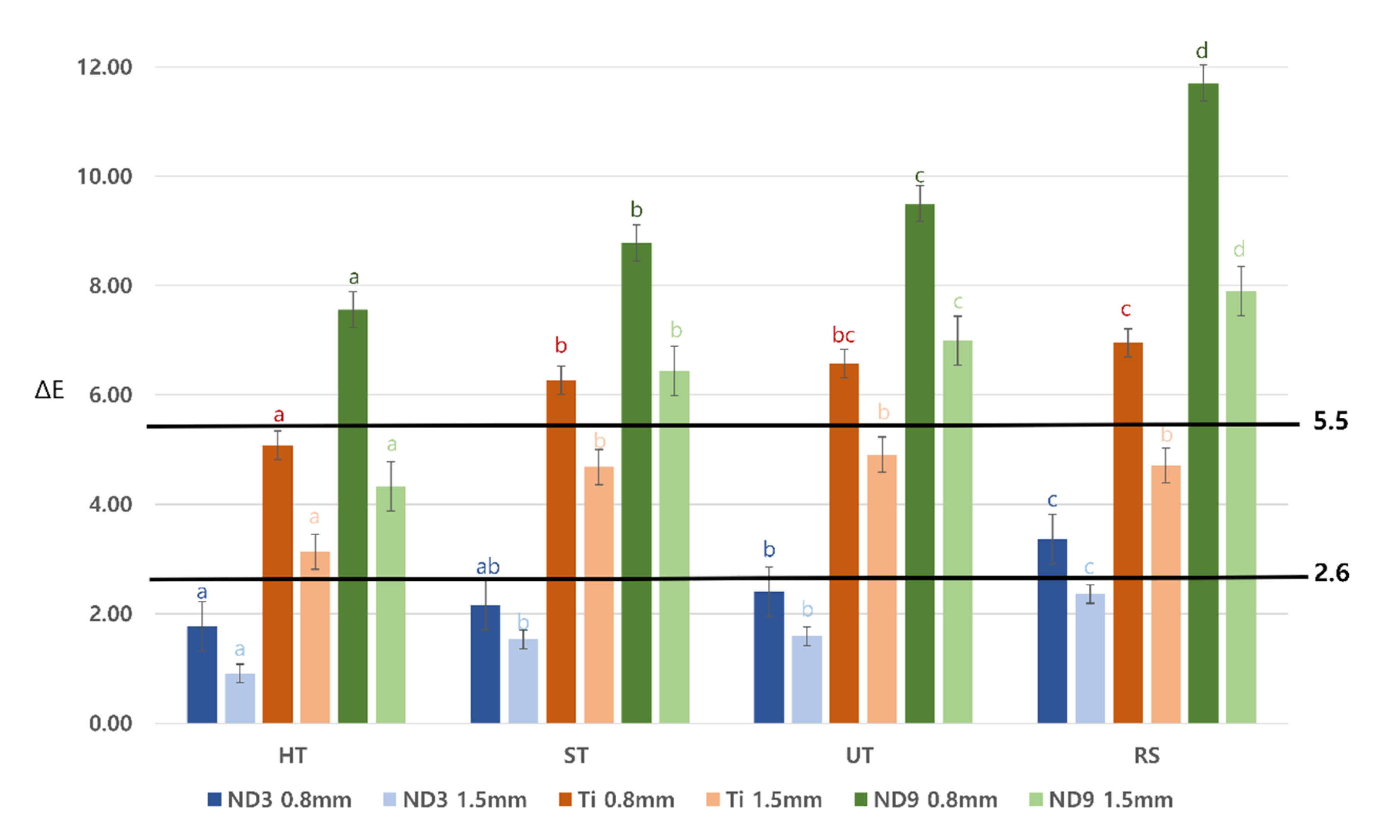
| Group | Mean ± Standard Deviation | |
|---|---|---|
| 0.8 mm Thickness | 1.5 mm Thickness | |
| Katana HT | 7.56 ± 0.33 a | 4.33 ± 0.45 a |
| Katana ST | 8.78 ± 0.61 b | 6.44 ± 0.31 b |
| Katana UT | 9.50 ± 0.55 c | 6.99 ± 0.35 c |
| Rosetta SM | 11.71 ± 0.28 d | 7.90 ± 0.31 d |
Publisher’s Note: MDPI stays neutral with regard to jurisdictional claims in published maps and institutional affiliations. |
© 2020 by the authors. Licensee MDPI, Basel, Switzerland. This article is an open access article distributed under the terms and conditions of the Creative Commons Attribution (CC BY) license (http://creativecommons.org/licenses/by/4.0/).
Share and Cite
Cho, Y.-E.; Lim, Y.-J.; Han, J.-S.; Yeo, I.-S.L.; Yoon, H.-I. Effect of Yttria Content on the Translucency and Masking Ability of Yttria-Stabilized Tetragonal Zirconia Polycrystal. Materials 2020, 13, 4726. https://doi.org/10.3390/ma13214726
Cho Y-E, Lim Y-J, Han J-S, Yeo I-SL, Yoon H-I. Effect of Yttria Content on the Translucency and Masking Ability of Yttria-Stabilized Tetragonal Zirconia Polycrystal. Materials. 2020; 13(21):4726. https://doi.org/10.3390/ma13214726
Chicago/Turabian StyleCho, Young-Eun, Young-Jun Lim, Jung-Suk Han, In-Sung Luke Yeo, and Hyung-In Yoon. 2020. "Effect of Yttria Content on the Translucency and Masking Ability of Yttria-Stabilized Tetragonal Zirconia Polycrystal" Materials 13, no. 21: 4726. https://doi.org/10.3390/ma13214726
APA StyleCho, Y.-E., Lim, Y.-J., Han, J.-S., Yeo, I.-S. L., & Yoon, H.-I. (2020). Effect of Yttria Content on the Translucency and Masking Ability of Yttria-Stabilized Tetragonal Zirconia Polycrystal. Materials, 13(21), 4726. https://doi.org/10.3390/ma13214726