Fabrication of Bioprobe Self-Assembled on Au–Te Nanoworm Structure for SERS Biosensor
Abstract
1. Introduction
2. Materials and Methods
2.1. Preparation of Au–Te Nanoworms
2.2. Immobilization of Au–Te on ITO
2.3. Fabrication of Target/Biomolecules/Au–Te on ITO
2.4. Surface Morphology Analysis
2.5. Measurement of Surface-Enhanced Raman Scattering
3. Results
3.1. Investigation of Immobilized Biomolecules/Au–Te on the ITO Substrate
3.2. Enhancement Factor of Au–Tenanoworms
3.3. Optimization of Au–Te Immobilization on ITO
3.4. Detection of Biomolecules by SERS
4. Conclusions
Author Contributions
Funding
Conflicts of Interest
Appendix A
References
- Song, Y.; Luo, Y.; Zhu, C.; Li, H.; Du, D.; Lin, Y. Recent advances in electrochemical biosensors based on graphene two-dimensional nanomaterials. Biosens. Bioelectron. 2016, 76, 195–212. [Google Scholar] [CrossRef] [PubMed]
- Nuzaihan, M.N.M.; Hashim, U.; Arshad, M.K.; Kasjoo, S.R.; Rahman, S.F.A.; Ruslinda, A.R.; Fathil, M.F.M.; Adzhri, R.; Shahimin, M.M. Electrical detection of dengue virus (DENV) DNA oligomer using silicon nanowire biosensor with novel molecular gate control. Biosens. Bioelectron. 2016, 83, 106–114. [Google Scholar] [CrossRef]
- Sreekanth, K.V.; Alapan, Y.; Elkabbash, M.; Ilker, E.; Hinczewski, M.; Gurkan, U.A.; Luca, A.D.; Strangi, G. Extreme sensitivity biosensing platform based on hyperbolic metamaterials. Nat. Mater. 2016, 15, 621–627. [Google Scholar] [CrossRef] [PubMed]
- Masson, J.-F. Surface plasmon resonance clinical biosensors for medical diagnostics. ACS Sens. 2017, 2, 16–30. [Google Scholar] [CrossRef] [PubMed]
- Songa, E.A.; Okonkwo, J.O. Recent approaches to improving selectivity and sensitivity of enzyme-based biosensors for organophosphorous pesticides: A review. Talanta 2016, 155, 289–304. [Google Scholar] [CrossRef] [PubMed]
- Zhang, D.; Liu, Q. Biosensors and bioelectronics on smartphone for portable biochemical detection. Biosens. Bioelectron. 2016, 75, 273–284. [Google Scholar] [CrossRef]
- Cinti, S.; Minotti, C.; Moscone, D.; Palleschi, G.; Arduini, F. Fully integrated ready-to-use paper-based electrochemical biosensor to detect nerve agents. Biosens. Bioelectron. 2017, 93, 46–51. [Google Scholar] [CrossRef]
- Hu, L.; Xu, J.; Qin, Z.; Hu, N.; Zhou, M.; Huang, L.; Wang, P. Detection of bitterness in vitro by a novel male mouse germ cell-based biosensor. Sens. Actuators B Chem. 2016, 223, 461–469. [Google Scholar] [CrossRef]
- Sheikhzadeh, E.; Chamsaz, M.; Turner, A.P.F.; Jager, E.W.H.; Beni, V. Label-free impedimetric biosensor for salmonella typhimurium detection based on poly [pyrrole-co-3-carboxyl-pyrrole] copolymer supported aptamer. Biosens. Bioelectron. 2016, 80, 194–200. [Google Scholar] [CrossRef]
- Yoo, S.M.; Lee, S.Y. Optical biosensors for the detection of pathogenic microorganisms. Trends Biotechnol. 2016, 34, 7–25. [Google Scholar] [CrossRef]
- Lee, T.; Kim, G.H.; Kim, S.M.; Hong, K.; Kim, Y.; Park, C.; Sohn, H.; Min, J. Label-free localized surface plasmon resonance biosensor composed of multi-functional DNA 3 way junction on hollow Au spike-like nanoparticles (HAuSN) for avian influenza virus detection. Colloids Surf. B Biointerfaces 2019, 182, 110341. [Google Scholar] [CrossRef]
- Jianrong, C.; Yuqing, M.; Nongyue, H.; Xiaohua, W.; Sijiao, L. Nanotechnology and biosensors. Biotechnol. Adv. 2004, 22, 505–518. [Google Scholar] [CrossRef]
- Loo, A.H.; Sofer, Z.; Bouša, D.; Ulbrich, P.; Bonanni, A.; Pumera, M. Carboxylic carbon quantum dots as a fluorescent sensing platform for DNA detection. ACS Appl. Mat. Interfaces 2016, 8, 1951–1957. [Google Scholar] [CrossRef]
- Chemla, Y.R.; Grossman, H.L.; Poon, Y.; McDermott, R.; Stevenes, R.; Alper, M.D.; Clarke, J. Ultrasensitive magnetic biosensor for homogeneous immunoasssay. Proc. Natl. Acad. Sci. USA 2000, 97, 14268–14272. [Google Scholar] [CrossRef]
- Fleischmann, M.; Hendra, P.J.; McQuillan, A.J. Raman spectra of pyridine adsorbed at a silver electrode. Chem. Phys. Lett. 1974, 26, 163–166. [Google Scholar] [CrossRef]
- Caro, C.; Sayagues, M.J.; Franco, V.; Conde, A.; Zaderenko, P.; Gámez, F. A hybrid silver-magnetite detector based on surface enhanced raman scattering for differentiating organic compounds. Sens. Actuators B Chem. 2016, 228, 124–133. [Google Scholar] [CrossRef]
- Leopold, N.; Lendl, B. A new method for fast preparation of highly surface-enhanced raman scattering (SERS) active silver colloids at room temperature by reduction of silver nitrate with hydroxylamine hydrochloride. J. Phys. Chem. B 2003, 107, 5723–5727. [Google Scholar] [CrossRef]
- Alvarez-Puebla, R.A.; Liz-Marzán, L.M. SERS-based diagnosis and biodetection. Small 2010, 6, 604–610. [Google Scholar] [CrossRef]
- Brus, L. Noble metal nanocrystals: Plasmon electron transfer photochemistry and single-molecule raman spectroscopy. Acc. Chem. Res. 2008, 41, 1742–1749. [Google Scholar] [CrossRef]
- Moisoiu, V.; Badarinza, M.; Stefancu, A.; Lancu, S.D.; Serban, O.; Leopold, N.; Fodor, D. Combining surface-enhanced raman scattering (SERS) of saliva and two-dimensional shear wave elastography (2D-SWE) of the parotid glands in the diagnosis of Sjögren’s syndrome. Spectrochim. Acta A 2020, 235, 118267. [Google Scholar] [CrossRef] [PubMed]
- Moskovits, M. Surface-enhanced spectroscopy. Rev. Mod. Phys. 1985, 57, 783. [Google Scholar] [CrossRef]
- Camden, J.P.; Dieringer, J.A.; Zhao, J.; Van Duyne, R.P. Controlled plasmonic nanostrucctures for surface-enhanced spectroscopy and sensing. Acc. Chem. Res. 2008, 41, 1653–1661. [Google Scholar] [CrossRef]
- Caro, C.; Gámez, F.; Zaderenko, A.P. Preparation of Surface-Enhanced Raman Scattering Substrates Based on Immobilized Silver-Capped Nanoparticles. J. Spectrosc. 2018, 2018, 9. [Google Scholar] [CrossRef]
- Huang, X.; Neretina, S.; El-Sayed, M.A. Gold nanorods: From synthesis and properties to biological and biomedical applications. Adv. Mat. 2009, 21, 4880–4910. [Google Scholar] [CrossRef]
- Kodiyath, R.; Wang, J.; Combs, Z.A.; Chang, S.; Gupta, M.K.; Anderson, K.D.; Brown, R.J.C.; Tsukruk, V.V. Sers effects in silver-decorated cylindrical nanopores. Small 2011, 24, 3452–3457. [Google Scholar] [CrossRef]
- Truong, S.L.; Levi, G.; Bozon-Verduraz, F.; Petrovskaya, A.V.; Simakin, A.V.; Shafeev, G.A. Generation of Ag nanospikes via laser ablation in liquid environment and their activity in SERS of organic molecules. Appl. Phys. A 2007, 89, 373–376. [Google Scholar] [CrossRef]
- Yang, Y.; Matsubara, S.; Xiong, L.; Hayakawa, T.; Nogami, M. Solvothermal synthesis of multiple shapes of silver nanoparticles and their SERS properties. J. Phys. Chem. C 2007, 111, 9095–9104. [Google Scholar] [CrossRef]
- Mulvihill, M.J.; Ling, X.Y.; Henzie, J.; Yang, P. Anisotropic etching of silver nanoparticles for plasmonic structures capable of single-particle SERS. J. Am. Chem. Soc. 2009, 132, 268–274. [Google Scholar] [CrossRef]
- Senapati, T.; Senapati, D.; Singh, A.K.; Fan, Z.; Kanchanapally, R.; Ray, P.C. Highly selective SERS probe for Hg(Ⅱ) detection using tryptophan-protected popcorn shaped gold nanoparticles. Chem. Commun. 2011, 47, 10326–10328. [Google Scholar] [CrossRef] [PubMed]
- Hwang, J.; Lee, S.; Choo, J. Application of a SERS-based lateral flow immunoassay strip for the rapid and sensitive detction of staphylococcal enterotoxin B. Nanoscale 2016, 8, 11418–11425. [Google Scholar] [CrossRef]
- Fu, X.; Cheng, Z.; Yu, J. A SERS-based lateral flow assay biosensor for highly sensitive detection of HIV-1 DNA. Biosens. Bioelectron. 2016, 78, 530–537. [Google Scholar] [CrossRef] [PubMed]
- Wang, Y.; Lee, K.; Irudayaraj, J. SERS aptasensor from nanorod-nanoparticle junction for protein detection. Chem. Commun. 2010, 46, 613–615. [Google Scholar] [CrossRef] [PubMed]
- Zhou, H.; Yang, D.; Ivleva, N.P.; Mircescu, N.E.; Niessner, R.; Haisch, C. SERS Detection of bacteria in water by in situ coating with Ag nanoparticles. Anal. Chem. 2014, 86, 1525–1533. [Google Scholar] [CrossRef] [PubMed]
- Lin, Z.-H.; Chang, H.-T. Preparation of gold-tellurium hybrid nanomaterials for surface-enhanced raman spectroscopy. Langmuir 2008, 24, 365–367. [Google Scholar] [CrossRef]
- Smitha, S.L.; Gopchandran, K.G.; Ravindran, T.R.; Prasad, V.S. Gold nanorods with finely tunable longitudinal surface plasmon resonance as SERS substrates. Nanotechnology 2011, 22, 265705. [Google Scholar] [CrossRef]
- Li, J.; Guo, H.; Li, Z.Y. Microscopic and macroscopic manipulation of gold nanorod and its hybrid nanostructures. Photonics Res. 2013, 1, 28–41. [Google Scholar] [CrossRef]
- Kang, S.; Gil, Y.-G.; Kim, Y.-J.; Kim, Y.-K.; Min, D.-H.; Jang, H. Environmentally friendly synthesis of Au-Te-Clustered nanoworms via galvanic replacement for wavelength-selective combination cancer therapy. ACS Appl. Mat. Interfaces 2020, 12, 5511–5519. [Google Scholar] [CrossRef]
- Dar, N.; Wen-Jing, W.; Kuo-Hao, L.; In-Gann, C. High Surface-Enhanced Raman Scattering (SERS) sensitivity of R6G by fabrication of silver nanoparticles over GaN nanowires. In Proceedings of the 2011 11th IEEE International Conference on Nanotechnology, Portland, OR, USA, 15–18 August 2011; pp. 297–300. [Google Scholar]
- Zhang, Q.; Li, X.; Ma, Q.; Zhang, Q.; Bai, H.; Yi, W.; Liu, J.; Han, J.; Xi, G. A metallic molybdenum dioxide with high stability for surface enhanced Raman spectroscopy. Nat. Commun. 2017, 8, 14903. [Google Scholar] [CrossRef]
- Nowicka, A.B.; Czaplicka, M.; Kowalska, A.A.; Sszmborski, T.; Kaminska, A. Flexible PET/ITO/Ag SERS Platform for Label-Free Detection of Pesticides. Biosensors 2019, 9, 111. [Google Scholar] [CrossRef]
- Gupta, R.; Weimer, W.A. High enhancement factor gold films for Surface Enhanced Raman Spectroscopy. Chem. Phys. Lett. 2003, 374, 3–4. [Google Scholar] [CrossRef]
- Banaee, M.G.; Crozier, K.B. Gold nanorings as substrates for Surface-Enhanced Raman Scattering. Opt. Lett. 2010, 35, 760–762. [Google Scholar] [CrossRef]
- Borah, M.M.; Devi, T.G. Vibrational studies of thyroxine hormone: Comparative study with quantum chemical calculations. J. Mol. 2017, 1148, 293–313. [Google Scholar] [CrossRef]
- Zhao, Y.; Li, X.; Du, Y.; Chen, G.; Qu, Y.; Jiang, J.; Zhu, Y. Strong light-matter interactions in sub-nanometer gaps defined by monolayer graphene: Toward highly sensitive SERS substrates. Nanoscale 2014, 6, 11112–11120. [Google Scholar] [CrossRef] [PubMed]
- Fenn, M.B.; Xanthopoulos, P.; Pyrgiotakis, G.; Grobmyer, S.R.; Pardalos, P.M.; Hench, L.L. Raman spectroscopy for clinical oncology. Adv. Opt. Technol. 2011, 2011, 20. [Google Scholar] [CrossRef][Green Version]
- Owens, P.; Phillipson, N.; Perumal, J.; O’Connor, G.M.; Olivo, M. Sensing of p53 and EGFR biomarkers using high efficiency SERS substrates. Biosensor 2015, 5, 664–677. [Google Scholar] [CrossRef]
- Kho, K.W.; Dinish, U.S.; Kumar, A.; Olivo, M. Frequency shifts in SERS for biosensing. ACS Nano 2012, 6, 4892–4902. [Google Scholar] [CrossRef]
- Almehmadi, L.M.; Curley, S.M.; Tokranova, N.A.; Tenenbaum, S.A.; Lednev, L.K. Surface Enhanced Raman Spectroscopy for Single Molecule Protein Detection. Sci. Rep. 2019, 9, 1–9. [Google Scholar] [CrossRef] [PubMed]
- Lee, J.-H.; Kim, B.-C.; Byeung-Keun, O.H.; Choi, J.-W. Rapid and sensitive determination of HIV-1 virus based on surface enhanced raman spectroscopy. J. Biomed. Nanotechnol. 2015, 11, 2223–2230. [Google Scholar] [CrossRef]
- Lord, R.C.; Yu, N.T. Laser-excited Raman spectroscopy of biomolecules: I. Native lysozyme and its constituent amino acids. J. Mol. Biol. 1970, 50, 509–524. [Google Scholar] [CrossRef]
- Baniukevic, J.; Boyaci, I.H.; Bozkurt, A.G.; Tamer, U.; Ramanavicius, A.; Ramanaviciene, A. Magnetic gold nanoparticles in SERS-based sandwich immunoassay for antigen detection by well oriented antibodies. Biosens. Bioelectron. 2013, 43, 281–288. [Google Scholar] [CrossRef]
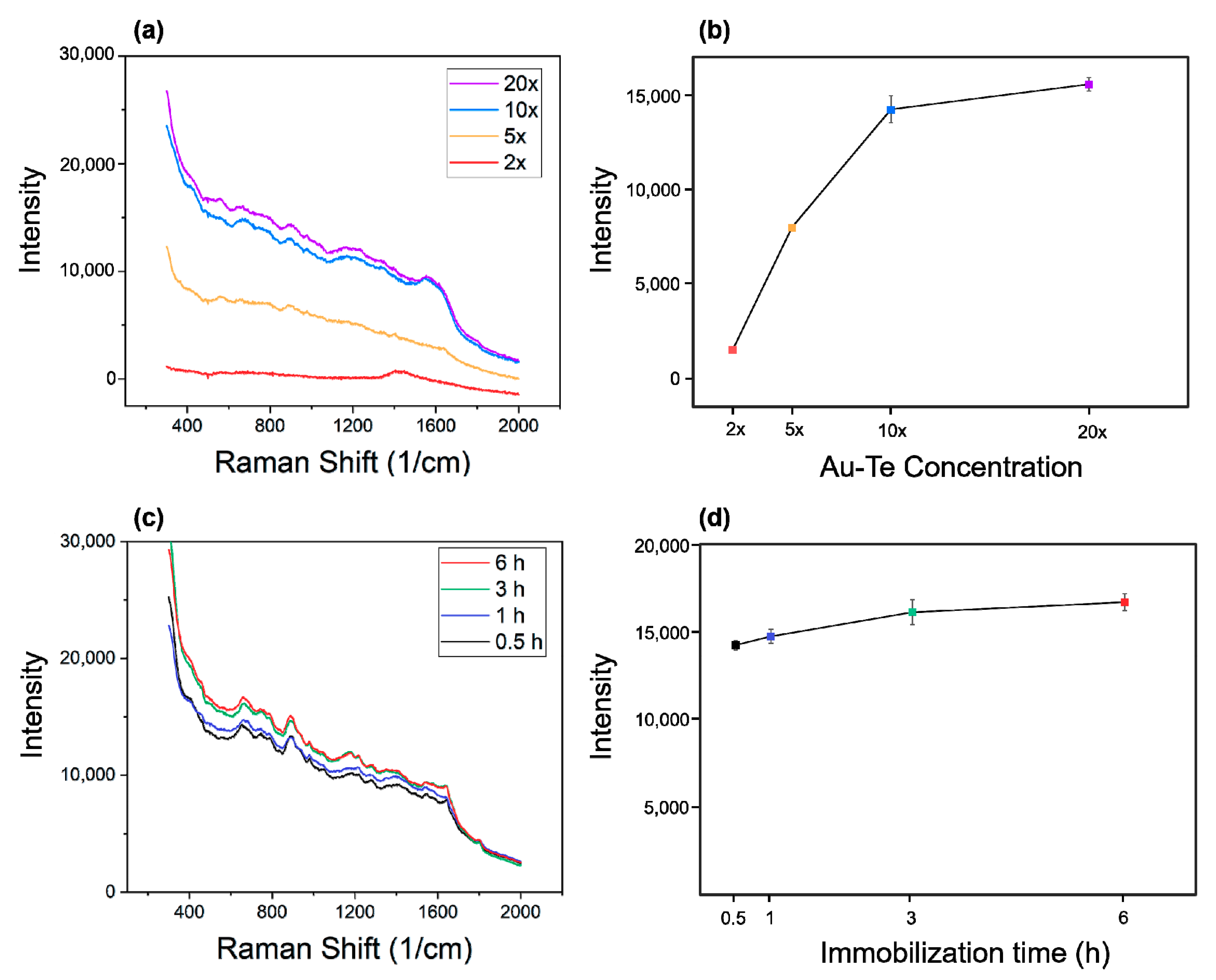
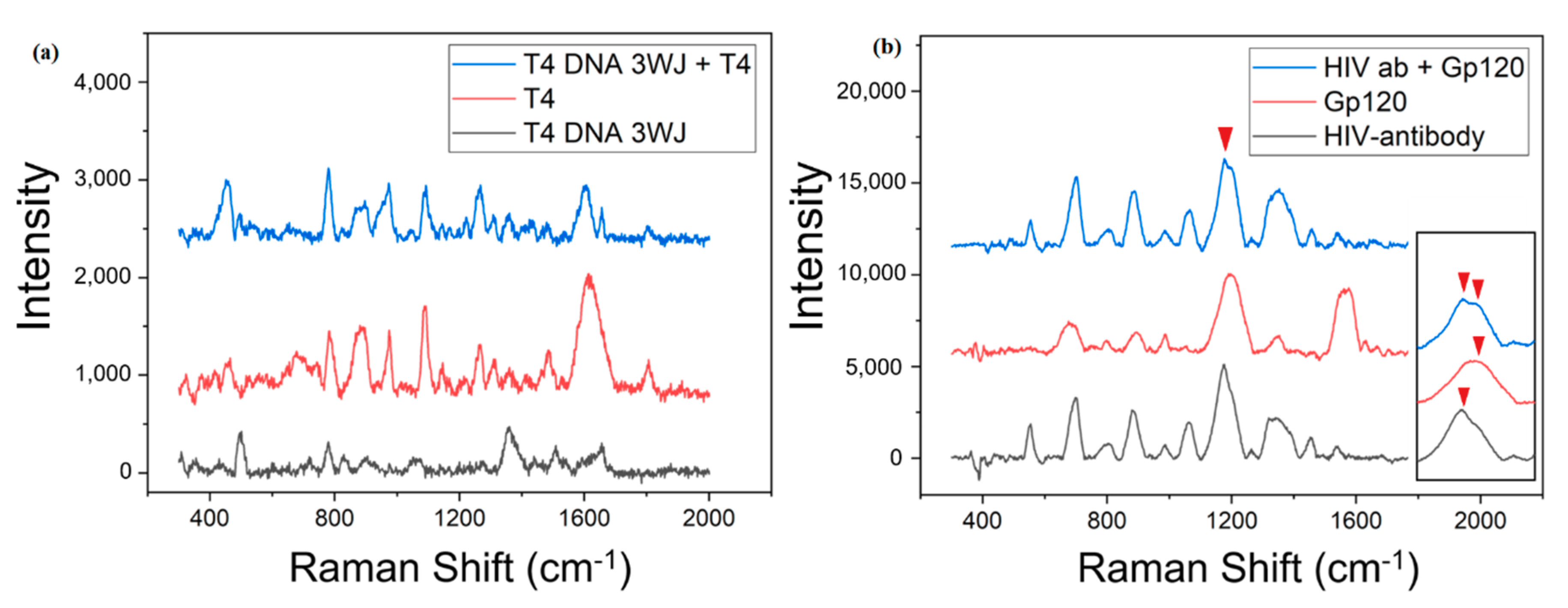
| Wave Number (cm−1) | Vibrational Assignments |
|---|---|
| 783 | (C3—O)10, (C32—C27)10, β(C2—C1—C6), β(C4—C5—C6)10, β(C5—C6—O17)13 |
| 891 | τ(H15—C10—C11—C12)48 |
| 974 | (C27—N), y(C24—C27)17, β(H30—N29—C27)20, τ(H7—C1—C2—C3)10 |
| 1087 | (C9—C24)11, β(H16—C14—C13)45 |
| 1615 | β(N29H2)84, τ(H30—N29—C27—C24), τ(H31—N29—C27—C24)13 |
| GP120 | |
|---|---|
| Wave Number (cm−1) | Assignment |
| 675 | D-Mannose |
| 798 | C, U, Thr |
| 891 | β-C1 config. Trp, Val Man |
| 987 | Man, ribose |
| 1200 | Tyr, Phe |
| 1354 | Trp |
| 1561 | GlcNac(Amid II) Amide II, Trp |
| 1576 | G, A, Trp |
© 2020 by the authors. Licensee MDPI, Basel, Switzerland. This article is an open access article distributed under the terms and conditions of the Creative Commons Attribution (CC BY) license (http://creativecommons.org/licenses/by/4.0/).
Share and Cite
Kim, S.M.; Lee, T.; Gil, Y.-G.; Kim, G.H.; Park, C.; Jang, H.; Min, J. Fabrication of Bioprobe Self-Assembled on Au–Te Nanoworm Structure for SERS Biosensor. Materials 2020, 13, 3234. https://doi.org/10.3390/ma13143234
Kim SM, Lee T, Gil Y-G, Kim GH, Park C, Jang H, Min J. Fabrication of Bioprobe Self-Assembled on Au–Te Nanoworm Structure for SERS Biosensor. Materials. 2020; 13(14):3234. https://doi.org/10.3390/ma13143234
Chicago/Turabian StyleKim, Soo Min, Taek Lee, Yeong-Gyu Gil, Ga Hyeon Kim, Chulhwan Park, Hongje Jang, and Junhong Min. 2020. "Fabrication of Bioprobe Self-Assembled on Au–Te Nanoworm Structure for SERS Biosensor" Materials 13, no. 14: 3234. https://doi.org/10.3390/ma13143234
APA StyleKim, S. M., Lee, T., Gil, Y.-G., Kim, G. H., Park, C., Jang, H., & Min, J. (2020). Fabrication of Bioprobe Self-Assembled on Au–Te Nanoworm Structure for SERS Biosensor. Materials, 13(14), 3234. https://doi.org/10.3390/ma13143234


