Nano-Montmorillonite Regulated Crystallization of Hierarchical Strontium Carbonate in a Microbial Mineralization System


 and
and 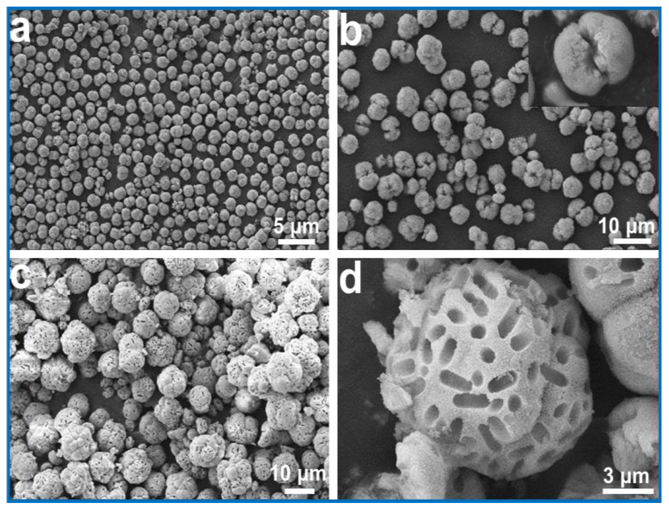
Abstract
1. Introduction
2. Materials and Methods
2.1. Materials
2.2. Strain Culture
2.3. Preparation of Nano-MMT Solution
2.4. Preparation of Strontium Carbonate Nanocrystals by Microbial Mineralization in the Presence of Nano-MMT
2.5. Characterization
3. Results and Discussion
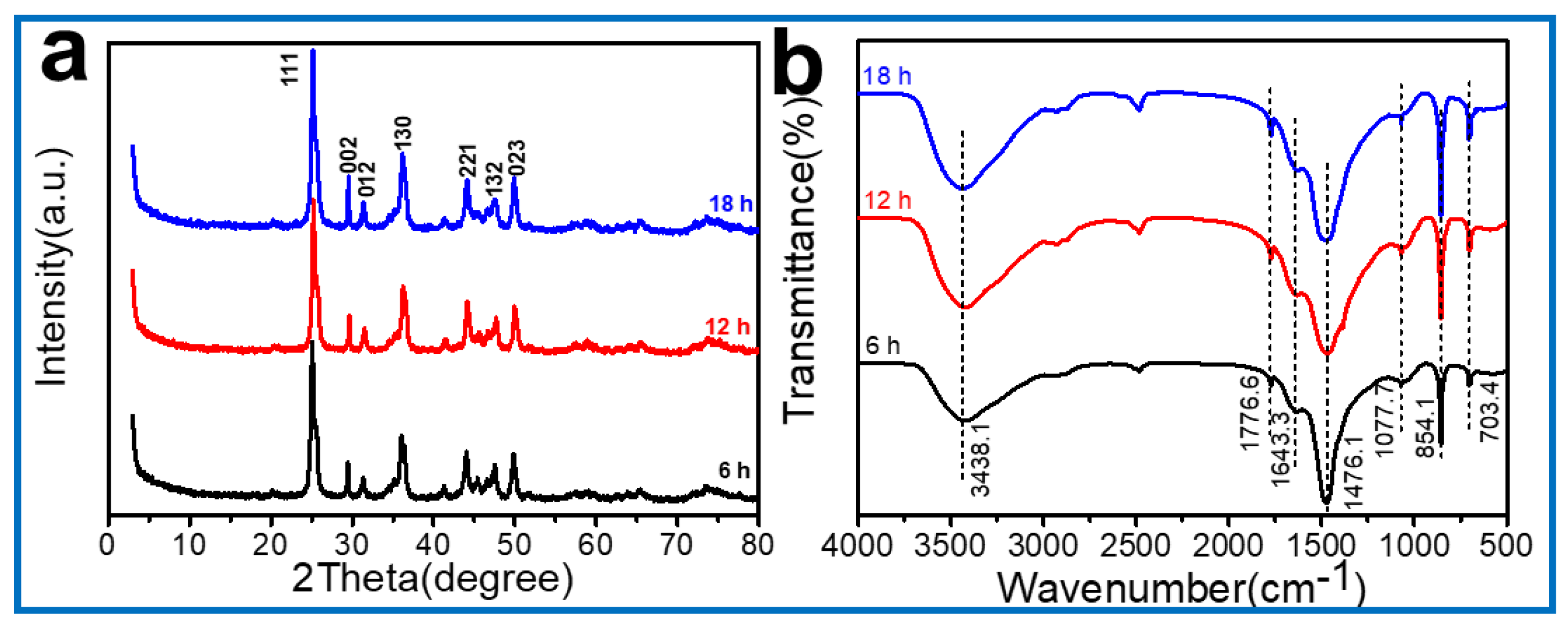
3.1. Effects of Mineralization Time on the Morphology of Strontium Carbonate in the Absence of Nano-MMT
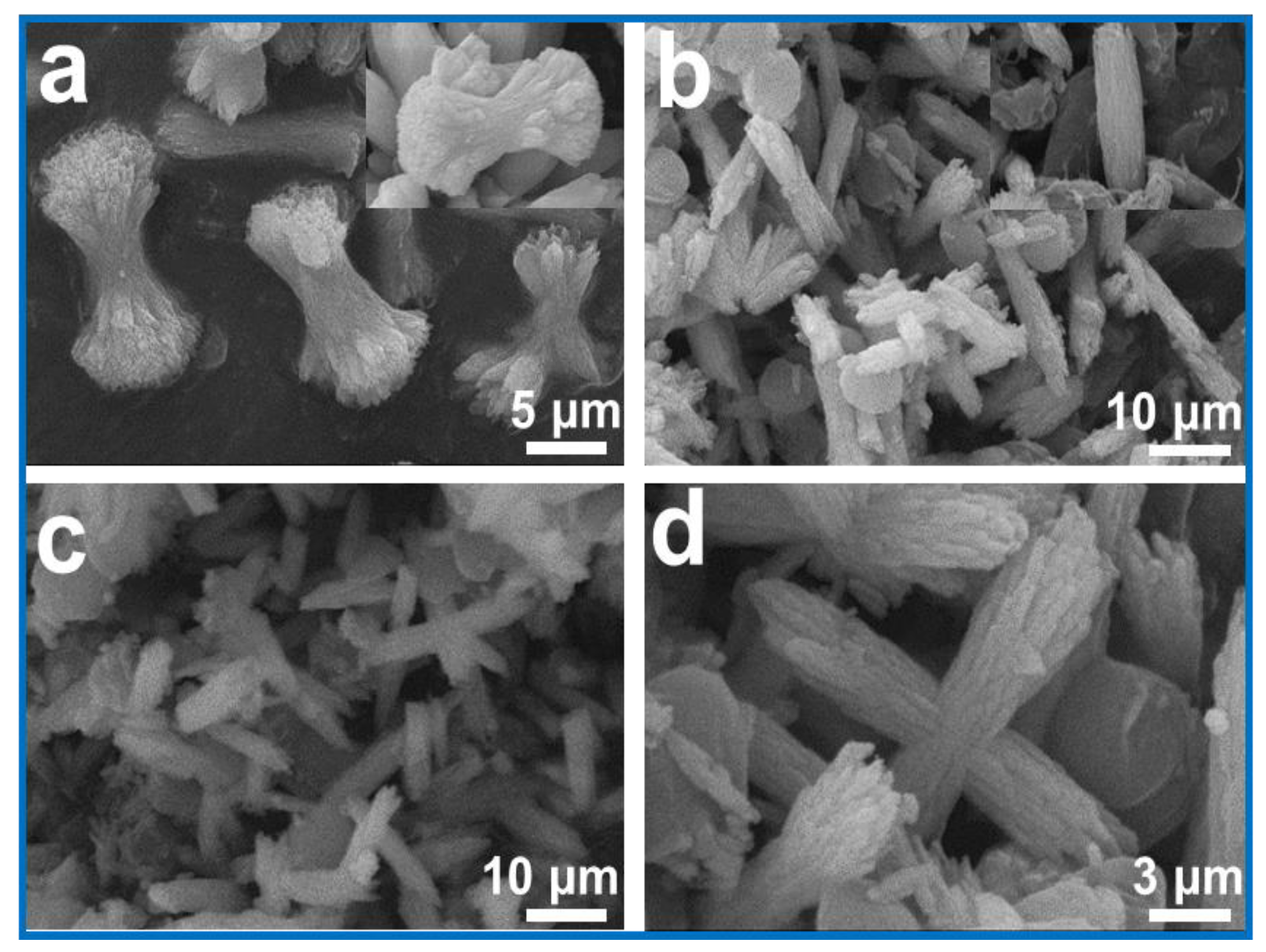
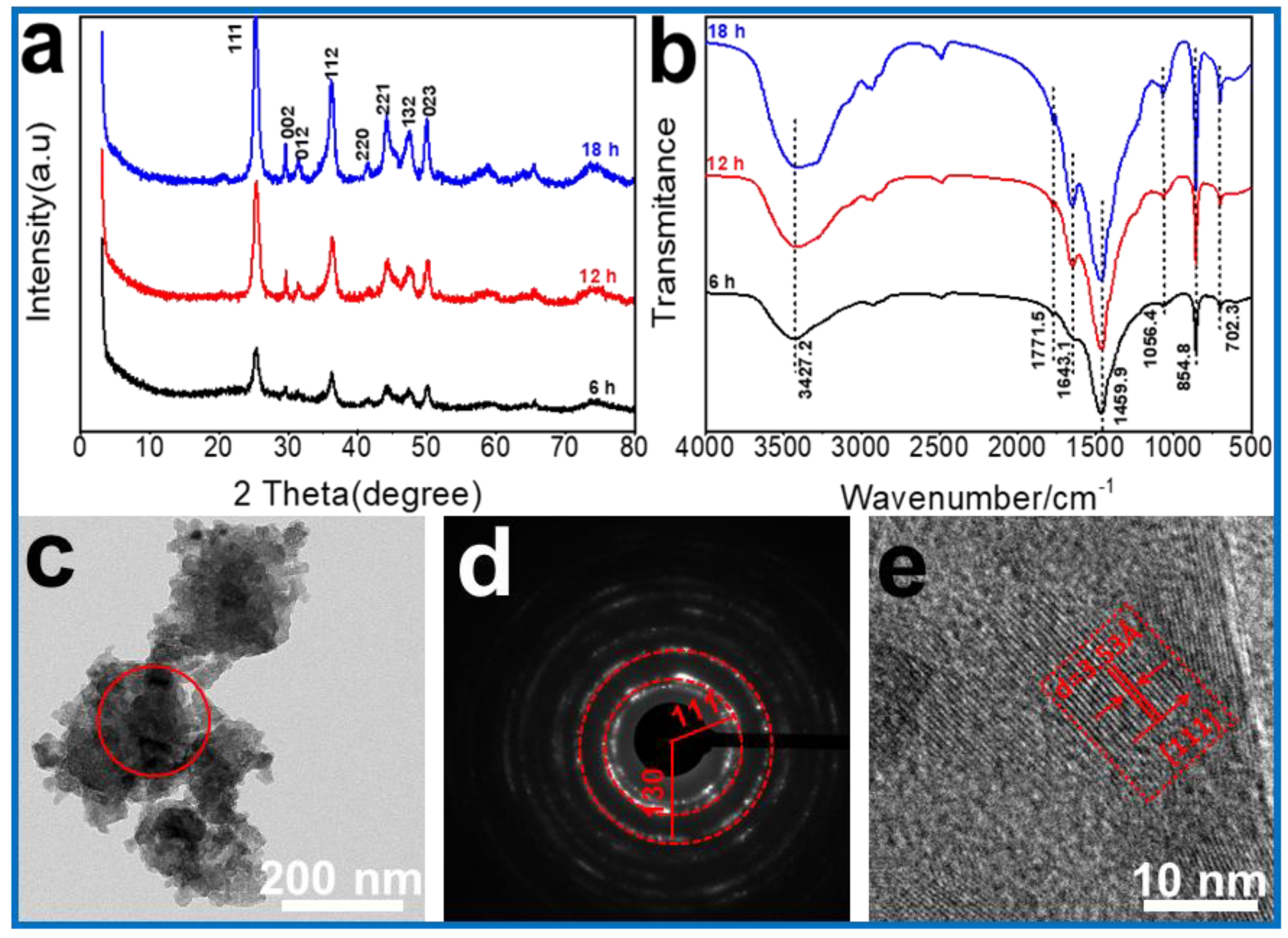
3.2. Effects of Mineralization Time on Morphology of Strontium Carbonate in the Presence of Nano-MMT
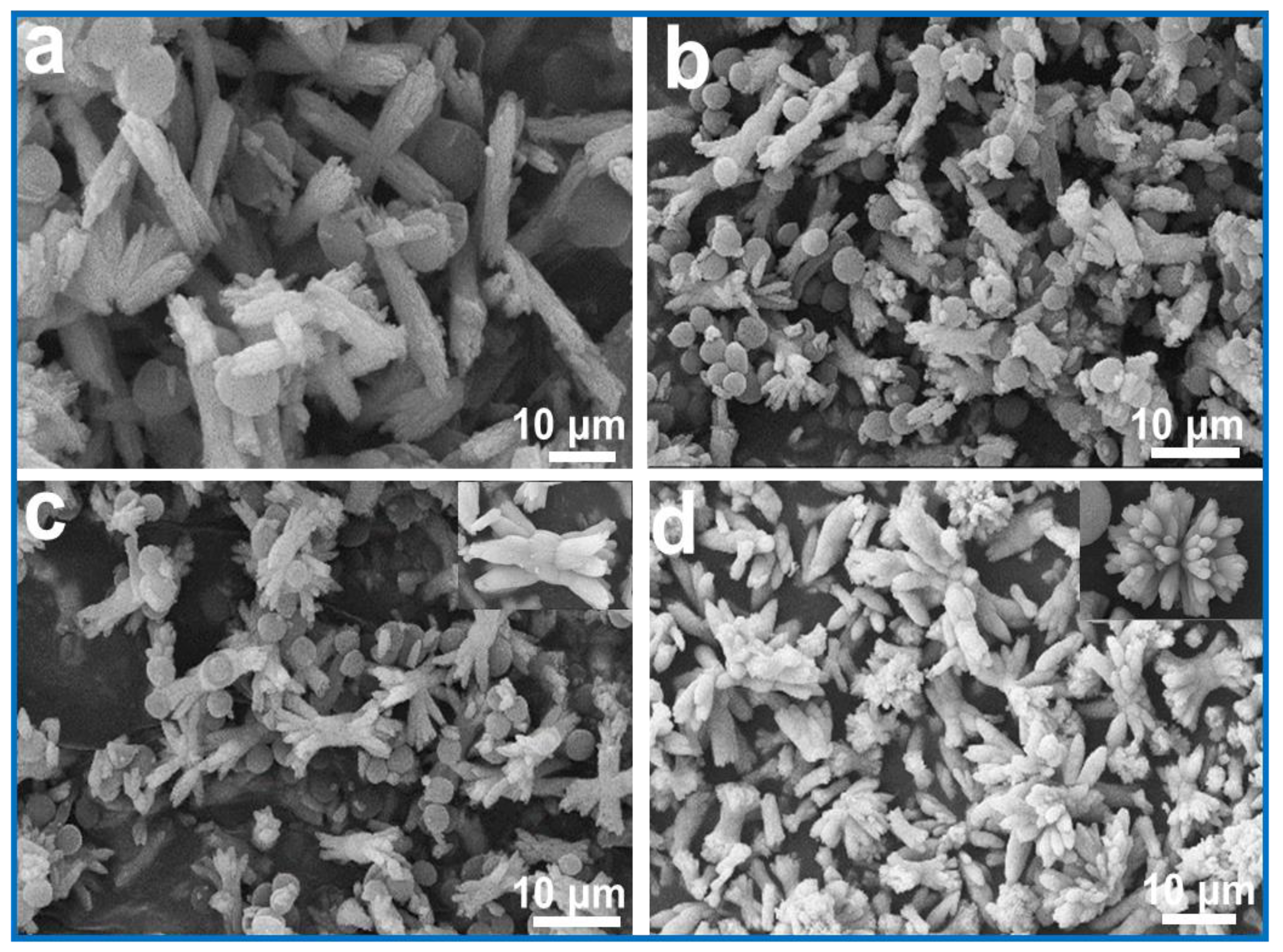
3.3. Effects of Nano-MMT Concentrations on Morphology of Strontium Carbonate
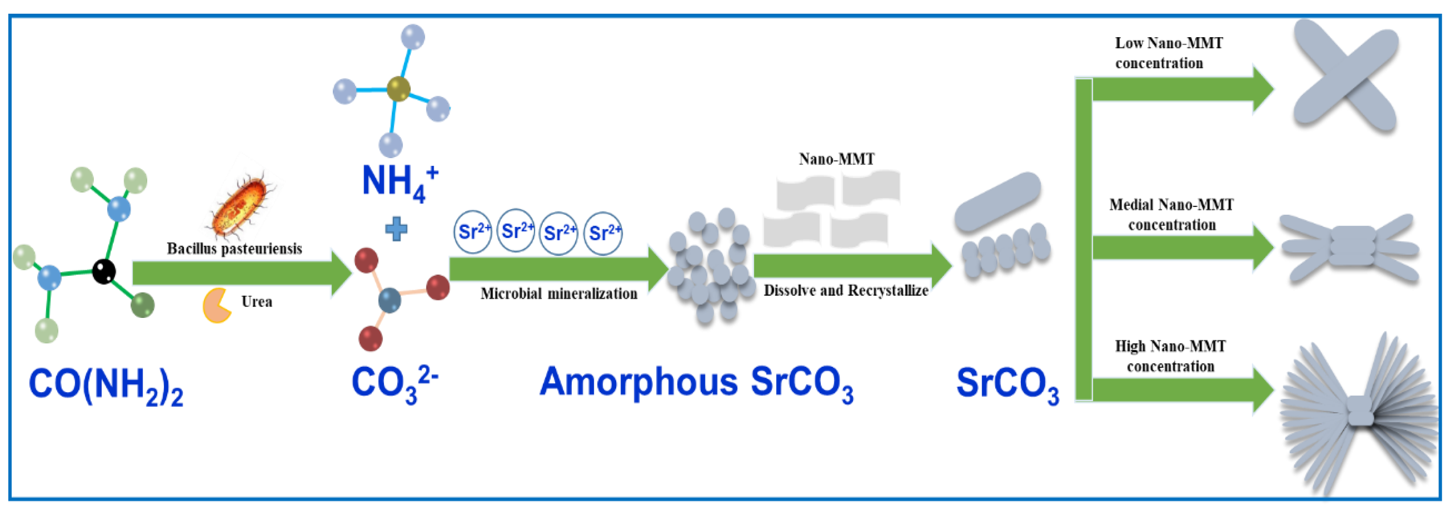
3.4. Mineralization Mechanism
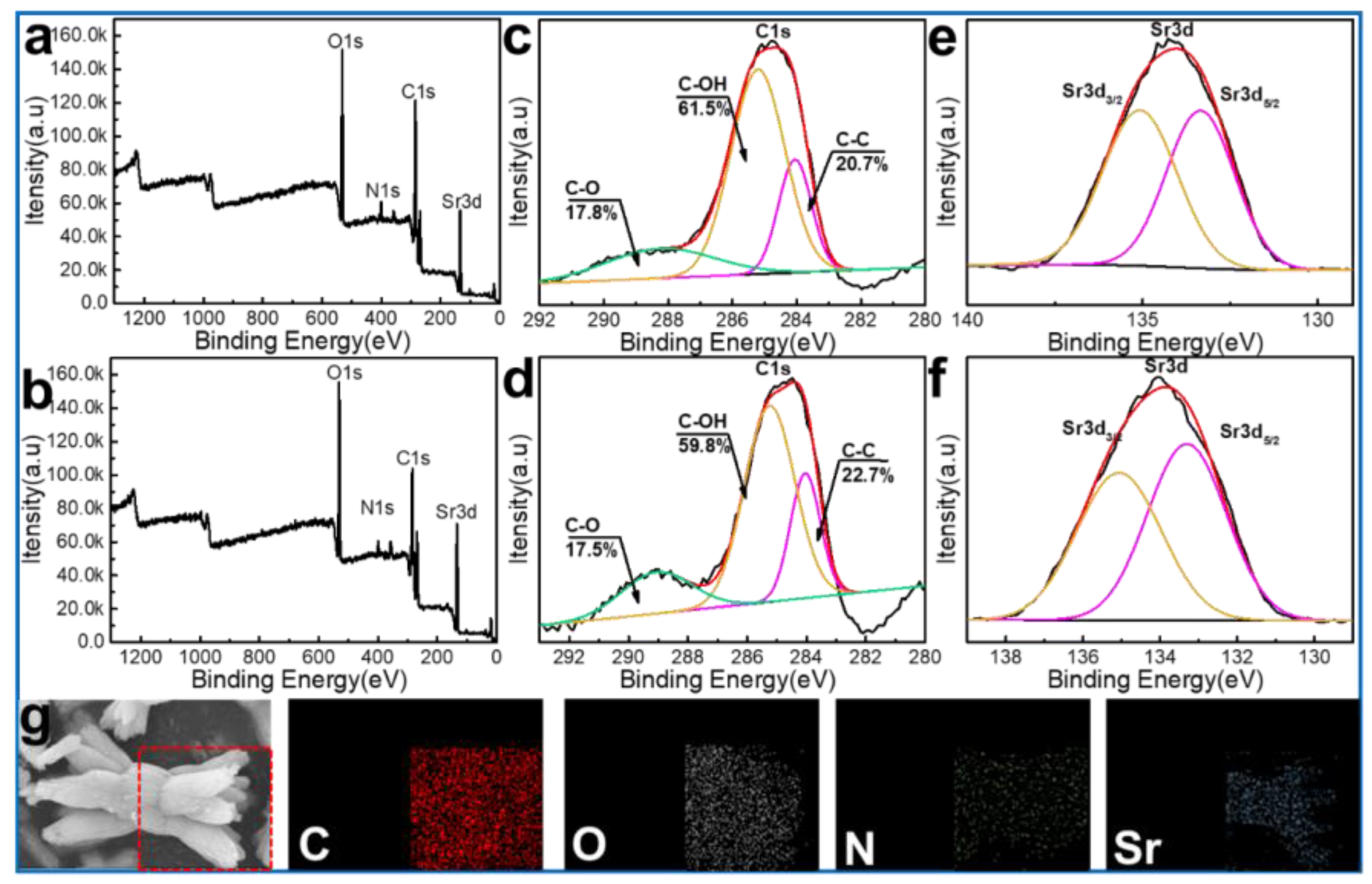
3.5. Mineralization of Strontium Ions under Microbial Mineralization Systems
4. Conclusions
Supplementary Materials
Author Contributions
Funding
Conflicts of Interest
References
- Zhu, W.K.; Luo, X.G.; Zhang, C.; Duan, T.; Zhou, J. Regulation of microstructure of calcium carbonate crystals by egg white protein. Chem. Res. Chin. Univ. 2012, 28, 180–185. [Google Scholar]
- Chen, T.; Shi, P.; Zhang, J.; Li, Y.; Tian, X.; Lian, J.; Duan, T.; Zhu, W. Bioinspired enhancement of chitosan nanocomposite films via mg-acc crystallization, their robust, hydrophobic and biocompatible. Appl. Surf. Sci. 2018, 459, 129–137. [Google Scholar] [CrossRef]
- Shelobolina, E.S.; Avakyan, Z.A.; Bulygina, E.S.; Turova, T.P.; Lysenko, A.M.; Osipov, G.A.; Karavaiko, G.I. Description of a new species of mucilaginous bacteria, bacillus edaphicus sp. Nov., and confirmation of the taxonomic status of bacillus mucilaginosus avakyan et al. 1986 based on data from phenotypic and genotypic analysis. Microbiology 1997, 66, 681–689. [Google Scholar]
- Ehlmann, B.L.; Mangold, N.; Michalski, J.R.; Catling, D.C.; Ruff, S.W.; Chassefière, E.; Niles, P.B.; Chevrier, V.; Poulet, F. Geochemical consequences of widespread clay mineral formation in mars’ ancient crust. Space Sci. Rev. 2013, 174, 329–364. [Google Scholar] [CrossRef]
- Ehlmann, B.L.; Mustard, J.F.; Murchie, S.L.; Bibring, J.P.; Meunier, A.; Fraeman, A.A.; Langevin, Y. Subsurface water and clay mineral formation during the early history of mars. Nature 2011, 479, 53–60. [Google Scholar] [CrossRef]
- Brookshaw, D.R.; Pattrick, R.A.D.; Lloyd, J.R.; Vaughan, D.J. Microbial effects on mineral–radionuclide interactions and radionuclide solid-phase capture processes. Mineral. Mag. 2012, 76, 777–806. [Google Scholar] [CrossRef]
- Savage, D.; Noy, D.; Mihara, M. Modelling the interaction of bentonite with hyperalkaline fluids. Appl. Geochem. 2002, 17, 207–223. [Google Scholar] [CrossRef]
- Norrfors, K.; Bouby, M.; Marsac, R.; Heck, S.; Schäfer, T.; Geckeis, H.; Wold, S. Montmorillonite colloids ii: Dependency of colloidal size on sorption of radionuclides. Appl. Clay. Sci. 2016, 123, 292–303. [Google Scholar] [CrossRef]
- Wang, X.; Yang, Y.; Yang, Y.; Yang, J.; Huang, Q. Analysis of hazardous characteristics of strontium salt residue in the production of strontium carbonate. J. Environ. Eng. Tech. 2018, 8, 297–301. [Google Scholar]
- Liu, Y.; Jing, X.; Hu, X.; Wu, G. Morphology of strontium carbonate particle adjusted by phthalic acid and isophthalic acid. IOP Conf. Ser. Mater. Sci. Eng. 2018, 389, 012020. [Google Scholar] [CrossRef]
- Tobías, G.; Oró-Solé, J.; Beltrán-Porter, D.; Fuertes, A. Synthesis and crystal structure of novel ruddlesden–popper strontium niobium oxynitrides. Cryst. Eng. 2002, 5, 479–485. [Google Scholar] [CrossRef]
- Brinza, L.; Mosselmans, J.F.W.; Schofield, P.F.; Quinn, P.D.; Hodson, M.E. Strontium incorporation into carbonate granules secreted by earthworms. J. Neurochem. 2011, 79, 463–484. [Google Scholar]
- Harris, D.; Porter, L.K.; Paul, E.A. Continuous flow isotope ratio mass spectrometry of carbon dioxide trapped as strontium carbonate. Commun. Soil Sci. Plant Anal. 1997, 28, 747–757. [Google Scholar] [CrossRef][Green Version]
- Chen, T.; Shi, P.; Zhang, J.; Li, Y.; Duan, T.; Dai, L.; Wang, L.; Yu, X.; Zhu, W. Natural polymer konjac glucomannan mediated assembly of graphene oxide as versatile sponges for water pollution control. Carbohydr. Polym. 2018, 202, 425–433. [Google Scholar] [CrossRef] [PubMed]
- Zhu, Y.; Li, Y.; Lu, A.; Wang, H.; Yang, X.; Wang, C.; Cao, W.; Wang, Q.; Zhang, X.; Pan, D. Study of the interaction between bentonite and a strain of bacillus mucilaginosus. Acta Petrol. Mineral. 2011, 59, 538–545. [Google Scholar] [CrossRef]
- Biswas, B.; Sarkar, B.; Rusmin, R.; Naidu, R. Bioremediation of pahs and vocs: Advances in clay mineral–microbial interaction. Environ. Int. 2015, 85, 168–181. [Google Scholar] [CrossRef]
- Luan, F.; Liu, Y.; Griffin, A.M.; Gorski, C.A.; Burgos, W.D. Iron(iii)-bearing clay minerals enhance bioreduction of nitrobenzene by shewanella putrefaciens cn32. Environ. Sci. Tech. 2015, 49, 1418–1426. [Google Scholar] [CrossRef]
- Hauber, E.; Platz, T.; Reiss, D.; Deit, L.L.; Kleinhans, M.G.; Marra, W.A.; Haas, T.D.; Carbonneau, P. Asynchronous formation of hesperian and amazonian-aged deltas on mars and implications for climate. J. Geophys. Res. Planet. 2013, 118, 1529–1544. [Google Scholar] [CrossRef]
- Dong, H.; Lu, A. Geomicrobiology research in china: Mineral-microbe interactions. Geomicrobiol. J. 2012, 29, 197–198. [Google Scholar] [CrossRef]
- Klaus-Joerger, T.; Joerger, R.; Olsson, E.; Granqvist, C.-G. Bacteria as workers in the living factory: Metal-accumulating bacteria and their potential for materials science. Trends Biotechnol. 2001, 19, 15–20. [Google Scholar] [CrossRef]
- Stephen, J.R.; Macnaughtont, S.J. Developments in terrestrial bacterial remediation of metals. Curr. Opin. Biotech. 1999, 10, 230–233. [Google Scholar] [CrossRef]
- Liu, S.-H.; Zeng, G.-M.; Niu, Q.-Y.; Liu, Y.; Zhou, L.; Jiang, L.-H.; Tan, X.-F.; Xu, P.; Zhang, C.; Cheng, M. Bioremediation mechanisms of combined pollution of pahs and heavy metals by bacteria and fungi: A mini review. Bioresour. Technol. 2017, 224, 25–33. [Google Scholar] [CrossRef]
- Kang, C.-H.; Kwon, Y.-J.; So, J.-S. Bioremediation of heavy metals by using bacterial mixtures. Ecol. Eng. 2016, 89, 64–69. [Google Scholar] [CrossRef]
- Chen, T.; Li, J.; Shi, P.; Li, Y.; Lei, J.; Zhou, J.; Hu, Z.; Duan, T.; Tang, Y.; Zhu, W. Effects of montmorillonite on the mineralization and cementing properties of microbiologically induced calcium carbonate. Adv. Mater. Sci. Eng. 2017, 2017, 1–13. [Google Scholar] [CrossRef]
- Chen, T.; Shi, P.; Li, Y.; Zhang, J.; Duan, T.; Yu, Y.; Zhou, J.; Zhu, W. Crystallization of calcium carbonate mineral with hierarchical structures regulated by silk fibroin in microbial mineralization system. J. Cryst. Growth 2018, 493, 51–57. [Google Scholar] [CrossRef]
- Chen, T.; Shi, P.H.; Li, Y.; Duan, T.; Yu, Y.; Li, X.; Zhu, W. Biomineralization of varied calcium carbonate crystal by synergistic effect of silk fibroin/magnesium ions in a microbial system. Crystengcomm 2018, 20, 2366–2373. [Google Scholar] [CrossRef]
- Li, Y.; Zhu, W.; Chen, T.; Lei, J.; Duan, T.; Zhou, J.; Tang, Y.; Hu, Z. Synergistic metallogenesis of simulated radionuclide strontium by carbonate-mineralization bacteria/nano-montmorillonite. J. Radioanal. Nuclear Chem. 2017, 314, 333–341. [Google Scholar] [CrossRef]
- Guo, X.H.; Xu, A.W.; Yu, S.H. Crystallization of calcium carbonate mineral with hierarchical structures in dmf solution under control of poly(ethylene glycol)-b-poly(l-glutamic acid): Effects of crystallization temperature and polymer concentration. Cryst. Growth Des. 2008, 8, 1233–1242. [Google Scholar] [CrossRef]
- Cantaert, B.; Kim, Y.Y.; Ludwig, H.; Nudelman, F.; Sommerdijk, N.A.; Meldrum, F.C. Think positive: Phase separation enables a positively charged additive to induce dramatic changes in calcium carbonate morphology. Adv. Funct. Mater. 2012, 22, 907–915. [Google Scholar] [CrossRef]
- Czemierska, M.; Szcześ, A.; Hołysz, L.; Wiater, A.; Jarosz-Wilkołazka, A. Characterisation of exopolymer r-202 isolated from rhodococcus rhodochrous and its flocculating properties. Eur. Polym. J. 2017, 88, 21–33. [Google Scholar] [CrossRef]
- Hood, M.A.; Landfester, K.; Muñoz-Espí, R. The role of residue acidity on the stabilization of vaterite by amino acids and oligopeptides. Cryst. Growth Des. 2014, 14, 1077–1085. [Google Scholar] [CrossRef]
- Szcześ, A.; Czemierska, M.; Jarosz-Wilkołazka, A. Calcium carbonate formation on mica supported extracellular polymeric substance produced by rhodococcus opacus. J. Solid State Chem. 2016, 242, 212–221. [Google Scholar] [CrossRef]
- Wenkun, Z.; Tao, M.; Tao, D.; Youkui, Z.; Xuegang, L. Metallogenesis of simulated radioactive sr2+ by carbonate-mineralization bacteria. Res. Environ. Sci. 2015, 2, S429–S430. [Google Scholar]


© 2019 by the authors. Licensee MDPI, Basel, Switzerland. This article is an open access article distributed under the terms and conditions of the Creative Commons Attribution (CC BY) license (http://creativecommons.org/licenses/by/4.0/).
Share and Cite
Zheng, K.; Chen, T.; Zhang, J.; Tian, X.; Ge, H.; Qiao, T.; Lei, J.; Li, X.; Duan, T.; Zhu, W. Nano-Montmorillonite Regulated Crystallization of Hierarchical Strontium Carbonate in a Microbial Mineralization System. Materials 2019, 12, 1392. https://doi.org/10.3390/ma12091392
Zheng K, Chen T, Zhang J, Tian X, Ge H, Qiao T, Lei J, Li X, Duan T, Zhu W. Nano-Montmorillonite Regulated Crystallization of Hierarchical Strontium Carbonate in a Microbial Mineralization System. Materials. 2019; 12(9):1392. https://doi.org/10.3390/ma12091392
Chicago/Turabian StyleZheng, Kui, Tao Chen, Jian Zhang, Xiuquan Tian, Huilin Ge, Tiantao Qiao, Jia Lei, Xianyan Li, Tao Duan, and Wenkun Zhu. 2019. "Nano-Montmorillonite Regulated Crystallization of Hierarchical Strontium Carbonate in a Microbial Mineralization System" Materials 12, no. 9: 1392. https://doi.org/10.3390/ma12091392
APA StyleZheng, K., Chen, T., Zhang, J., Tian, X., Ge, H., Qiao, T., Lei, J., Li, X., Duan, T., & Zhu, W. (2019). Nano-Montmorillonite Regulated Crystallization of Hierarchical Strontium Carbonate in a Microbial Mineralization System. Materials, 12(9), 1392. https://doi.org/10.3390/ma12091392