Article

Characterization of Phytochemical and Nutrient Compounds from the Leaves and Seeds of Moringa oleifera and Moringa peregrina

1
CAS Key Laboratory of Plant Germplasm Enhancement and Specialty Agriculture, Wuhan Botanical Garden, Chinese Academy of Sciences, Wuhan 430074, China
2
University of Chinese Academy of Sciences, Beijing 100049, China
3
Department of Botany, Agriculture and Biological Institute, National Research Centre, Dokki, Giza 12422, Egypt
4
Egyptian Deserts Gene Bank, North Sinai Research Station, Department of Genetic Resources, Desert Research Center, Cairo 11753, Egypt
5
Department of Resources Science of Traditional Chinese Medicines, School of Traditional Chinese Pharmacy, China Pharmaceutical University, Nanjing 211198, China
*
Author to whom correspondence should be addressed.
Horticulturae 2022, 8(11), 1081; https://doi.org/10.3390/horticulturae8111081
Submission received: 23 September 2022 / Revised: 5 November 2022 / Accepted: 7 November 2022 / Published: 16 November 2022
(This article belongs to the Section Medicinals, Herbs, and Specialty Crops)

Abstract

:
Moringa oleifera and M. peregrina are robust and fast-growing trees. These trees are considered some of the most highly valued trees worldwide because of their properties and uses. This study aimed to investigate and determine the content of phytochemical and nutrient compounds in the fresh leaves and the mature seeds of both M. oleifera and M. peregrina. The experimental data analysis showed that all four extracts were rich in proximate minerals, fatty acids (FA), and un-saponified and polyphenolic components. The total percentages of saturated and unsaturated fatty acids compounds obtained from the leaves and seeds of M. oleifera and M. peregrina were (45.02 and 54.93%), (10.80 and 89.19%), (37.13 and 62.8%), and (11.95 and 88.03%), respectively. The major polyphenols compositions were identified as gallic acid, chlorogenic acid, methyl gallate, and coffeic acid. The outcomes of the present study indicate that the leaves and seeds of Egyptian Moringa (M. oleifera and M. peregrina) contain various phytochemical and nutrient compounds, which can provide several health advantages and play an important role in the metabolism of the human body, especially in diseases such as atherosclerosis, heart disease, obesity, and high cholesterol and triglycerides.

1. Introduction

Moringa is a genus from the Moringaceae family, in which fourteen species have been described to be distributed across Egypt, India, the Philippines, Pakistan, the Caribbean Islands, Asia Minor, Africa, Central America, Cambodia, North America, South America, and the Western and Sub-Himalayan regions [1,2,3]. Most Moringa species are used in Indian and Chinese folk medicine, with many traditional names, such as Tree of Life, Miracle Tree, Horse Radish Tree, Ben Oil Tree, Ber Oil Tree, and Drumstick Tree. In addition, Moringa trees include M. oleifera, M. hildebrandtii, M. concanensis, M. drouhardii, M. stenopetala, M. arborea, M. ruspoliana, M. borziana, M. longituba, M. peregrina, M. pygmaea, M. rivae, M. ovalifolia, and M. pterygosperma [4]. The best-known, most widely distributed species in the Moringaceae family are M. oleifera and M. peregrina [5]. All parts of the Moringa tree, such as the leaf, seed, bark, gum, seed oil, meal, fruit (pods), flowers, root, stem bark, and root bark, are suitable for consumption and used by consumers in plant-based foods and medicines [4].
For these reasons, some parts of these species have drawn researchers’ attention and have been studied for their various therapeutic properties, including antibacterial, antifungal, and antiproliferation through apoptosis; the treatment of ovarian cancer, prostate cancer, breast cancer, phyto-oxidative damage, radioprotective issues, and circulatory/endocrine disorders; anti-anemic, anti-atherosclerotic, and diuretic activity; detoxification, analgesic, anti-aging, and antipyretic activity; antioxidant activity towards fluoride toxicity; an antidote for snakebites and scorpion bites; treatment for oxidative DNA damage; protective properties regarding digestive disorders; anti-colitis activity; cholagouge, diarrhea, and dysentery treatment; and purgative, laxative, flatulence/carminative, antinociceptive, asthma, abortifacient, and skin disorder applications [6,7,8,9,10,11,12,13,14,15,16,17,18,19,20,21,22,23,24].
Moreover, the leaves and seeds of Moringa species are rich in various phytochemicals, such as phenolic acids (e.g., chlorogenic acid, caffeic acid, sinapic acid, verbascoside, chicoric acid, salvianolic acid, and caffeoyl shikimic acid), flavonoids (e.g., rutin, kaempferol malonyl-glucoside, morin, silymarin, apigenin, epicatechin, pinocembrin, eriocitrin, hispidulin, geraldine, biochanin A, myricitrin, and cirsilineol), and other polyphenols (e.g., umbelliferone, coumarin, isolariciresinol glucoside, estragole, 6-gingerol, rosmanol, and bergapten) [25,26,27,28,29,30,31,32].
Furthermore, Moringa plant species offer various bioactive compounds, such as kaempferol, quercetin, isothiocyanates, zeatin, glucosinolates, caffeoylquinic acid, ascorbic acid, amino acids, carotenoids, alkaloids, moringine, phytoestrogens, and moringinine [1,33,34]. On the other hand, the fruits, leaves, seeds, and pods have been reported as a rich source of crude protein, fat, carbohydrate, fiber, chlorophyll, tryptophan, vitamins (A, B1, B2, B3, C, and E), and essential elements (sulfur, calcium, magnesium, copper, phosphorus, iron, and potassium) [24,35].
In developing countries, almost all parts of the Moringa seeds, leaves, fruits, flowers, bode, and roots are suitable for people who wish to use therapeutic plants and inexpensive foods for their daily life and well-being needs [33,36]. In this context, most parts of Moringa have been used in areas such as food security, water purification, renewable feedstock, and the support of sustainable and rural development, and they are a natural source of oil and fat in the context of global industrialization [33,36]. In addition, the knowledge and aggregated data on the content of Moringa parts from bioactive compounds may help to achieve health benefits and health-promoting effects. Most studies on Moringa are based on M. oleifera, whose polyphenols and fatty acid content have been estimated from various tree parts [37,38,39]. Recently, M. peregrina has become an attractive medicinal plant in biochemistry research due to its nutritional and pharmaceutical properties. For example, Mohammad pour et al. [40] used gas chromatography and mass spectrometry (GC–MS) to calculate the significant anticancer activity in M. peregrina roots extracted and dissolved in different solvents. In addition, M. peregrina was reported as having the largest fatty acid composition among two Egyptian species of M. peregrina and M. oleifera [41,42]. Moreover, the polyphenol content in M. peregrina was the highest among three Moringa species (M. stenopetala, M. peregrina, and M. oleifera) grown at Orman Botanical Garden, Giza, Egypt [43]. In the current study, we aimed to perform the full screening for proximate and bioactive compounds in M. oleifera and M. peregrina grown at Orman Botanical Garden, Giza, Egypt, using leaves and mature seeds, by using chromatography techniques, which could allow researchers to broaden their scientific research and explore the content of Moringa parts that are used as herbal medicines and nutrition supplements.

2. Materials and Methods

2.1. Materials and Sample Preparation

Fresh leaves and mature seeds of M. oleifera and M. peregrina were obtained from the Orman Garden (30°01′45″ N 31°12′47″ E), Ministry of Agriculture, Egypt, in August 2021, and fresh samples were collected at the flowering stage. Three replicates from collected samples were dried in an oven at 40 °C. The oven-dried seeds and leaves were ground into a fine powder using a clean coffee grinding machine for 30 s, followed by a mortar, pestle, and later a blender.

2.2. Proximate and Mineral Composition

For the total oil content (W1), (2 g) of each of the samples was weighed on filter paper (W2). The filter papers containing the samples were tied and then placed into a Soxhlet extractor. Then, they were placed in the extraction chamber and suspended above an already-weighed receiving flask containing petroleum ether (40–60 °C) below the condenser. The flask was heated for 6 h to extract the crude oil. After the extraction, the flask containing the crude oil was disconnected from the Soxhlet extractor and oven-dried at 100 °C for 24 h. Afterward, it was cooled in a desiccator and weighed until a constant weight was obtained (W3). The difference in weight was expressed as a percentage of crude oil content [44], while total ash content was calculated in (2 g) of each sample, which was weighed into small dry crucibles of known weight. The samples in crucibles were placed in a furnace and ashed at 550 °C, which was kept constant for 3 h. The ashed samples were removed from the furnace, cooled, and kept in a desiccator until constant weights were obtained [44]. The total nitrogen (N) content (2 g) of each sample was weighed, and the samples were analyzed by using the micro Kjeldahl method. The N content was converted to CP by multiplying it by 6.25. The method involved the digestion of the samples in concentrated (98%) sulfuric acid, a distillation of the digests into weak acids (4% boric acid), and titration of the distillates with 0.1 µ hydrochloric (HCL) acid using a mixed indicator (Methyl and Bromocresol Green) as an indicator according to AOAC [45]. The crude protein was calculated depending on the nitrogen content by multiplying the value by 6.25. Hence, the total carbohydrate content was determined by the method known as ‘estimation by difference’, where the sum of the oil, ash, protein, and carbohydrate content was subtracted from one hundred (100). For total calcium (TCa) and potassium (TK) analysis, samples of 1 g of each plant were digested with 5 mL of concentrated nitric acid (HNO3) and 1 mL each of concentrated sulfuric acid (H2SO4) and 60–62% perchloric acid (HCLO4) and heated until white fumes of perchloric acid formed. The volume of the digest was reduced by heating, but not to dryness. The flask was set aside to cool, after which the content was diluted with distilled deionized water and then filtered into a 50 mL volumetric flask. The content was made up to mark with deionized water and stored until it was analyzed for mineral content using an atomic absorption spectrophotometer (AAS) [46,47]. Meanwhile, total phosphorous (TP) in the dried samples (1 g/each sample) was determined using hydrogen peroxide–sulfuric acid digestion at 360 °C [46,47], followed by automated colorimetric measurements of the TP concentrations in the digests (AutoAnalyzerII).

2.3. Gas Chromatographic Fatty Acid (FA) Analysis

For fatty acid (FA) composition analysis, the ground samples (10 g) from leaves and (2.5 g) from seeds of M. oleifera and M. peregrina were dissolved in 200 mL of n-hexane for 72 h, and then incubated in an orbital shaker at 75 rpm at room temperature. After the incubation, the upper phase extraction was filtered using Whatman paper (No. 1). After this, the residues were re-extracted with the same solvent and under the same conditions. The solvent was removed using a rotary evaporator under a vacuum at 40 °C. For fatty acid (FA) analysis, we added 12 mL of 0.5 µ from sodium hydroxide (NaOH) with the residue extract in a 25 mL flask. The mixture was heated in a Digital Thermostat Water Bath until the disappearance of all the fat was. Then, we added 2 mL of boron trifluoride–methanol solution (BF3. CH3OH) to the mixture, and then boiled it for 2 min, and placed it at room temperature until the mixture cooled. After this, we added 25 mL of saturated sodium chloride (NaCl) solution to prepare the fatty acid methyl esters (FAMEs). The profiling of the FAMEs was carried out using an Agilent model 7890B gas chromatograph (Santa Clara, CA, USA), fitted with a flame ionization detection (FID) system and a DB-Wax capillary column (60 m × 0.25 mm internal diameter, film thickness 0.25 μm). A sample of 1 μL was injected into the column using the split sample injection mode, and helium gas was used as a carrier at a flow rate of 1.2 mL/min in split-less mode, with the flow rate of 1.0 mL/min at a split ratio of 10:1 and injection volume of 1 µL, with the following temperature program: start with 240 °C; rising at 10 °C/min to 265 °C and held for 1 min; rising at 15 °C/min to 300 °C and held for 25 min. Meanwhile, the injector and detector were held at 280 and 290 °C. The column oven temperature was initially 50 and was raised to 175 °C at 4 °C/min, with initial and final hold times of 1 and 5 min, respectively, while the injector and detector were set at 235/20 min and the injector and detector were held at 260 to 280 °C, respectively. The FA composition was reported as a relative percentage of the total peak area.

2.4. Un-Saponified Compounds Profiling

For un-saponified compound profiling, the residue extract was subjected to saponification with potassium hydroxide (KOH) and then extracted with heptane/diethyl ether and finally dried. After this, we added 50 µL of n, o-bis (trimethylsilyl) trifluo (BSTFA) to the Moringa extraction and then incubated the samples at 70 °C for 30 min in a dry block heater; the residue was dissolved in 50 mL of distilled water and treated with dichloromethane until the GC–FID run. The profiling of the un-saponified compounds was carried out using an Agilent model 59977A gas chromatograph (Santa Clara, CA, USA), fitted with a flame ionization detection (FID) system and an HP-5MS column (30 m × 0.25 mm, film thickness 0.25 μm). Analyses were carried out using hydrogen as the carrier gas at a flow rate of 1.0 mL/min at a split ratio of 10:1 and injection volume of 1 µL, and the following temperature program was used: 240 °C; rising at 10 °C/min to 265 °C and held for 1 min; rising at 15 °C/min to 300 °C and held for 25 min. The injector and detector were held at 280 and 290 °C, respectively. Mass spectra were obtained by electron ionization (EI) at 70 eV, using a spectral range of m/z 50–550 and solvent delay for 3 min. The mass temperature was 230 °C and Quad 150 °C. Identification of different constituents was performed by comparing the spectrum fragmentation pattern with those stored in the Wiley and NIST Mass Spectral Library databases.

2.5. Total Polyphenolic Contents Assay

The total polyphenolic content of the extracts was determined based on the method of Goupy et al. [48], with some modifications. Separation and determination of leaves and mature seeds’ polyphenolic content were performed via HPLC (Agilent 1260) using a column eclipse C18 (25 cm × 4.6 mm). The mobile phase used water (as component A) and 0.05% trifluoroacetic acid in acetonitrile (component B), operating in a gradient mode. The mobile phase gradient was 18% B (0 min), 20% B (0–5 min), 20–40% B (5–8 min), 40% B (8–12), 18% B (12–15 min), and 18% B (15–20 min). The column temperature was set at 40 °C, and the flow rate was 0.9 mL /min. Polyphenols were detected at 280 nm. HPLC fractions were collected in multiples and analyzed

2.6. Statistical Analysis

Data analysis was performed using the General Linear Model (GLM) procedure of the SPSS software package (SPSS, 2010) by Arbuckle [49], version 20.0. All the data were analyzed based on a completely randomized design using one-way ANOVA. The differences among groups were evaluated by Duncan’s [50] multiple comparison tests to evaluate the significant differences (p < 0.05) between the content values obtained via different extraction procedures.

3. Results

3.1. Proximate and Mineral Composition Analysis

Some proximate and mineral characteristics of the leaves and seeds from M. oleifera and M. peregrina are presented in Figure 1 and Table 1. M. oleifera and M. peregrina have been found to be potentially beneficial sources of proximate and mineral compounds. The proximate analysis reported the following concentrations (%) in leaves and seeds from M. oleifera and M. peregrina: total protein (33.6, 24, 35.3, and 29%), total carbohydrates (38.6, 33.6, 34.3, and 24%), total oil (22, 36, 25.1, and 42%), and total ash (5.3, 6.3, 5.6 and 4.9%), respectively (Table 1). On the other hand, total nitrogen (TN; 5.4, 3.8, 5.6 and 4.6%), phosphorus (TP; 0.34, 0.52, 0.27 and 0.32%), potassium (TK; 2.53, 0.87, 1.9 and 0.58%), and calcium (TCa; 1.97, 3.6, 0.94 and 2.8%) were in the leaves and seeds from M. oleifera and M. peregrina, respectively (Table 1).

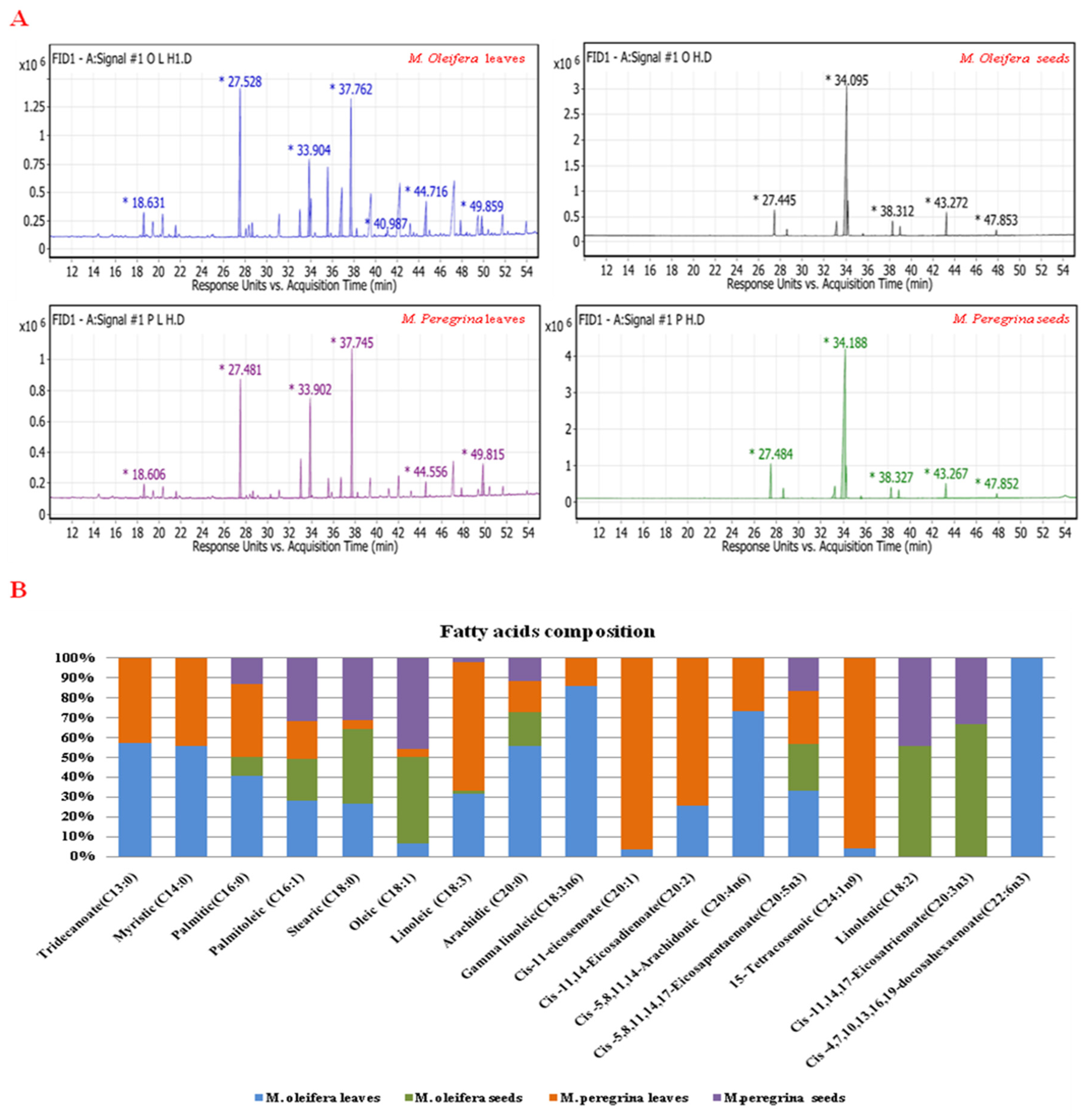
3.2. Fatty Acid (FA) Compositions

The fatty acid (FA) compositions of the extracts, as revealed by GC–MS analysis, are shown in Table 2. Seventeen fatty acids (FA) compounds were identified using n-hexane extracts from the leaves and seeds of M. oleifera and M. peregrina. The percentages of saturated fatty and unsaturated fatty acid compounds obtained from the leaves and seeds of M. oleifera and M. peregrina were (45.02 and 54.93%), (10.80 and 89.19%), (37.13 and 62.8%), and (11.98 and 88.03%), respectively. The qualitative and quantitative analyses of all fatty acid (FA) compounds from the four extracts are reported in (Figure 2A and Table 2). In the leaves of M. oleifera, palmitic (C16:0) was shown as the main FA compound (24.68%), followed by gamma linoleic (C18:3n6) (20.24%), arachidic (C20:0) (11.26%), oleic acid (C18:1) (10.89%), and linoleic (C18:3) (9.38%). The main FA compounds in the seeds of M. oleifera were oleic acid (C18:1), cis-11, 14, 17-eicosatrienoate (C20:3n3), palmitic (C16:0), stearic (C18:0), and arachidic (C20:0)) represented (74.15, 6.47, 5.81, 5.0 and 3.58%), respectively. Furthermore, in the leaves of M. peregrina, the cis-11-eicosenoate (C20:1; 28.05%) compound was observed to be the main FA compound, followed by palmitic (C16:0; 22.21%), linoleic (C18:3; 18.95%), 15-tetracosenoic (C24:1n9; 7%), and Oleic acid (C18:1; 6.66%), as presented in (Figure 2B and Table 2). Moreover, the main FA compounds in the seeds of M. peregrina were oleic acid (C18:1), palmitic (C16:0), stearic (C18:0), cis-11, 14, 17-eicosatrienoate (C20:3n3), and arachidic (C20:0), which represented [(77.47), (7.82), (4.07), (3.22), and (2.33)%], respectively. On the other hand, the four n-hexane extracts from the leaves and seeds have unique and common FA compounds. For example, the extract of leaves from M. oleifera had one unique FA compound, seven common FA compounds shared with extracts from the leaves of M. peregrina, and a further seven common FA compounds shared with all four n-hexane extracts from the leaves and seeds of M. oleifera and M. peregrina, as presented in (Figure 2 and Table 2). Finally, the extract of seeds from M. oleifera had two FA common compounds shared with an extract from the seeds of M. peregrina.

3.3. Un-Saponified Composition

The un-saponified compositions of the extracts from leaves and seeds of M. oleifera and M. peregrina are displayed in (Figure 3 and Table 3); results were obtained through GC–TIC. The dominant un-saponified compound in the leaves of M. oleifera was nonacosane (38.68%), followed by heptacosane (24.84%), octacosane (9.4%), Sitosterol (5.78%), and beta-sitosterol (4.77%). Moreover, sitosterol (29.24%), stigmasterol (16.38%), heptacosane (8.84%), nonacosane (8.24%), octacosane (8.14%), and fucosterol (7.95%) were the most abundant of the un-saponified compounds found in M. oleifera seeds. Additionally, the main un-saponified compounds of M. peregrina leaves were nonacosane (30.11%), Stigmasterol (15.59%), Stigmasterol-7-en-ol, (3β,5α) (13.81%), heptacosane (10.8%), and octacosane (8.86%), as presented in Figure 3A and Table 3. Furthermore, the main un-saponified compounds in the seeds of M. peregrina were sitosterol (34.13%), campesterol (21.83%), fucosterol (15.85%), and stigmasterol (14.06%) (Figure 3A and Table 3). In this context, the four extracts from the leaves and seeds contained unique and common un-saponified compounds. For example, the extract of M. oleifera leaves had two unique un-saponified compounds, two common un-saponified compounds shared with an extract from M. peregrina leaves, two common un-saponified compounds shared with extracts from M. oleifera seeds and M. peregrina leaves, and eight common un-saponified compounds shared with the other three extracts from the seeds and leaves of M. oleifera and M. peregrina (Figure 3B and Table 3). Lastly, one unique un-saponified compound was detected in the seeds of M. oleifera, and the extract from the leaves of M. peregrina had one unique un-saponified compound (Figure 3B and Table 3).

3.4. Determination of Polyphenol Content

The polyphenol content in the leaves and seeds of M. oleifera and M. peregrina is described in Figure 4 and Table 4. The results indicate that catechin (3.67%), methyl gallate (3.4%), cinnamic acid (3.0%), syringic acid (2.59%), and rutin (2.31%) were the major polyphenol compounds in the leaves of M. oleifera. In addition, chlorogenic acid (6.5%), cinnamic acid (6.02%), rutin (6%), gallic acid (5.6%), and catechin (4.48%) were the most abundant of the polyphenol compounds found in M. oleifera seeds (Figure 4 and Table 4). Moreover, the main polyphenol compounds in the leaves of M. peregrina were caffeic acid (4.67%), syringic acid (1.9%), ellagic acid (1.58%), rutin (1.54%), and methyl gallate (1.45%). Furthermore, in the seeds of M. peregrina, gallic acid (7.30%) was observed to be the main polyphenol compound, followed by chlorogenic acid (5.88%), cinnamic acid (4.03%), catechin (3.90%), caffeic acid (3.86%), and rutin (2.8%) (Figure 4A and Table 4). Moreover, the extract of M. oleifera leaves had two unique polyphenol compounds, two common polyphenol compounds shared with an extract from M. peregrina leaves and seeds, one common polyphenol compound shared with an extract from M. peregrina leaves, one common polyphenol compound shared with an extract from M. peregrina seeds, three common compounds shared with both extracts from M. oleifera and M. peregrina seeds, and seven common compounds shared with the other three extracts of seeds and leaves from M. oleifera and M. peregrina (Figure 4B). Finally, the extract from M. peregrina seeds had one common polyphenol compound shared with both extracts from M. peregrina leaves and seeds.

4. Discussion

Medicinal plant research and applications are growing each day owing to their beneficial phytochemicals, which can stimulate the development of innovative medicines. Phytochemicals are biologically active, naturally occurring chemical compositions originating in several parts of plants that stimulate human health and protect against diseases. Today, around 80% of the population in the developing world uses phytochemicals as traditional medicines for healthcare. The majority of these phytochemicals are also present in the Moringa tree. Indeed, the various biological properties and the disease-preventive possibilities of Moringa are mainly supposed to be due to the presence of these phytochemicals [51].

4.1. Proximate and Mineral Composition Analysis

In the findings of the proximate content, M. Peregrina was richer in protein than M. Oleifera. However, dried seeds of M. Oleifera were reported as a significant mineral source [52]. On the other hand, in Moringa, the fruits, leaves, seeds, and pods have been reported as a rich source of crude protein, fat, carbohydrate, fiber, chlorophyll, tryptophan, vitamins (A, B1, B2, B3, C, and E), and essential elements (sulfur, calcium, magnesium, copper, phosphorus, iron, and potassium) [24,35]. Regarding the total protein concentration, our results agreed with those of Anwar et al. [45], who found that the protein content in the M. oleifera seed ranged from 29.6 to 31.3%. Our results were not in line with those of various studies, such as those by Aja et al. [53], Ranhotra et al. [54], Singh and Singh [55], and Bullock et al. [56], who recorded total protein ranging between 7.8 and 25 g/100 g in M. oleifera seeds. On the contrary, M. oleifera leaves contained the highest level of crude protein and thus could be used as an animal food source [57]. Al-Dabbas et al. [58] noticed that Moringa seeds had good estimated protein (24%) and mineral content, with abundant essential amino acids. Moreover, the total carbohydrate and ash results were higher than the concentrations of total carbohydrates and ash found in M. peregrina by Somali et al. [59] and lower than the results presented by Aja et al. [53]. In addition, M. Oleifera leaves were estimated to contain total ash and protein of 15.3 and 15.6%, respectively [60]. In line with Olusanya et al. [61], it was suggested that M. oleifera leaf powder, as an effective source of minerals, could be used to enhance the nutritional profile. Furthermore, we recorded that the total oil in the four samples was lower than the findings presented by Abd El Baky and El-Baroty [62], while Anwar et al. [63] found that the oil content ranged from 38.0 to 42.0% in M. oleifera seeds, and it was 42.23% for M. peregrina. Moreover, M. Oleifera leaves growing in Nigeria were studied and estimated for total protein (17.01%), ash (7.93%), Ca (1.91%), and K (0.97%) by Ogbe and Affiku [64]. In this context, Nel [65] found variations in some proximate and mineral concentrations, which were affected by the extraction methods used and genetic and environmental factors.
Additionally, Osman and Abohassan [66] suggested that dried leaves of M. Peregrina, distributed in Saudi Arabia, were a more important source of protein and minerals than M. Oleifera. M. Oleifera dried powder leaves were found to contain 25% calcium, potassium, iron, and phosphorus [67].

4.2. Fatty Acid (FA) Composition Profile

Moringa spp. contains various FA compounds, which are utilized in food materials or to combat environmental and physiological stress in plants [67,68,69]. These compounds are unsaturated and saturated fatty acids. A complete FA profile for the leaves and seeds of M. oleifera and M. peregrina is shown in Table 2 and Figure 3. These two species contain a high level of saturated and unsaturated fatty acids, such as oleic, palmitic, palmitoleic, stearic, and linolenic acids. Hence, M. oleifera and M. peregrina were evaluated as vital sources of seed oil and protein under the Mediterranean climate; results indicated that M. peregrina has the highest level of oil and protein in seeds under drought stress [70]. Similarly, Boukandoul et al. [71] found that Moringa peregrina (Forssk.) Fiori contained a significant oil yield, with high quality and quantity. This finding is similar to those of Ashraf and Gilani [72], who reported that M. oleifera seeds’ oleic acid, arachidic acid, and stearic and palmitic acid content was 73.22, 4.08, 5.50, and 6.45%. Additionally, some of the FA compounds that have been detected in both M. oleifera and M. peregrina were found in other Moringa ssp. species, such as M. hildebrandtii, M. ovalifolia, M. concanensis, and M. drouhardii [58,73,74,75,76,77]. These FA compounds, such as oleic acid, myristic acid, linoleic acid, stearic acid, palmitic acid, palmitoleic acid, and arachidic acid, were detected with a high concentration in Moringa species’ tissues, which can be applied in cosmetic, edible, and biodiesel applications, such as lubrication for machinery, as well as some other purposes [4,62,73,74,75,76,77]. For example, the M. peregrine’s aerial parts were identified as having the highest content among thirteen FAs compared with other Moringa species using gas-liquid chromatography (GLC) analysis [78]. Our findings agreed with those of Belo et al. [79], who stated that the majority of M. Peregrina seed oil consists of unsaturated fatty acids, named Ben oil. In this regard, the seeds of M. peregrina are recorded as a rich oil source (42–54%) by El-Hak et al. [80]. Furthermore, the oil percentage constituted the main composition of M. peregrina seeds, as indicated by El-Awady et al. [81].

4.3. The Un-Saponified Matter

The un-saponified compositions include components of fatty substances (e.g., oil, fat, wax) that fail to form soaps when treated with alkali and remain insoluble in water but soluble in organic solvents [79]. These compositions are present in many vegetable oils and usually composed of alkanes, sterols esters, sterols, triterpenes, wax esters, fatty alcohols, tocopherols, tocopherol esters, carotenoids, and hydrocarbons (squalene), which have individual biological importance [79,82]. Remarkable variation and high content of un-saponified compounds mean that the leaves and seeds from M. oleifera and M. peregrina are a good source of these compounds (Table 3 and Figure 4A,B). The un-saponified composition values in our studies are lower than the values published by Dhara et al. [82], who also found that the main un-saponified compounds in M. oleifera extract were β-sitosterol (56.76%), campsterol (23.24%), and sigmasterol (8.11%). Moreover, our results are in line with the study of Lalas and Tsaknis [83], who reported that M. oleifera oil has highly varying content of β-sitosterol, stigmasterol, and campesterol. These results were similar to the results of [67]. Elbatran et al. [77] used gas-liquid chromatography (GLC) analysis and identified eighteen un-saponified components from the aerial parts of M. peregrina. Meanwhile, C Maiyo et al. [84] recorded that sitosterol had the highest content in the leaves and seeds of M. oleifera. Moreover, Pedraza-Hernández et al. [85] observed that heneicosane (35.69%) and heptacosane (18.26%) had the highest content in the leaves of M. oleifera using GC–MS. Furthermore, β-sitosterol was indicated as the major component in M. peregrina [62]. Nevertheless, un-saponified components such as β-sitosterol are evaluated as having antibacterial and bioactive characteristics by Sahay et al. [86]. In addition, β-sitosterol was found to present the largest percentage in M. oleifera leaves and seeds [85]. In agreement with this, M. oleifera seeds extracted in South Africa were reported as the major sterol components by Özcan [87].

4.4. Polyphenols Compositions Profile

Polyphenols are major natural products that are naturally synthesized in many organisms, especially in plants, and the most well-known polyphenols are tannic acid, flavonoids, and ellagitannin [88,89,90]. Table 4 shows various polyphenols found in the leaves and seeds of M. oleifera and M. peregrina. In general, these results show that M. oleifera leaves and seeds were richer in polyphenols compared to M. peregrina leaves and seeds. In agreement with Gopalakrishnan et al. [91], M. oleifera contained a significant level of flavonoids and polyphenols. Similarly, Moringa oleifera leaves were a good source of phenols and flavonoid content [92]. Moreover, M. oleifera dried leaves (PKM-1) were recorded as a more valuable source of phenol composites than fresh leaves [93]. These findings agreed with those of Juhaimi et al. [94]; they reported M. oleifera as having a larger polyphenol concentration than M. peregrine. Moreover, the obtained results are closely related to [95,96,97,98,99,100], which suggested that M. oleifera has a higher polyphenol amount. In the mass analysis regarding each polyphenol, it was found that M. oleifera and M. peregrina extracts contain gallic acid, chlorogenic acid, catechins, coffeic acid, rutin, and cinnamic acid, as shown in Table 4 and Figure 4A. Furthermore, our findings are in agreement with many studies that found variations in polyphenols extracted from M. oleifera [25,32,100]. Our findings suggested that the gallic ratio was the highest in Moringa peregrina seeds, similar to findings recorded by Dehshahri et al. [101]. Moreover, M. peregrina dried leaves were determined as an essential and safe source of antioxidant compounds [102].

5. Conclusions

Moringa species’ parts are recognized as important sources of phytochemicals and nutrients; thus, they can be used as additive ingredients in the food and/or pharmaceutical industries. The results obtained indicate that the fresh leaves and mature seeds of M. oleifera and M. peregrina can be considered as a rich source of proximate, mineral, fatty acid (FA), un-saponified, and polyphenolic components, which can contribute to the development of nutraceuticals and functional foods. The purpose of this exploratory study was to determine the nature and extent of differences in the content of the fresh leaves and the mature seeds for both M. oleifera and M. peregrina in terms of their physicochemical and natural bioactive compositions. Finally, the results of the present study suggest that both M. oleifera and M. peregrina have great potential due to their active compounds. Furthermore, ancient Arabian populations used both species as primitive medicines, with a focus on the M. peregrina species, which is used worldwide to support the immune systems of both humans and animals.

Author Contributions

H.A.M.A. and L.C. conceived and designed the study. H.A.M.A. performed phytochemical and nutrient compounds analysis. M.A. wrote the manuscript. M.H.A., L.C. and Q.-F.W. revised the final manuscript. All authors have read and agreed to the published version of the manuscript.

Funding

H.A.M.A. was supported by the CAS Key Laboratory of Plant Germplasm Enhancement and Specialty Agriculture, Wuhan Botanical Garden, Chinese Academy of Sciences, Wuhan 430074, China. This work was funded by the National Natural Science Foundation of China (no. 319611430) and the Sino-Africa Joint Research Center.

Institutional Review Board Statement

This study did not involve humans or animals.

Informed Consent Statement

Not applicable.

Data Availability Statement

All data generated or analyzed during this study are included in this published article and its Supplementary Information files. The datasets used and/or analyzed during the current study are available from the corresponding author upon reasonable request.

Acknowledgments

The authors thank all members of the CAS Key Laboratory of Plant Germplasm Enhancement and Specialty Agriculture, Wuhan Botanical Garden, for their encouragement and assistance with experiments. Our sincere appreciation for supporting the Academy of Scientific Research and Technology (ASRT) with the National Natural Science Foundation of China and with the Egyptian Deserts Gene Bank, Desert Research Center. H.A.M.A. would like to thank the Chinese Academy of Sciences (CAS) for the scholarship and support.

Conflicts of Interest

The authors declare no conflict of interest.

References

  1. Fahey, J.W. Moringa oleifera: A review of the medical evidence for its nutritional, therapeutic, and prophylactic properties. Part 1. Trees Life J. 2005, 1, 1–15. [Google Scholar]
  2. Mughal, M.H.; Ali, G.; Srivastava, P.; Iqbal, M. Improvement of drumstick (Moringa pterygosperma Gaertn.)—A unique source of food and medicine through tissue culture. Hamdard Med. 1999, 42, 37–42. [Google Scholar]
  3. Morton, J.F. The horseradish tree, Moringa pterygosperma (Moringaceae)—A boon to arid lands? Econ Bot. 1991, 45, 318–333. [Google Scholar] [CrossRef]
  4. Dhakad, A.K.; Ikram, M.; Sharma, S.; Khan, S.; Pandey, V.V.; Singh, A. Biological, nutritional, and therapeutic significance of Moringa oleifera Lam. Phytother. Res. 2019, 33, 2870–2903. [Google Scholar]
  5. Salama, M.A.E.-B.; Owon, M.; Osman, M.; Ibrahim, A.; Matthäus, B. Effect of germination and roasting on oil profile of Moringa oleifera and Moringa peregrina seeds. J. Food Meas. Charact. 2020, 14, 2220–2229. [Google Scholar] [CrossRef]
  6. Ramabulana, T.; Mavunda, R.; Steenkamp, P.A.; Piater, L.; Dubery, I.; Madala, N. Perturbation of pharmacologically relevant polyphenolic compounds in Moringa oleifera against photo-oxidative damages imposed by gamma radiation. J. Photochem. Photobiol. B Biol. 2016, 156, 79–86. [Google Scholar] [CrossRef]
  7. Ingale, S.P.; Gandhi, F.P. Effect of aqueous extract of Moringa oleifera leaves on pharmacological models of epilepsy and anxiety in mice. Int. J. Epilepsy 2016, 3, 12–19. [Google Scholar] [CrossRef]
  8. Zayas-Viera, M.D.M. Anticancer effect of Moringa oleifera leaf extract in human cancer cell lines. J. Health Disparities Res. Pract. 2016, 9, 102. [Google Scholar]
  9. Mun’im, A.; Puteri, M.U.; Sari, S.P. Anti-anemia effect of standardized extract of Moringa oleifera Lamk. Leaves on aniline induced rats. Pharmacogn. J. 2016, 8, 255–258. [Google Scholar] [CrossRef] [Green Version]
  10. Al-Asmari, A.K.; Albalawi, S.M.; Athar, M.T.; Khan, A.Q.; Al-Shahrani, H.; Islam, M. Moringa oleifera as an anti-cancer agent against breast and colorectal cancer cell lines. PLoS ONE 2015, 10, e0135814. [Google Scholar] [CrossRef]
  11. Waterman, C.; Rojas-Silva, P.; Tumer, T.B.; Kuhn, P.; Richard, A.J.; Wicks, S.; Stephens, J.M.; Wang, Z.; Mynatt, R.; Cefalu, W.; et al. Isothiocyanate-rich Moringa oleifera extract reduces weight gain, insulin resistance, and hepatic gluconeogenesis in mice. Mol Nutr Food Res. 2015, 59, 1013–1024. [Google Scholar] [CrossRef] [PubMed] [Green Version]
  12. Ahmad, W.; Ejaz, S.; Anwar, K.; Ashraf, M. Exploration of the in vitro cytotoxic and antiviral activities of different medicinal plants against infectious bursal disease (IBD) virus. Open Life Sci. 2014, 9, 531–542. [Google Scholar] [CrossRef] [Green Version]
  13. Pontual, E.V.; de Lima Santos, N.D.; de Moura, M.C.; Coelho, L.C.; do Amaral Ferraz Navarro, D.M.; Napoleão, T.H.; Paiva, P.M. Trypsin inhibitor from Moringa oleifera flowers interferes with survival and development of Aedes aegypti larvae and kills bacteria inhabitant of larvae midgut. Parasitol Res. 2014, 113, 727–733. [Google Scholar] [CrossRef] [PubMed]
  14. Berkovich, L.; Earon, G.; Ron, I.; Rimmon, A.; Vexler, A.; Lev-Ari, S. Moringa oleifera aqueous leaf extract down-regulates nuclear factor-kappaB and increases cytotoxic effect of chemotherapy in pancreatic cancer cells. BMC Complement Altern Med. 2013, 13, 212. [Google Scholar] [CrossRef] [PubMed]
  15. Gismondi, A.; Canuti, L.; Impei, S.; Di Marco, G.; Kenzo, M.; Colizzi, V.; Canini, A. Antioxidant extracts of African medicinal plants induce cell cycle arrest and differentiation in B16F10 melanoma cells. Int. J. Oncol. 2013, 43, 956–964. [Google Scholar] [CrossRef] [PubMed] [Green Version]
  16. Waiyaput, W.; Payungporn, S.; Issara-Amphorn, J.; Panjaworayan, N.T. Inhibitory effects of crude extracts from some edible Thai plants against replication of hepatitis B virus and human liver cancer cells. BMC Complement Altern Med. 2012, 12, 246. [Google Scholar] [CrossRef] [Green Version]
  17. Gupta, V.K.; Arya, V. A review on potential diuretics of Indian medicinal plants. J. Chem. Pharm. Res. 2011, 3, 613–620. [Google Scholar]
  18. Hamza, A.A. Ameliorative effects of Moringa oleifera Lam seed extract on liver fibrosis in rats. Food Chem. Toxicol. 2010, 48, 345–355. [Google Scholar] [CrossRef]
  19. Singh, B.N.; Singh, B.R.; Singh, R.L.; Prakash, D.; Dhakarey, R.; Upadhyay, G.; Singh, H.B. Oxidative DNA damage protective activity, antioxidant and anti-quorum sensing potentials of Moringa oleifera. Food Chem. Toxicol. 2009, 47, 1109–1116. [Google Scholar] [CrossRef]
  20. Sutar, N.G.; Bonde, C.; Patil, V.; Narkhede, S.; Patil, A.; Kakade, R. Analgesic activity of seeds of Moringa oleifera Lam. Int. J. Green Pharm. 2008, 2, 108. [Google Scholar] [CrossRef]
  21. Agrawal, B.; Mehta, A. Antiasthmatic activity of Moringa oleifera Lam: A clinical study. Indian J. Pharmacol. 2008, 40, 28. [Google Scholar] [PubMed] [Green Version]
  22. Armand-Stussi, I.; Basocak, V.; Pauly, G.; McCaulley, J. Moringa oleifera: An interesting source of active ingredients for skin and hair care. SÖFW-J. 2003, 129, 45–52. [Google Scholar]
  23. Rao, A.V.; Devi, P.U.; Kamath, R. In vivo radioprotective effect of Moringa oleifera leaves. Indian J. Exp. Biol. 2001, 39, 858–863. [Google Scholar] [PubMed]
  24. Fuglie, L.J. The Miracle Tree, Moringa oleifera: Natural Nutrition for the Tropics; Church World Service: New York, NY, USA, 1999. [Google Scholar]
  25. Bennour, N.; Mighri, H.; Eljani, H.; Zammouri, T.; Akrout, A. Effect of solvent evaporation method on phenolic compounds and the antioxidant activity of Moringa oleifera cultivated in Southern Tunisia. S. Afr. J. Bot. 2020, 129, 181–190. [Google Scholar] [CrossRef]
  26. Rocchetti, G.; Pagnossa, J.P.; Blasi, F.; Cossignani, L.; Piccoli, R.H.; Zengin, G.; Montesano, D.; Cocconcelli, P.S.; Lucini, L. Phenolic profiling and in vitro bioactivity of Moringa oleifera leaves as affected by different extraction solvents. Int. Food Res. J. 2020, 127, 108712. [Google Scholar] [CrossRef] [PubMed]
  27. Rocchetti, G.; Blasi, F.; Montesano, D.; Ghisoni, S.; Marcotullio, M.C.; Sabatini, S.; Cossignani, L.; Lucini, L. Impact of conventional/non-conventional extraction methods on the untargeted phenolic profile of Moringa oleifera leaves. Food Res. Int. 2019, 115, 319–327. [Google Scholar] [CrossRef]
  28. Oldoni, T.; Merlin, N.; Karling, M.; Carpes, S.T.; Alencar, S.M.; Morales, R.; Silva, E.; Pilau, E.J. Bioguided extraction of phenolic compounds and UHPLC-ESI-Q-TOF-MS/MS characterization of extracts of Moringa oleifera leaves collected in Brazil. Food Res. Int. 2019, 125, 108647. [Google Scholar] [CrossRef]
  29. Prabakaran, M.; Kim, S.-H.; Sasireka, A.; Chandrasekaran, M.; Chung, I.-M. Polyphenol composition and antimicrobial activity of various solvent extracts from different plant parts of Moringa oleifera. Food Biosci. 2018, 26, 23–29. [Google Scholar] [CrossRef]
  30. Cuellar-Nuñez, M.; Luzardo-Ocampo, I.; Campos-Vega, R.; Gallegos-Corona, M.; De Mejía, E.G.; Loarca-Piña, G. Physicochemical and nutraceutical properties of moringa (Moringa oleifera) leaves and their effects in an in vivo AOM/DSS-induced colorectal carcinogenesis model. Food Res. Int. 2018, 105, 159–168. [Google Scholar] [CrossRef]
  31. Nouman, W.; Anwar, F.; Gull, T.; Newton, A.; Rosa, E.; Domínguez-Perles, R. Profiling of polyphenolics, nutrients and antioxidant potential of germplasm’s leaves from seven cultivars of Moringa oleifera Lam. Ind. Crops Prod. 2016, 83, 166–176. [Google Scholar] [CrossRef]
  32. Rodríguez-Pérez, C.; Quirantes-Piné, R.; Fernández-Gutiérrez, A.; Segura-Carretero, A. Optimization of extraction method to obtain a phenolic compounds-rich extract from Moringa oleifera Lam leaves. Ind. Crops Prod. 2015, 66, 246–254. [Google Scholar] [CrossRef]
  33. Kashyap, P.; Kumar, S.; Riar, C.S.; Jindal, N.; Baniwal, P.; Guiné, R.; Correia, P.; Mehra, R.; Kumar, H. Recent advances in Drumstick (Moringa oleifera) leaves bioactive compounds: Composition, health benefits, bioaccessibility, and dietary applications. Antioxidants 2022, 11, 402. [Google Scholar] [CrossRef] [PubMed]
  34. Siddhuraju, P.; Becker, K. Antioxidant properties of various solvent extracts of total phenolic constituents from three different agroclimatic origins of drumstick tree (Moringa oleifera Lam.) leaves. J. Agric. Food Chem. 2003, 51, 2144–2155. [Google Scholar] [CrossRef] [PubMed]
  35. Moyo, B.; Masika, P.J.; Hugo, A.; Muchenje, V. Nutritional characterization of Moringa (Moringa oleifera Lam.) leaves. Afr. J. Biotechnol. 2011, 10, 12925–12933. [Google Scholar]
  36. Rahman, I.M.; Barua, S.; Nazimuddin, M.; Begum, Z.A.; Rahman, M.A.; Hasegawa, H. Physicochemical properties of Moringa oleifera lam. Seed oil of the indigenous-cultivar of Bangladesh. J. Food Lipids 2009, 16, 540–553. [Google Scholar] [CrossRef]
  37. Shakour, Z.T.A.; Radwa, H.; Elshamy, A.I.; El Gendy, A.E.N.G.; Wessjohann, L.A.; Farag, M.A. Dissection of Moringa oleifera leaf metabolome in context of its different extracts, origin and in relationship to its biological effects as analysed using molecular networking and chemometrics. Food Chem. 2022, 399, 133948. [Google Scholar] [CrossRef]
  38. Bassey, K.; Mabowe, M.; Mothibe, M.; Witika, B.A. Chemical Characterization and Nutritional Markers of South African Moringa oleifera Seed Oils. Molecules 2022, 27, 5749. [Google Scholar] [CrossRef]
  39. Sayed, A.M.E.; Omar, F.A.; Emam, M.M.A.A.; Farag, M.A. UPLC-MS/MS and GC-MS based metabolites profiling of Moringa oleifera seed with its anti-Helicobacter pylori and anti-inflammatory activities. Nat. Prod. Res. 2022, 1–6. [Google Scholar] [CrossRef]
  40. Mohammadpour, H.; Sadrameli, S.M.; Eslami, F.; Asoodeh, A. Optimization of ultrasound-assisted extraction of Moringa peregrina oil with response surface methodology and comparison with Soxhlet method. Ind. Crops Prod. 2019, 131, 106–116. [Google Scholar] [CrossRef]
  41. Senthilkumar, A.; Karuvantevida, N.; Rastrelli, L.; Kurup, S.S.; Cheruth, A.J. Traditional Uses, Pharmacological Efficacy, and Phytochemistry of Moringa peregrina (Forssk.) Fiori.—A Review. Front. Pharmacol 2018, 9, 465. [Google Scholar] [CrossRef] [Green Version]
  42. Sardabiab, F.; Azizia, M.H.; Gavlighia, H.A.; Rashidinejad, A. Potential benefits of Moringa peregrina defatted seed: Effect of processing on nutritional and anti-nutritional properties, antioxidant capacity, in vitro digestibility of protein and starch, and inhibition of α-glucosidase and α-amylase enzymes. Food Chem. adv. 2022, 1, 1–9. [Google Scholar]
  43. Abo El-Fadl, S.; Osman, A.; Al-Zohairy, A.M.; Dahab, A.A.; Abo El Kheir, Z.A. 2020. Assessment of total phenolic, flavonoid content, antioxidant potential and HPLC profile of three Moringa species leaf extracts. Sci. J. Flowers Ornam. Plants 2020, 7, 53–70. [Google Scholar] [CrossRef]
  44. Lee, J.; Durst, R.; Wrolstad, R. AOAC official method 2005.02: Determination of total monomeric anthocyanin pigment content of fruit juices, beverages, natural colorants, and wines by the pH differential method Collaborative Study. J. AOAC Int. 2005, 88, 1269–1278. [Google Scholar] [CrossRef] [PubMed] [Green Version]
  45. AOAC (Association of Official Analytical Chemists). Official Methods of Analysis. AOAC: Washington, DC, USA, 1990. [Google Scholar]
  46. Zhigang, Y.; Thomas, R.; Gitta, H.; Uwe, B. Continuous flow analysis of dissolved total phosphorus in seawater by UV-K 2 S 2 O 8 online digestion method. Acta Oceanol. Sin. 2004, 4, 637–645. [Google Scholar]
  47. Maqueda, C.; Morillo, E. Determination of calcium by atomic-absorption spectrometry in samples dissolved by acid mixtures. Fresenius J. Anal Chem. 1990, 338, 253–254. [Google Scholar] [CrossRef] [Green Version]
  48. Goupy, P.; Hugues, M.; Boivin, P.; Amiot, M.J. Antioxidant composition and activity of barley (Hordeum vulgare) and malt extracts and of isolated phenolic compounds. J. Sci. Food Agric. 1999, 79, 1625–1634. [Google Scholar] [CrossRef]
  49. Arbuckle, J.L. IBM SPSS Amos 19 User’s Guide; Amos Development Corporation: Crawfordville, FL, USA, 2010; Volume 635. [Google Scholar]
  50. Duncan, D.B. Multiple range and multiple F tests. Biometrics 1955, 11, 1–42. [Google Scholar] [CrossRef]
  51. Barakat, H.; Ghazal, G.A. Physicochemical properties of Moringa oleifera seeds and their edible oil cultivated at different regions in Egypt. Food Nutr. Sci. 2016, 7, 472. [Google Scholar]
  52. Mahmud, G.F.; El Ghadban, E.A.E.; Shahat, M.S. Chemical and Nutrient Evaluation of Moringa Oleifera Seed and Oil Cultivated in Egypt. Bull. Nat. Nutr. Inst. A. R. Egypt 2017, 49, 91. [Google Scholar]
  53. Aja, P.; Ibiam, U.; Uraku, A.; Orji, O.; Offor, C.; Nwali, B. Comparative proximate and mineral composition of Moringa oleifera leaf and seed. Glob. Adv. Res. J. Agric. Sci. 2013, 2, 137–141. [Google Scholar]
  54. Ranhotra, G.; Gelroth, J.; Glaser, B.; Stallknecht, G. Nutritional profile of three spelt wheat cultivars grown at five different locations. Cereal Chem. 1996, 73, 533–535. [Google Scholar]
  55. Singh, U.; Singh, B. Tropical grain legumes as important human foods. Econ. Bot. 1992, 46, 310–321. [Google Scholar] [CrossRef] [Green Version]
  56. Bullock, D.; Raymer, P.; Savage, S. Variation of protein and fat concentration among commercial corn hybrids grown in the southeastern USA. J. Prod. Agric. 1989, 2, 157–161. [Google Scholar] [CrossRef]
  57. Su, B.; Chen, X. Current status and potential of Moringa oleifera leaf as an alternative protein source for animal feeds. Front. Vet. Sci. 2020, 7, 53. [Google Scholar] [CrossRef] [PubMed] [Green Version]
  58. Al-Dabbas, M.M. Antioxidant activity of different extracts from the aerial part of Moringa peregrina (Forssk.) Fiori, from Jordan. Pak. J. Pharm Sci. 2017, 30, 2151–2157. [Google Scholar]
  59. Somali, M.; Bajneid, M.; Al-Fhaimani, S. Chemical composition and characteristics of Moringa peregrina seeds and seeds oil. J Am. Oil Chem. Soc. 1984, 61, 85–86. [Google Scholar] [CrossRef]
  60. Debebe, M.; Eyobel, M. Determination of proximate and mineral compositions of Moringa oleifera and Moringa stenopetala leaves cultivated in Arbaminch Zuria and Konso, Ethiopia. Afr. J. Biotechnol. 2017, 16, 808–818. [Google Scholar] [CrossRef] [Green Version]
  61. Olusanya, R.N.; Kolanisi, U.; Van Onselen, A.; Ngobese, N.Z.; Siwela, M. Nutritional composition and consumer acceptability of Moringa oleifera leaf powder (MOLP)-supplemented mahewu. S. Afr. J. Bot. 2020, 129, 175–180. [Google Scholar] [CrossRef]
  62. Abd El Baky, H.H.; El-Baroty, G.S. Characterization of Egyptian Moringa peregrine seed oil and its bioactivities. Int. J. Manage. Sci. Bus Res. 2013, 2, 98–108. [Google Scholar]
  63. Anwar, F.; Zafar, S.N.; Rashid, U. Characterization of Moringa oleifera seed oil from drought and irrigated regions of Punjab, Pakistan. Grasas Y Aceites 2006, 57, 160–168. [Google Scholar] [CrossRef] [Green Version]
  64. Ogbe, A.; Affiku, J.P. Proximate study, mineral and anti-nutrient composition of Moringa oleifera leaves harvested from Lafia, Nigeria: Potential benefits in poultry nutrition and health. J. Microbiol. Biotechnol. Food Sci. 2011, 1, 296–308. [Google Scholar]
  65. Nel, A.A. Determination of Sunflower Seed Quality for Processing. Ph.D Thesis, Dept. of Plant Production and Soil Sciences. University of Pretoria, Pretoria, South Africa, 2001; pp. 40–56. [Google Scholar]
  66. Osman, H.E.; Abohassan, A.A. Morphological and analytical characterization of Moringa peregrina populations in western Saudi Arabia. Int. J. Theor. Appl. Sci. 2012, 4, 174–184. [Google Scholar]
  67. Silva, A.R.; Silva, M.M.; Ribeiro, B.D. Health issues and technological aspects of plant-based alternative milk. Food Res. Int. 2020, 131, 108972. [Google Scholar] [CrossRef] [PubMed]
  68. Saini, R.K.; Sivanesan, I.; Keum, Y.-S. Phytochemicals of Moringa oleifera: A review of their nutritional, therapeutic and industrial significance. 3 Biotech 2016, 6, 203. [Google Scholar] [CrossRef] [PubMed] [Green Version]
  69. Khoddami, A.; Wilkes, M.A.; Roberts, T.H. Techniques for analysis of plant phenolic compounds. Molecules 2013, 18, 2328–2375. [Google Scholar] [CrossRef]
  70. Vaknin, Y.; Mishal, A. The potential of the tropical “miracle tree” Moringa oleifera and its desert relative Moringa peregrina as edible seed-oil and protein crops under Mediterranean conditions. Sci. Hortic. 2017, 225, 431–437. [Google Scholar] [CrossRef]
  71. Boukandoul, S.; Casal, S.; Zaidi, F. The potential of some moringa species for seed oil production. Agriculture 2018, 8, 150. [Google Scholar] [CrossRef]
  72. Ashraf, F.; Gilani, S. Fatty acids in Moringa oleifera oil. J. Chem. Soc. Pak. 2007, 29, 343–345. [Google Scholar]
  73. Nibret, E.; Wink, M. Trypanocidal and antileukaemic effects of the essential oils of Hagenia abyssinica, Leonotis ocymifolia, Moringa stenopetala, and their main individual constituents. Phytomedicine 2010, 17, 911–920. [Google Scholar] [CrossRef]
  74. Rashid, U.; Anwar, F.; Moser, B.R.; Knothe, G. Moringa oleifera oil: A possible source of biodiesel. Bioresour. Technol. 2008, 99, 8175–8179. [Google Scholar] [CrossRef]
  75. Kleiman, R.; Ashley, D.A.; Brown, J.H. Comparison of two seed oils used in cosmetics, moringa and marula. Ind. Crops Prod. 2008, 28, 361–364. [Google Scholar] [CrossRef]
  76. Faizi, S.; Sumbul, S.; Versiani, M.A.; Saleem, R.; Sana, A.; Siddiqui, H. GC/GCMS analysis of the petroleum ether and dichloromethane extracts of M oringa oleifera roots. Asian Pac. J. Trop. Biomed. 2014, 4, 650–654. [Google Scholar] [CrossRef] [PubMed] [Green Version]
  77. Elbatran, S.A.; Abdel-Salam, O.M.; Abdelshfeek, K.A.; Nazif, N.M.; Ismail, S.I.; Hammouda, F.M. Phytochemical and pharmacological investigations on Moringa peregrina (Forssk) Fiori. Nat. Prod. Sci. 2005, 11, 199–206. [Google Scholar]
  78. Belitz, H.-D.; Grosch, W.; Schieberle, P. Food Chemistry; Springer Science & Business Media: Berlin/Heidelberg, Germany, 2008. [Google Scholar]
  79. Belo, Y.N.; Al-Hamimi, S.; Chimuka, L.; Turner, C. Ultrahigh-pressure supercritical fluid extraction and chromatography of Moringa oleifera and Moringa peregrina seed lipids. Anal. Bioanal. Chem. 2019, 411, 3685–3693. [Google Scholar] [CrossRef] [Green Version]
  80. El-Hak, H.N.G.; Moustafa, A.R.A.; Mansour, S.R. Toxic effect of Moringa peregrina seeds on histological and biochemical analyses of adult male Albino rats. Toxicol. Rep. 2018, 5, 38–45. [Google Scholar] [CrossRef]
  81. El-Awady, M.; Hassan, M.; El-Sayed, S.; Gaber, A. Comparison of the antioxidant activities, phenolic and flavonoids contents of the leaves-crud extracts of Moringa peregrine and Moringa oleifera. Int. J. Biosci. 2016, 8, 55–62. [Google Scholar]
  82. Dhara, R.; Bhattacharyya, D.K.; Ghosh, M. Analysis of sterol and other components present in unsaponifiable matters of mahua, sal and mango kernel oil. J. Oleo Sci. 2010, 59, 169–176. [Google Scholar] [CrossRef] [Green Version]
  83. Lalas, S.; Tsaknis, J. Extraction and identification of natural Antioxidant from the seeds of the Moringa oleifera tree variety of Malaw. J. Am. Oil Chem. Soc. 2002, 79, 677–683. [Google Scholar] [CrossRef]
  84. Maiyo, F.C.; Moodley, R.; Singh, M. Cytotoxicity, Antioxidant and Apoptosis Studies of Quercetin-3-O Glucoside and 4-(β-D-Glucopyranosyl-1→4-α-L-Rhamnopyranosyloxy)-Benzyl Isothiocyanate from Moringa oleifera. Anticancer Agents Med. Chem. 2016, 16, 648–656. [Google Scholar] [CrossRef]
  85. Pedraza-Hernández, J.; Elghandour, M.M.; Khusro, A.; Salem, M.Z.; Camacho-Diaz, L.M.; Barbabosa-Pliego, A.; Salem, A. Assessment on bioactive role of Moringa oleifera leaves as anthelmintic agent and improved growth performance in goats. Trop Anim. Health Prod. 2021, 53, 318. [Google Scholar] [CrossRef]
  86. Sahay, S.; Yadav, U.; Srinivasamurthy, S. Potential of Moringa oleifera as a functional food ingredient: A review. Int. J. Food Sci. Nutr. 2017, 2, 31–37. [Google Scholar]
  87. Özcan, M.M. Moringa spp.: Composition and bioactive properties. S. Afr. J. Bot. 2020, 129, 25–31. [Google Scholar] [CrossRef]
  88. Quideau, S.; Deffieux, D.; Douat-Casassus, C.; Pouységu, L. Plant polyphenols: Chemical properties, biological activities, and synthesis. Nat. Prod. 2011, 50, 586–621. [Google Scholar] [CrossRef] [PubMed]
  89. Manach, C.; Scalbert, A.; Morand, C.; Rémésy, C.; Jiménez, L. Polyphenols: Food sources and bioavailability. Am. J. Clin. Nutr. 2004, 79, 727–747. [Google Scholar] [CrossRef] [PubMed] [Green Version]
  90. Nonaka, G. Isolation and structure elucidation of tannins. Pure Appl. Chem. 1989, 61, 357–360. [Google Scholar] [CrossRef]
  91. Gopalakrishnan, L.; Doriya, K.; Kumar, D.S. Moringa oleifera: A review on nutritive importance and its medicinal application. Food Sci. Hum. Wellness 2016, 5, 49–56. [Google Scholar] [CrossRef] [Green Version]
  92. Djemoui, D.; Saidi, M.; Rahmani, Z.; Djemoui, A. Influence of phenolic compounds on antioxidant capacity of leaves extracts of Moringa oleifera from tamanrasset region. Int. J. Fundam. Appl. 2019, 11, 280–293. [Google Scholar]
  93. Rajput, H.; Prasad, S.; Srivastav, P.; Singh, N.; Suraj, L.; Chandra, R. Chemical and phytochemical properties of fresh and dried Moringa oliferiea (PKM-1) leaf powder. Chem. Sci. Rev. Lett. 2017, 6, 1004–1009. [Google Scholar]
  94. Juhaimi, F.A.; Ghafoor, K.; Ahmed, I.M.; Babiker, E.E.; Özcan, M.M. Comparative Study of Mineral and Oxidative Status of Sonchus oleraceus, Moringa oleifera and Moringa peregrina Leaves. J. Food Meas. Charact. 2017, 11, 1745–1751. [Google Scholar] [CrossRef]
  95. Park, M.-O.; Park, C.-I.; Jin, S.-J.; Park, M.-R.; Choi, I.-Y.; Park, C.-H.; Adnan, M. Comparison in Content of Total Polyphenol, Flavonoid, and Antioxidant Capacity from Different Organs and Extruded Condition of Moringa oleifera Lam. Processes 2022, 10, 819. [Google Scholar] [CrossRef]
  96. Abdel-Nabey, A.A.; Abou-Tor, E.-S.M.; Magda, S.S. Chemical and Technological Studies of Moringa oliefera Lam. Leaves and Its Phenolic Extracts. Alex. J. Food Sci. Technol. 2015, 12, 1–12. [Google Scholar]
  97. Nascimento, J.A.; Araujo, K.L.G.V.; Epaminonolas, P.S.; Soledade, L.E.B.; Gueiroz, N.; Souza, A.G. Ethanolic extracts of Moringa oleifera Lam. Evaluation of its potential as an antioxidant additive for fish oil. J. Therm. Anal. Calorim. 2013, 114, 833–838. [Google Scholar] [CrossRef]
  98. Vongsak, B.; Sithisarn, P.; Mangmool, S.; Thongpra- ditchote, S.; Wongkrajang, Y.; Gritsanapan, W. Maximizing total phenolics, total flavonoids contents and antioxidant activity of Moringa oleifera leaf extract by the appro- priate extraction methods. Ind. Crops Prod. 2013, 44, 566–571. [Google Scholar] [CrossRef]
  99. Moyo, M.; Oyedemi, S.; Masika, P.J.; Muchenje, V. Polyphenolic content and antioxidant properties of Moringa oleifera leaf extracts and enzymatic activity of liver from goats supplemented with Moringa oleifera leaves/sunflower seed cake. Meat Sci. 2012, 91, 441–447. [Google Scholar] [CrossRef] [PubMed]
  100. Iqbal, S.; Bhanger, M.I. Effect of season and production location on antioxidant activity of Moringa oleifera leaves grown in Pakistan. J. Food Compos. Anal. 2006, 19, 544–551. [Google Scholar] [CrossRef]
  101. Dehshahri, S.; Wink, M.; Afsharypuor, S.; Asghari, G.; Mohagheghzadeh, A. Antioxidant activity of methanolic leaf extract of Moringa peregrina (Forssk.) Fiori. Res. Pharm. Sci. 2012, 7, 111. [Google Scholar]
  102. Asghari, G.; Palizban, A.; Bakhshaei, B. Quantitative analysis of the nutritional components in leaves and seeds of the Persian Moringa peregrina (Forssk.) Fiori. Pharmacogn. Res. 2015, 7, 242. [Google Scholar]
Figure 1. Leaves and seeds phenotype of M. oleifera and M. peregrina plants. (A,C) represent the leaves, (B,D) represent the seeds.
Horticulturae 08 01081 g001
Figure 2. Typical GC-FID chromatogram for fatty acids (FA) from leaves and seeds of M. oleifera and M. peregrina. (A) GC-FID Peak of the extracts, (B) the percentage of each fatty acids (FA) compounds in the four extracts.
Horticulturae 08 01081 g002
Figure 3. Typical GC-TIC chromatogram for un-saponified from the leaves and seeds of M. oleifera and M. peregrina. (A) GC-TIC Peak of the extracts, (B) Four-way Venn diagram to show the number of unique and common compounds in the extracts from M.oleifera leaves (a), M. oleifera seeds (b), M. peregrina leaves (c), and M. peregrina seeds (d).
Horticulturae 08 01081 g003
Figure 4. HPLC profile analyses polyphenols from the leaves and seeds of M. oleifera and M. peregrina. (A) HPLC peak of the extracts, (B) four-way Venn diagram to show the number of unique and common compounds in the extracts from M. oleifera leaves (a), M. oleifera seeds (b), M. peregrina leaves(c), and M. peregrina seeds (d).
Horticulturae 08 01081 g004
Table 1. Proximate and mineral composition (%) from leaves and seeds of M. oleifera and M. peregrina.
No.ComponentM. oleiferaM. peregrina
LeavesSeedsLeavesSeeds
1Total Protein33.6 a ± 2.0824 c ± 135.3 a ± 3.529 b ± 1
2Total Carbohydrates38.6 a ± 1.233.6 b ± 1.1534.3 b ± 0.5724 c ± 1
3Total oil22 d ± 136 b ± 125.1 c ± 1.2542 a ± 1
4Total ASH5.3 ± 0.66.3 ± 0.575.6 ± 1.24.9 ± 0.45
5Total Nitrogen (TN)5.4 a ± 0.553.8 b ± 0.265.6 a ± 0.774.6 b ± 0.65
6Total Phosphorus (TP)0.34 b ± 0.0070.52 a ± 0.080.27 b ± 0.040.32 b ± 0.07
7Total Potassium (TK)2.53 a ± 0.200.87 c ± 0.0721.9 b ± 0.10.58 d ± 0.015
8Total Calcium (TCa)1.97 c ± 0.283.6 a ± 0.20.94 d ± 0.0752.8 b ± 0.26
Letters a, b, c, and d. while a is the highly significant and d is the low significant.
Table 2. Fatty acid composition (%) from leaves and seeds extracts of M. oleifera and M. peregrina.
No.Fatty AcidsFormulaM. oleiferaM. peregrina
LeavesSeedsLeavesSeeds
1Tridecanoate (C13:0)C13H26O23.91 a ± 0.3002.91 b ± 0.100
2Myristic (C14:0)C14H28O21.68 a ± 0.2001.37 b ± 0.200
3Palmitic (C16:0)C16H32O224.68 a ± 0.505.81 d ± 0.2022.21 b ± 0.237.82 c ± 0.14
4Palmitoleic (C16:1)C16H30O21.86 a ± 0.211.35 b ± 0.051.24 b ± 0.152.12 a ± 0.04
5Stearic (C18:0)C18H36O23.50 c ± 0.505.0 a ± 0.10.63 d ± 0.154.07 b ± 0.02
6Oleic acid (C18:1)C18H34O210.89 c ± 0.4074.15 b ± 0.156.66 d ± 0.3577.47 a ± 0.14
7Linoleic (C18:3)C18H32O29.38 b ± 0.100.44 d ± 0.0118.95 a ± 0.060.59 c ± 0.03
8Arachidic (C20:0)C20H40O211.26 a ± 0.253.58 b ± 0.203.15 b ± 0.152.33 c ± 0.05
9Gamma linoleic (C18:3n6)C18H30O220.24 a ± 0.2703.33 b ± 0.150
10Cis-11-eicosenoate (C20:1)C20H38O21.05 b ± 0.04028.05 a ± 0.150
11Cis-11,14-Eicosadienoate (C20:2)C21H38O20.27 b ± 0.02500.78 a ± 0.030
12Cis-5,8,11,14-Arachidonic Acid (C20:4n6)C20H32O26.69 a ± 0.2002.48 b ± 0.200
13Cis-5,8,11,14,17 Eicosapentaenoate (C20:5n3)C21H32O21.6 a ± 0.201.14 b ± 0.051.25 b ± 0.050.83 c ± 0.06
1415-Tetracosenoic (C24:1n9)C25H48O20.31 b ± 0.02507 a ± 0.10
15Linolenic acid(C18:2)C18H30O202.06 a ± 0.1501.65 b ± 0.02
16Cis-11,14,17-Eicosatrienoate (C20:3n3)C21H36O206.47 a ± 0.3003.22 b ± 0.085
17Cis-4,7,10,13,16,19 docosahexaenoate (C22:6n3)C24H36O22.73 a ± 0.20000
Total saturated fatty acids45.02 ± 0.1110.80 ± 0.0237.11 a ± 0.1111.98 d ± 0.02
Total unsaturated fatty acids54.93 ± 0.9589.19 ± 0.3062.9 d ± 0.9588.02 a ± 0.30
Letters a, b, c, and d. while a is the highly significant and d is the low significant.
Table 3. Un-saponified composition (%) from leaves and seeds of M. oleifera and M. peregrina.
No.ComponentFormulaM. oleiferaM. peregrina
LeavesSeedsLeavesSeeds
1HeptacosaneC27H5624.84 a ± 0.158.42 c ± 0.06510.8 b ± 0.102.86 d ± 0.035
22-OleoylglycerolC21H40O40.33 c ± 0.0570.69 b ± 0.0150.86 a ± 0.010
3OctacosaneC28H589.4 a ± 0.118.14 c ± 0.0158.78 b ± 0.122.95 d ± 0.049
4NonacosaneC29H6038.68 a ± 0.238.24 c ± 0.05530.11 b ± 0.122.92 d ± 0.055
5Hexadecyl esterC27H38F15NO30.42 a ± 0.05700.29 b ± 0.0010
6HexatriacontaneC36H744.68 b ± 0.195.42 a ± 0.0703.94 c ± 0.602.10 d ± 0.10
7HentriacontaneC31H644.27 a ± 0.0250.53 d ± 0.0204.09 b ± 0.1071.68 c ± 0.010
8Acetic acidC31H48O30.70 b ± 0.100.55 c ± 0.350.92 a ± 0.0150
9CampesterolC28H48O1.01 d ± 0.0116.76 b ± 0.0282.81 c ± 0.02021.83 a ± 0.026
10StigmasterolC29H48O1.01 d ± 0.00716.38 a ± 0.02615.59 b ± 0.07514.06 c ± 0.025
11Stigmasterol-7-en-ol, (3β,5α)C29H48O03.02 b ± 0.02513.81 a ± 0.0601.56 c ± 0.045
12SitosterolC29H50O5.78 c ± 0.1129.24 b ± 0.115.86 c ± 0.1034.13 a ± 0.075
13FucosterolC29H50O0.346 d ± 0.057.95 b ± 0.0360.533 c ± 0.01515.85 a ± 0.12
141,25-Dihydroxyvitamin D3C27H44O30.440 b ± 0.0100.58 a ± 0.0200.443 b ± 0.0150
15Tetracosanol-1-olC24H50O0.44 b ± 0.01503.35 a ± 0.0700
16Gamma-SitosterolC29H50O3.64 a ± 0.02000
17Beta-SitosterolC29H50O4.77 a ± 0.10000
18l-Alanine, n-pentadecafluorooctanoyl-, hexadecyl esterC27H38F15NO300.47 a ± 0.02000
197-Methyl-Z-tetradecane-1-ol acetateC17H32O2000.85 a ± 0.01000
Letters a, b, c, and d. while a is the highly significant and d is the low significance.
Table 4. Polyphenols composition (%) from leaves and seeds of M. oleifera and M. peregrina.
No.ComponentFormulaM. oleiferaM. peregrina
LeavesSeedsLeavesSeeds
1 Gallic acidC7H6O50.11 d ± 0.0155.6 b ± 0.300.70 c ± 0.0107.30 a ± 0.10
2 Chlorogenic acidC16H18O90.14 d ± 0.016.5 a ± 0.150.32 c ± 0.0105.88 b ± 0.010
3 CatechinC15H14O63.67 c ± 0.0904.48 a ± 0.0503.90 b ± 0.10
4 Methyl gallateC8H8O53.4 a ± 0. 4601.45 b ± 0.0100.50 c ± 0.10
5 Caffeic acidC9H8O42.27 c ± 0.00504.67 a ± 0.0103.86 b ± 0.03
6 Syringic acidC9H10O52.59 a ± -0.090.58 d ± 0.0151.9 b ± 0.11.76 c ± 0.01
7 RutinC27H36O192.31 c ± 0.036 a ± 0.11.54 d ± 0.012.8 b ± 0.1
8 Ellagic acidC14H6O81.67 a ± 0.1901.58 a ± 0.010
9 Coumaric acidC9H8O302.3 a ± 0.10.41 c ± 0.010.8 b ± 0.14
10 VanillinC8H8O30.11 b ± 0.0050.75 a ± 0.010.08 c ± 0.010.013 d ± 0.005
11 Ferulic acidC10H10O40.38 b ± 0.010.10 c ± 0.0110.59 a ± 0.010.6 a ± 0.014
12 NaringeninC15H12O50. 59 c ± 0.0141.28 a ± 0.010.65 b ± 0.010.026 d ± 0.001
13 DaidzeinC15H10O40. 5 b ± 0.0142.64 a ± 0.0100.24 c ± 0.07
14 QuercetinC15H10O70. 52 b ± 0.0141.92 a ± 0.0100.36 c ± 0.02
15 Cinnamic acidC9H8O23 c ± 0.16.02 a ± 0.0104.03 b ± 0.02
16 ApigeninC15H10O50.016 b ± 0.011000.43 a ± 0.0028
17 KaempferolC15H10O60. 42 a ± 0.014000
18 HesperetinC16H14O60. 69 a ± 0.014000
Letters a, b, c, and d. while a is the highly significant and d is the low significant.
Publisher’s Note: MDPI stays neutral with regard to jurisdictional claims in published maps and institutional affiliations.

