Next Article in Journal
Maxillofacial Fractures Surgically Managed at Aalesund Hospital Between 2002 and 2009
Previous Article in Journal
Analysis of 1545 Fractures of Facial Region—A Retrospective Study
 
 
Craniomaxillofacial Trauma & Reconstruction is published by MDPI from Volume 18 Issue 1 (2025). Previous articles were published by another publisher in Open Access under a CC-BY (or CC-BY-NC-ND) licence, and they are hosted by MDPI on mdpi.com as a courtesy and upon agreement with Sage.
Article

Minimizing the Submandibular Incision in Endoscopic Subcondylar Fracture Repair

by
Yasser Abdallah Aboelatta
,
Amir S. Elbarbary
*,
Sarah Abdelazeem
,
Karim S. Massoud
and
Ikram I. Safe
Department of Plastic & Reconstructive Surgery, Ain-Shams University, Cairo, Egypt
*
Author to whom correspondence should be addressed.
Craniomaxillofac. Trauma Reconstr. 2015, 8(4), 315-320; https://doi.org/10.1055/s-0035-1549010
Submission received: 13 April 2014 / Revised: 27 December 2014 / Accepted: 27 December 2014 / Published: 30 March 2015

Abstract

:
Endoscope-assisted treatment of mandibular condylar fractures is an evolving surgical technique of this controversial subject. The approach is performed through an intraoral and additional submandibular incision. This study presents a technique for minimizing the length of the optional submandibular incision. Ten patients with displaced subcondylar fractures andmalocclusion underwent endoscope-assisted open reduction and internal fixation (ORIF). A limited (<1 cm) submandibular incision (dissected under endoscopic guidance from within) was needed in eight patients to complement the intraoral incision and facilitate the reduction in the fractures. Satisfactory small scar could be obtained in all patients with neither wound complications nor facial nerve injuries. Our technique depends on dissection first then incision. Performing the external incision after complete intraoral dissection is safe for the facial nerve and minimizes scarring markedly. This very limited submandibular incision facilitates reduction in relatively difficult cases and enables clear visualization of posterior border of the mandible to confirm adequate fracture reduction.

Fractures of the mandibular condyle are very common and account for 9 to 45% of all mandibular fractures [1,2,3,4]. Despite this fact, the controversy in their management remains as common as their incidences. Although closed treatment is the method most commonly used, anatomic reduction is rarely accomplished [1]. Open reduction and internal fixation (ORIF) is needed for proper anatomic condylar segments alignment. Classically, ORIF is achieved through external approaches such as the retromandibular, or a combined preauricular and submandibular approach. Nevertheless, complications including damage to the facial nerve and the creation of visible scars hinder its wide acceptance. Intraoral approaches to the mandibular condyle can reduce the risk to the facial nerve and eliminate facial scarring [1] but they lack the adequate access for proper reduction and fixation. Endoscopeassisted treatment of condyle fractures offers an appealing alternative to overcome both problems and limitations while achieving ORIF. However, with the exception of pure intraoral approach advocated by Schön et al. [1] most surgeons still need to use an additional submandibular incision to reduce a difficult fracture displacement.
This pilot study presents a modified surgical technique for minimizing the submandibular incision to only few millimeters during the endoscopic treatment of mandibular condyle fractures. This technique relies on complete intraoral dissection first then making the external incision. This limited incision makes the procedure effective and easy to perform.

Patients and Methods

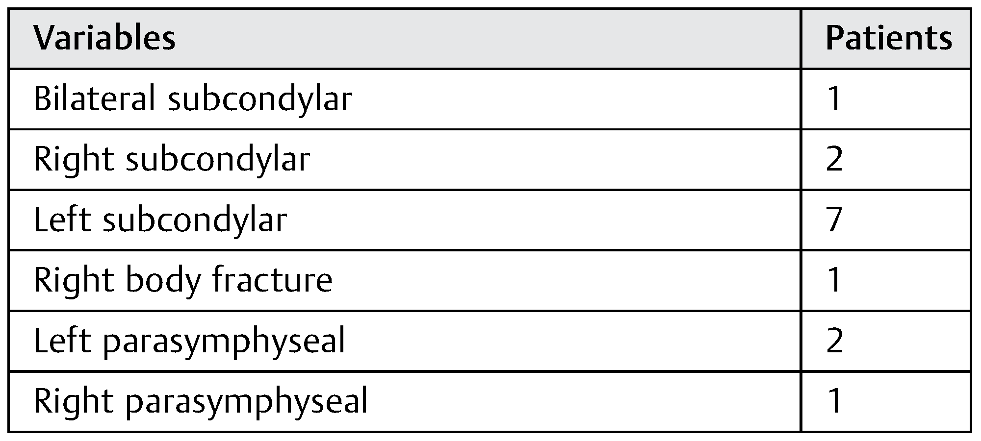
This study included 10 patients (7 males and 3 females) presenting with posttraumatic mandibular subcondylar fractures admitted to Plastic Surgery Department at Ain-Shams University Hospitals and underwent endoscope-assisted mandibular condylar fracture over a period of 1 year from January 2013 till December 2013. The patients’ mean age was 28 ± 12 years. Five patients had associated mandibular fractures and two patients had other associated maxillofacial fracture. The anatomical distribution of the mandibular fractures is shown in Table 1. All patients were referred from the emergency department after exclusion of other associated trauma. History taking and physical examination were performed to confirm the diagnosis. Preoperative radiographs were done including panoramic X-ray and CT scan facial bones with thin cuts and 3D reconstructions.
Patients with displaced subcondylar fractures (fracture line passing at or just below the deepest level of the sigmoid notch that allows for placement of two screws on the proximal segment of an adult mandible) and malocclusion were included in the study. Exclusion criteria comprised intracapsular, comminuted, and condylar head fractures, patients presenting with fractures without malocclusion, and those with associated comorbidities such as cardiopulmonary disorders, bleeding tendency, or patients receiving antiplatelet or anticoagulant medications.
All surgical procedures were done by the same surgical team. Upper and lower arch bars were applied first, followed by intraoral exposure of any other associated fractures. ORIF was achieved using 2.3 plates and screws. Exposure of the affected condylar segment was done by an intraoral incision along the anterior border of the mandibular ramus. Intraoral subperiosteal dissection was performed till an adequate optical cavity was developed. A 4-mm, 30-degree rigid endoscope (Karl Storz, Germany) fitted with either a standard endoforehead sheath or a specialized intraoral sheath retractor was used. Subperiosteal dissection was performed along the lateral side of the ramus till the whole posterior border is exposed. Subperiosteal dissection of the proximal segment was performed exposing the whole neck and subcondylar area as far as the capsule of the temporomandibular joint (TMJ). In cases where endoscopic ORIF could be achieved through the intraoral incision alone, the procedure was completed and closure of the intraoral incision was done.
If reduction in the fracture could not be achieved, an additional modified submandibular incision was used. The intraoral dissection was extended inferiorly toward the angle of the mandible under endoscopic guidance till the pterygomasseteric sling is reached. Blunt dissection of the soft tissue toward the angle of the mandible and parallel to the direction of the facial nerve was performed, guided by the tip of a periosteal elevator pushing the pterygomasseteric sling through the intraoral incision. Upon reaching the skin at angle of the mandible, a small stab incision (<1 cm) was done over the tip of the dissector and the external incision was connected with the intraoral incision. An interosseus wire was introduced through a drill hole at the angle of the mandible. Traction was applied using this wire that facilitated the reduction and maintained the fractured segments in proper anatomical alignment. Fixation was then achieved using 2.0 miniplates applied through the intraoral incision and percutaneous miniscrews. Intraoral incision closure, release of MMF, and ensuring that proper occlusion has been achieved completed the procedure.
The patients were instructed on soft diet for a couple of weeks. The occlusion of the patient was rechecked on the second postoperative day and the arch bars were removed within the first week if the occlusion was satisfactory. Followup was scheduled weekly in the first month then postoperative visits were arranged at 3 and 6 months. Assessment included evaluation of occlusion and measurements of maximal incisal opening. Any clinical symptoms such as pain, clicking, TMJ dysfunction, or the presence of any abnormality in mouth opening were recorded. Postoperative radiographs were obtained immediately following the surgical intervention and at 6 months postoperatively.
Scar assessment criteria were adopted from the Patient and Observer Scar Assessment Scale (POSAS) and The Stony Brook Scar Evaluation Scale [5,6]. The assessment criteria included scar erythema, height, width, pigmentation, pliability, pain, itching, or pruritus that interferes or not with quality of life, and scar visibility to patient’s relatives.

Results

Comparison of preoperative mouth opening (24 ± 8 mm) and postoperative mouth opening (40 ± 7 mm) showed highly significant improvement (p < 0.001). Furthermore, there was also statistically significant improvement in recorded pre- and postoperative clinical findings, including pain, clicking, mouth deviation, and occlusion (p < 0.05). This clinical improvement was also confirmed radiologically, which revealed also statistically highly significant improvement between pre- and postoperative radiographs (Figure 1, Table 2).
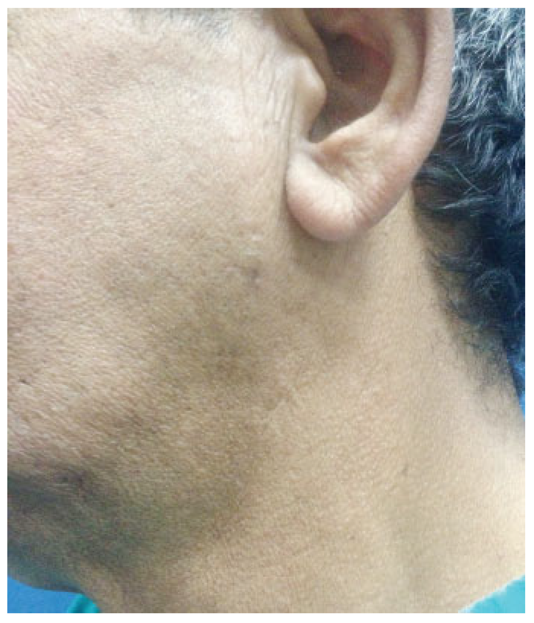
The intraoral exposure was adequate to complete the procedure in two patients whereas the minimized submandibular incision was needed in eight patients to achieve fracture alignment and proper occlusion. Scar assessment revealed that no patient had hypertrophic scars, widened scars, scar dyspigmentation, and no associated symptoms such as pain or pruritus. Scars were slightly firm in early postoperative follow-up. However, all scars were nearly not felt at all at 6 months follow-up. In addition, all patients had satisfactory results with overall good scar appearance. Regarding scar visibility, no patient had visible attracting scar, four patients had visible scar to patient’s relatives from near distances, and four patients had nearly nonvisible scars even from near distances (Figure 2).
Regarding postoperative complications; the patient with bilateral subcondylar fracture had a minimal anterior open bite and mouth deviation postoperatively that was corrected by the use of class II guiding dental elastics worn at night for 1 month. Another patient with an associated parasymphyseal fracture had a minor midline shift and was managed similarly by class II guiding elastics worn at night for 1 month. In addition, intraoral parasymphyseal (not intraoral condylar) wound dehiscence occurred in one patient that required secondary suturing.

Discussion

The management of mandibular subcondylar fractures remains controversial [7]. Although closed reduction and MMF is the most commonly used treatment, it showed a higher percentage of anatomic displacement when compared with ORIF [8]. In addition, it is associated with higher complications rate (39%) in comparison to ORIF (4%) [9]. After reviewing of literature, Haug and Brandt [10] determined that “it seems rather conclusive that ORIF of the mandibular condyle is superior to closed reduction and MMF.”
However, the drawbacks of an external approach limited its routine use in condylar fractures. The endoscope-assisted ORIF is a relatively new technology that allows anatomical reduction and at the same time has the potential of reducing the risk of facial nerve injury, eliminating the need for MMF, and limiting the problem of scaring [11,12,13,14,15].
The used approaches for endoscope-assisted ORIF include pure transoral, transoral with cheek trocar, or submandibular incisions [13,16,17,18,19,20,21,22,23,24,25,26]. The advantage of the intraoral approach is the absence of skin incision, but the optical cavity is smaller. The submandibular approach requires a 1.5-cm skin incision at the angle of the mandible, similar to a Risdon incision, placing the facial nerve at minimal risk. However, the optical cavity created has a larger working space with better endoscopic orientation [27].
Lee and colleagues [28] presented the results of 20 patients who underwent endoscope-assisted ORIF. They used an intraoral approach with a transfacial portal. However, one patient had facial nerve palsy. In 1999, Lauer and Schmelzeisen [11] used a 2- to 3-cm submandibular incision and two additional facial portals in four patients. In 2001, Honda and associates [20] used two incisions: one submandibular 20 to 25 mm long and another preauricular 7.0 to 10 mm long. They reported no conspicuous scars. Troulis and Kaban [29] used 1.5-cm transfacial Risdon incision, and they had satisfactory outcome with no complications. In 2002, Schön et al. [17] presented endoscope-assisted ORIF for subcondylar fractures in 17 patients. Nine patients were treated with a 4- to 5-cm submandibular incision and eight patients were treated by transbuccal approach. They reported nearly 70% overall incidence of perceived some form of scaring. In 2003, Kellman [30] used an intraoral incision and transcutaneous ports for fixation. However, one patient required revision, the procedures were converted to a full-open approach in two patients, and four patients were reduced but could not be completed. He concluded that this approach is a feasible but challenging technique. In 2003, Miloro used [22] a 15- to 20-mm modified Risdon incision in six patients. He reported stable occlusions with no complications and acceptable scar perception. In 2004, Troulis [24] reported 4.5% complication rate of temporary marginal mandibular nerve weakness.
Chen and coworker [19] used transbuccal intraoral incision and transcutaneous ports. They claimed that the transfacial ports healed inconspicuously but without any details. Martin and Lee [22] used a nearby technique as they used stab incision immediately over the palpated location of the posterior aspect of the fracture. This dissection is carried through the parotid gland and masseter muscle. The sleeved trocar is inserted and the cheek retractor is mounted on the trocar sleeve to allow the maintenance of the optical cavity by gentle traction. The endoscope is placed through the intraoral incision. Schön et al. [17] first recommended using intraoral approach for laterally dislocated condyles whereas external approach is used for fractures with medial displacement of the condylar head, condylar fractures with medial override, and for comminuted condylar fractures. After 5 years of experience, they recommended using the transoral approach even for fractures with medial override [1]. Moreover, Schön et al. [23] stated that the transoral approach is less time consuming than the submandibular approach.
Apparently, most of the surgeons tend to use an external approach or a combined intraoral/external approach for endoscope-assisted ORIF with the exception of pure intraoral approach by Schön et al. [1] The technique presented in this work uses an intraoral approach to visualize and reduce the dislocated segments. As long as the dislocated segments can be reduced and proper occlusion obtained, the procedure was completed through intraoral incisions in accordance with Schön et al. [1] Yet, it was difficult to reduce the fractured segments appropriately in many circumstances (80% in our study) through intraoral incisions solely. Moreover, maintenance of the reduced segments is sometimes more difficult than the reduction itself. In these circumstances, the minimal submandibular incision served this purpose. It allowed for the introduction of an interosseus wires that facilitated both the reduction and its maintenance till the plate and screws were applied. This very limited (<1-cm incision) is totally satisfactory for all patients without any annoying apparent scars.
Martin and Lee [31] described a nearby technique, but they perform the submandibular incision first. To the contrary, our technique uses dissection first then incision. The intraoral incision is used for complete dissection, which could prove sufficient in some cases for ORIF. If it was not enough, completing the dissection bluntly takes place under complete visualization by endoscope to avoid any possible facial nerve injury. As the dermis is reached, the skin is incised safely on the tip of the dissector in a very simple, and at the same time very limited fashion.
Biglioli and Colletti described a 20-mm transmasseter mini-retromandibular approach for treatment of condylar fractures in 25 and 33 patients with very satisfactory results. However, they reported improper reduction and fixation when they used 15-mm incision that required another surgical intervention [32,33]. Recently, Colletti et al. [34] reported their experience in 100 cases and concluded that this approach seems a safe approach with minimal morbidity. Nicolai et al. [35] used combined intraoral approach and retromandibular incision. In addition, Schiel et al. [36] used endoscopically assisted transoral route only for pediatric patients.
The merits of this very limited submandibular incision include placement of the endoscope through the external incision that reduces the load of surgical instruments used during fracture fixation. Second, it facilitates greatly the reduction in displaced condylar segments and achieves proper alignment of the relatively difficult reductions. Third, the incision is used also to place the endoscope that visualizes the posterior border of the mandible clearly to ensure proper and adequate reduction in the displaced segments. This anatomical reduction in the fractured segments allows for avoiding complications associated with endoscopic procedures such as higher rate of hardware loosening [11], a possible higher rate of nonunion, refracture [12], and possibly malocclusion [4].
Endoscope-assisted surgical techniques have been developed over the past decade to address some of the surgical problems of the facial skeleton; however, it remains in its infancy [7]. Minimizing the submandibular incision can be considered as a step in surgical techniques. This step can continue as it is, can be modified later on, or even be replaced totally.

Conclusion

The endoscope-assisted ORIF described herein depends on complete dissection through intraoral approach that may be sufficient to obtain proper reduction and fixation. However, in many circumstances, a very small submandibular incision less than 1 cm is usually needed to achieve good results. Performing the external incision after complete intraoral dissection seems very safe to facial nerve and minimizes scarring markedly. This incision facilitates reduction in relatively difficult cases and enables visualization of the posterior border of the mandible clearly.

References

  1. Schön, R.; Fakler, O.; Gellrich, N.C.; Schmelzeisen, R. Five-year experience with the transoral endoscopically assisted treatment of displaced condylar mandible fractures. Plast Reconstr Surg 2005, 116, 44–50. [Google Scholar] [CrossRef] [PubMed]
  2. Mitchell, D.A. A multicentre audit of unilateral fractures of the mandibular condyle. Br J Oral Maxillofac Surg 1997, 35, 230–236. [Google Scholar] [CrossRef]
  3. Silvennoinen, U.; Iizuka, T.; Lindqvist, C.; Oikarinen, K. Different patterns of condylar fractures: an analysis of 382 patients in a 3-year period. J Oral Maxillofac Surg 1992, 50, 1032–1037. [Google Scholar] [CrossRef] [PubMed]
  4. Stacey, D.H.; Doyle, J.F.; Mount, D.L.; Snyder, M.C.; Gutowski, K.A. Management of mandible fractures. Plast Reconstr Surg 2006, 117, 48e–60e. [Google Scholar] [CrossRef]
  5. Draaijers, L.J.; Tempelman, F.R.; Botman, Y.A.; et al. The patient and observer scar assessment scale: a reliable and feasible tool for scar evaluation. Plast Reconstr Surg 2004, 113, 1960–1965, discussion 1966–1967. [Google Scholar] [CrossRef] [PubMed]
  6. Singer, A.J.; Arora, B.; Dagum, A.; Valentine, S.; Hollander, J.E. Development and validation of a novel scar evaluation scale. Plast Reconstr Surg 2007, 120, 1892–1897. [Google Scholar] [CrossRef]
  7. Boutros, S.G. Closed reduction and fluoroscopically assisted percutaneous stabilization of displaced subcondylar mandible fractures. Plast Reconstr Surg 2005, 116, 971–977. [Google Scholar] [CrossRef]
  8. Silverman, S.L. New operation for displaced fractures at the neck of the condyle. Int J Orthod 1925, 67, 876. [Google Scholar]
  9. Wassmund, M. Uber Luxations frakturen des Kiefernglenkes. Dtsch Kieferchir. 1934, 1, 27. [Google Scholar]
  10. Haug, R.H.; Brandt, M.T. Closed reduction, open reduction, and endoscopic assistance: current thoughts on the management of mandibular condyle fractures. Plast Reconstr Surg 2007, 120 (Suppl. 2), 90S–102S. [Google Scholar] [CrossRef]
  11. Lauer, G.; Schmelzeisen, R. Endoscope-assisted fixation of mandibular condylar process fractures. J Oral Maxillofac Surg 1999, 57, 36–39, discussion 39–40. [Google Scholar] [CrossRef]
  12. Lee, C.; Stiebel, M.; Young, D.M. Cranial nerve VII region of the traumatized facial skeleton: optimizing fracture repair with the endoscope. J Trauma 2000, 48, 423–431, discussion 431–432. [Google Scholar] [CrossRef]
  13. Sandler, N.A.; Andreasen, K.H.; Johns, F.R. The use of endoscopy in the management of subcondylar fractures of the mandible: a cadaver study. Oral Surg Oral Med Oral Pathol Oral Radiol Endod 1999, 88, 529–531. [Google Scholar] [CrossRef] [PubMed]
  14. Schön, R.; Schmelzeisen, R. Endoscopic fracture treatment. Ann R Australas Coll Dent Surg 2002, 16, 40–45. [Google Scholar] [PubMed]
  15. Mueller, R. Endoscopic treatment of facial fractures. Facial Plast Surg 2008, 24, 78–91. [Google Scholar] [CrossRef] [PubMed]
  16. Schmelzeisen, R.; Lauer, G.; Wichmann, U. Endoscope-assisted fixation of condylar fractures of the mandible [in German]. Mund Kiefer Gesichtschir 1998, 2 (Suppl. 1), S168–S170. [Google Scholar] [CrossRef]
  17. Schön, R.; Gutwald, R.; Schramm, A.; Gellrich, N.C.; Schmelzeisen, R. Endoscopy-assisted open treatment of condylar fractures of the mandible: extraoral vs intraoral approach. Int J Oral Maxillofac Surg 2002, 31, 237–243. [Google Scholar] [CrossRef]
  18. Kellman, R.M. Endoscopic approach to subcondylar mandible fractures. Facial Plast Surg 2004, 20, 239–247. [Google Scholar] [CrossRef]
  19. Chen, C.T.; Lai, J.P.; Tung, T.C.; Chen, Y.R. Endoscopically assisted mandibular subcondylar fracture repair. Plast Reconstr Surg 1999, 103, 60–65. [Google Scholar] [CrossRef]
  20. Honda, T.; Nozaki, M.; Isono, N.; Sasaki, K. Endoscope-assisted facial fracture repair. World J Surg 2001, 25, 1075–1083. [Google Scholar] [CrossRef]
  21. Sandler, N.A. Endoscopic-assisted reduction and fixation of a mandibular subcondylar fracture: report of a case. J Oral Maxillofac Surg 2001, 59, 1479–1482. [Google Scholar] [CrossRef] [PubMed]
  22. Miloro, M. Endoscopic-assisted repair of subcondylar fractures. Oral Surg Oral Med Oral Pathol Oral Radiol Endod 2003, 96, 387–391. [Google Scholar] [CrossRef] [PubMed]
  23. Schön, R.; Schramm, A.; Gellrich, N.C.; Schmelzeisen, R. Follow-up of condylar fractures of the mandible in 8 patients at 18 months after transoral endoscopic-assisted open treatment. J Oral Maxillofac Surg 2003, 61, 49–54. [Google Scholar] [CrossRef]
  24. Troulis, M.J. Endoscopic open reduction and internal rigid fixation of subcondylar fractures. J Oral Maxillofac Surg 2004, 62, 1269–1271. [Google Scholar] [CrossRef]
  25. Vural, E. Treatment of adult subcondylar mandibular fractures: closed vs open vs endoscopic approach. Arch Otolaryngol Head Neck Surg 2004, 130, 1228–1230. [Google Scholar] [CrossRef] [PubMed]
  26. Mueller, R.V.; Czerwinski, M.; Lee, C.; Kellman, R.M. Condylar fracture repair: use of the endoscope to advance traditional treatment philosophy. Facial Plast Surg Clin North Am 2006, 14, 1–9. [Google Scholar] [CrossRef]
  27. Aziz, S.R.; Ziccardi, V.B. Endoscopically assisted management of mandibular condylar fractures. Atlas Oral Maxillofac Surg Clin North Am 2009, 17, 71–74. [Google Scholar] [CrossRef] [PubMed]
  28. Lee, C.; Mueller, R.V.; Lee, K.; Mathes, S.J. Endoscopic subcondylar fracture repair: functional, aesthetic, and radiographic outcomes. Plast Reconstr Surg 1998, 102, 1434–1443, discussion 1444–1445. [Google Scholar] [CrossRef] [PubMed]
  29. Troulis, M.J.; Kaban, L.B. Endoscopic approach to the ramus/condyle unit: clinical applications. J Oral Maxillofac Surg 2001, 59, 503–509. [Google Scholar] [CrossRef]
  30. Kellman, R.M. Endoscopically assisted repair of subcondylar fractures of the mandible: an evolving technique. Arch Facial Plast Surg 2003, 5, 244–250. [Google Scholar] [CrossRef]
  31. Martin, M.; Lee, C. Endoscopic mandibular condyle fracture repair. Atlas Oral Maxillofac Surg Clin North Am 2003, 11, 169–178. [Google Scholar] [CrossRef] [PubMed]
  32. Biglioli, F.; Colletti, G. Transmasseter approach to condylar fractures by mini-retromandibular access. J Oral Maxillofac Surg 2009, 67, 2418–2424. [Google Scholar] [CrossRef] [PubMed]
  33. Biglioli, F.; Colletti, G. Mini-retromandibular approach to condylar fractures. J Craniomaxillofac Surg 2008, 36, 378–383. [Google Scholar] [CrossRef] [PubMed]
  34. Colletti, G.; Battista, V.M.; Allevi, F.; Giovanditto, F.; Rabbiosi, D.; Biglioli, F. Extraoral approach to mandibular condylar fractures: our experience with 100 cases. J Craniomaxillofac Surg 2014, 42, e186–e194. [Google Scholar] [CrossRef]
  35. Nicolai, G.; Lorè, B.; De Marinis, L.; Calabrese, L. Combined surgical approach retromandibular and intraoral to subcondylar mandibular fractures. J Craniofac Surg 2011, 22, 1354–1357. [Google Scholar] [CrossRef]
  36. Schiel, S.; Mayer, P.; Probst, F.; Otto, S.; Cornelius, C.P. Transoral open reduction and fixation of mandibular condylar base and neck fractures in children and young teenagers—a beneficial treatment option? J Oral Maxillofac Surg 2013, 71, 1220–1230. [Google Scholar] [CrossRef]
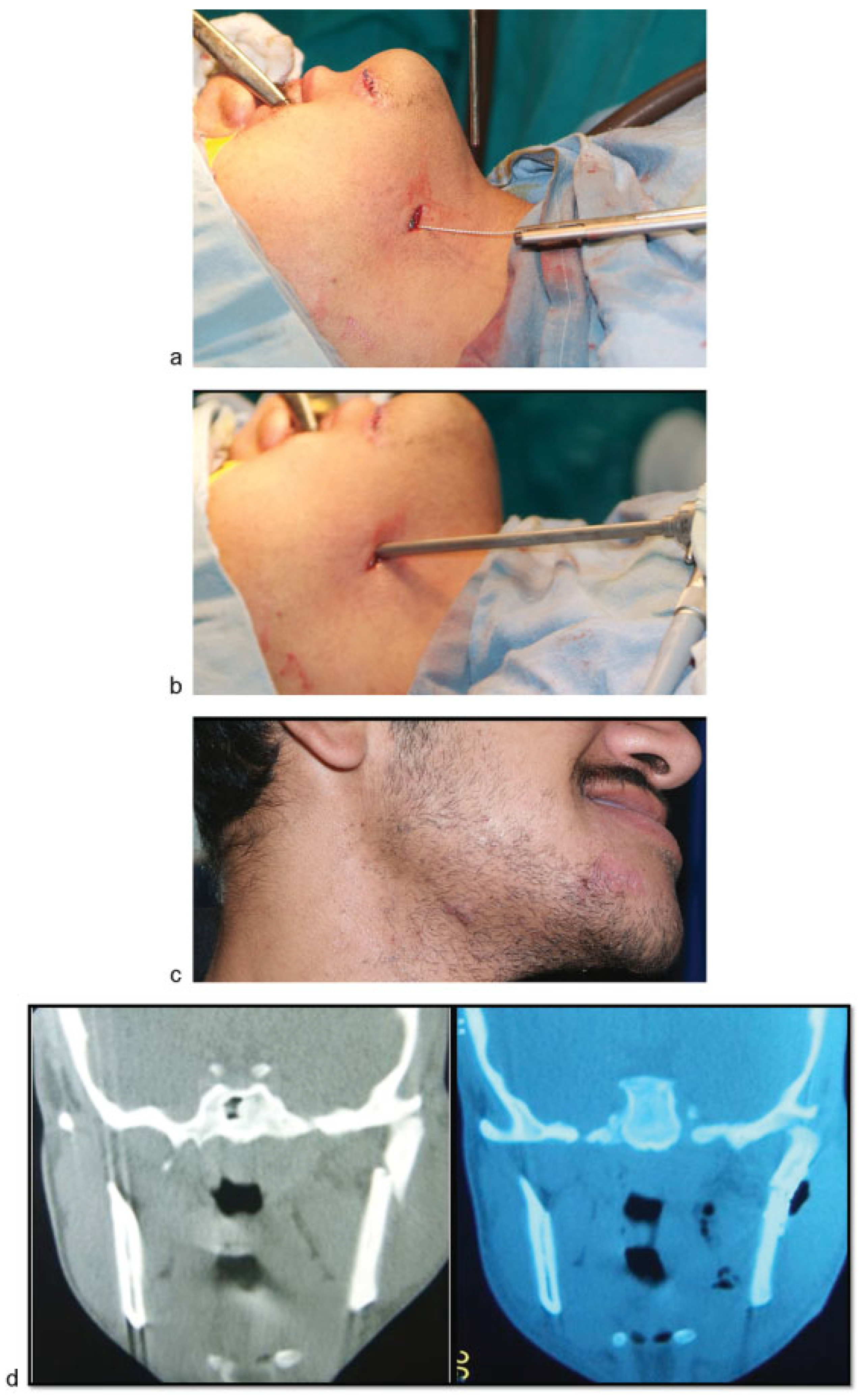
Figure 1. (a) The extraoral incision is performed over the tip of the dissector after complete intraoral dissection, and an interosseus wire is applied and used for traction to facilitate reduction. (b) The endoscope is applied through the extraoral incision to visualize the posterior border of the mandible clearly. (c) The extraoral incision 2 weeks postoperative. (d) Pre- and postoperative CT scan coronal views.
Cmtr 08 4b2 g001
Figure 2. A 60-year-old male patient with an 18-month postoperative photo after endoscopic ORIF of subcondylar fracture. It shows excellent result with nearly nonvisible scar.
Cmtr 08 4b2 g002
Table 1. Anatomical distribution of mandibular fractures.
Cmtr 08 4b2 t001
Table 2. Statistically significant radiologic improvement among patients (McNemar test).
Cmtr 08 4b2 t002