Next Article in Journal
Bilateral Alloplastic Prostheses for Temporomandibular Joint Reconstruction in a Patient with Ankylosing Spondylitis
Previous Article in Journal
Rapid Prototyping Technology in Orbital Floor Reconstruction: Application in Three Patients
 
 
Craniomaxillofacial Trauma & Reconstruction is published by MDPI from Volume 18 Issue 1 (2025). Previous articles were published by another publisher in Open Access under a CC-BY (or CC-BY-NC-ND) licence, and they are hosted by MDPI on mdpi.com as a courtesy and upon agreement with Sage.
Case Report

Extraction of Fronto-Orbital Shower Hook Through Transcranial Orbitotomy

by
Maxwell D. Elia
1,*,
Murat Gunel
2,
Juan J. Servat
1 and
Flora Levin
1
1
Department of Ophthalmology and Visual Sciences, Yale School of Medicine, New Haven, CT 06510, USA
2
Department of Neurosurgery, Yale School of Medicine, New Haven, CT 06510, USA
*
Author to whom correspondence should be addressed.
Craniomaxillofac. Trauma Reconstr. 2014, 7(2), 147-148; https://doi.org/10.1055/s-0034-1371545
Submission received: 24 April 2013 / Revised: 25 April 2013 / Accepted: 25 April 2013 / Published: 4 March 2014

Abstract

:
Transorbital foreign bodies threaten both the integrity of the globe and the brain. We present an unusual case of a penetrating right frontal lobe-orbital metallic shower hook. Extensive intracranial involvement necessitated transcranial orbitotomy allowing for the removal of the object without loss of the globe.

Case Report

A 64-year-old man with extensive cardiovascular disease and alcoholism presented to the emergency department at Yale New Haven Hospital 2 days after a bathroom fall. On arrival patient was awake and oriented with a metallic shower hook protruding from his right orbit.
Uncorrected visual acuity was light perception (LP) in the right eye (OD) and 20/30 in the left eye (OS) with a right relative afferent pupillary defect. There was a penetrating wound on the right lower eyelid (Figure 1A), at the point of entry of the shower hook, 6 mm inferior to the lid margin. Extraocular movements in the right eye were markedly limited in all directions, and there was marked proptosis OD. Slit-lamp examination revealed 360 degrees chemosis and subconjunctival hemorrhage. The cornea was clear and the anterior chamber formed. The right fundoscopic view was notable for chorioretinal folds inferiorly. The left eye was unremarkable.
Computed tomography (CT) of the head revealed a foreign body entering the right inferolateral orbit, penetrating the right orbital roof 30 mm posterior to the superior segment of the orbital rim and extending 22 mm into the right frontal lobe (Figure 1B,C). There was an adjacent intraparenchymal hemorrhage. Cerebral angiography demonstrated no arterial or venous injury (Figure 1D).
The patient underwent emergent craniotomy with the removal of a portion of the right orbital roof to allow the removal of the hook from the brain, orbital roof, and the orbit. The exposed brain was covered using a synthetic dural graft, and the anterior fossa skull base was reconstructed. A vascularized paracranial flap was laid over the dural graft, and the area was filed with TISSEEL (Baxter, Deerfield, IL) to prevent cerebrospinal fluid leakage. Out of concern for potential infection, the bone flap from the craniotomy was left out, and the area was covered with a synthetic mesh which was secured in place using a titanium plating system. A lumbar drain was placed to prevent postoperative increases in intracranial pressure and was removed after 6 days.
After the procedure, the patient had no LP in the right eye, but his globe remained formed and his extraocular motility improved. Because of the high risk for infection, this patient was treated with 2 weeks of intravenous vancomycin and ceftazidime, and he was subsequently transitioned to 2 weeks of oral trimethoprim-sulfamethoxazole.

Discussion

Orbitocranial foreign bodies necessitate emergent ophthalmologic and neurosurgical evaluation. Possible ophthalmic complications include globe rupture, traumatic or compressive optic neuropathy, retinal detachment, or cranial nerve palsies [1]. This patient’s vision loss has likely resulted from a traumatic/compressive optic neuropathy.
In addition to direct ocular trauma, intracranial neural and vascular damage are possible. CT is the optimal imaging modality to determine the extent of penetration. CT angiography may also help determine the extent vascular compromise [2]. In this case, conventional diagnostic cerebral arteriography was used to determine whether major vessels were involved before removal.
The shower hook in this patient penetrated the orbital roof. This has previously been described to be the most likely path to intracranial penetration secondary to the weakness of the orbital roof [3]. Penetration of the orbital roof allows for inoculation of bacteria, posing a serious potential complication. Brain abscess has previously been reported in this type of injury [4].

Comment

Orbitocranial foreign body injuries are exceedingly rare, and frequently result in mortality [5]. Had the shower hook proceeded farther into the brain, this likely would have been fatal. Unfortunately, the patient delayed medical attention, compromising his visual potential from his right eye due to optic neuropathy. At the time of discharge, the patient’s only persistent neurologic deficits were left hand weakness and right eye blindness. Physicians evaluating patients with penetrating orbital trauma should be aware of the potential for intracranial injury.

References

  1. Irshad, K.; McAuley, D.; Khalaf, K.; Ricard, D. Unsuspected penetrating maxillo-orbitocranial injury: a case report. Can J Surg 1998, 41, 393–397. [Google Scholar] [PubMed]
  2. Walid, M.S.; Yelverton, J.C.; Robinson, J.S., Jr. Penetrating orbital trauma with internal carotid injury. South Med J 2009, 102, 116–117. [Google Scholar] [PubMed]
  3. Wesley, R.E.; Anderson, S.R.; Weiss, M.R.; Smith, H.P. Management of orbital-cranial trauma. Adv Ophthalmic Plast Reconstr Surg 1987, 7, 3–26. [Google Scholar] [PubMed]
  4. Gupta, A.; Chacko, A.; Anil, M.S.; Karanth, S.S.; Shetty, A. Pencil in the brain: a case of temporal lobe abscess following an intracranial penetrating pencil injury. Pediatr Neurosurg 2011, 47, 307–308. [Google Scholar] [PubMed]
  5. Dunya, I.M.; Rubin, P.A.; Shore, J.W. Penetrating orbital trauma. Int Ophthalmol Clin 1995, 35, 25–36. [Google Scholar] [PubMed]
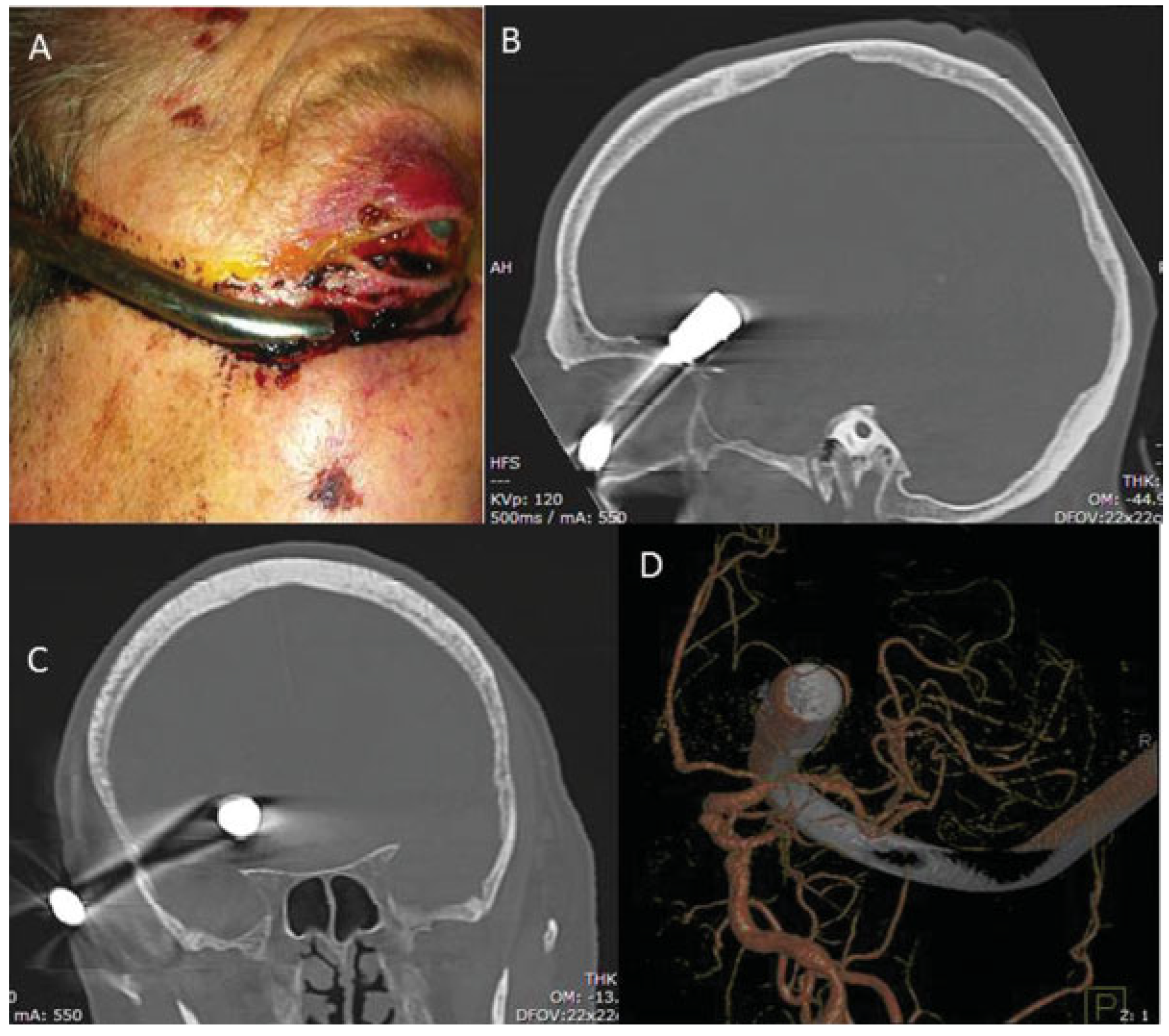
Figure 1. (A) Metallic shower hook with original entry wound. Computed tomography in (B) sagittal and (C) coronal planes revealing extension of the shower hook. (D) Diagnostic cerebral arteriography demonstrating no arterial or venous injury.
Cmtr 07 00036 g001