Abstract
The AO classification system for fractures in the adult craniomaxillofacial (CMF) skeleton is organized in anatomic modules in a 3 precision-level hierarchy with account for an increasing complexity and details. Level-1 is most elementary and identifies no more than the presence of fractures in 4 separate anatomical units: the mandible (code 91), midface (92), skull base (93) and cranial vault (94). Level-2 relates the detailed topographic location of the fractures within defined regions of the mandible, central and lateral midface, internal orbit, endo- and exocranial skull base, and the cranial vault. Level-3 is based on an even more refined topographic assessment and focuses on the morphology—fragmentation, displacement, and bone defects—within specified subregions. An electronic fracture case collection complements the preceding tutorial papers, which explain the features and options of the AOCMF classification system in this issue of the Journal. The electronic case collection demonstrates a range of representative osseous CMF injuries on the basis of diagnostic images, narrative descriptions of the fracture diagnosis and their classification using the icons for illustration and coding of a dedicated software AOCOIAC (AO Comprehensive Injury Automatic Classifier). Ninety four case examples are listed in two tables for a fast overview of the electronic content. Each case can serve as a guide to getting started with the new AOCMF classification system using AOCOIAC software and to employ it in the own clinical practice.
Fractures of the craniomaxillofacial skeleton occur in an endless array of different patterns. To design a standardized classification system to categorize fractures in a way that is meaningful, validated and clinically relevant is a challenging long-term project that requires an iterative development process.
A key prerequisite in the development of a classification system is to establish a reproducible visual language and coding to ensure referral to identical fracture entities across medical disciplines. [1]
The new AO classification system for fractures of the craniomaxillofacial (CMF) skeleton in adults is organized in several anatomic modules in a precision-level hierarchy relating to the rendition of topographical details and fracture morphology.
The introduction of precision levels offers versatility and staged procedures during the developmental phase, the validation process and the finalization of a classification proposal.
Three precision levels enable to refine the documentation of fractures according to their complexity:
- Level-1 is most elementary and identifies no more than the presence of fractures in 4 separate anatomical units: the mandible (code 91), midface (92), skull base (93) and cranial vault (94).
- Level-2 relates the detailed topographic location of the fractures within defined regions of the mandible, central and lateral midface, and internal orbit. The endocranial surface of the skull base is divided in 9 regions: a central part further subdivided into 3 components (cribriform plate and ethmoido-sphenoidal planum, sellar/parasellar compartment, clivus) adjoined by two lateral parts each encompassing an anterior, middle and posterior component. The regions of the exocranial skull base and the cranial vault are defined according to common anatomical nomenclature into paired and non-paired bones: frontal, parietal, temporal, sphenoid and occipital.
- Level-3 is based on an even more refined topographic assessment and focuses on the morphology – fragmentation, displacement, and bone defects – within the specified subregions, parts and components.
Objective
The purpose of this paper is to present an electronic case collection complementing the series of tutorial papers [2,3,4,5,6,7,8] about the AOCMF classification system preceding in this issue of the Journal. The electronic cases demonstrate a range of representative osseous CMF injuries on the basis of diagnostic images, narrative descriptions of the fracture diagnosis and their classification using the icons for illustration and coding of a dedicated software AOCOIAC (AO Comprehensive Injury Automatic Classifier). [9]

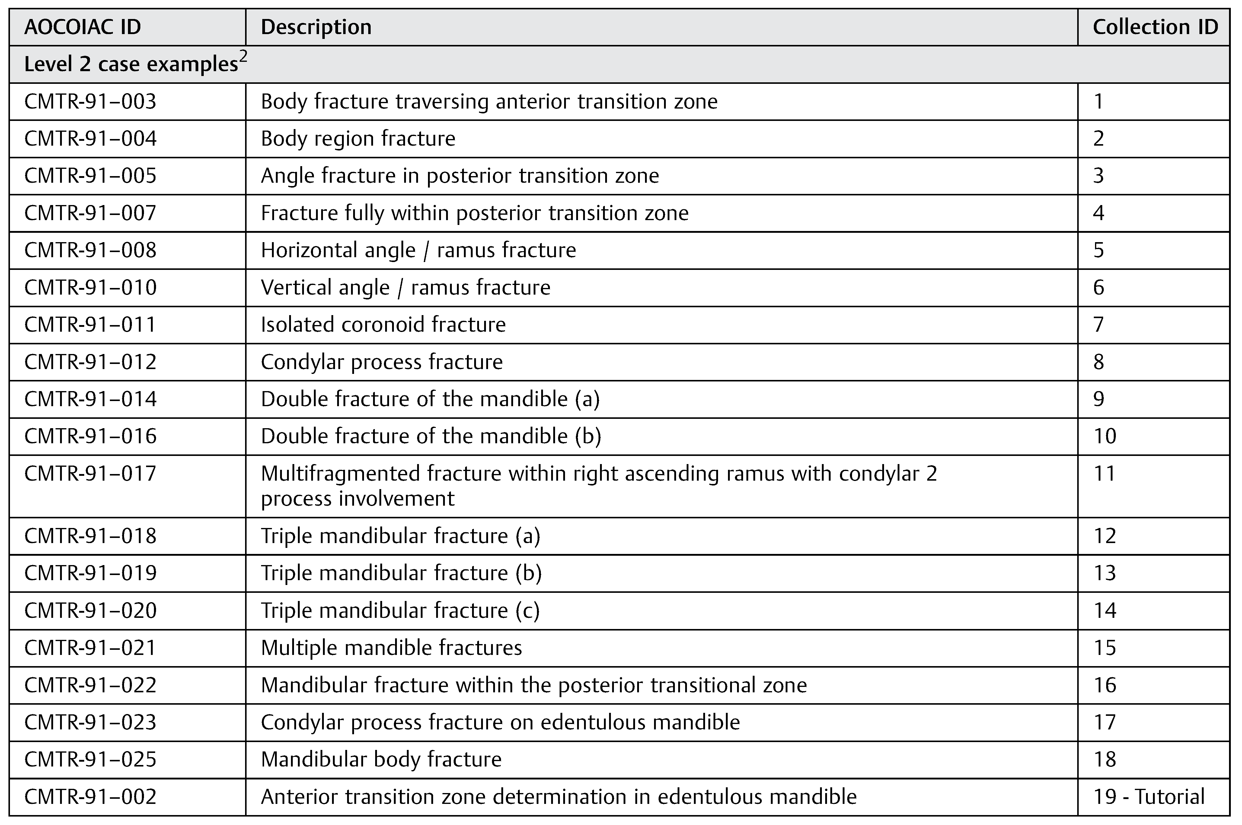
Table 1.
Case examples of mandibular fractures.
Table 1.
Case examples of mandibular fractures.
 |
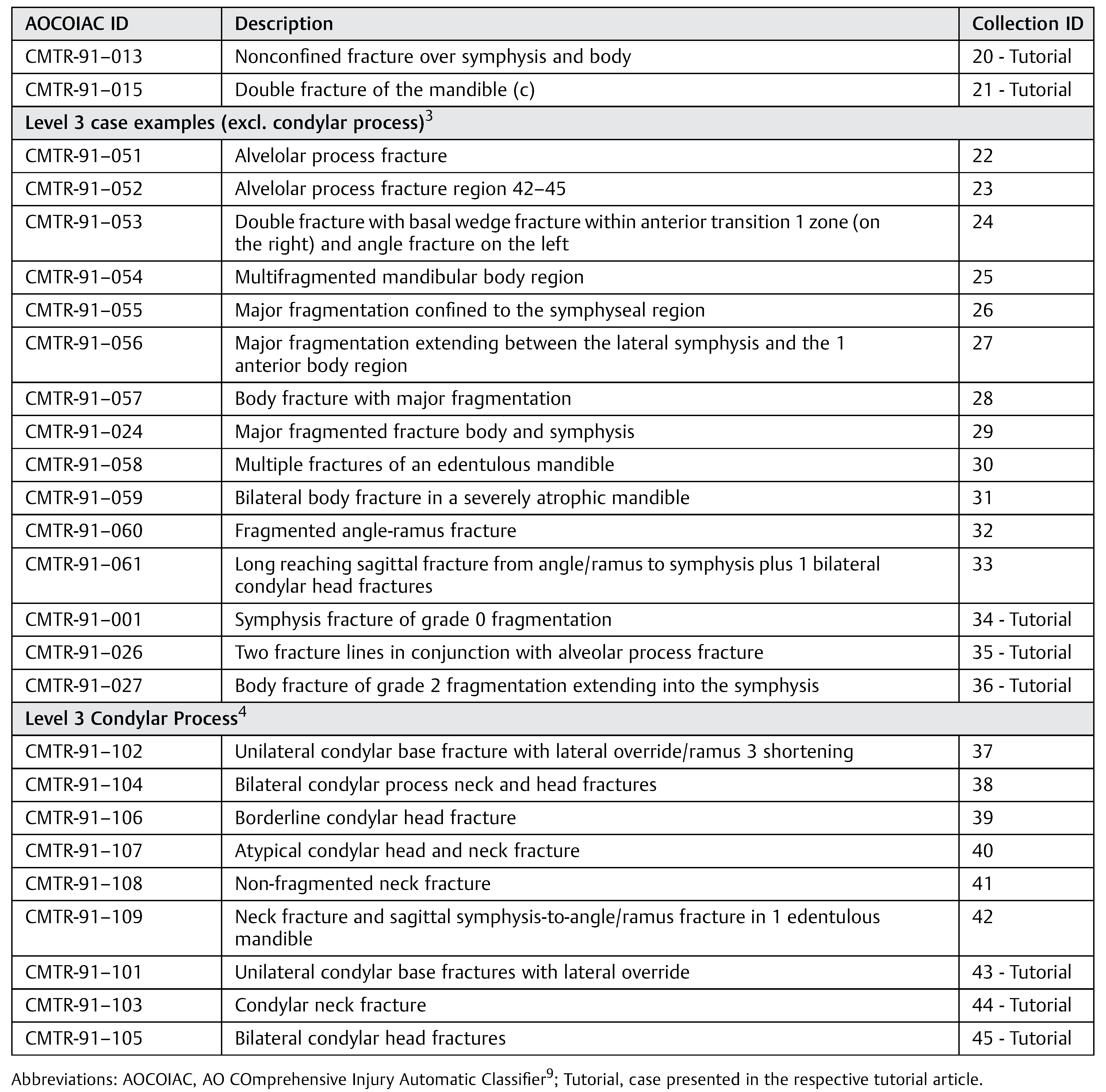 |
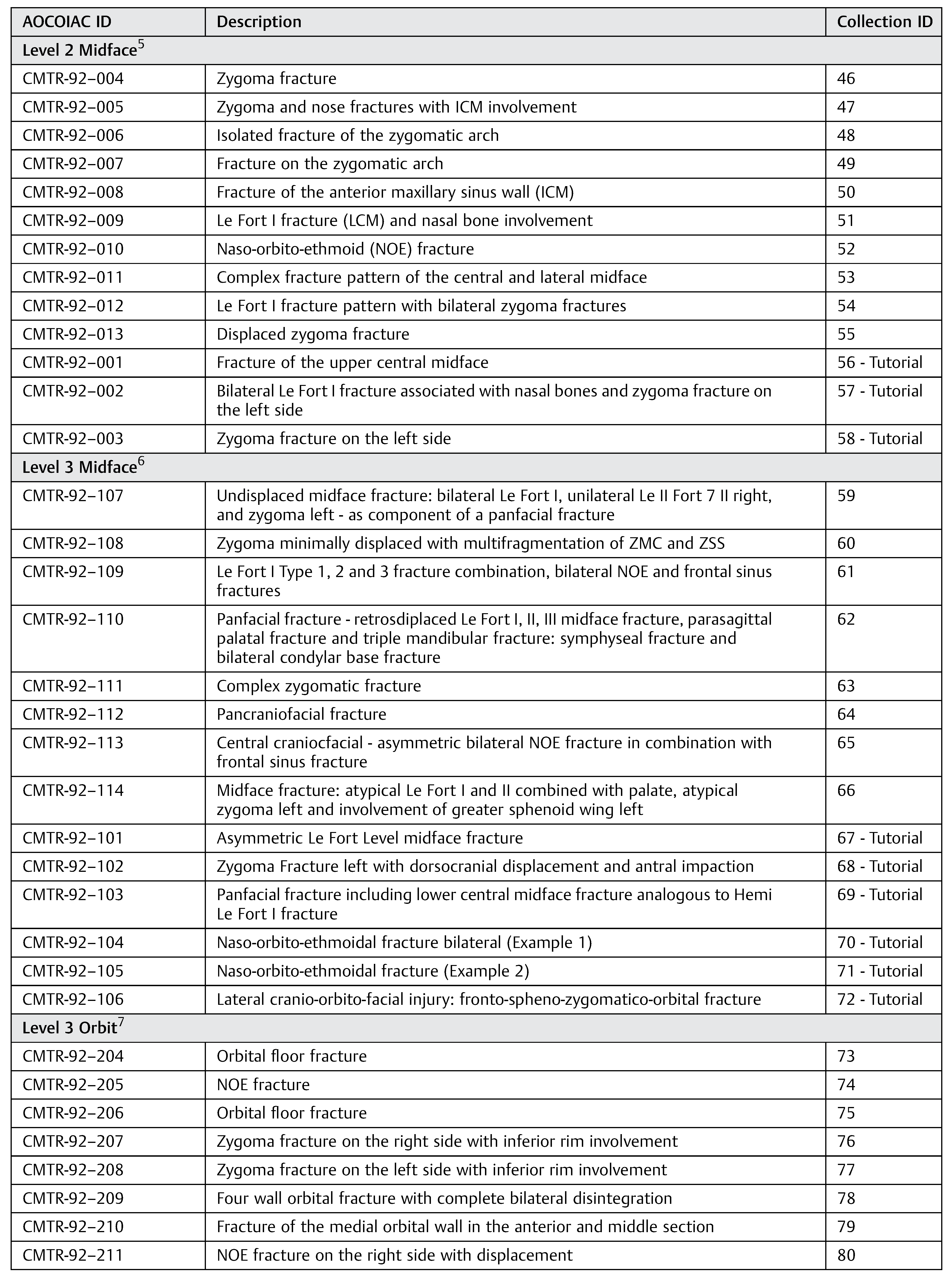
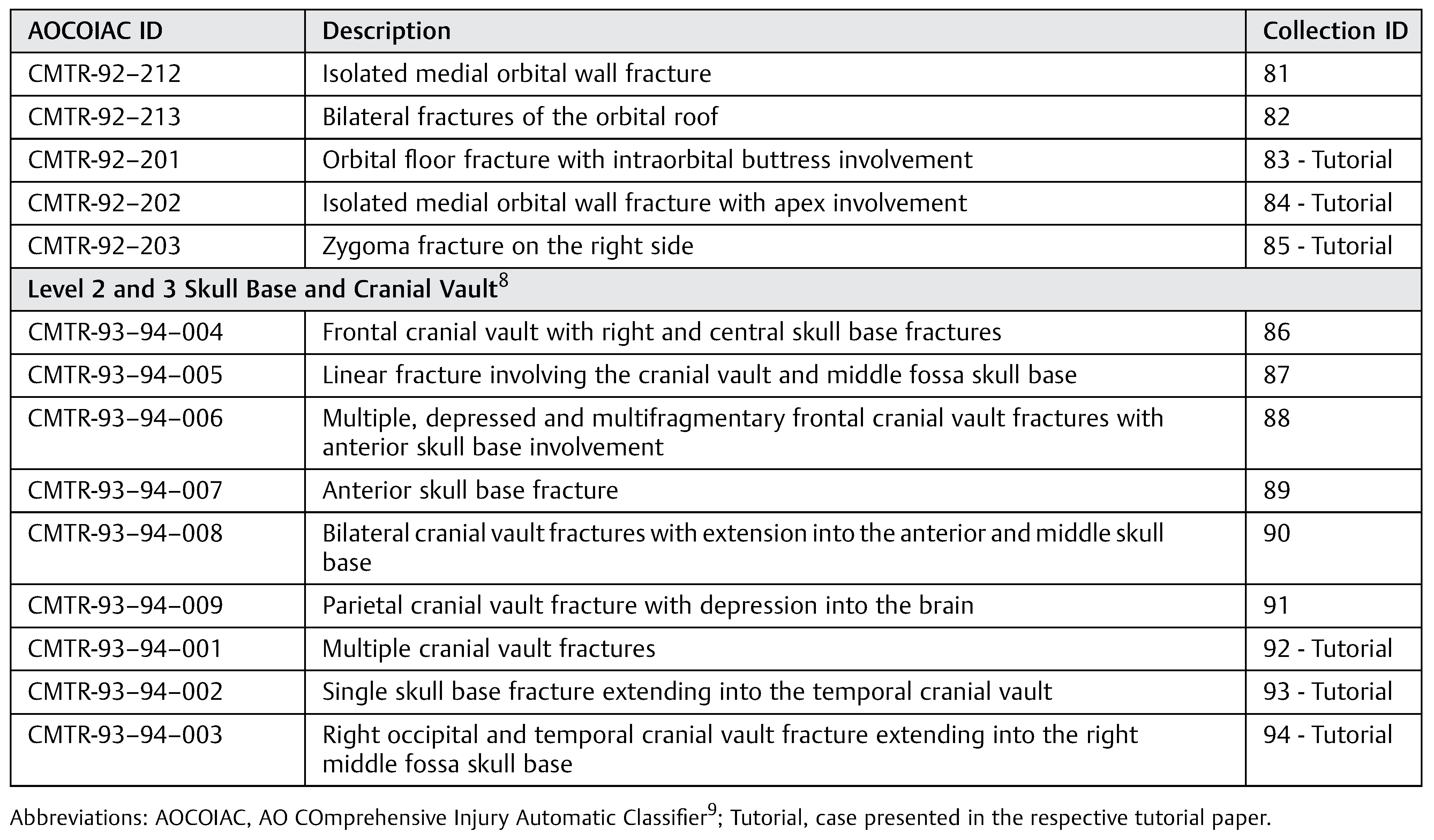
The electronic case collection is intended to serve as a guide in getting started and familiar with all the nuances of the AOCMF classification system. A total of 94 case examples listed in two tables (Table 1 and Table 2) provides a fast overview to select appropriate fracture patterns for a first orientation or comparison in reference to injuries in the own clinical setting. The case examples are grouped according to the sequence of the preceding tutorial papers, which accounts for the anatomical regions/ subregions and the 3 precision levels. The tables list the documentation ID used for documentation within the integrated database in AOCOIAC and indicate case examples simultaneously published in the tutorial papers.
Access to Additional Electronic Content
Each fracture case example is available electronically from a Website of AOCMF (www.aocmf.org/classification) for viewing and editing by use of the new CMF fracture module of AOCOIAC software. [9] A freeware copy of AOCOIAC (Version 4.0) and its user manuals can be obtained at the following Website: www.aofoundation.org/aocoiac.
Note and Disclaimer
While the developers thrived to make AOCOIAC 4.0 software as user-friendly and applicable as possible, they cannot guarantee it is problem-free and will work adequately on all computers. The user manuals should be read carefully before installation and use. The AO Foundation cannot take responsibility for any damages or inconveniences that may occur by using the available most current software version. For use of the AOCOIAC software, its manuals and all case examples, the AO Foundation terms, conditions, and disclaimers apply (https://www.aofoundation.org/Structure/the-ao-foundation/Pages/legal.aspx).
Table 2.
Case examples of midface, cranio-facial skull base and cranial vault fractures.
Table 2.
Case examples of midface, cranio-facial skull base and cranial vault fractures.
 |
 |
References
- Audigé, L.; Cornelius, C.P.; Di Ieva, A.; et al. The first AO classification system for fractures of the craniomaxillofaxial skeleton: rationale, methodological background, developmental process and objectives. Craniomaxillofac Trauma Reconstr 2014, 7 (Suppl 1), S6–S14. [Google Scholar]
- Cornelius, C.P.; Audigé, L.; Kunz, C.; et al. The comprehensive AOCMF classification system: mandible fractures - level 2 tutorial. Craniomaxillofac Trauma Reconstr 2014, 7 (Suppl 1), S15–S30. [Google Scholar] [CrossRef] [PubMed]
- Cornelius, C.P.; Audigé, L.; Kunz, C.; et al. The comprehensive AOCMF classification system: mandible fractures - level 3 tutorial. Craniomaxillofac Trauma Reconstr 2014, 7 (Suppl 1), S31–S43. [Google Scholar] [CrossRef] [PubMed]
- Neff, A.; Cornelius, C.P.; Rasse, M.; et al. The comprehensive AOCMF classification system: condylar process fractures - level 3 tutorial. Craniomaxillofac Trauma Reconstr 2014, 7 (Suppl 1), S44–S58. [Google Scholar] [CrossRef] [PubMed]
- Kunz, C.; Audigé, L.; Cornelius, C.P.; et al. The comprehensive AOCMF classification system: midface fractures - level 2 tutorial. Craniomaxillofac Trauma Reconstr 2014, 7, S59–S567. [Google Scholar] [CrossRef] [PubMed]
- Cornelius, C.P.; Audigé, L.; Kunz, C.; et al. The comprehensive AOCMF classification system: midface fractures - level 3 tutorial. Craniomaxillofac Trauma Reconstr 2014, 7 (Suppl 1), S68–S91. [Google Scholar] [CrossRef] [PubMed]
- Kunz, C.; Audigé, L.; Cornelius, C.P.; et al. The comprehensive AOCMF classification system: orbital fractures - level 3 tutorial. Craniomaxillofac Trauma Reconstr 2014, 7 (Suppl 1), S92–S102. [Google Scholar] [CrossRef] [PubMed]
- Di Ieva, A.; Audigé, L.; Kellman, R.M.; et al. The comprehensive AOCMF classification system: skull base and cranial vault fractures - level 2 and 3 tutorial. Craniomaxillofac Trauma Reconstr 2014, 7 (Suppl 1), S103–S113. [Google Scholar] [CrossRef]
- Audigé, L.; Cornelius, C.P.; Buitrago-Téllez, C.; et al. The comprehensive AOCMF classification system: classification and documentation within AOCOIAC software. Craniomaxillofac Trauma Reconstr 2014, 7 (Suppl 1), S114–S122. [Google Scholar] [CrossRef] [PubMed]
© 2014 by the author. The Author(s) 2014.