Next Article in Journal
The Comprehensive AOCMF Classification System: Classification and Documentation within AOCOIAC Software
Previous Article in Journal
The Comprehensive AOCMF Classification System: Orbital Fractures—Level 3 Tutorial
 
 
Craniomaxillofacial Trauma & Reconstruction is published by MDPI from Volume 18 Issue 1 (2025). Previous articles were published by another publisher in Open Access under a CC-BY (or CC-BY-NC-ND) licence, and they are hosted by MDPI on mdpi.com as a courtesy and upon agreement with Sage.
Article

The Comprehensive AOCMF Classification: Skull Base and Cranial Vault Fractures—Level 2 and 3 Tutorial

by
Antonio Di Ieva
1,
Laurent Audigé
2,3,*,
Robert M. Kellman
4,
Kevin A. Shumrick
5,
Helmut Ringl
6,
Joachim Prein
7 and
Christian Matula
8
1
Division of Neurosurgery, St. Michael’s Hospital, University of Toronto, Toronto, ON M5S 1A1, Canada
2
AO Clinical Investigation and Documentation, AO Foundation, Dübendorf, Switzerland
3
Research and Development Department, Schulthess Clinic, Zürich, Switzerland
4
Department of Otolarynology-Head and Neck Surgery, SUNY Upstate Medical University, Syracuse, NY 13210, USA
5
Group Health Associates, Cincinnati, OH 45230, USA
6
Department of Radiology, Medical University of Vienna, Vienna, Austria
7
Clinic for Oral and Craniomaxillofacial Surgery, University Hospital Basel, Basel, Switzerland
8
Department of Neurosurgery, Medical University of Vienna, Vienna, Austria
*
Author to whom correspondence should be addressed.
Craniomaxillofac. Trauma Reconstr. 2014, 7(s1), 103-113; https://doi.org/10.1055/s-0034-1389563
Submission received: 1 September 2014 / Revised: 1 October 2014 / Accepted: 1 November 2014 / Published: 1 December 2014

Abstract

:
The AOCMF Classification Group developed a hierarchical three-level craniomaxillofacial classification system with increasing level of complexity and details. The highest level 1 system distinguish four major anatomical units, including the mandible (code 91), midface (code 92), skull base (code 93), and cranial vault (code 94). This tutorial presents the level 2 and more detailed level 3 systems for the skull base and cranial vault units. The level 2 system describes fracture location outlining the topographic boundaries of the anatomic regions, considering in particular the endocranial and exocranial skull base surfaces. The endocranial skull base is divided into nine regions; a central skull base adjoining a left and right side are divided into the anterior, middle, and posterior skull base. The exocranial skull base surface and cranial vault are divided in regions defined by the names of the bones involved: frontal, parietal, temporal, sphenoid, and occipital bones. The level 3 system allows assessing fracture morphology described by the presence of fracture fragmentation, displacement, and bone loss. A documentation of associated intracranial diagnostic features is proposed. This tutorial is organized in a sequence of sections dealing with the description of the classification system with illustrations of the topographical skull base and cranial vault regions along with rules for fracture location and coding, a series of case examples with clinical imaging and a general discussion on the design of this classification.

The lack of universally accepted and validated classification systems for skull base and cranial vault fractures hampers our ability to compare results among surgeons and institutions, and thus it makes it difficult to determine the best treatment approaches for patients with head injuries. Therefore, we are proposing a classification system for fractures of the skull base and cranial vault for use in clinical settings to support their diagnosis and documentation, which eventually would help in their management.
The AOCMF fracture classification is based on different precision levels. Level 1, an elementary system for fracture location, is appropriate for all trauma surgeons to describe the fractures that affect the midface as one of the four main anatomical units of the facial skeleton: mandible (code 91), midface (code 92), skull base (code 93), and cranial vault (code 94).[1] In this tutorial, the level 2 system allocation of fractures to topographical regions of the skull base and cranial vault is described. Further characterization of the fracture morphology is presented considering the presence of fracture fragmentation, fracture displacement, and bone loss (level 3 system). In addition, the recording of associated intracranial features is considered for complete clinically relevant description of the injuries. Illustrations are provided along with rules to assist the reader in making appropriate choices and the associated coding.

Level 2 and Level 3 Classification System for Skull Base and Cranial Vault Fractures

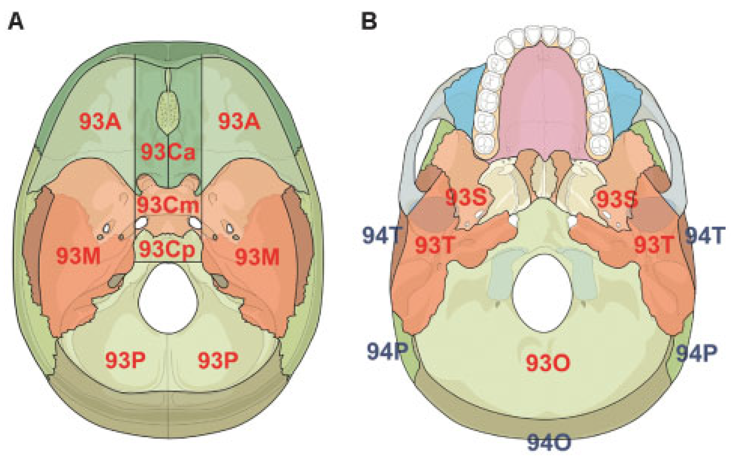
Level 2 System for Fracture Location

The skull base is defined with its endocranial (inner) surface, which constitutes the floor of the cranial cavity, on which the brain rests, and its exocranial (external) surface (Figure 1). The anatomic definition of anterior, middle, and posterior skull base is used. The clinical consideration that the whole central part of the skull base, from the frontal sinuses to the great occipital foramen and below, is surgically reachable by means of transsphenoidal endoscopic approaches, led us to delineate the anatomy of the central compartment of the skull base. Tracing two sagittal lines parallel to the crista galli along the frontoethmoid rims, the skull base is divided into one central and two lateral (right and left) parts (Figure 1A).
The anterior part (the median part of the anterior skull base) covers the upper nasal cavity and the sphenoid sinus; the middle part contains the cavernous sinuses laterally, with the carotid arteries inside (parasellar compartments); the posterior part includes the clivus, reaching the anterior margin of the great occipital foramen. The endocranial surface of the skull base is therefore divided into nine “quadrants.” The foramina through which nerves, arteries, and veins pass were considered for each quadrant because of their clinical relevance regarding the relationship between anatomic fracture location and clinical outcome. Moreover, since the dural venous sinuses can be involved in skull base fractures and can cause massive bleeding, the anatomy of bones along with dural venous sinuses were also considered; although the soft tissue parts are not included in this bone fracture classification system, nonetheless, this includes for instance the involvement of the transverse sinuses in fractures of the occipital bone or of the superior sagittal sinus associated with frontal or parietal fractures.
Above the exocranial surface of the skull base (Figure 1B), no quadrant was delineated. Both the occipital and temporal bones were split between the skull base (code 93) and the cranial vault (code 94) (Figure 2). The squamous portion of the temporal bone (94T) is a well-defined anatomic region and is clearly part of the cranial vault. The division line between the occipital squamous part (squama occipitalis) and the skull base part of the occipital bone at the outer surface is the superior nuchal line with confluence on both sides in the midline at the level of the so-called torus occipitalis or “occipital spur,” at the insertion of the nuchal ligament (also known as midpart of external occipital protuberance). This occipital spur is sometimes very prominent and clear to identify in computed tomography (CT) and lateral skull X-rays. Other than the squamous portion, the temporal bone contributes primarily to the floor of the middle cranial fossa. The petrous portion houses the middle and inner ear structures, and the numerous foramina transmit important neural and vascular structures from the intracranial region to their extracranial connections. The tympanic portion of the temporal bone is small and often difficult to recognize on CT scans. The mastoid portion, is more of an appendage, but should be considered as part of the external cranial vault.
Similarly, the occipital bone contributes to both the skull base and the cranial vault, though as noted above, its main contribution is to the cranial vault. However, as it extends inferoanteriorly, it forms the foramen magnum and houses the hypoglossal canals bilaterally. Regarding other regions of the cranial vault, the classical anatomical borders of the bones were outlined and used for level 2.

Level 3 System for Fracture Morphology

Within each region the anatomy of the fractures can be described by a series of diagnostic features making a more focused level 3 system (Table 1). The fractures can be single, crossing one or more regions or bones, or multiple, in the same bone or in different bones. Within each defined region, the fractures are defined with:
  • 1 = Nonfragmented (single fracture line [linear and branched], ignoring small fracture cracks), or
  • 2 = Fragmented
Fractures are further defined regarding their level of displacement according to one of the three categories:
  • 0 = Nondisplaced
  • 1 = Displaced but not depressed
  • 2 = Depressed (when the broken bones are displaced inward toward the meninges and brain for more than 3 mm)
Fractures of the walls of the paranasal sinuses with the presence or the absence of opacification of the paranasal sinuses on CT imaging (i.e., blood or other fluids) are also documented. Illustrative examples are presented in Figure 3 and Figure 4A.

Associated Intracranial Features

A list of clinically relevant intracranial features that may be associated with skull base and/or cranial vault fractures is proposed in addition to the fracture classification system to support the diagnosis and decision process (Table 2). These features include the presence of intracranial air (0 = none, 1 = single, or multiple air bubbles [up to 3 mm diameter], 2 = pneumocephalus [larger air pockets > 3mm]) (Figure 4B–D), intracranial mass lesion (epidural, subdural, subarachnoid, intraventricular, and/or intraparenchymal hematoma, brain contusions, foreign bodies) (Figure 4E–H), as well as radiological or clinical evidence of dural rupture, with following cerebrospinal fluid (CSF) leak.

Fracture Coding and Topographical Distribution
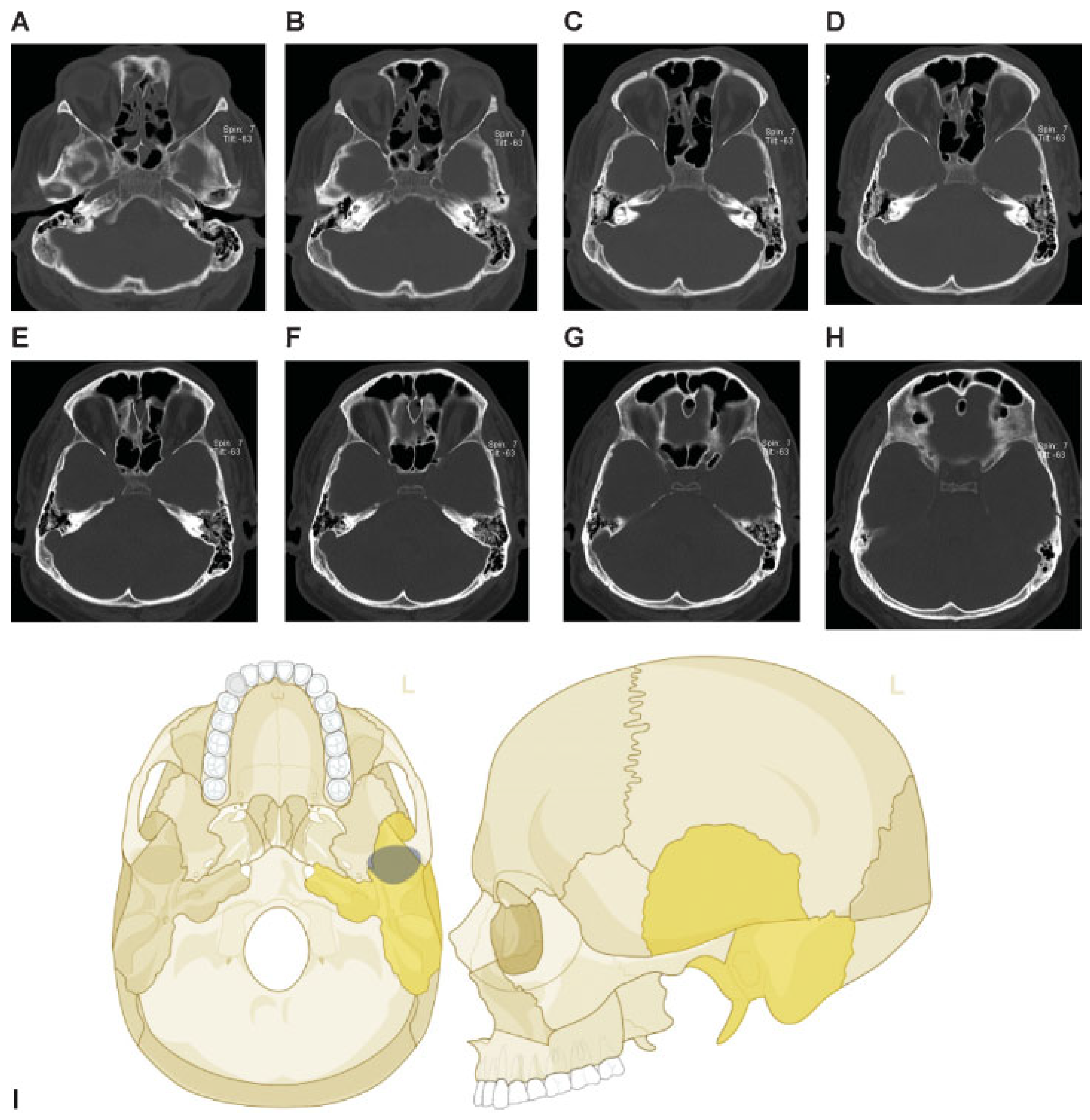
Fractures of the skull base and cranial vault units are identified with the two digit code 93 and 94, respectively.[1] In coding the fractures according to their location in the level 2 system, each fractured region is identified by letters (Figure 1 and Figure 2), which stand for the skull base (level 1 code 93): A = anterior, M = middle, P = posterior, Ca = central anterior, Cm = central middle, Cp = central posterior, and for the cranial vault (level 1 code 94): F = frontal bone, P = parietal bone, O = occipital bone, S = sphenoid bone, and T = temporal bone.
The letters specifying the involved regions are added after the initial code 93 or 94. The regions are coded in the order from the patient’s right side to the patient’s left side considering in order for the skull base (93) O, T, S, A, M, P (right), Ca, Cm, Cp (central), P, M, A, S, T, O (left), and for the cranial vault (94): O, P, T, F (right); F, T, P, O (left). In the overall fracture code, the small letter “m” (abbreviation for “middle”), or the letter for any involved central region (i.e., for the skull base “Ca,” “Cm,” and “Cp”), marks the limit between the two sides. Hence, letters specified before and after the “m” or any central region refer to the patient’s right and left side, respectively. In the code, a point “.” is inserted in between region letters, which in this context does not indicate whether the fractures are confined or not confined within the involved regions, as opposed to the mandibular system.[2]
The level 3 system is coded according to the two main items describing fracture morphology (Table 1). For instance, a nonfragmented nondisplaced fracture of the frontal bone on the right side is coded “94 F10.m;” a fragmented and depressed fracture of the frontal bone on the left side is coded “94 m.F23.”

Case Examples

This coding system allows description of most relevant fracture patterns as illustrated in the case examples, such as the differentiation between unilateral or bilateral fracture patterns. In a series of three case examples, we illustrate the coding of a case with multiple cranial vault fractures (Figure 5), a single skull base fracture extending into the temporal cranial vault (Figure 6) and a right occipital and temporal cranial vault fracture extending into right middle fossa skull base (Figure 7). A range of additional fracture patterns are presented in a special case appendix as electronic supplement to this issue of the Journal (www.aocmf.org/classification).

Discussion

Considering that head trauma is one of the leading causes of disability and death in the Western countries,[3,4,5] great efforts have been directed at determining the best treatments, including the search for the most reliable diagnostic and prognostic tools. Head injuries require a highly specialized multidisciplinary evaluation. Skull base fractures should always be considered as an “epiphenomenon,” that is, they occur alongside of a primary phenomenon (e.g., an associated body trauma with or without neurological lesions), and they are among the most difficult fractures to evaluate and manage. Skull base fractures can lead to death or comatose states as well to immediate or delayed CSF leakage, neurovascular damage, meningitis, cranial nerve injury, or delayed complications.[6,7]
In the course of history, many “treatises” have been written on the classification and treatment of head injuries, from Hippocrates to Avicenna, from Celsus to Lanfranc. However, the first textbook on head injuries, in the modern meaning of the word, was written in the 16th Century by the Italian physician Berengario da Carpi.[8] In the “Treatise on Fractures of the Calvaria or Cranium” the author did not only describe an entire set of surgical instruments to be used for cranial operations to treat head traumas, but he also systematically reviewed the existing literature and added many personal “case reports” to cover the mechanisms, classification, and medical and surgical treatments. Berengario, according to the literature of the time and his personal experience, wrote a classification system of the head injuries intended to be useful in everyday practice (it is interesting to note that the description of haesena resembles that of what is now called a ping–pong fracture). This textbook became a reference book for generations of physicians during the following centuries.
Audigé et al. [1] listed the most relevant classification systems for the craniomaxillofacial (CMF) skeleton, including the famous classification system of Le Fort at the beginning of the 20th century. [9,10,11] Among the few most pertinent fracture classification systems for the skull base, Sakas et al. [12] proposed a radiological-based classification system in which four major types were described: I, cribriform; II, frontoethmoidal; III, lateral frontal; and IV, complex (any combination of the other three types) also considering the amount of bone displacement. The results showed a gradation of risk of the described pattern relating to some complications (rate of infection, prolonged duration of rhinorrhea), suggesting some guidelines to improve the management of posttraumatic CSF fistulas of the anterior cranial base. Further algorithms to manage posttraumatic CSF leakage treatment algorithm in anterior skull base fractures were later suggested by Sherif et al.[13]
Madhusudan et al. [14] considered patients with cranial and facial injuries due to high-velocity accidents. The fractures were classified as frontal, basal, and combined. According to the site of the impact, the fractures were classified as occurring in the central region or in the lateral one, or combined. In consideration of the site and the involvement of the midfacial structures, a nine-type scoring system was proposed to describe the incidence of each specific fracture pattern and the related complications. Later, Bächli et al. [15] proposed a semiautomatic classification based on the high-resolution CT of the whole craniofacial region, looking for correlation with clinical findings and the involvement of skull base. The classification system defined a hierarchical level of increasing severity based on previous work by Buitrago-Téllez et al.,[16] and a good correlation was found between the scoring and the rate of complications and the kind of treatment (conservative treatment vs. surgical operation). Simultaneously, Manson et al. [17] proposed a classification system of frontobasal and midface fractures involving the skull base, integrating experiments on cadavers with clinical data. The categorization of the fractures was based on their pattern, location, involvement of the midface, and related complications. The authors described three kind of fracture patterns: isolated linear cranial base fractures (type I); vertical-linear fractures of the skull vault (frontal bone) in combination with base fractures (type II); comminution of the frontolateral skull vault and orbital roof in association with a linear base fractures (type III). The evaluation of these fracture patterns in more than 200 clinical cases showed an increasing rate of complications associated with more complex fracture patterns.
Despite the existence of many classification systems, no comprehensive and structured classifications of fractures of the CMF skeleton including the skull base and cranial vault have been proposed. A clinically relevant, well-structured and reliable classification provides a universal language and coding that facilitates worldwide communication and collaboration. We believe the proposed system offers a minimum set of clinically relevant features that will help surgeons in making their decisions in clinical settings. An extension to the fracture classification system allows documentation of additional diagnostic features highly clinically relevant, thus addressing the current limits of existing systems. This includes the pterygoid processes defined in the midface fracture Level 2 classification system 17 Kunz C, Cornelius CP, Prein J, et al. The Comprehensive AOCMF Classification System: Midface Fractures - Level 2 Tutorial.[18] and the occipital condylar processes, cause of craniovertebral instability, when fractured. The styloid processes of the temporal bone and the tip of the mastoid were considered, however left out of the present system to avoid excessive complexity. It is obvious that a simplification was required and that a practical classification cannot include all conceivable factors and patient details into the classification.
The strength of our system is that it includes the specific complications that may occur related to the site of the skull base fractures. The fractures of the anterior skull base, which involve the paranasal sinuses or the orbit, for example, can cause CSF leakage and/or injury to the olfactory and/or optic nerves, or the ones involving the carotid canal may result in carotid-cavernous fistula. Fractures of the petrous bone can cause facial palsy and deafness, CSF leakage with otorrhoea or paradoxical (oto)rhinorrhea. Fracture of the posterior fossa may lacerate the major venous sinuses and affect the craniocervical stability.[6]
The present classification system and evaluation sessions were limited by the type and quality of the images; the approach was pragmatic to ensure the final system could be used in practice, as well as in hospitals where highresolution CT scans are not available.
In the early development phase, other terminology and definitions were considered for inclusion in the system, such as “comminuted fracture” (when the bone is shattered into many pieces) and “diastatic fracture” (involving a horizontal displacement of the bones at the margin of the fracture > 3 mm, which can typically occur along the cranial sutures). In the evaluation process, participants realized that simplification was necessary, along with the clarification of definitions for terms commonly used. The cases with the highest levels of disagreement were collectively reviewed, to identify the limits of the system. No disagreement was noted regarding the anatomical localization of the fractures; the use of the anatomical terminology and of the quadrants on the skull base was supported. On the other hand, high levels of disagreement were noted on the use of some terms regarding the type and morphology of the fractures. The use of “single fracture line” versus “multiple fracture lines” with or without comminution was considered highly controversial, particularly when a small fracture had many “cracks,” forming several small lines of fracture. For this reason, it was decided to exclude the term “comminution,” as well as consider any “associated cracks” as clinically irrelevant (Figure 8A). The item “diastatic fracture” was also eliminated due to lack of a clear agreement, particularly when the fracture was associated with a suture (Figure 8B). Finally, contrary to fractures of the mandible,[2] a large consensus prevailed among the authors that whether fractures are confined within a region or extending across several regions had limited clinical relevance and could not be documented reliably. Hence, the term “contiguous” (subsequently replaced by “not confined”) to describe a fracture extending over two or more regions was not supported.

Conclusion

We propose an objective and clinically meaningful classification system of the fractures of skull base and cranial vault. While the validation process of this system is in progress, the inclusion of this system within an electronic tool and database such as the AO comprehensive injury automatic classifier software [18] is essential to facilitate teaching, classification, and documentation of head injuries. In turn this will support the clinical assessment and management of patients affected by head trauma.

Acknowledgments

The CMF classification project was funded by the AO Foundation and its AOCMF Specialty. Illustrations were prepared by AO Education (Publishing) by Jecca Reichmuth and her colleagues. The authors are grateful to Prof. Geoff Manley (San Francisco, CA) for his valuable input during a classification review meeting. We would also like to thank M. Wilhelmi for the editing support of this article.

References

  1. Audigé, L.; Cornelius, C.P.; Di Ieva, A.; Prein, J. CMF Classification Group. The first AO classification system for fractures of the craniomaxillofaxial skeleton: rationale, methodological background, developmental process and objectives. Craniomaxillofac Trauma Reconstr 2014, 7 (Suppl 1), S6–S14. [Google Scholar]
  2. Cornelius, C.P.; Audigé, L.; Kunz, C.; et al. The comprehensive AOCMF classification system: mandible fractures - level 2 tutorial. Craniomaxillofac Trauma Reconstr 2014, 7 (Suppl 1), S15–S30. [Google Scholar] [CrossRef] [PubMed]
  3. Masson, F.; Vecsey, J.; Salmi, L.R.; et al. Disability and handicap 5 years after a head injury: a population-based study. J Clin Epidemiol 1997, 50, 595–601. [Google Scholar] [CrossRef] [PubMed]
  4. Annegers, J.F.; Grabow, J.D.; Kurland, L.T.; Laws, E.R., Jr. The incidence, causes, and secular trends of head trauma in Olmsted County, Minnesota, 1935-1974. Neurology 1980, 30, 912–919. [Google Scholar] [CrossRef] [PubMed]
  5. Kraus, J.F.; Black, M.A.; Hessol, N.; et al. The incidence of acute brain injury and serious impairment in a defined population. Am J Epidemiol 1984, 119, 186–201. [Google Scholar] [CrossRef] [PubMed]
  6. Samii, M.; Tatagiba, M. Skull base trauma: diagnosis and management. Neurol Res 2002, 24, 147–156. [Google Scholar] [CrossRef] [PubMed]
  7. de Boussard, C.N.; Bellocco, R.; af Geijerstam, J.L.; et al. Delayed intracranial complications after concussion. J Trauma 2006, 61, 577–581. [Google Scholar] [CrossRef] [PubMed]
  8. Di Ieva, A.; Gaetani, P.; Matula, C.; et al. Berengario da Carpi: a pioneer in neurotraumatology. J Neurosurg 2011, 114, 1461–1470. [Google Scholar] [CrossRef] [PubMed]
  9. Le Fort, R. Étude expérimentale sur les fractures de la machoire supérieure (Part l). Rev Chir 1901, 23, 208–227. [Google Scholar]
  10. Le Fort, R. Étude expérimentale sur les fractures de la machoire supérieure (Part II). Rev Chir 1901, 23, 360–379. [Google Scholar]
  11. Le Fort, R. Étude expérimentale sur les fractures de la machoire supérieure (Part lll). Rev Chir 1901, 23, 479–507. [Google Scholar]
  12. Sakas, D.E.; Beale, D.J.; Ameen, A.A.; et al. Compound anterior cranial base fractures: classification using computerized tomography scanning as a basis for selection of patients for dural repair. J Neurosurg 1998, 88, 471–477. [Google Scholar] [CrossRef] [PubMed]
  13. Sherif, C.; Di Ieva, A.; Gibson, D. A management algorithm for cerebrospinal fluid leak associated with anterior skull base fractures: detailed clinical and radiological follow-up. Neurosurg Rev 2012, 35, 227–237, discussion 237–238. [Google Scholar] [CrossRef] [PubMed]
  14. Madhusudan, G.; Sharma, R.K.; Khandelwal, N.; Tewari, M.K. Nomenclature of frontobasal trauma: a new clinicoradiographic classification. Plast Reconstr Surg 2006, 117, 2382–2388. [Google Scholar] [CrossRef] [PubMed]
  15. Buitrago-Téllez, C.H.; Schilli, W.; Bohnert, M.; et al. A comprehensive classification of craniofacial fractures: postmortem and clinical studies with twoand three-dimensional computed tomography. Injury 2002, 33, 651–668. [Google Scholar] [CrossRef] [PubMed]
  16. Bächli, H.; Leiggener, C.; Gawelin, P.; et al. Skull base and maxillofacial fractures: two centre study with correlation of clinical findings with a comprehensive craniofacial classification system. J Craniomaxillofac Surg 2009, 37, 305–311. [Google Scholar] [CrossRef] [PubMed]
  17. Manson, P.N.; Stanwix, M.G.; Yaremchuk, M.J.; et al. Frontobasal fractures: anatomical classification and clinical significance. Plast Reconstr Surg 2009, 124, 2096–2106. [Google Scholar] [CrossRef] [PubMed]
  18. Kunz, C.; Cornelius, C.P.; Prein, J.; et al. The comprehensive AOCMF classification system: midface fractures - level 2 tutorial. Craniomaxillofac Trauma Reconstr 2014, 7 (Suppl 1), S59–S67. [Google Scholar] [CrossRef] [PubMed]
Figure 1. Skull base, (A) endocranial and (B) exocranial surface. Tracing two sagittal lines parallel to the crista galli along the fronto-ethmoid rims, the endocranial surface of the skull base can be divided in a central and in two (right and left) parts. (A) The nine quadrants used for the anatomical location of the fractures. A, anterior skull base; C, central skull base; Ca, Central anterior skull base (Cribriform plate and etmoido-sphenoidal planum); Cm, Central middle skull base (sellar and parasellar comparments); Cp, Central posterior skull base (clivus); M, middle skull base; P, posterior skull base; a, anterior; m, middle; p, posterior. (B) F, frontal bone; O, occipital bone; P, parietal bone; S, sphenoid bone; T, temporal bone.
Cmtr 07 00021 g001
Figure 2. Cranial vault, (A) frontal and (B) lateral view. F: Frontal bone; P: Parietal bone; S: Sphenoid bone; T: Temporal bone; O: Occipital bone.
Cmtr 07 00021 g002
Figure 3. Examples of fractures and definitions. (A) Depressed fracture (displaced toward the brain) of the parietal bone. (B) Multiple fractures, depressed on the right frontal bone, displaced on the left frontal bone, with involvement and opacification of the frontal sinus bilaterally. (C) Transverse fracture line through the right petrous bone, another fracture line through the right apex petrosis, opacification of the left mastoid with fracture at the rostral rim. (D) Transverse maximum intensity projection (MIP) – reconstruction of the skull base with fragmented fracture of the right middle skull base fossa. (E): Nonfragmented fracture of the left parietal bone. (F) Curved MIP—reconstruction of the occipital bones with fragmented fracture, displaced but not depressed.
Cmtr 07 00021 g003
Figure 4. Examples of associated diagnostic features with skull base and cranial vault fractures. (A) Partial opacification in the sphenoid sinus with air–fluid level in computed tomographic imaging (representing possible the presence of blood or fluid). (B–D) Presence of intracranial air: single or multiple air-bubbles in the subdural spaces (arrows in B) or in the subarachnoid space (arrow in C); (D) pneumocephalus. (E–H) Intracranial mass lesions: (E) epidural hematoma in the left occipital region (arrowheads) with small mixed (epidural and subdural) hematoma in the right temporal region (arrows), (F) subdural hematoma of the right hemisphere (arrows) with extension along the falx (arrowhead), (G) intraventricular haematoma, (H) brain contusions.
Cmtr 07 00021 g004
Figure 5. Multiple cranial vault fractures. (A–F) Multiple cranial vault fractures. The right sided fractures involve the frontal and parietal vault with comminution and depression and extension inferiorly into the temporal cranial vault. Similarly, on the left, there is a linear parietal vault fracture that appears to continue into the temporal bone. (G) There is extension of these fractures into the left and right sphenoid bone. Imaging: Computed tomographic axial views. (H) Level 3 code: 93 S0.m.S0, 94 F1.P1.T0.m.T0.P0, This case example CMTR-93-94-001 is made available electronically for viewing using the AOCOIAC software at www.aocmf.org/classification.
Cmtr 07 00021 g005
Figure 6. Single skull base fracture extending the temporal cranial vault. (A–C) Fracture through the skull base involving the left temporal bone, (D–I) traversing the glenoid fossa, and extending into the temporal cranial vault. The fracture is linear, unbranched and non-fragmented. Imaging: Computed tomographic axial views. (I) Level 3 code: 93 m.T0, 94 m.T0, This case example CMTR-93-94-002 is made available electronically for viewing using the AOCOIAC software at www.aocmf.org/classification.
Cmtr 07 00021 g006
Figure 7. Right occipital and temporal cranial vault fracture extending into the right middle fossa skull base. (A–C) Right occipital cranial vault fracture extending inferiorly into the right temporal bone, (D–H) with extension across the right middle fossa skull base through the skull base portion of the right temporal bone. There does not appear to be any fragmentation. (D) A probable left sphenoid fracture may be noted as well. Imaging: Computed tomographic axial views. (I) Level 3 code: 93 T0.M0.m.S, 94 O0.m, This case example CMTR-93-94-003 is made available electronically for viewing using the AOCOIAC software at www.aocmf.org/classification.
Cmtr 07 00021 g007
Figure 8. Examples of cases in which the definitions of the first session of the classifications system caused disagreement. (A) According to the definitions of the first session, the fracture marked with the arrow was classified in different ways by the different participants of the pilot session: single branched or multiple and comminuted. The disagreement was solved in the following classification form, defining it single, ignoring the multiple cracks, because considered clinically irrelevant. (B) The fracture marked with the arrow was cause of controversies in the assignment to the definition “diastatic”. Considering the term “diastatic” clinically irrelevant and cause of disagreement, in the new form of the classification system that fracture was defined single fracture and displaced not depressed.
Cmtr 07 00021 g008
Table 1. Level 3 focused classification of fractures within the skull base and cranial vault.
CodeSubdivisionsLevel 3 itemsCategories
AllFracture line(s)1 = Single fracture line (ignoring associated cracks)
2 = Multiple fracture lines
Displacement0 = Nondisplaced
1 = Displaced but not depressed
2 = Depressed (displaced toward the brain)
93AAnterior SB
Frontal sinusFracture0 = No; 1 = Yes
Opacification0 = No; 1 = Yes
93MMiddle SB
Ethmoid sinusFracture0 = No; 1 = Yes
Opacification0 = No; 1 = Yes
93CaCentral anterior SB
CribriformFracture0 = No; 1 = Yes
93CmCentral middle SB
Sphenoid sinusFracture0 = No; 1 = Yes
Opacification0 = No; 1 = Yes
93OOccipital bone
Occipital condylar processFracture0 = No; 1 = Yes
94FFrontal bone
Frontal sinusFracture0 = No; 1 = Yes
Opacification0 = No; 1 = Yes
93SSphenoid bone
93TTemporal bone
Glenoid fossaaFracture0 = No; 1 = Yes
Abbreviation: SB, skull base. aThe glenoid fossa involvement is classified separately for the sphenoid bone and the temporal bone. Accordingly, any involved subdivision is classified as being fractured.
Table 2. Level 3 focused documentation of associated intracranial diagnostic features.
Item groupsItemsCategories
Intracranial air a 0 = None
1 = Bubbles (up to 3 mm diameter)
2 = Pneumocephalus (>3 mm diameter)
Intracranial mass lesionEpidural hematoma0 = No; 1 = Yes
Subdural hematoma0 = No; 1 = Yes
Intracerebral hematoma0 = No; 1 = Yes
Traumatic SAH0 = No; 1 = Yes
Intraventricular0 = No; 1 = Yes
Contusion0 = No; 1 = Yes
Foreign bodies0 = No; 1 = Yes
Cranial nerve involvementNerve 10 = No; 1 = Yes
Nerve 20 = No; 1 = Yes
Nerve 30 = No; 1 = Yes
Nerve 40 = No; 1 = Yes
Nerve 50 = No; 1 = Yes
Nerve 60 = No; 1 = Yes
Nerve 70 = No; 1 = Yes
Nerve 80 = No; 1 = Yes
Nerve 90 = No; 1 = Yes
Nerve 100 = No; 1 = Yes
Nerve 110 = No; 1 = Yes
Nerve 120 = No; 1 = Yes
Vascular structure involvementTraumatic aneurysm0 = No; 1 = Yes
Traumatic CCF0 = No; 1 = Yes
Traumatic occlusion0 = No; 1 = Yes
Venous sinusesTransverse sinus0 = No; 1 = Yes
Sigmoid sinus0 = No; 1 = Yes
Superior sagittal sinusa0 = No; 1 = Yes
Confluence sinusa0 = No; 1 = Yes
Cerebrospinal fluid leak
PreviousRhinorrhea0 = No; 1 = Yes
Otorrhea0 = No; 1 = Yes
Other location0 = No; 1 = Yes
ActiveRhinorrhea0 = No; 1 = Yes
Otorrhea0 = No; 1 = Yes
Other location0 = No; 1 = Yes
Abbreviation: SAH, subarachnoid hemorrhage. a Items documented only once for the whole intracranial space. All other items are to be recorded separately for both the sides.

