Next Article in Journal
The Comprehensive AOCMF Classification System: Midface Fractures—Level 3 Tutorial
Previous Article in Journal
The Comprehensive AOCMF Classification System: Midface Fractures—Level 2 Tutorial
 
 
Craniomaxillofacial Trauma & Reconstruction is published by MDPI from Volume 18 Issue 1 (2025). Previous articles were published by another publisher in Open Access under a CC-BY (or CC-BY-NC-ND) licence, and they are hosted by MDPI on mdpi.com as a courtesy and upon agreement with Sage.
Short Note

Three-Dimensional Volumetric Restoration by Structural Fat Grafting

by
Luigi C. Clauser
*,
Giuseppe Consorti
,
Giovanni Elia
,
Manlio Galié
and
Riccardo Tieghi
Unit of Cranio Maxillo Facial Surgery, Center for Craniofacial Deformities and Orbital Surgery, Ferrara, Italy
*
Author to whom correspondence should be addressed.
Craniomaxillofac. Trauma Reconstr. 2014, 7(1), 63-69; https://doi.org/10.1055/s-0033-1356757
Submission received: 9 January 2013 / Revised: 10 January 2013 / Accepted: 10 January 2013 / Published: 23 October 2013

Abstract

:
The use of adipose tissue transfer for correction of maxillofacial defects was reported for the first time at the end of the 19th century. Structural fat grafting (SFG) was introduced as a way to improve facial esthetics and in recent years has evolved into applications in craniomaxillofacial reconstructive surgery. Several techniques have been proposed for harvesting and grafting the fat. However, owing to the damage of many adipocytes during these maneuvers, the results have not been satisfactory and have required several fat injection procedures for small corrections. The author’s (L.C.) overview the application of SFG in the management of volumetric deficit in the craniomaxillofacial in patients treated with a long-term follow-up.

The use of adipose tissue transfer for the correction of maxillofacial defects was reported for the first time at the end of the 19th century. Since 1893, some physicians have used fat grafts, the results of which, however, have been unpredictable and extremely technique dependent.
Several reports in the literature of the early 20th century [1] have described tissue augmentation with different injectable fillers such as gold, rubber latex, gutta–percha, paraffin, collagen, Goretex, silicon, and more recently, polylactic acid and byoalcamid [2].
Focusing attention on the face, in 1991 Sydney R. Coleman systematized the technique.
This was based on the belief that fat was the ideal filler because it was natural, stable, and without the complications of earlier fillers. Structural fat grafting (SFG) differs from other fat grafting techniques, given its delicate aspiration to protect the fragile adipocytes, purification of the material, and its reinsertion using microinjections to redefine facial contours and create a more harmonious and esthetically appealing proportion of the face [3,4].
Human adipose tissue represents a rich source of mesenchymal stem cells because they exhibit multilineage potential and secrete angiogenic and antiapoptotic factors [5,6,7]. The main indications for SFG are the restoration and soft tissue facial reconstruction after trauma, tumor resection, in congenital deformities and clefts, Parry–Romberg syndrome and scleroderma, orbital and periorbital surgery, facial palsy, burns, and scars [8,9,10,11,12,13,14,15].
SFG is also used for the rejuvenation of the face in areas such as forehead, zygomas, lips, nose, chin, and mandible [16,17,18].

Patients and Methods

The authors present four clinical cases to confirm that SFG technique is part of the complex reconstructive planning. All patients received general anesthesia.
A comparison of frontal, three-fourth view, lateral, subcranial, and close-up photos were taken preoperatively and during the follow-up.

Technique

The preoperative plan includes a photographic study according to Coleman’s guidelines [3]. Colored pens were used on these photographs in the areas that were to be injected, those not injected, and, if necessary, areas from which excessive fat tissue was needed to be removed. These markings were duplicated on the patient’s face preoperatively (Figure 1).
Common donor sites are trochanteric region, inner thighs and knees, periumbilical area, and flanks. Usually, buttocks are the preferred donor area in children.
Patient was operated on under general anesthesia or local anesthesia with mild sedation.
The fat is aspirated using a very thin liposuction cannula attached to a 10-mL Luer-Lock syringe for suction. During the removal of fat, care is taken to minimize mechanical trauma to the adipocytes. Once the fat has been removed, every 10 mL syringe is carefully placed into a sterilized sleeve in a sterilized central rotor of a centrifuge and spun at approximately 1,300 rpm for 5 minutes. The fat separates into three, the top being composed primarily of oil from ruptured parcels of fat, the bottom of blood, and the middle layer of usable subcutaneous tissue. The top oil is decanted and the dense lowest layer is drained. The refined parcels of fat are then transferred to a 1-mL syringe and layered into the areas requiring enhancement, working from the underlying bone up to the skin surface [19,20].
Cannulas with different tip shapes, diameters, lengths, and curves can be used. The use of blunt cannulas allows placement of the fat parcels in a more stable and less traumatic manner. However, less blunt cannulas may give the surgeon more control for placement in the immediate subdermal plane, in fibrous tissue, and in scars. A cannula with pointed or sharp elements is used to free up adhesions. Fatty tissue should be injected only as the cannula is withdrawn. With linear deposition, the fat is layered into the area requiring enhancement, working from the underlying bone up to the skin surface to produce a three-dimensional grill. The largest amount of tissue that should be placed with each withdrawal of a cannula is 1/10 mL, but in some areas, such as the eyelid, the maximum placed should be from 1/30 to 1/50 mL per withdrawal.
One step correction, using more fat tissue, can cause poor vascularization and more reabsorption, particularly in areas covered by a thin layer of soft tissue like the maxillofacial area [21,22].
SFG allows the fat to be carefully layered into minuscule strands creating a large surface area of contact with the augmented site, and this ensures that nutrition can be exchanged through capillaries in the enveloping tissue (the so-called Structure) [23]. This is essential for the fat to survive. By using the patient’s own fat, there is no risk of allergic reactions or rejections by the body. The deeply embedded fat does not shift or migrate.
After surgery compression, dressings are applied around the infiltrated areas and the patient wears an abdominal binder if the fat is harvested from the abdomen. For 24 to 36 hours postoperatively, ice packs are applied continuously on all infiltrated sites.
Postoperative care involves use of antibiotics for several days and anti-inflammatory therapy and massage of donor area to relieve swelling and bruising. Patients must be instructed that postoperative swelling can last for 12 to 16 weeks [11]. Recovery time will depend on the extent of the procedure [23].
The fat is slowly absorbed by the body, although the amount of absorption is variable and frequently unpredictable [24,25]. More than half of the fat used in injectable treatments except SFG is absorbed. However, this percentage varies from patient to patient.
If a significant amount of fat is absorbed, a second procedure may be needed to achieve the final result.
When dealing with craniofacial deformities, one must bear in mind that patients have altered growth potential. Fat reabsorption is higher in these patients compared with others, and SFG should be done as many as 4 to 5 times to improve the final result, with a lapse of 10 to 14 months.

Case 1

Due to a deficit in her upper jaw and lip, a 25-year-old woman was seen as the outcome of numerous operations made for the correction of palate and lip cleft sequela (Figure 2).
She had previously undergone upper lip and palate reconstruction, upper alveolus iliac bone graft and rhinoplasty. SFG (with a total amount of 30 cc of pure fat) was performed in the upper and lower lips, white roll, philtrum, cheeks, and tip of the nose to obtain a more pleasant facial appearance. The follow-up was 2 years.

Case 2

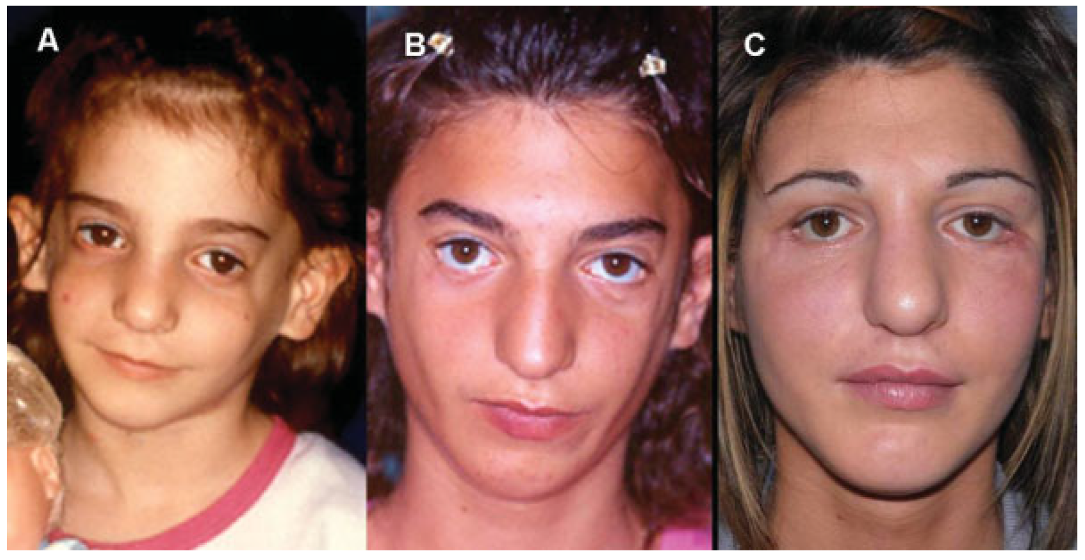
Patient affected by Treacher Collins syndrome (Figure 3). The main reconstructive steps were as follows: orbital reconstruction using cranial bone grafts, rotation of skin muscular flap from the upper eyelids to the lower eyelids and advanced jumping genioplasty. To improve the soft tissue, three stages of SFG (with a total amount of 180 cc fat) of the frontozygomatic, cheeks, and temporal area, jaw line, and chin was performed. The outcome can be considered stable after 3 years.

Case 3

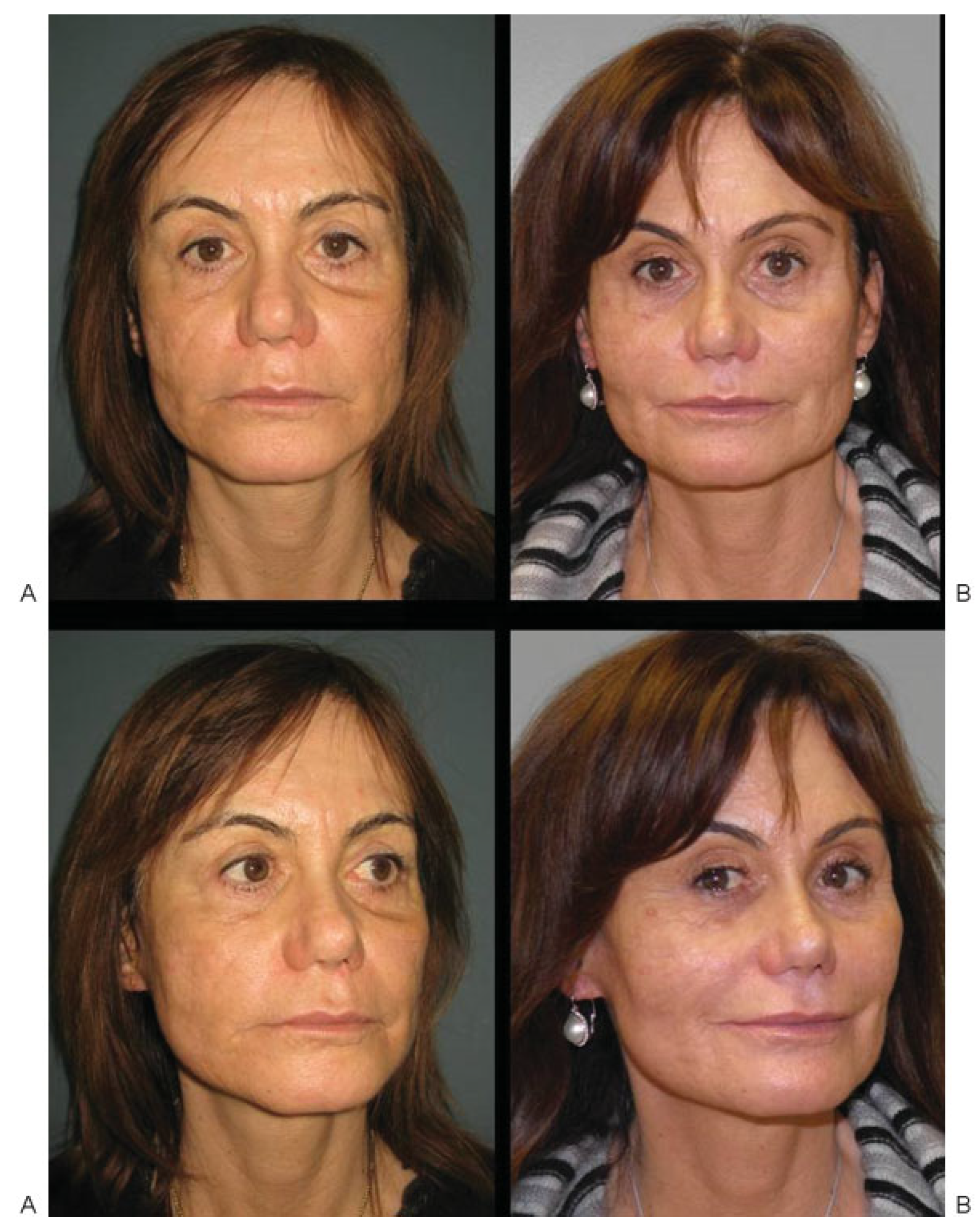
A 52-year-old woman treated 20 years ago for complex facial trauma (Le Fort I, naso-ethmoidal fracture) (Figure 4). In the years after trauma, the patient underwent different reconstructive procedure. She came back 2 years ago complaining about lack of facial volume and premature aging face. Two different stages of total facial SFG (lips, forehead, cheeks, naso-labial folds, chin, and border of the mandible) with a total amount of 160 cc fat were injected.

Case 4

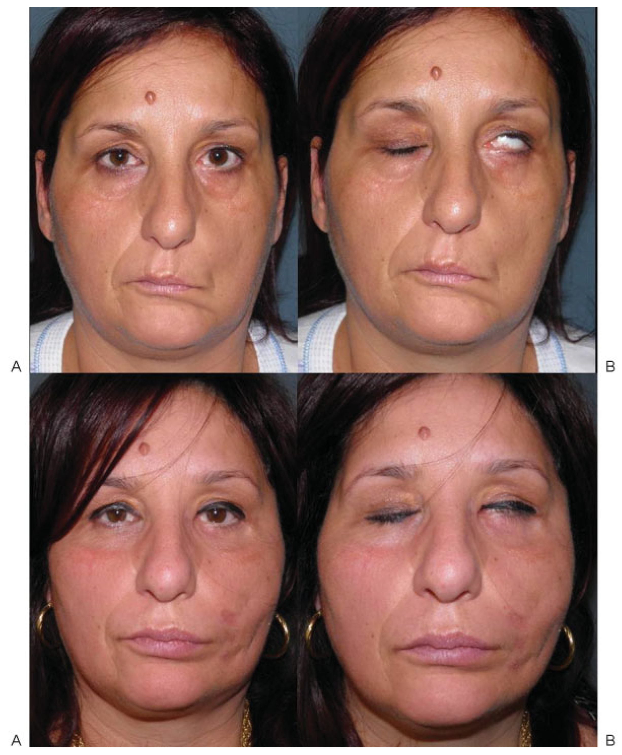
A patient with sequela of VII left cranial nerve palsy (Figure 5). Before SFG, blink restoration with temporalis muscle fascial flap rotation, static suspension of the oral commissure was performed.
SFG improved the hemifacial atrophy due to the facial palsy. A total amount of 100 cc of fat in two procedures was injected in the patient. Functional and morphologic result was stable after 2 years.

Discussion

SFG was introduced as a way to improve facial esthetics and in recent years has evolved into applications in more complex reconstructive procedures [12]. Fat grafting provides a technique whose biocompatibility easily adapts to the growth of the face over time. It has lower morbidity compared with many other alloplastic materials, which often lead to the reactions caused by foreign bodies, making it the ideal tool for the correction of localized tissue atrophy; loss of substance due to trauma, posttumor and congenital complex craniofacial anomalies; burns, as well as hemifacial atrophy, that is, Parry–Romberg syndrome and scleroderma. Results are natural and natural long-lasting [8,10,13,14].
SFG differs from other fat grafting techniques for harvesting, purification, and the placement of the fat. Different kinds of needles have been used for harvesting because minimum trauma to the adipocytes is of paramount importance [19]. There are four main ways to purify the adipose tissue: centrifugation, filtration, decantation, and washing. The authors compared centrifugation with other techniques such as percolator and decantation; however, according to Mojallal et al. centrifugation is the best way to purify adipose tissue, because there is no deterioration or damage to the adipocytes, and because it increases the fat density by eliminating red blood and cellular debris [17,19,21].
With this technique, there are no consequences on the cellular structure, and there are no differences in cellular structures between centrifuged adipose cells and those that are not centrifuged adipose cells. Using a blunt cannula and centrifugation, more than 90% of adipocytes survive and have normal enzyme activity [22].
SFG is a useful technique in craniofacial and maxillofacial surgery because it can be obtained in relatively large quantities with minimal risk. Although it is a safe procedure, complications may occur. The most frequent complications are overcorrection, undercorrection, lumps, bumps, and fat migration, infection, damage to underlying structures, and ischemic necrosis. Fat embolism has been reported in a few cases [26,27,28].
Particular applications of SFG are recalcitrant ectropion, anoftalmic socket, orthognathic surgery and scars or burn wounds.
Ectropion (posttrauma, congenital, post–different reconstructions) represents a challenging situation. Many techniques have been described and reported, some as successful and others as not. The basic problem of ectropion is the shortage of skin, muscles, and internal lamellae. Palatal mucosal graft to rebuild the internal lamella is very useful but it does not restore skin and orbicularis muscle. Lengthening of the internal lamella can be combined (in a second stage) with microparcels of fat. The result is lengthening of the lower eyelid with improvement in volume and texture [29].
In anophthalmic socket, SFG is a real pearl. In the so-called post enucleation socket syndrome, there can be a lack of soft tissue, even after positioning an endo-orbital implant. Appearance can be improved by injecting the intraconal part as well as the upper sulcus on the lower eyelid [9].
SFG in orthognathic surgery represents a new application. It is well known that orthognathic surgery moves the skeletal bases (maxilla, mandible, and chin). In dentofacial deformities there can be a lack of soft tissue that does not improve by moving bones. SFG of the periorbital area, zygomas, upper and lower marionettes, and chin, gives more texture and lightness to the face. The other possibility is to insert implants during surgery or as a second step [8].
Different studies have proved that human adipose tissue represents a rich source of mesenchymal stem cells (ASCs), exhibiting multilineage potential and featuring secretion of angiogenic and antiapoptotic factors [5,6,7].
Adipose-derived stem cells (formerly termed adiposederived stromal cells or adipose-derived regenerative cells) are isolated from the vascular stromal component of lipoaspirate. A standard raw lipoaspirate is composed of mature adipocytes, extracellular matrix, ASCs, endothelial cells, and mural cells (pericytes and vascular smooth muscle cells). When enzymatically digested, the nonbuoyant cellular fraction forms the stromal vascular fraction and contains ASCs, vascular progenitor cells, pericytes, and endothelial cells. Although ASCs are of mesodermal origin, they have the potential, under the appropriate conditions, to differentiate into multiple lineages of adipogenic, osteogenic, chondrogenic, myogenic, cardiomyogenic, and neurogenic cells. Recent studies also show that ASCs are able to differentiate into tissues of ectoand endodermal lineages such as neural cells, hepatocytes, pancreatic islet cells, endothelial cells, and epithelial cells. Evidence exists that stem cells contribute to the restoration of tissue vascularization and organ function. For these reasons, adipose tissue represents a potential clinical use for cellular therapy, tissue engineering, and gene transfer applications in regenerative medicine [30].
The study proposed by Rigotti et al. demonstrates the therapeutic efficacy of ASCs for the treatment of radiationinduced damage through a process of replacement of damaged tissue with reconstructed normal tissue [31].
Adipose-derived stem cells could be considered in the treatment of neurologic or different types of disorders, including maxillofacial deformities [32].
The various surgical operations involving SFG performed by the authors have led to satisfactory morphologic and functional rehabilitation of the reconstructed area. Because the use of autologous adipocytes means that part of this tissue will be spontaneously reabsorbed, further SFG stages are required.

Conclusion

SFG, introduced as a way of improving facial esthetics, has evolved in recent years into more complex reconstructive procedures [12,13,14,15]. The technique should be part of the armamentarium of the reconstructive surgeon not only as an ancillary treatment, but also as a basic complementary reconstructive surgery. SFG represents an excellent tool for craniomaxillofacial surgery as it accomplishes natural results that stand the test of time.
Today, tissue engineering and regenerative medicine are a multidisciplinary science that is evolving along with biotechnological advances. The proposed uses for ASCs in tissue repair/regeneration are quite impressive. Recent works on ASCs would suggest that this adult stem cell may prove to be an equally powerful tool in treating congenital and acquired disorders.
However, the availability and the processes for obtaining stem cells remain a challenge for both surgeon and scientist pursuing regenerative medicine.

References

  1. Coleman, S.R. Facial recontouring with lipostructure. Clin Plast Surg 1997, 24, 347–367. [Google Scholar] [CrossRef]
  2. Coleman, S.R. Structural fat grafts: the ideal filler? Clin Plast Surg 2001, 28, 111–119. [Google Scholar] [CrossRef] [PubMed]
  3. Coleman, S.R. (Ed.) Structural Fat Grafting; Quality Medical Publishing Inc: Saint Louis, MO, 2004. [Google Scholar]
  4. Rigotti, G.; Marchi, A.; Sbarbati, A. Adipose-derived mesenchymal stem cells: past, present, and future. Aesthetic Plast Surg 2009, 33, 271–273. [Google Scholar] [CrossRef] [PubMed]
  5. Strem, B.M.; Hicok, K.C.; Zhu, M.; et al. Multipotential differentiation of adipose tissue-derived stem cells. Keio J Med 2005, 54, 132–141. [Google Scholar] [CrossRef] [PubMed]
  6. Fraser, J.K.; Wulur, I.; Alfonso, Z.; Hedrick, M.H. Fat tissue: an underappreciated source of stem cells for biotechnology. Trends Biotechnol 2006, 24, 150–154. [Google Scholar] [CrossRef]
  7. Moseley, T.A.; Zhu, M.; Hedrick, M.H. Adipose-derived stem and progenitor cells as fillers in plastic and reconstructive surgery. Plast Reconstr Surg 2006, 118 (Suppl. 3), 121S–128S. [Google Scholar] [CrossRef]
  8. Clauser, L.C.; Tieghi, R.; Galiè, M.; Carinci, F. Structural fat grafting: facial volumetric restoration in complex reconstructive surgery. J Craniofac Surg 2011, 22, 1695–1701. [Google Scholar] [CrossRef]
  9. Clauser, L.; Sarti, E.; Dallera, V.; Galiè, M. Integrated reconstructive strategies for treating the anophthalmic orbit. J Craniomaxillofac Surg 2004, 32, 279–290. [Google Scholar] [CrossRef]
  10. Clauser, L.; Tieghi, R. Optimizing maxillofacial and craniofacial results. Presented at: American Society of Plastic Surgeons Annual Meeting, Seattle, WA, October 2009. [Google Scholar]
  11. Clauser, L. Optimizing maxillofacial and craniofacial results. In Fat Injection From Filling to Regeneration; Coleman, S.R., Mazzola, R.F., Eds.; Quality Medical Publishing Inc: Saint Louis, MO, 2009; pp. 475–500. [Google Scholar]
  12. Clauser, L.; Polito, J.; Mandrioli, S.; et al. Structural fat grafting in complex reconstructive surgery. J Craniofac Surg 2008, 19, 187–191. [Google Scholar] [CrossRef]
  13. Clauser, L.C.; Tieghi, R.; Consorti, G. Parry-Romberg syndrome: volumetric regeneration by structural fat grafting technique. J Craniomaxillofac Surg 2010, 38, 605–609. [Google Scholar] [CrossRef]
  14. Consorti, G.; Tieghi, R.; Clauser, L.C. Frontal linear scleroderma: longterm result in volumetric restoration of the fronto-orbital area by structural fat grafting. J Craniofac Surg 2012, 23, e263–e265. [Google Scholar] [CrossRef] [PubMed]
  15. Giugliano, C.; Benitez, S.; Wisnia, P.; Sorolla, J.P.; Acosta, S.; Andrades, P. Liposuction and lipoinjection treatment for congenital and acquired lipodystrophies in children. Plast Reconstr Surg 2009, 124, 134–143. [Google Scholar] [CrossRef]
  16. Coleman, S.R.; Saboeiro, A.; Sengelmann, R. A comparison of lipoatrophy and aging: volume deficits in the face. Aesthetic Plast Surg 2009, 33, 14–21. [Google Scholar] [CrossRef]
  17. Foyatier, J.L.; Mojallal, A.; Voulliaume, D.; Comparin, J.-P. [Clinical evaluation of structural fat tissue graft (Lipostructure) in volumetric facial restoration with face-lift About 100 cases]. Ann Chir Plast Esthet 2004, 49, 437–455. [Google Scholar] [CrossRef]
  18. Bucky, L.P.; Kanchwala, S.K. The role of autologous fat and alternative fillers in the aging face. Plast Reconstr Surg 2007, 120 (Suppl. 6), 89S–97S. [Google Scholar] [CrossRef] [PubMed]
  19. Mojallal, A.; Foyatier, J.L. [The effect of different factors on the survival of transplanted adipocytes]. Ann Chir Plast Esthet 2004, 49, 426–436. [Google Scholar] [CrossRef] [PubMed]
  20. Pu, L.L.; Coleman, S.R.; Cui, X.; Ferguson, R.E., Jr.; Vasconez, H.C. Autologous fat grafts harvested and refined by the Coleman technique: a comparative study. Plast Reconstr Surg 2008, 122, 932–937. [Google Scholar] [CrossRef] [PubMed]
  21. Mojallal, A.; Auxenfans, C.; Lequeux, C.; Braye, F.; Damour, O. Influence of negative pressure when harvesting adipose tissue on cell yield of the stromal-vascular fraction. Biomed Mater Eng 2008, 18, 193–197. [Google Scholar] [CrossRef]
  22. Mojallal, A.; Shipkov, C.; Braye, F.; Breton, P.; Foyatier, J.L. Influence of the recipient site on the outcomes of fat grafting in facial reconstructive surgery. Plast Reconstr Surg 2009, 124, 471–483. [Google Scholar] [CrossRef]
  23. Kawamoto, H.K. Fat injection and craniofacial surgery. In Fat Injection From Filling to Regeneration; Coleman, S.R., Mazzola, R.F., Eds.; Quality Medical Publishing Inc: Saint Louis, MO, 2009; pp. 447–474. [Google Scholar]
  24. Coleman, S.R. Long-term survival of fat transplants: controlled demonstrations. Aesthetic Plast Surg 1995, 19, 421–425. [Google Scholar] [CrossRef]
  25. Fagrell, D.; Eneström, S.; Berggren, A.; Kniola, B. Fat cylinder transplantation: an experimental comparative study of three different kinds of fat transplants. Plast Reconstr Surg 1996, 98, 90–96, discussion 97–98. [Google Scholar] [CrossRef]
  26. Gutowski, K.A. ASPS Fat Graft Task Force. Current applications and safety of autologous fat grafts: a report of the ASPS fat graft task force. Plast Reconstr Surg 2009, 124, 272–280. [Google Scholar]
  27. Glasgold, R.A.; Glasgold, M.J.; Lam, S.M. Complications following fat transfer. Oral Maxillofac Surg Clin North Am 2009, 21, 53–58, vi. [Google Scholar] [CrossRef] [PubMed]
  28. Lee, D.H.; Yang, H.N.; Kim, J.C.; Shyn, K.H. Sudden unilateral visual loss and brain infarction after autologous fat injection into nasolabial groove. Br J Ophthalmol 1996, 80, 1026–1027. [Google Scholar] [CrossRef] [PubMed]
  29. Caviggioli, F.; Klinger, F.; Villani, F.; Fossati, C.; Vinci, V.; Klinger, M. Correction of cicatricial ectropion by autologous fat graft. Aesthetic Plast Surg 2008, 32, 555–557. [Google Scholar] [CrossRef] [PubMed]
  30. Zuk, P.A. The adipose-derived stem cell: looking back and looking ahead. Mol Biol Cell 2010, 21, 1783–1787. [Google Scholar] [CrossRef]
  31. Rigotti, G.; Marchi, A.; Galiè, M.; et al. Clinical treatment of radiotherapy tissue damage by lipoaspirate transplant: a healing process mediated by adipose-derived adult stem cells. Plast Reconstr Surg 2007, 119, 1409–1422, discussion 1423–1424. [Google Scholar] [CrossRef]
  32. Kokai, L.E.; Rubin, J.P.; Marra, K.G. The potential of adipose-derived adult stem cells as a source of neuronal progenitor cells. Plast Reconstr Surg 2005, 116, 1453–1460. [Google Scholar] [CrossRef]
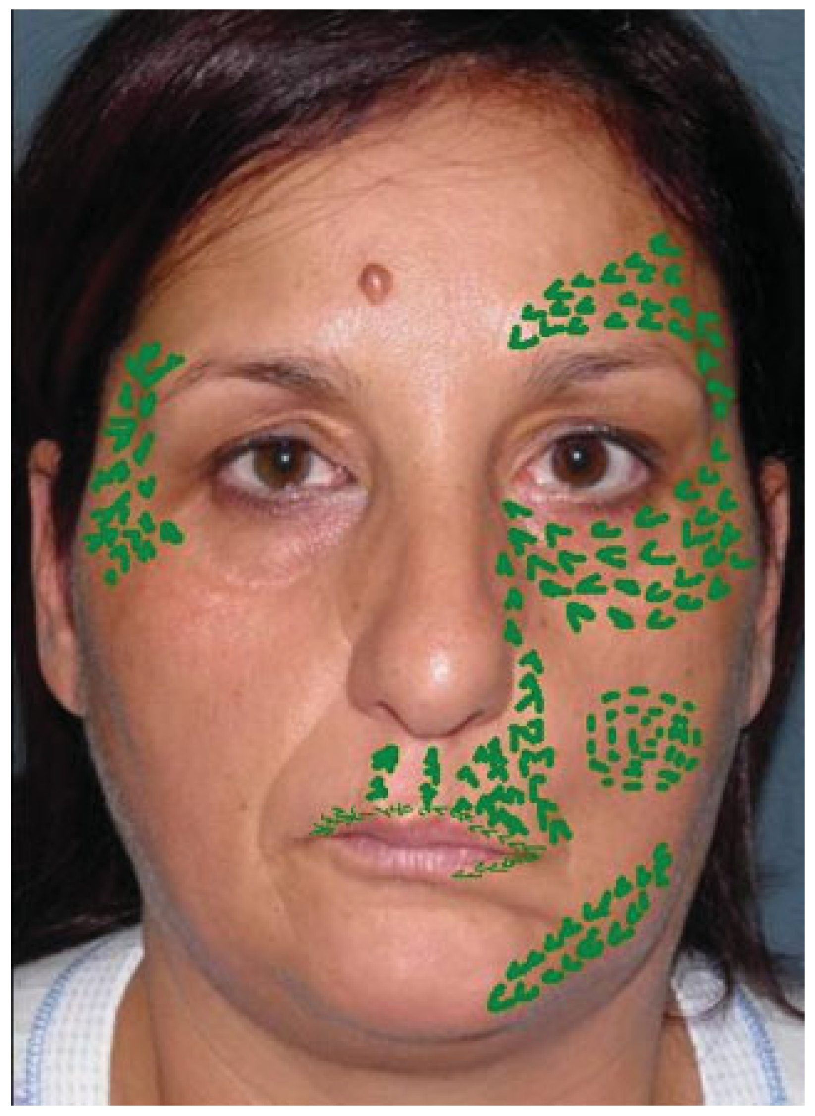
Figure 1. Preoperative program with the injection areas outlined.
Cmtr 07 00016 g001
Figure 2. Frontal and worm’s-eye views of a patient with unilateral right cleft lip and palate sequelae. (A) The patient before SFG. Depression and disharmony of the upper vermillius and the missing philtrum. (B) 2 years after SFG with recovery of the cupid bow and fullness of the perioral area. SFG, structural fat grafting.
Cmtr 07 00016 g002
Figure 3. Treacher Collins Syndrome. (A) The patient at age 6 before any reconstructive procedure. (B) After orbital bony reconstruction with soft tissue deficiency. (C) After SFG with improvement of the orbital-zygomatic region and temporal area. SFG, structural fat grafting.
Cmtr 07 00016 g003
Figure 4. (A) Patient with trauma sequela. (B) After SFG with younger appearance well defined jaw line and improvement of the orbital, zygomatic, cheek area, and the skin quality and texture. SFG, structural fat grafting.
Cmtr 07 00016 g004
Figure 5. (A) Left facial palsy. (B) Result after blink restoration. SFG was performed as a later stage with improvement of facial volumes and texture. SFG, structural fat grafting.
Cmtr 07 00016 g005