Next Article in Journal
Combined Orbital Floor and Medial Wall Fractures Involving the Inferomedial Strut: Repair Technique and Case Series Using Preshaped Porous Polyethylene/Titanium Implants
Previous Article in Journal
Pathologic Fractures in Bisphosphonate-Related Osteonecrosis of the Jaw—Review of the Literature and Review of Our Own Cases
 
 
Craniomaxillofacial Trauma & Reconstruction is published by MDPI from Volume 18 Issue 1 (2025). Previous articles were published by another publisher in Open Access under a CC-BY (or CC-BY-NC-ND) licence, and they are hosted by MDPI on mdpi.com as a courtesy and upon agreement with Sage.
Review

Frontal Sinus Fractures: A Conservative Shift

by
William M. Weathers
*,
Erik M. Wolfswinkel
,
Daniel A. Hatef
,
Edward I. Lee
,
Rodger H. Brown
and
Larry H. Hollier, Jr.
Department of Plastic Surgery, Baylor College of Medicine, 6701 Fannin St., Suite 610, Houston, TX 77030-2399, USA
*
Author to whom correspondence should be addressed.
Craniomaxillofac. Trauma Reconstr. 2013, 6(3), 155-160; https://doi.org/10.1055/s-0033-1349210
Submission received: 7 November 2012 / Revised: 9 November 2012 / Accepted: 9 November 2012 / Published: 26 June 2013

Abstract

:
This article reflects on the changing management of frontal sinus fractures. Severity of these injuries has decreased tremendously since the universal adoption of seat belts and air bags. Recently, there has been a shift from aggressive surgical management to more conservative management strategies, some forgoing surgery all together. New tech-nologies, such as bioabsorbable plates and endoscopic sinus surgery, are leading the way in improved surgical management strategies and offer promising alternatives to the more traditional approaches.

Frontal sinus fractures are relatively uncommon, associated with only 5 to 12% of all facial injuries.[1,2] Majority of these injuries are secondary to assault and motor vehicle accidents (MVA) [3]. Airbags and safety belts have led to a decrease in severity and frequency of MVA-related frontal sinus fractures over the past 30 years [4]. Most of the guidelines we use for management were derived from old studies where the inju-ries were typically more devastating. Decreased severity of injuries and advancements in technology may give us the opportunity to employ more conservative approaches to fracture management.
Studies by Rohrich and Hollier and Manson attempted to establish guidelines for modern management of frontal sinus fractures [2,5,6].With improvement in multiple technologies, the reconstructive surgeons’ armamentarium has grown over the past 20 years. This article highlights the advancements in frontal sinus fracture management and documents the trend from aggressive to conservative management strategies.

History

Management of frontal sinus fractures has evolved tremen-dously over the past 100 years. The early surgical techniques for management of frontal sinus fractures were derived from the treatment of frontal sinusitis and resultant complications, such as mucopyocele. The ablation technique was used for management of sinusitis as early as 1898. This procedure involved permanent removal of the anterior table, resulting in significant disfigurement [7,8]. The osteoplastic flap, created in 1955, represented a significant advancement in frontal sinus management. This flap removed the frontal wall of the sinus along with a pericranial pedicle. This allowed replace-ment of the bone after exploration of the sinus and resulted in improved cosmesis [9]. This flap was modified for use in frontal sinus trauma and has been associated with minimal morbidi-ty [10,11,12,13]. These procedures paved the way for open reduction and internal fixation (ORIF), sinus obliteration, and cranial-ization of the frontal sinus.
Seatbelts and airbags have changed the nature and inci-dence of frontal sinus fractures. Until around 1986, 70% of frontal sinus fractures were caused by MVA [14]. This decreased significantly to around 52% in 2002, with a larger proportion attributable to assault [4]. Over this same time period, a de-crease in severity of frontal sinus fractures was observed. “Through and through” injuries with violation of the anterior cranial fossa fell from 40 to 11% from 1987 to 2002. Improve-ments in motor vehicle safety standards have contributed significantly to the decline in facial trauma. Roden et al, found that facial fractures from motor vehicle collisions decreased from 40 to 27% from 2005 to 2010 [15]. As the incidence has declined, experience in caring for these injuries has also dropped. Truly devastating frontal sinus fractures are rarely seen. Airbags and safety belts have lessened the injuries sustained in car crashes and aggravated assault cannot generate the same destructive energy as a car crash. This decrease in severity has led to the increased use of conserva-tive management for frontal sinus injuries.

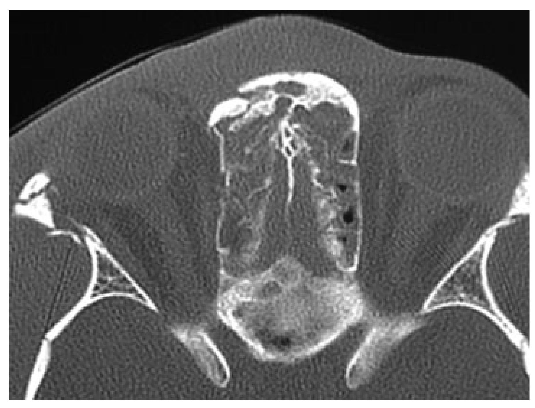
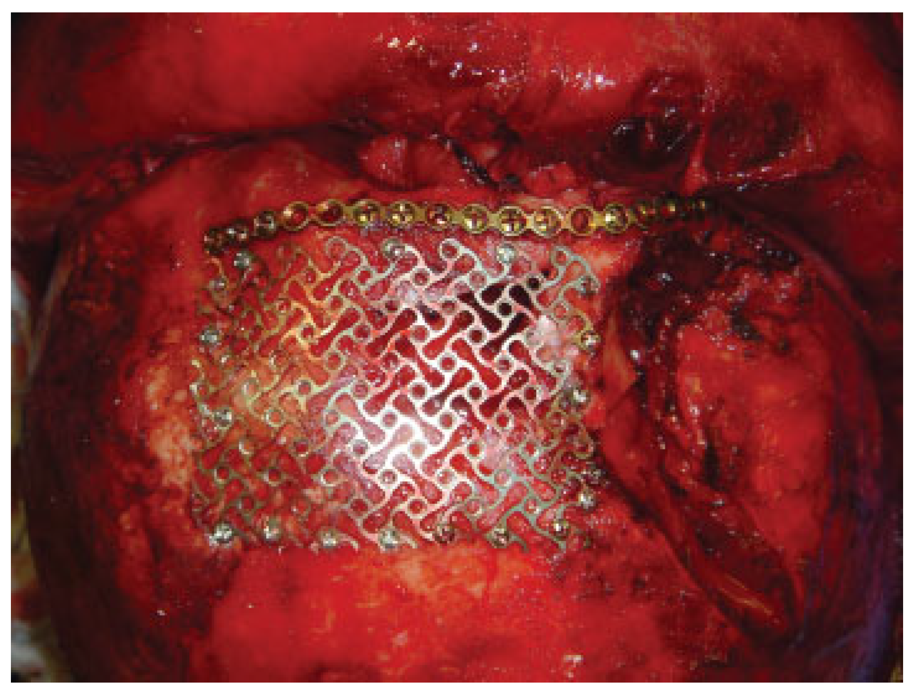
Anterior Table Fractures

The principles of management of anterior table fractures have changed very little over the years. New technologies and techniques have expanded the surgeon’s tool box for restora-tion of forehead cosmesis. Since the primary aim of surgery in isolated anterior table fractures is cosmetic, adequate counseling of the patient regarding observation and various surgical options should be undertaken.
Isolated anterior table fractures are the most common frontal sinus injuries, representing one-third of all frontal sinus fractures [16]. The decision to operate is dependent on two things: (1) amount of aesthetic deformity and (2) involve-ment of the nasofrontal duct [2,7,17]. Duct involvement should be assumed if the fracture involves the ethmoids, medial superior orbital rim or orbital roof (Figure 1) [2,7]. Minimally displaced anterior table fractures can be observed if there is no involvement of the nasofrontal duct [7,18]. For simple frac-tures without duct involvement, surgical correction can be left to patient preference and the desire to restore forehead contour.
ORIF of isolated anterior table injuries is the most com-monly employed treatment. Microplates have been used for fixation of fracture fragments, including severely comminut-ed fractures, with great success (Figure 2) [19]. Despite their success, these metallic microplates have some drawbacks, such as palpability, migration, and extrusion [20]. The develop-ment of bioabsorbable plates for fracture fixation may help address these issues while still providing rigidity and strength during the healing process. Bioabsorbable plates have been shown to be effective for anterior table fracture fixation in several limited studies[21,22]. Kim et al used bio-absorbable mesh plates for fixation of 14 patients. At a mean follow-up of 17.5 months there were no reported complica-tions. The authors state that mesh bioabsorbables are easier to work with than the traditional bioabsorbable plate and screw system. These meshes allow the surgeon to place a screw anywhere within the implant allowing easy fixation of complex fractures [21]. Despite the benefits of bioabsorbable plates, there are pitfalls that the surgeon should be aware of. These plates have been associated with late-onset sterile abscess and granuloma formation [23,24,25]. The thick convention-al bioabsorbable plates can be difficult to handle and use in the frontal sinus region [21]. There are pros and cons to both bioabsorbable and metallic plates. Ultimately, the type of plate used in repair of frontal sinus fractures is determined by surgeon preference.
Advancements in endoscopic surgery have led to promis-ing minimally invasive techniques for simple anterior table fracture fixation. Simple anterior table fractures have been successfully reduced and fixated using endoscopes [26]. This technique was first described in 1996 by Graham and Spring [27]. Endoscopic fixation of anterior table injuries is based on the endoscopic brow lift procedure. One additional incision is made over the fracture for placement of a screw for elevation of the fracture fragments [28,29]. This technique is advantageous since it avoids the coronal incisions used in ORIF. Coronal incisions have been associated with alopecia, paresthesias, and temporal wasting [29]. Coronal incisions can be avoided in open frontal sinus fractures as well. The laceration overlying the frontal sinus fracture can be extend-ed laterally to provide adequate exposure with fixation performed through the laceration [30].

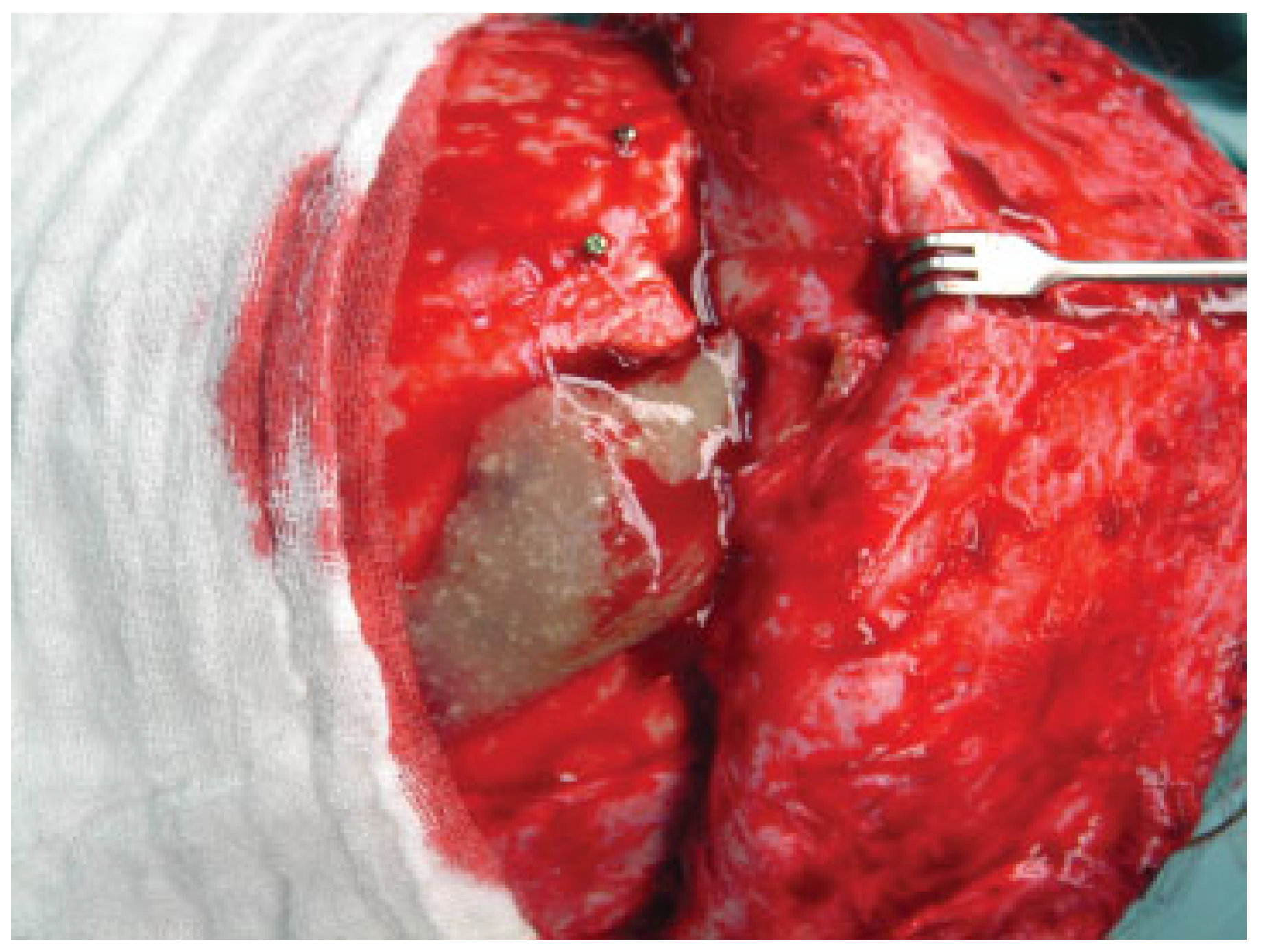
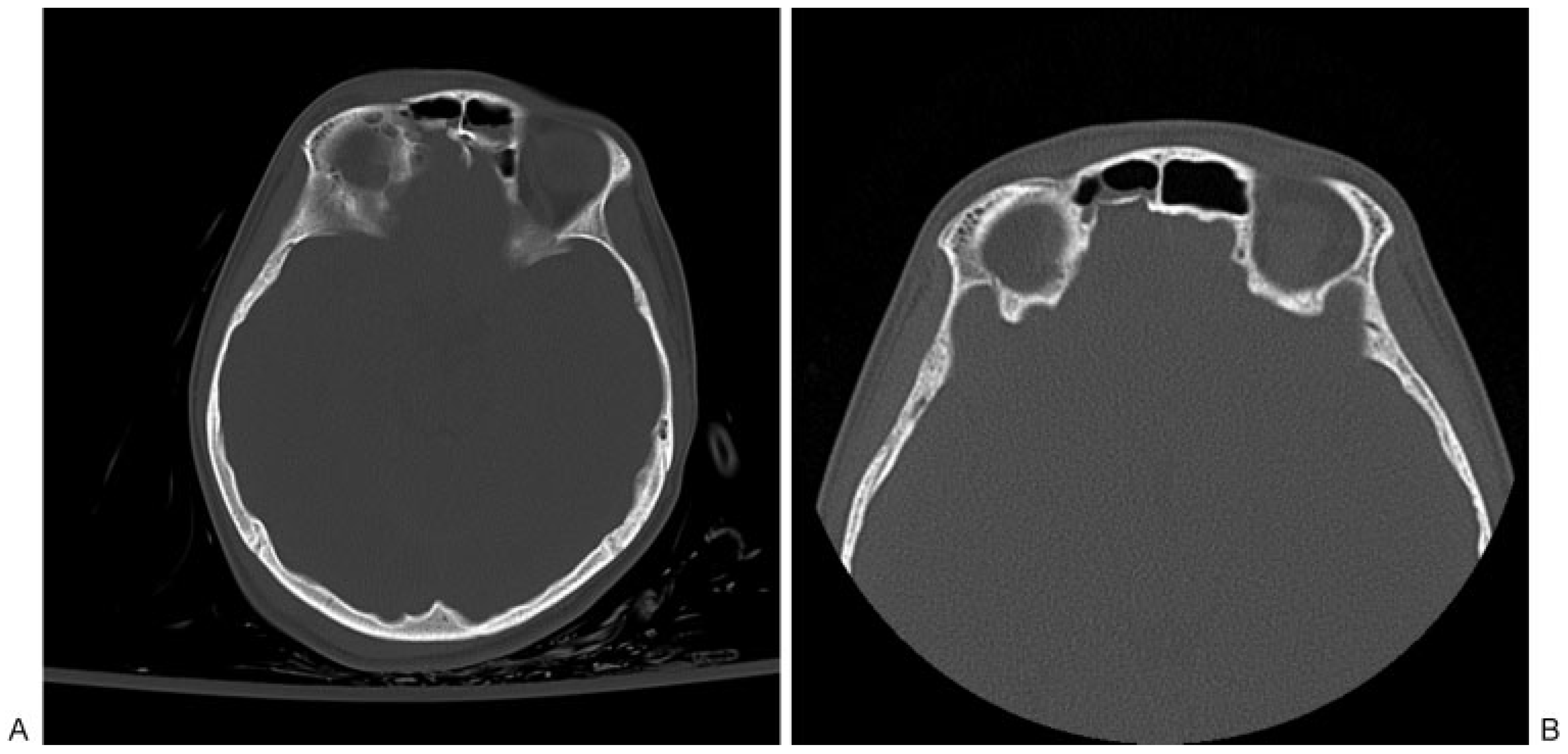
Posterior Table Fractures

Posterior table fractures are frequently associated with severe anterior table, fovea ethmoidalis, and cribriform plate frac-tures [5]. The anterior table can withstand from 800 to 2200 lb of force before fracture, making it one of the strongest bones of the face [2,31]. This quality acts as protection for the posterior table, adjacent dura, and brain parenchyma in low-velocity injuries. Only with a substantial amount of force can injuries to the posterior table occur. As a result of this, they are often associated with intracranial injuries, dural tears, and other injuries associated with high-impact trauma [14,31,32,33,34]. The major sequelae of a dural tear and a posterior table fracture is the creation of a communication between the once isolated intracranial contents and the sinuses. This injury creates a pathway for infections to ascend into the intracranial region and has historically required surgical intervention [2,7,14].
Complications from this injury have been classified as early (< 6 mo) and late (≥ 6 mo). Early complications include sinusitis, cerebrospinal fluid (CSF) leaks and meningitis. Late complications include mucoceles, mucopyoceles, and brain abscesses (Figure 3) [2,35]. The goal of management is to prevent these early and late complications, but the best way to do this is under considerable debate.
CSF leaks are some of the most common problems follow-ing combined anterior and posterior table fractures. After 7 days, persistent CSF leaks increase the likelihood of menin-gitis [36]. This is important since meningitis can be easily missed in patients with severe head trauma. Therefore, persistent CSF leaks are generally treated aggressively with cranialization. Rohrich and Hollier (1992) came up with an algorithm for cranialization that is still used today. They recommend urgent cranialization for posterior table fractures with severe com-minution, displacement greater than one table width, and a CSF leak [2]. Posterior table fractures with CSF leaks in the absence of these other findings are generally observed for 7 days before attempting cranialization.2,35 It has been ob-served that many of these leaks will resolve on their own. A minimally or nondisplaced posterior table fracture without CSF leak requires no intervention [2]. Associated nasofrontal duct injuries and anterior wall fractures are treated as they would be typically.
Recently, an argument has been made for conservative management of these injuries despite meeting criteria for cranialization. Cranialization requires craniotomy, removal of the posterior table, removal of all sinus mucosa, closure of the dura, and closure of any communication with the sinonasal tract (Figure 4) [2,33,34,37,38,39,40]. This procedure is aggressive and not without risk. It requires a plastic surgeon, a neuro-surgeon, and significant operative time. Recently, Choi et al reviewed 59 patients with posterior table fractures. Of these, seven patients met the above criteria for cranialization. However, none of these seven patients underwent craniali-zation and there were no complications at a mean follow-up of 92 days [3]. This article demonstrates that conservative management could be a safe and effective strategy for pa-tients who traditionally meet the requirements for craniali-zation. A significant limitation of this approach is that close follow-up is necessary to ensure that patients do not develop complications (Figure 5). This study promotes a significant departure from the established school of thought. It replaces the idea of radical surgery with a watch and wait approach for severe posterior table fractures with CSF leaks. The incidence of long term complications, such as mucoceles, in these patients remains to be seen and further follow-up is warranted.

Nasofrontal Duct Injury

Nasofrontal duct injuries are seen with approximately 25 to 50% of anterior table fractures that involve the supraorbital rim or the nasal–orbital–ethmoidal region.11–13,41 Patency of the nasofrontal duct is paramount in preventing serious early and late complications, such as formation of a mucopyocele or infection [18,42]. Stents have been used with limited success and up to 30% fail to maintain patency after removal.2,40 The current mainstay of treatment is to obliterate the nasofrontal duct and frontal sinus, isolating the frontal sinus from the sinus tract. The principles of this technique are extirpation of all frontal sinus mucosa using a microscope, removal of the inner cortex of the sinus wall, and permanent occlusion of the nasofrontal duct with muscle, fascia or bone chips [2,43]. Once these steps are done, obliteration of the frontal sinus is completed by filling the dead space of the sinus cavity. It is believed that filling the frontal sinus may mitigate the risk of infection and prevent accumulation of serosanguineous fluid [9,19].
Spontaneous osteogenesis, bone, bone substitutes, autol-ogous fat, pericranial flaps, and muscle have all been used to effectively fill the frontal sinus [44,45,46,47,48,49]. Autologous fat is one of the most studied and commonly used materials [9,50,51,52]. It is thought that some of the fat graft becomes vascularized by the blood supply of the frontal sinus after removal of the inner bony cortex [9]. This viable fat tissue may be more resistant to infection while the nonviable fat graft becomes fibrous tissue filling the dead space of the frontal sinus [9,53]. Spontaneous osteogenesis, another commonly used technique, is based on the principle that a cavity surrounded by healthy, bleeding bone will fill in with new bone obliterating the dead space [19,45,54,55]. Animal studies have not demonstrated a significant difference between the use of fat, muscle, bone or spontaneous osteogenesis for filling the frontal sinus dead space [45]. Human studies are limited to noncomparative stud-ies without uniform operative techniques. Since no clear superior choice has emerged in the literature, filling material is usually based on the surgeon’s preference. Use of synthetic bone substitutes should generally never be used, especially in acute or contaminated fractures. The risk of infection and reoperation is too high [7,47,56,57,58,59,60]. Advancements in endoscopic sinus surgery have led to promising new techniques in limited situations [61,62,63].Smith et al described a new algorithm for the treatment of simple anterior table fractures with nasofrontal duct injury. In this algorithm, open repair and internal fixation was performed without obliteration. Patients were closely followed under-going serial computed tomography and 4 weeks of antibiotic therapy. The majority of the patients they observed sponta-neously regained patency of the nasofrontal duct. Two pa-tients underwent endoscopic sinus surgery for persistent frontal sinus outflow obstruction without significant compli-cations or restenosis at 21 and 25 months after injury [61]. Despite these results, Smith’s work is still a cautionary tale. Reliance on patient follow-up is a big limitation seen with these more conservative approaches. Also, manipulation of the nasofrontal duct with an endoscope can lead to edema in the acute setting and scarring/stenosis in the long term [64]. Ung et al described the use of endopscopic surgery for obliteration of the frontal sinus in chronic frontal sinusitis. They also compared this technique to the traditional open technique and found decreased morbidity using the endoscopic tech-nique [63]. How endoscopic obliteration will fare in the setting of trauma remains to be seen and further studies are still needed. The watch and wait approach with close follow-up and intervention as needed is becoming more mainstream for all types of frontal sinus injuries.
In summary, the current management of frontal sinus fractures with associated nasofrontal duct injury calls for obliteration of the frontal sinus. Untreated obstruction of the nasofrontal duct, incomplete removal of sinus mucosa, and failure to close the nasofrontal duct can lead to mucopyocele and mucocele formation, even years after the injury [19,40,43,45]. The basic tenets of frontal sinus obliteration described by Rohrich and Hollier (1992) are still the guiding principles in the treatment of nasofrontal duct injuries. However, watch and wait approaches for management of frontal sinus frac-tures are promising when close follow-up can be assured.

Conclusions

The management of frontal sinus fractures has evolved tremendously over the years. Multiple new techniques and technologies have the potential to revolutionize frontal sinus fracture management. Decreased reliance on major procedures, such as cranialization, with increased utiliza-tion of minimally invasive techniques will provide surgeons more options to safely and effectively care for these injuries. Endosopic procedures are promising and will likely lead the way. Not only are the techniques and technologies chang-ing, but so are the management strategies. Careful patient selection to ensure close follow-up may allow the utiliza-tion of conservative management strategies when appro-priate. The mechanisms of injury have changed over the years as have the severity of injuries. New treatment algorithms should be established to include these newer techniques and devices.

References

  1. May, M.; Ogura, J.H.; Schramm, V. Nasofrontal duct in frontal sinus fractures. Arch. Otolaryngol. 1970, 92, 534–538. [Google Scholar] [CrossRef] [PubMed]
  2. Rohrich, R.J.; Hollier, L.H. Management of frontal sinus fractures. Changing concepts. Clin. Plast. Surg. 1992, 19, 219–232. [Google Scholar] [CrossRef] [PubMed]
  3. Choi, M.; Li, Y.; Shapiro, S.A.; Havlik, R.J.; Flores, R.L. A 10-year review of frontal sinus fractures: clinical outcomes of conservative manage-ment of posterior table fractures. Plast. Reconstr. Surg. 2012, 130, 399–406. [Google Scholar] [CrossRef] [PubMed]
  4. Strong, E.B.; Pahlavan, N.; Saito, D. Frontal sinus fractures: a 28-year retrospective review. Otolaryngol. Head. Neck Surg. 2006, 135, 774–779. [Google Scholar] [CrossRef]
  5. Manson, P.N.; Crawley, W.A.; Hoopes, J.E. Frontal cranioplasty: risk factors and choice of cranial vault reconstructive material. Plast. Reconstr. Surg. 1986, 77, 888–904. [Google Scholar] [CrossRef]
  6. Manson, P.N.; Markowitz, B.; Mirvis, S.; Dunham, M.; Yaremchuk, M. Toward CT-based facial fracture treatment. Plast. Reconstr. Surg. 1990, 85, 202–212. [Google Scholar] [CrossRef]
  7. Manolidis, S.; Hollier, L.H., Jr. Management of frontal sinus fractures. Plast. Reconstr. Surg. 2007, 120 (Suppl. S2), 32S–48S. [Google Scholar] [CrossRef]
  8. Jacobs, J.B. 100 years of frontal sinus surgery. Laryngoscope 1997, 107 Pt. 2, 1–36. [Google Scholar] [CrossRef]
  9. Bergara, A.R.; Itoiz, A.O. Present state of the surgical treatment of chronic frontal sinusitis. AMA Arch. Otolaryngol. 1955, 61, 616–628. [Google Scholar] [CrossRef]
  10. Stanley, R.B., Jr. Management of frontal sinus fractures. Facial Plast. Surg. 1988, 5, 231–235. [Google Scholar] [CrossRef]
  11. Stanley, R.B., Jr. Fractures of the frontal sinus. Clin. Plast. Surg. 1989, 16, 115–123. [Google Scholar] [PubMed]
  12. Stanley, R.B., Jr. Management of severe frontobasilar skull fractures. Otolaryngol. Clin. North. Am. 1991, 24, 139–150. [Google Scholar] [CrossRef]
  13. Stanley, R.B., Jr; Becker, T.S. Injuries of the nasofrontal orifices in frontal sinus fractures. Laryngoscope 1987, 97, 728–731. [Google Scholar] [CrossRef]
  14. Wallis, A.; Donald, P.J. Frontal sinus fractures: a review of 72 cases. Laryngoscope 1988, 98 Pt. 1, 593–598. [Google Scholar]
  15. Roden, K.S.; Tong, W.; Surrusco, M.; Shockley, W.W.; Van Aalst, J.A.; Hultman, C.S. Changing characteristics of facial fractures treated at a regional, level 1 trauma center, from 2005 to 2010: an assessment of patient demographics, referral patterns, etiology of injury, anatomic location, and clinical outcomes. Ann Plast Surg 2012, 68, 461–466. [Google Scholar] [PubMed]
  16. Sataloff, R.T.; Sariego, J.; Myers, D.L.; Richter, H.J. Surgical management of the frontal sinus. Neurosurgery 1984, 15, 593–596. [Google Scholar] [PubMed]
  17. Rohrich, R.J.; Hollier, L.H. The role of the nasofrontal duct in frontal sinus fracture management. J. Craniomaxillofac Trauma. 1996, 2, 31–40. [Google Scholar]
  18. Metzinger, S.E.; Guerra, A.B.; Garcia, R.E. Frontal sinus fractures: management guidelines. Facial Plast. Surg. 2005, 21, 199–206. [Google Scholar]
  19. Tiwari, P.; Higuera, S.; Thornton, J.; Hollier, L.H. The management of frontal sinus fractures. J. Oral. Maxillofac. Surg. 2005, 63, 1354–1360. [Google Scholar]
  20. Berryhill, W.E.; Rimell, F.L.; Ness, J.; Marentette, L.; Haines, S.J. Fate of rigid fixation in pediatric craniofacial surgery. Otolaryngol. Head. Neck Surg. 1999, 121, 269–273. [Google Scholar]
  21. Kim, C.H.; Kim, D.H.; Kim, H.; Song, S.Y. Treatment of frontal sinus fracture using bioabsorbable mesh plates. J. Craniofac Surg. 2012, 23, 549–551. [Google Scholar] [PubMed]
  22. Bhanot, S.; Alex, J.C.; Lowlicht, R.A.; Ross, D.A.; Sasaki, C.T. The efficacy of resorbable plates in head and neck reconstruction. Laryngoscope 2002, 112, 890–898. [Google Scholar] [PubMed]
  23. Bergsma, J.E.; de Bruijn, W.C.; Rozema, F.R.; Bos, R.R.; Boering, G. Late degradation tissue response to poly(L-lactide) bone plates and screws. Biomaterials 1995, 16, 25–31. [Google Scholar]
  24. Eppley, B.L.; Morales, L.; Wood, R.; et al. Resorbable PLLA-PGA plate and screw fixation in pediatric craniofacial surgery: clinical experience in 1883 patients. Plast Reconstr Surg 2004, 114, 850–856. [Google Scholar]
  25. Landes, C.A.; Ballon, A.; Roth, C. Maxillary and mandibular osteosyn-theses with PLGA and P(L/DL)LA implants: a 5-year inpatient biocompatibility and degradation experience. Plast. Reconstr. Surg. 2006, 117, 2347–2360. [Google Scholar] [PubMed]
  26. Strong, E.B.; Buchalter, G.M.; Moulthrop, T.H. Endoscopic repair of isolated anterior table frontal sinus fractures. Arch. Facial Plast. Surg. 2003, 5, 514–521. [Google Scholar]
  27. Graham, H.D.I.I.I.; Spring, P. Endoscopic repair of frontal sinus fracture: case report. J. Craniomaxillofac Trauma. 1996, 2, 52–55. [Google Scholar]
  28. Chen, D.J.; Chen, C.T.; Chen, Y.R.; Feng, G.M. Endoscopically assisted repair of frontal sinus fracture. J. Trauma. 2003, 55, 378–382. [Google Scholar]
  29. Mueller, R. Endoscopic treatment of facial fractures. Facial Plast. Surg. 2008, 24, 78–91. [Google Scholar]
  30. Gonty, A.A.; Marciani, R.D.; Adornato, D.C. Management of frontal sinus fractures: a review of 33 cases. J. Oral. Maxillofac. Surg. 1999, 57, 372–379. [Google Scholar]
  31. Nahum, A.M. The biomechanics of maxillofacial trauma. Clin. Plast. Surg. 1975, 2, 59–64. [Google Scholar] [PubMed]
  32. Duvall, A.J.I.I.I.; Porto, D.P.; Lyons, D.; Boies, L.R., Jr. Frontal sinus fractures. Analysis of treatment results. Arch. Otolaryngol. Head. Neck Surg. 1987, 113, 933–935. [Google Scholar]
  33. Gerbino, G.; Roccia, F.; Benech, A.; Caldarelli, C. Analysis of 158 frontal sinus fractures: current surgical management and complications. J. Craniomaxillofac Surg. 2000, 28, 133–139. [Google Scholar] [PubMed]
  34. Gossman, D.G.; Archer, S.M.; Arosarena, O. Management of frontal sinus fractures: a review of 96 cases. Laryngoscope 2006, 116, 1357–1362. [Google Scholar] [PubMed]
  35. Chen, K.T.; Chen, C.T.; Mardini, S.; Tsay, P.K.; Chen, Y.R. Frontal sinus fractures: a treatment algorithm and assessment of outcomes based on 78 clinical cases. Plast. Reconstr. Surg. 2006, 118, 457–468. [Google Scholar]
  36. Appelbaum, E. Meningitis following trauma to the head and face. JAMA 1960, 173, 1818–1822. [Google Scholar]
  37. Bell, R.B. Management of frontal sinus fractures. Oral. Maxillofac. Surg. Clin. North. Am. 2009, 21, 227–242. [Google Scholar]
  38. Day, T.A.; Meehan, R.; Stucker, F.J.; Nanda, A. Management of frontal sinus fractures with posterior table involvement: a retrospective study. J. Craniomaxillofac Trauma. 1998, 4, 6–9. [Google Scholar]
  39. Donald, P.J. Frontal sinus ablation by cranialization. Report of 21 cases. Arch. Otolaryngol. 1982, 108, 142–146. [Google Scholar]
  40. Luce, E.A. Frontal sinus fractures: guidelines to management. Plast. Reconstr. Surg. 1987, 80, 500–510. [Google Scholar]
  41. Stevens, M.; Kline, S.N. Management of frontal sinus fractures. J. Craniomaxillofac Trauma. 1995, 1, 29–37. [Google Scholar]
  42. Rodriguez, E.D.; Stanwix, M.G.; Nam, A.J.; et al. Twenty-six-year experi-ence treating frontal sinus fractures: a novel algorithm based on anatomical fracture pattern and failure of conventional techni-ques. Plast. Reconstr. Surg. 2008, 122, 1850–1866. [Google Scholar] [PubMed]
  43. Rohrich, R.J.; Mickel, T.J. Frontal sinus obliteration: in search of the ideal autogenous material. Plast. Reconstr. Surg. 1995, 95, 580–585. [Google Scholar]
  44. Friedman, C.D.; Costantino, P.D.; Jones, K.; Chow, L.C.; Pelzer, H.J.; Sisson GASr Hydroxyapatite cement, I.I. Obliteration and reconstruction of the cat frontal sinus. Arch. Otolaryngol. Head. Neck Surg. 1991, 117, 385–389. [Google Scholar] [PubMed]
  45. Mickel, T.J.; Rohrich, R.J.; Robinson, J.B., Jr. Frontal sinus obliteration: a comparison of fat, muscle, bone, and spontaneous osteoneogen-esis in the cat model. Plast. Reconstr. Surg. 1995, 95, 586–592. [Google Scholar]
  46. Peltola, M.J.; Aitasalo, K.M.; Suonpää, J.T.; Yli-Urpo, A.; Laippala, P.J.; Forsback, A.P. Frontal sinus and skull bone defect obliteration with three synthetic bioactive materials. A comparative study. J. Biomed. Mater. Res. B Appl. Biomater. 2003, 66, 364–372. [Google Scholar] [PubMed]
  47. Ross, D.A.; Marentette, L.J.; Thompson, B.G.; Haller, J.S. Use of hydroxy-apatite bone cement to prevent cerebrospinal fluid leakage through the frontal sinus: technical report. Neurosurgery 1999, 45, 401–402. [Google Scholar]
  48. Weber, R.; Draf, W.; Keerl, R.; et al. Magnetic resonance imaging following fat obliteration of the frontal sinus. Neuroradiology 2002, 44, 52–58. [Google Scholar]
  49. Ducic, Y.; Stone, T.L. Frontal sinus obliteration using a laterally based pedicled pericranial flap. Laryngoscope 1999, 109, 541–545. [Google Scholar]
  50. Montgomery, W.W. The fate of adipose implants in a bony cavity. Laryngoscope 1964, 74, 816–827. [Google Scholar]
  51. Sessions, R.B.; Alford, B.R.; Stratton, C.; Ainsworth, J.Z.; Shill, O. Current concepts of frontal sinus surgery: an appraisal of the osteoplastic flap-fat obliteration operation. Laryngoscope 1972, 82, 918–930. [Google Scholar] [CrossRef]
  52. Montgomery, W.W.; Pierce, D.L. Anterior osteoplastic fat obliteration for frontal sinus: Clinical experience and animal studies. Trans. Am. Acad. Ophthalmol. Otolaryngol. 1963, 67, 46–57. [Google Scholar]
  53. Peer, L.A. Loss of weight and volume in human fat grafts. Plast. Reconstr. Surg. 1950, 5, 217–230. [Google Scholar] [CrossRef]
  54. MacBeth, R. The osteoplastic operation for chronic infection of the frontal sinus. J. Laryngol. Otol. 1954, 68, 465–477. [Google Scholar] [CrossRef]
  55. Bosley, W.R. Osteoplastic obliteration of the frontal sinuses. A review of 100 patients. Laryngoscope 1972, 82, 1463–1476. [Google Scholar] [CrossRef] [PubMed]
  56. Xie, C.; Mehendale, N.; Barrett, D.; Bui, C.J.; Metzinger, S.E. 30-year retrospective review of frontal sinus fractures: The Charity Hospi-tal experience. J. Craniomaxillofac Trauma. 2000, 6, 7–15. [Google Scholar] [PubMed]
  57. Dujovny, M.; Aviles, A.; Anger, C. An innovative approach for cranio-plasty using hydroxyapatite cement. Surg. Neurol. 1997, 48, 294–297. [Google Scholar] [CrossRef]
  58. Kveton, J.F.; Friedman, C.D.; Piepmeier, J.M.; Costantino, P.D. Reconstruc-tion of suboccipital craniectomy defects with hydroxyapatite cement: a preliminary report. Laryngoscope 1995, 105, 156–159. [Google Scholar] [CrossRef]
  59. Maniker, A.; Cantrell, S.; Vaicys, C. Failure of hydroxyapatite cement to set in repair of a cranial defect: case report. Neurosurgery 1998, 43, 953–954. [Google Scholar] [CrossRef]
  60. Snyderman, C.H.; Scioscia, K.; Carrau, R.L.; Weissman, J.L. Hydroxyapa-tite: an alternative method of frontal sinus obliteration. Otolar-yngol Clin. North. Am. 2001, 34, 179–191. [Google Scholar] [CrossRef]
  61. Smith, T.L.; Han, J.K.; Loehrl, T.A.; Rhee, J.S. Endoscopic management of the frontal recess in frontal sinus fractures: a shift in the para-digm? Laryngoscope 2002, 112, 784–790. [Google Scholar] [PubMed]
  62. Thong, J.F.; Lee, J. Single-stage strategy for management of anterior table frontal sinus fractures involving the frontal sinus outflow tract. Eur. J. Plast. Surg. 2011, 34, 299–303. [Google Scholar] [CrossRef]
  63. Ung, F.; Sindwani, R.; Metson, R. Endoscopic frontal sinus obliteration: a new technique for the treatment of chronic frontal sinusitis. Otolaryngol. Head. Neck Surg. 2005, 133, 551–555. [Google Scholar] [PubMed]
  64. Smith, T.L.; Han, J.K.; Loehrl, T.A.; Rhee, J.S. Endoscopic management of the frontal recess in frontal sinus fractures: a shift in the para-digm? Laryngoscope 2002, 112, 784–790. [Google Scholar]
Figure 1. Computed tomography slice demonstrating involvement of the medial orbital wall and the ethmoids associated with a frontal sinus fracture. It should be assumed that the nasofrontal duct is injured with these findings.
Cmtr 06 00025 g001
Figure 2. Microplate fixation of an anterior table fracture.
Cmtr 06 00025 g002
Figure 3. Operative photo of a mucocele that formed a year after initial repair of frontal sinus fracture.
Cmtr 06 00025 g003
Figure 4. (A) Intraoperative picture of a cranialization procedure before exclusion of the sinonasal tract. (B) Intraoperative picture demonstrating exclusion of the sinonasal tract using a pericranial flap.
Cmtr 06 00025 g004
Figure 5. Computed tomography (CT) scan follow-up after conservative management following anterior and posterior table injuries. (A) Initial CT scan at the time of injury. (B) A 3-month follow-up CT scan demonstrating no complications and adequate bony healing. If the frontal sinus is opacified, this may indicate an occult nasofrontal duct injury.
Cmtr 06 00025 g005

