Abstract
A unique case of a vertical fracture of the mandibular posterior ramus border secondary to the stress of the rigid internal fixation material is described in this study. We think that the surgeon’s experience and awareness play a key role in avoidance of such a complication.
The use of rigid fixation with orthognathic surgery was greeted by both excitement and healthy concern when it began to find its way into the literature ~25 years ago [1,2]. However, there are several studies about the complications related to the tensile and stress forces secondary to the use of rigid internal fixation materials [3]. Here we report a unique case of a vertical fracture of the mandibular posterior ramus border secondary to the stress of the rigid internal fixation material.
Case Report
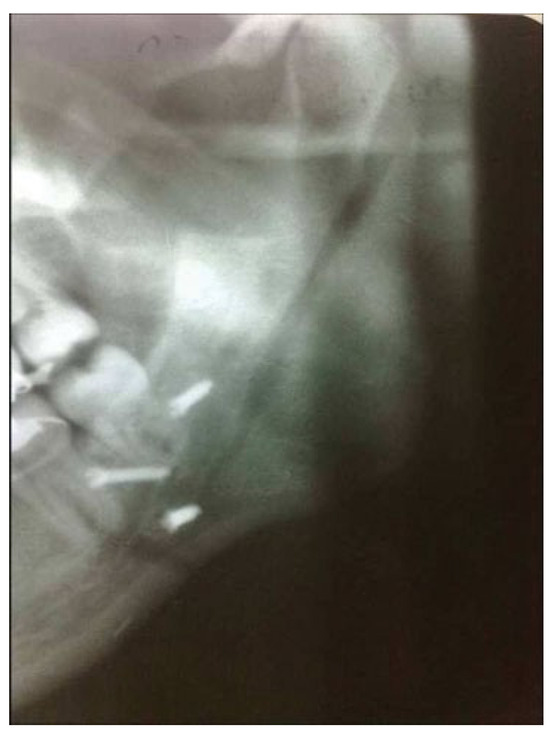
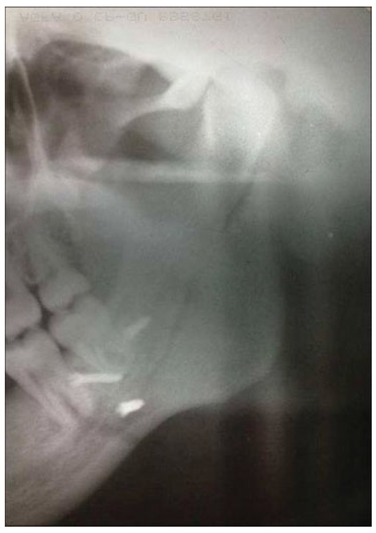
A 20-year-old man was admitted to our department with the complaints of difficulty in chewing and distorted facial view. After consultations with the Department of Orthodontics, it was decided to perform a mandibular set back procedure. The patient had an uneventful surgery. However, during the routine radiological control examination on the first postoperative day, a vertical fracture line on the mandibular ramus was observed (Figure 1). The fracture line has extended from the titanium screw located next to the border of the mandible to the subcondylar region and the authors have hypothesized that the patient actively moved postoperatively to cause the fracture, thus the surgeons didn’t notice a sudden loosening of the titanium screw during placement. The patient was examined very carefully. The fragments were stable and the fracture was left untreated. At the end of the second postoperative month, it was detected that the osseous healing was almost completed (Figure 2).

Figure 1.
The radiological control examination on the first postoperative day revealed the presence of a vertical fracture line on the left mandibular ramus.

Figure 2.
At 2 months postsurgery, the osseous healing at the fracture site was almost completed.
Discussion
The use of rigid fixation of bony segments in orthognathic surgery has become a standard of care. However, undesirable forces secondary to rigid fixation materials can result in condylar changes [3] or osseous complications [4,5]. In recent years, because of the problems with various methods of rigid osteosynthesis, semirigid fixation methods have been developed to stabilize the osteotomized fragments for bone healing with enough flexibility to avoid the problems arising from rigid fixation [6].
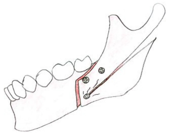
The location of the lateral osteotomy cut during sagittal split osteotomy varies according to the surgeon’s experience and preference, and no consensus has been reached regarding the ideal and most safe location from the perspective of biomechanics (Figure 3). In a study of Takahashi et al. [7] Trauner-Obwegeser, Obwegeser, and Obwegeser-Dal Pont lateral osteotomy designs for bilateral sagittal split osteotomy were compared and biomechanical evaluation with three-dimensional finite element analysis was performed. According to the results of their study, Obwegeser-Dal Pont method showed the least central incisor displacement, least maximum bone mechanical stress in the screw vicinity. One might conclude that the fracture could have propagated during creation of the osteotomy rather than as a result of the screw placement. However, the fracture line is far from the bone cuts created for SSRO (sagittal split ramus osteotomy) and the surgeons did not apply any external force or experience any difficulties during splitting of the mandible. In addition, if an uncontrolled fracture could have propagated during creation of the osteotomy, the screw placed in the fracture line would not be stable and would be noticed by the surgeon. Therefore, we think that, the surgeon’s experience and awareness play a key role in avoidance of such a complication.

Figure 3.
Schematic illustration of the fracture.
Acknowledgments
Competing interests: None declared
Funding: None
Ethical approval: Not required
References
- O’Connell, J.; Murphy, C.; Ikeagwuani, O.; Adley, C.; Kearns, G. The fate of titanium miniplates and screws used in maxillofacial surgery: A 10 year retrospective study. Int J Oral Maxillofac Surg 2009, 38, 731–735. [Google Scholar] [CrossRef] [PubMed]
- Van Sickels, J.E.; Richardson, D.A. Stability of orthognathic surgery: A review of rigid fixation. Br J Oral Maxillofac Surg 1996, 34, 279–285. [Google Scholar] [CrossRef] [PubMed]
- Chung, I.H.; Yoo, C.K.; Lee, E.K.; et al. Postoperative stability after sagittal split ramus osteotomies for a mandibular setback with monocortical plate fixation or bicortical screw fixation. J Oral Maxillofac Surg 2008, 66, 446–452. [Google Scholar] [CrossRef] [PubMed]
- Herford, A.S.; Hoffman, R.; Demirdji, S.; et al. A comparison of synovial fluid pressure after immediate versus gradual mandibular advancement in the miniature pig. J Oral Maxillofac Surg 2005, 63, 775–785. [Google Scholar] [CrossRef] [PubMed]
- Kim, Y.I.; Cho, B.H.; Jung, Y.H.; Son, W.S.; Park, S.B. Cone-beam computerized tomography evaluation of condylar changes and stability following two-jaw surgery: Le Fort I osteotomy and mandibular setback surgery with rigid fixation. Oral Surg Oral Med Oral Pathol Oral Radiol Endod 2011, 111, 681–687. [Google Scholar] [CrossRef] [PubMed]
- Mavili, M.E.; Canter, H.I.; Saglam-Aydinatay, B. Semirigid fixation of mandible and maxilla in orthognathic surgery: Stability and advantages. Ann Plast Surg 2009, 63, 396–403. [Google Scholar] [CrossRef] [PubMed]
- Takahashi, H.; Moriyama, S.; Furuta, H.; Matsunaga, H.; Sakamoto, Y.; Kikuta, T. Three lateral osteotomy designs for bilateral sagittal split osteotomy: Biomechanical evaluation with three-dimensional finite element analysis. Head Face Med 2010, 6, 4. [Google Scholar] [CrossRef] [PubMed]
© 2012 by the author. The Author(s) 2012.