Next Article in Journal
Reconstruction of the Cranial Vault Contour Using Tissue Expander and Castor Oil Prosthesis
Previous Article in Journal
Injured Anterior Superior Alveolar Nerve Endoscopically Resected Within Maxillary Sinus
 
 
Craniomaxillofacial Trauma & Reconstruction is published by MDPI from Volume 18 Issue 1 (2025). Previous articles were published by another publisher in Open Access under a CC-BY (or CC-BY-NC-ND) licence, and they are hosted by MDPI on mdpi.com as a courtesy and upon agreement with Sage.
Case Report

Concomitant “Ear Bleed and Styloid Fracture”: An Unusual Complication of Impacted Mandibular Third Molar Removal

by
Krishnakumar Raja
1,
Gayathri Gopi
2,
Elavenil Panneerselvam
1,*,
Jegatheesan Ramamoorthy
3,
Guruprasad Thulasi Doss
1 and
Aditi Rajendra Sharma
1
1
Department of Oral and Maxillofacial Surgery, SRM Dental College, Chennai, Tamil Nadu, India
2
Oral and Maxillofacial Surgeon, Private Practice Chennai, Chennai, India
3
Endodontist, Private Practice, Chennai, India
*
Author to whom correspondence should be addressed.
Craniomaxillofac. Trauma Reconstr. 2017, 10(3), 212-215; https://doi.org/10.1055/s-0036-1592086
Submission received: 18 January 2016 / Revised: 30 January 2016 / Accepted: 10 April 2016 / Published: 26 August 2016

Abstract

:
The removal of impacted mandibular third molar is associated with potential complications such as dry socket, paresthesia, uncontrolled socket bleeding, angle fracture, etc., which are commonly encountered in dental practice. This article presents a peculiar case of “ear bleed” concomitant with “isolated styloid” fracture following removal of impacted mandibular third molar, not reported in the literature till date. Ear bleed is a bothersome clinical sign that requires thorough investigation and prompt treatment because it is frequently related to fractures of the skull base. Isolated fracture of the styloid process is rare; its diagnosis, impact on adjacent vital structures, and treatment protocol are less discussed in maxillofacial literature. The case report elucidates the etiopathogenesis of ear bleed and styloid fracture which have great clinical implications. The clinical correlation between the two entities and dental extraction is discussed in this report to guide a dental practitioner in its management.

The most prevalent complications associated with the removal of impacted mandibular third molar are dry socket, bleeding, trismus, dysesthesia, angle fracture, etc. [1]. The following is an unusual clinical report featuring a rare traumatic event in a dental office comprising concurrent “ear bleed and styloid fracture” as a complication of removal of impacted mandibular third molar.
Ear bleed is generally considered an ominous clinical sign that demands prompt diagnosis and astute management [2]. It is commonly linked to fractures of the base of skull [3] and requires immediate attention. Nevertheless, awareness regarding the other less worrying causes of ear bleeding is necessary, to avoid undue apprehension. Only one case of ear bleed following third molar extraction has been reported in the literature so far [4].
The report also features isolated fracture of ipsilateral styloid process during the removal of impacted mandibular molar, which is uncommon. Two cases of isolated styloid fracture in a dental operatory have been reported till date; one which occurred during administration of local anesthesia [5] and the other following extraction [6]. Styloid fractures usually occur secondary to trauma either in isolation or in association with other fractures such as condyle, angle, and body [5,6,7,8]. The sparse incidence of styloid fracture is because it is positioned deeper and angulated favorably that precludes its fracture. However, the styloid process is prone to fracture if it is elongated or shaped abnormally [9,10]. Fractures have also been evidenced due to sudden uncoordinated muscle spasms such as during epileptic seizures, laughter, and coughing [11,12].

Case Report

A 23-year-old female patient reported to a dental practitioner with complaints of pain and swelling in the right lower third molar region which was diagnosed as pericoronitis secondary to an impacted third molar. Radiographic examination revealed a distoangular impacted 48. A decision of surgical removal of the tooth was made and implemented. Incidentally, 30 min postoperatively, bleeding from the ipsilateral ear was noticed which subsided with a pressure pack within the external auditory canal (EAC) for an hour. The following day, the patient visited our institution for further management. Assessment of the patient revealed history of dysphagia, pain in the preauricular, pharyngeal, and retromandibular regions with restricted mouth opening, otalgia, tinnitus, and temporomandibular joint (TMJ) pain. Tenderness in the above-mentioned regions and characteristic increase in pain while turning the head to the affected side were elicited. Orthopantomography (OPG) (Figure 1) demonstrated fracture of right styloid process. Promptly, an otolaryngologist’s opinion was obtained to rule out perforation of the tympanic membrane. A computed tomography (CT) brain was also simultaneously taken and the presence of basal skull fracture was negated. The patient was advised soft diet, analgesics, and restriction in mouth opening for 2 weeks. Complete resolution of symptoms was observed on review.

Discussion

The report presents two peculiar observations following a dental extraction procedure: bleed from the ear and fracture of the ipsilateral styloid process.
Ear bleed, in general, is considered a grave clinical sign because it is commonly related to a traumatic event, especially fractures of the skull base [3]. It therefore mandates critical assessment when encountered. Nevertheless, ear bleed may occur due to other innocuous causes such as ruptured ear drum and lacerations in anterior aspect of external acoustic meatus associated with or without concomitant condylar fracture [2]. The precipitating etiology of ear bleed is invariably road traffic accident, assault, or sports injury. However, ear bleed following dental extraction, as reported here, is exceedingly rare.
In the case presented, ear bleed was observed immediately after extraction. On evaluation, it was found to be bright red in color and passive which subsided with a pressure pack in an hour. It was discounted as arising from a skull base fracture because of negative history of trauma, absent Battle sign, nil cerebrospinal fluid otorrhea, and bleed which was nonpersistent. Certain other characteristic features of skull base fracture such as ruptured tympanic membrane, facial paresis, and hearing impairment were also absent [3]. It is very important to differentiate ear bleed due to posterior displacement of condyle from bleed due to skull base fracture because any intervention or packing of EAC in the latter can introduce infection into the cranium and complicate the clinical scenario.
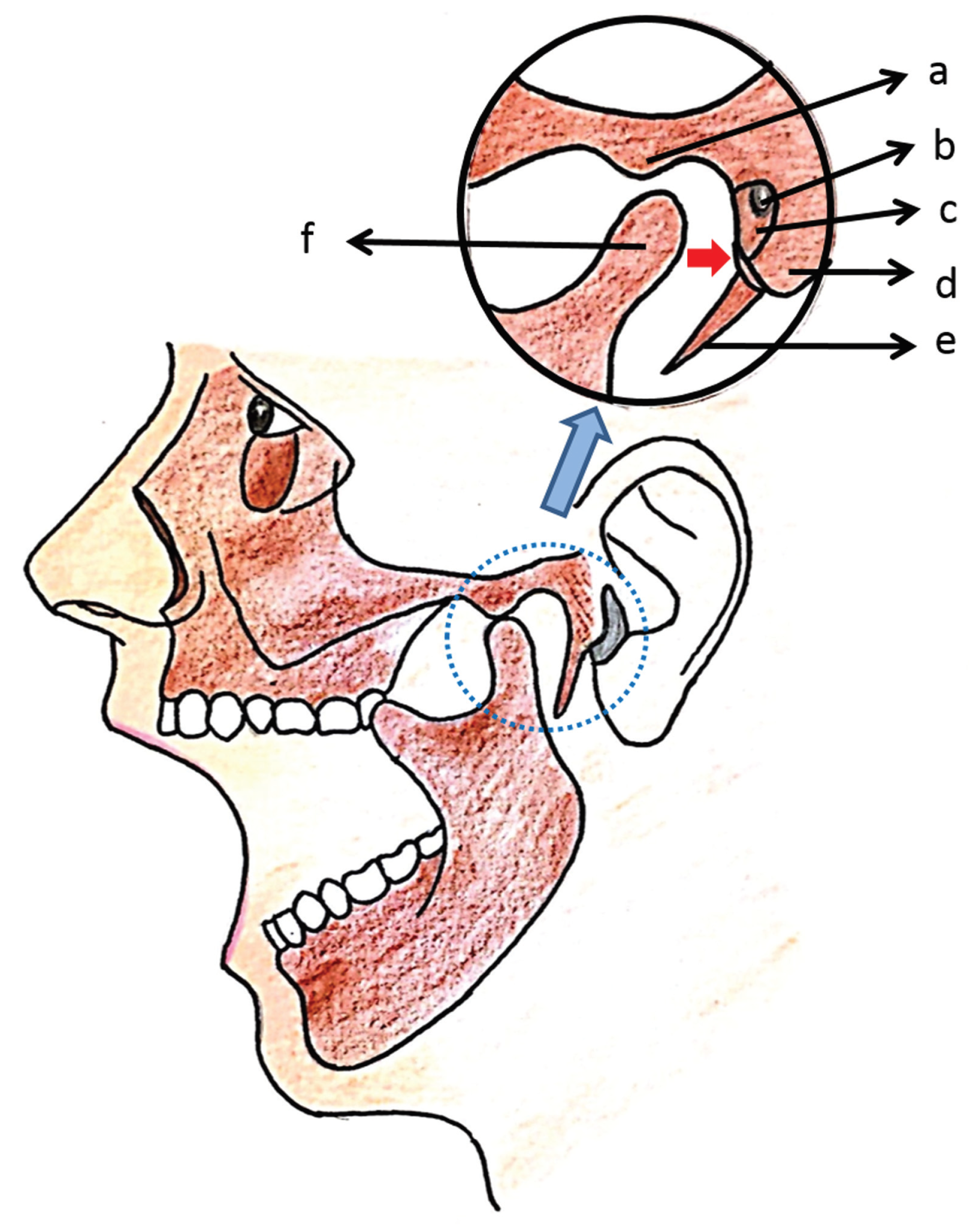
The possibility of a condylar fracture was suspected and promptly ruled out because of lack of derangement of occlusion or restriction of condylar movement. However, the traumatic impact of a posteriorly displaced nonfractured condyle on the EAC or the tympanic plate was considered [2]. This is because anatomically, the EAC is separated from the condyle by only a pad of fibrofatty tissue and the tympanic plate or cartilage. Also, the lateral third of the condyle does not have bony support posteriorly [13]. Thus, posterior displacement of the condyle by traumatic forces at the chin or angle can cause injury to the delicate cartilage bone junction of the EAC or sometimes even fracture of the tympanic plate [2,13] leading to ear bleed (Figure 2). This occurs especially when the mouth is wide open as during removal of an impacted tooth or extraction of posterior teeth. In this case, OPG demonstrating the fracture of styloid fracture is indicative of the traumatic displacement of the condyle during extraction. In tympanic plate fracture, positive findings are EAC stenosis and skin rupture of auditory canal [14]. Examination of the ear in this case revealed a breach in the anterior wall of the EAC (cartilaginous part). Ear bleed not associated with skull base fracture poses two problems: ear infection and aural stenosis, and the management strategy is aimed at preven-tion of these complications [15].
Fractures of the styloid process during dental extraction are rare, due to its relatively secure anatomic position. However, when it is abnormally elongated [16] (radiographic length on OPG more than one-third of the posterior ramal height) and shaped [10] as appreciated in this case, it fractures when force is directed on it by posteriorly displaced condyle. The styloid process may also possibly fracture due to the pull exerted by the stylomandibular ligament during extremes of mandibular movements. The primary diagnosis of styloid fracture is done clinically by symptoms such as dysphagia, otalgia, restricted mandibular movements, and pain in the preauricular, retromandibular, and TMJ region. Though OPG may be used to confirm the fracture, the displacement of the fractured fragment of styloid process is assessed by CT; this gives a clear spatial orientation of the fragment in relation to the adjacent vital structures.
It is important to realize the potential complications of displaced styloid by virtue of its close proximity to the carotid space which houses the internal carotid artery, internal jugular vein, the cranial nerves 9 to 12, and the sympathetic chain [17,18]. Anterior to the styloid are the lingual and auriculotemporal nerves. Pressure effects may be seen clinically such as glossopharyngeal neuralgia, syncope, etc., due to impingement of fractured styloid on any of these structures. Furthermore, even when undisplaced or asymptomatic immediately after trauma, the fibrosis or infection around a fractured styloid can later result in posttraumatic styloid syndrome [9], which is a painful condition similar to Eagle syndrome.
An undisplaced styloid fracture may be managed conservatively with soft diet, medication such as nonsteroidal antiinflammatory drugs, muscle relaxants, carbamazepine, and steroids, and local anesthetic injections—infiltrations or blocks [7]. The jaw and neck movements are restricted usually with intermaxillary fixation and cervical collar, respectively, to prevent the displacement of fractured fragments during jaw movements [7,10,19]. An immobilization period of 2 weeks would suffice to allow healing of styloid fracture. On the contrary, when conservative treatment fails or when there is potential harm to vital structures due to the fractured tip, surgical removal of the fragment is warranted. The procedure may be accomplished by intraoral or extraoral approach [17].
Regardless of the treatment protocol, such episodes of ear bleed or styloid fracture require prompt identification and management to alleviate patient anxiety and embarrassment to the dentist. Adequate care needs to be taken to prevent retropositioning of the mandible during extraction maneuvers.

Conclusion

During extraction procedures, excessive force delivered to the mandible especially when directed posteriorly, increases the chances of traumatic forces to the EAC, as well as the styloid process. This can precipitate styloid fracture and ear bleeding which may be managed conservatively. However, the ear bleed mandates critical evaluation and the patient needs to be under observation for development of potential pressure symptoms due to fractured styloid and treated surgically, if necessary.

Conflicts of Interest

None.

Patient Consent

Informed consent has been obtained for Figure 1 explaining her right to privacy, which has been waived.

References

  1. Chiapasco, M.; De Cicco, L.; Marrone, G. Side effects and complications associated with third molar surgery. Oral Surg Oral Med Oral Pathol 1993, 76, 412–420. [Google Scholar] [CrossRef] [PubMed]
  2. Martis, C.; Karakasis, D. Bleeding from the ear in maxillofacial injuries. J Maxillofac Surg 1974, 2, 126–128. [Google Scholar] [CrossRef] [PubMed]
  3. Lu, C.; He, D.; Yang, C. Which craniofacial fractures are associated with external auditory canal bleeding? J Oral Maxillofac Surg 2014, 72, 121–126. [Google Scholar] [CrossRef] [PubMed]
  4. Bayram, B.; Aydin, E.; Uckan, S. External auditory canal injury following removal of an impacted third molar: A case report. J Oral Maxillofac Surg 2007, 65, 1372–1374. [Google Scholar] [PubMed]
  5. Reichart, P.A.; Sooss, W. Fracture of the styloid process of the temporal bone: An unusual complication of dental treatment. Report of a case. Oral Surg Oral Med Oral Pathol 1976, 42, 150–154. [Google Scholar] [PubMed]
  6. Armao, T.A. Diagnosis and treatment of a styloid fracture. Oral Surg Oral Med Oral Path Oral Radiol 1960, 13, 1423–1424. [Google Scholar] [CrossRef]
  7. Dubey, K.N.; Bajaj, A.; Kumar, I. Fracture of the styloid process associated with the mandible fracture. Contemp Clin Dent 2013, 4, 116–118. [Google Scholar] [CrossRef] [PubMed]
  8. Miloro, M. Fracture of the styloid process: A case report and review of the literature. J Oral Maxillofac Surg 1994, 52, 1073–1077. [Google Scholar] [CrossRef] [PubMed]
  9. Smith, G.R.; Cherry, J.E. Traumatic Eagle’s syndrome: Report of a case and review of the literature. J Oral Maxillofac Surg 1988, 46, 606–609. [Google Scholar] [PubMed]
  10. Raman, U.; Samraj, T. Fracture of the styloid process associated with mandibular fracture. A case report. Int J Oral Maxillofac Surg 1990, 19, 350–351. [Google Scholar] [CrossRef] [PubMed]
  11. Babbitt, J.A. Fracture of the styloid process and its tonsil fossa complications, with report of a case. Ann Otol Rhinol Laryngol 1993, 42, 789–798. [Google Scholar] [CrossRef]
  12. Steinmann, E.P. A new light on the pathogenesis of the styloid syndrome. Arch Otolaryngol 1970, 91, 171–174. [Google Scholar] [CrossRef] [PubMed]
  13. Vasconcelos, B.C.; Rocha, N.S.; Cypriano, R.V. Posterior dislocation in intact mandibular condyle: An unusual case. Int J Oral Maxillofac Surg 2010, 39, 89–91. [Google Scholar] [CrossRef] [PubMed]
  14. Psimopoulou, M.; Antoniades, K.; Magoudi, D.; Karakasis, D. Tympanic plate fracture following mandibular trauma. Dentomaxillofac Radiol 1997, 26, 344–346. [Google Scholar] [PubMed]
  15. Loh, F.C.; Tan, K.B.; Tan, K.K. Auditory canal haemorrhage following mandibular condylar fracture. Br J Oral Maxillofac Surg 1991, 29, 12–13. [Google Scholar] [CrossRef] [PubMed]
  16. Goldstein, G.R.; Scopp, I.W. Radiographic interpretation of calcified stylomandibular and stylohyoid ligaments. J Prosthet Dent 1973, 30, 330–334. [Google Scholar] [CrossRef] [PubMed]
  17. Blythe, J.N.; Matthews, N.S.; Connor, S. Eagle’s syndrome after fracture of the elongated styloid process. Br J Oral Maxillofac Surg 2009, 47, 233–235. [Google Scholar] [CrossRef] [PubMed]
  18. Frommer, J. Anatomic variations in the stylohyoid chain and their possible clinical significance. Oral Surg Oral Med Oral Pathol 1974, 38, 659–667. [Google Scholar] [PubMed]
  19. Gayathri, G.; Elavenil, P.; Sasikala, B.; Pathumai, M.; Krishnakumar Raja, V.B. ‘Stylo-mandibular complex’ fracture from a maxillofacial surgeon’s perspective—Review of the literature and proposal of a management algorithm. Int J Oral Maxillofac Surg 2016, 45, 297–303. [Google Scholar] [CrossRef] [PubMed]
Figure 1. Orthopantomography demonstrating fracture.
Cmtr 10 00033 g001
Figure 2. Biomechanics of styloid fracture and injury to external auditory canal. (a) Articular eminence, (b) external auditory canal, (c) tympanic plate, (d) mastoid, (e) styloid, and (f) condyle.
Cmtr 10 00033 g002