Next Article in Journal
Management of a Life Threatening Bleeding Following Extraction of Deciduous Second Molar Related to a Capillary Haemangioma
Previous Article in Journal
Penetrating Craniomaxillofacial Injury Caused by a Pneumatic Nail Gun
 
 
Craniomaxillofacial Trauma & Reconstruction is published by MDPI from Volume 18 Issue 1 (2025). Previous articles were published by another publisher in Open Access under a CC-BY (or CC-BY-NC-ND) licence, and they are hosted by MDPI on mdpi.com as a courtesy and upon agreement with Sage.
Case Report

Basal Cell Adenoma of Retromolar Region from Minor Salivary Gland Origin in a 45-Year-Old Female: A Case Report

by
Velavan Krishnan
1,
Karthik Shunmugavelu
2,*,
Jamila Rose
3 and
Kumaravel Subramaniam
4
1
Department of Oral and Maxillofacial Surgery, Adiparasakthi Dental College and Hospital, Chennai, Tamilnadu, India
2
Department of Dentistry and Faciomaxillary Surgery, Kasthuri Multispeciality Hospital, Shanmugam Road, West Tambaram, Chennai, Tamilnadu 600045, India
3
Department of Pathology, Government Stanley Medical College and Hospital, Chennai, Tamilnadu, India
4
Department of Dentistry and Faciomaxillary Surgery, K.C. Multispeciality Hospital, Chennai, Tamil Nadu, India
*
Author to whom correspondence should be addressed.
Craniomaxillofac. Trauma Reconstr. 2017, 10(2), 162-165; https://doi.org/10.1055/s-0036-1584394
Submission received: 3 November 2015 / Revised: 1 January 2016 / Accepted: 24 January 2016 / Published: 18 July 2016

Abstract

:
Basal cell adenoma is a rare benign salivary gland neoplasm. The most common location is in parotid region. Clinically, gradual growth and firm consistency are seen. They account for 1 to 3% among all salivary gland neoplasms. Prominent basaloid cells with cluster of isomorphic cells and interspersed trabeculae are present histopathologically. In this article, we present a rare case of basal cell adenoma of retromolar region from minor salivary gland origin.

Among the head and neck neoplasms, salivary gland tumors constitute only 2 to 6.5%. Within the salivary gland tumors, only 25% and less is from that of minor origin [1,2]. Among the minor origin, diagnostic challenge was headed by the basaloid group of tumors, constituting 1 to 3%. Being known as monomorphic adenoma (due to resemblance to pleomorphic adenoma) in the past, Kleinsasser and Klein presented the nomenclature as basal cell adenoma in 1967. The gender predominance target females than males, usually in the fifth, sixth, and seventh decades. Histopathologically, this benign epithelial neoplasm lacks chondromyxoid matrix and consists of numerous basaloid cells [3,4,5,6,7]. Microscopically, they are classified as solid, trabecular, tubular, and membranous [3,8,9]. Low recurrence rate has been reported after excisional biopsy of the neoplasm. In this article, we present a rare case of basal cell adenoma of retromolar region from minor salivary gland origin in a 45-year-old female patient.

Case Presentation

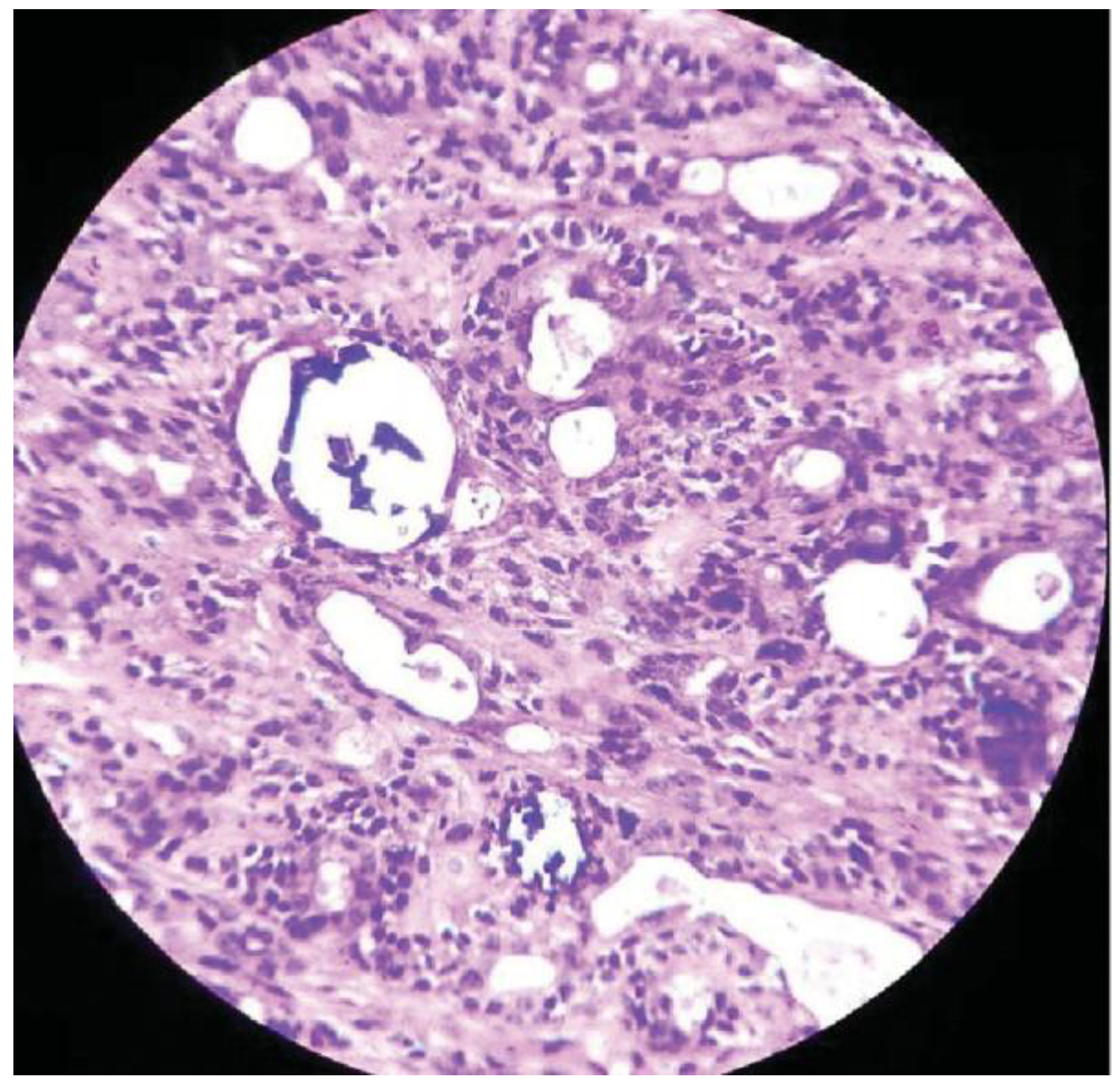
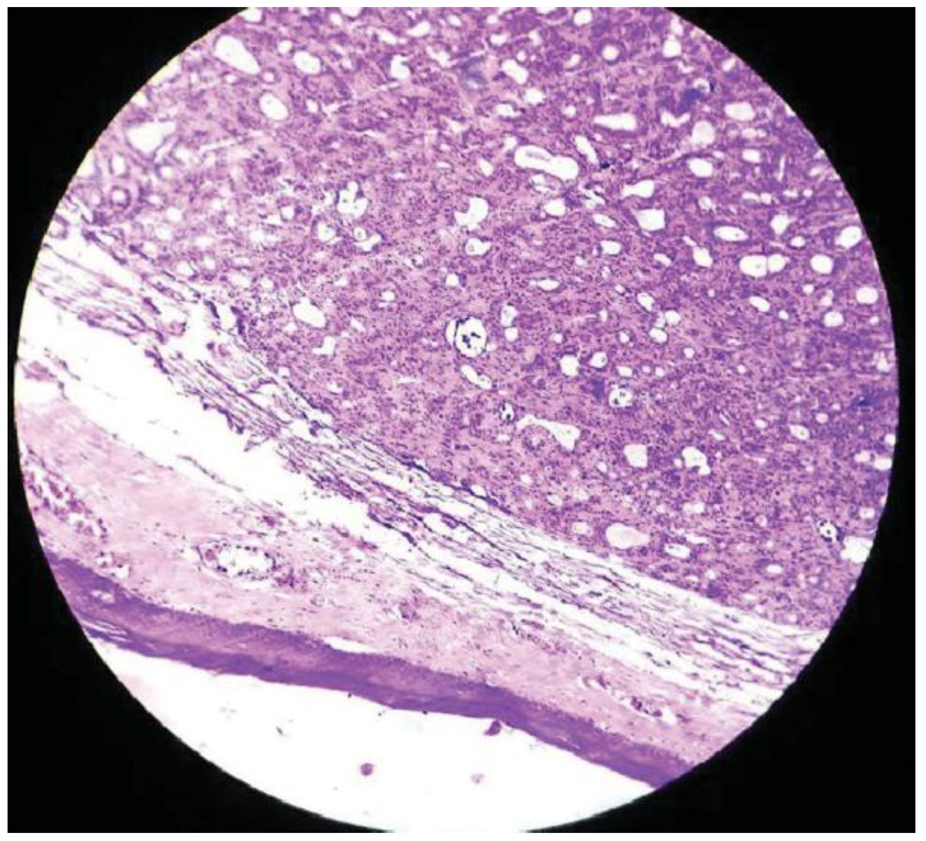
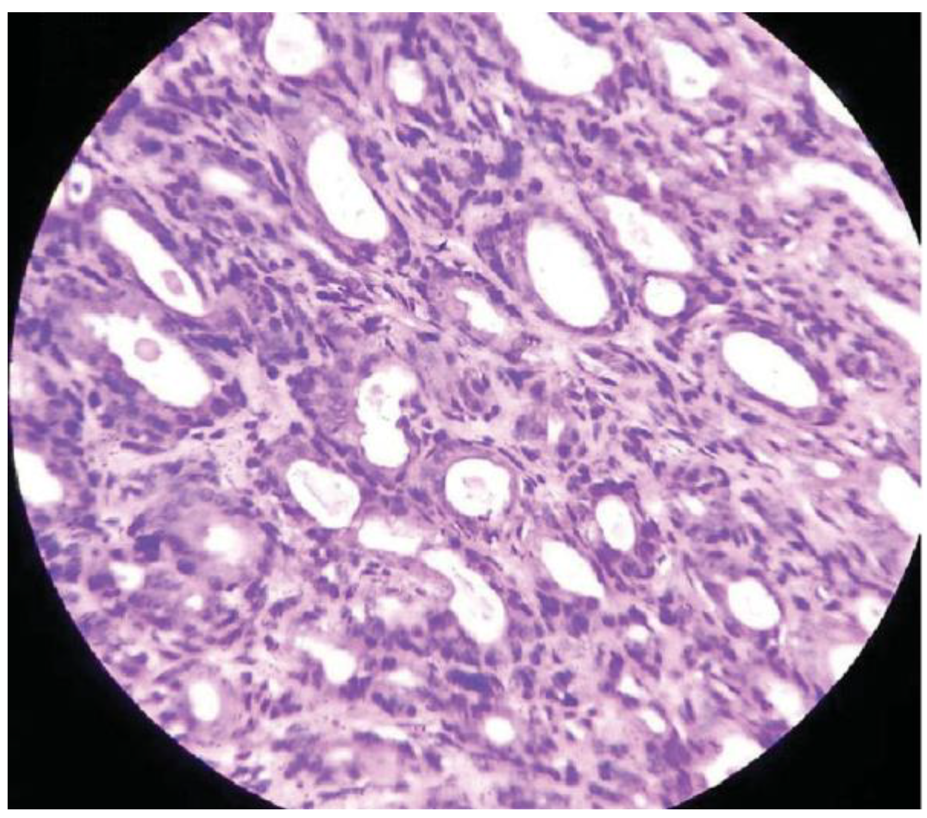
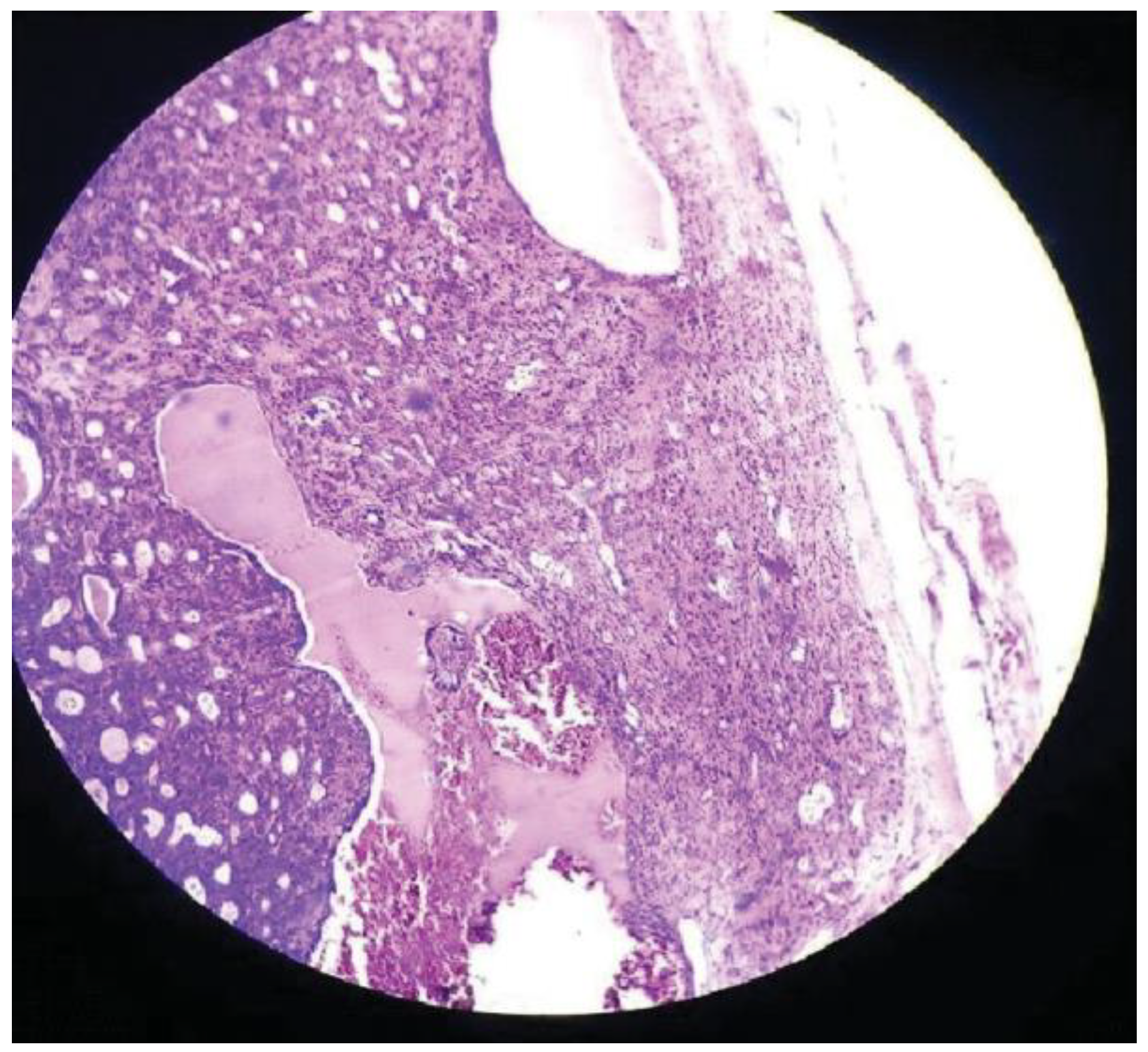
A 45-year-old female patient reported to the Department of Dentistry and Faciomaxillary Surgery in 2015, with slowly growing asymptomatic swelling on the right inner posterior part of the cheek present since 6 months. The patient also added that this growth has been progressing since 4 years, but she noticed only before 6 months. On inspection, the right retromolar region appeared normal, while on palpation a 2 × 2 cm, firm, well-circumscribed movable mass was present 2 cm posterior to the right mandibular third molar region (Figure 1) No ulceration was seen on the overlying mucosa. Differential diagnosis includes basal cell adenocarcinoma, pleomorphic adenoma, adenoid cystic carcinoma, chronic sialadenitis, cutaneous basal cell carcinoma, and metastatic basaloid squamous carcinoma. Treatment procedure was as follows: the growth was excised along with the adjoining normal borders under local anesthesia (Figure 2). Grossly, the lesion was grayish white measuring 2 × 2 cm, encapsulated, round and well demarcated and then the specimen was placed in 10% buffer formalin. The nature of the lesion during the procedure was that of a vascular one. The specimen was stained with eosin and hematoxylin. Histopathologically, underlying connective tissue was covered by orthokeratinized, stratified squamous epithelium. In depth, islands and cords of epithelial cells were seen along with wellcircumscribed, encapsulated tumor mass and fibrous stroma. Isomorphic basaloid cells were the pathognomonic feature in association with peripheral palisaded cuboidal to columnar cells in spite of round central cells. Hyperchromatism was observed in peripheral cells, whereas the central ones possessed pale stained nuclei. Based on the features, it was diagnosed as basal cell adenoma. Minor features such as squamous whorls and acinar cells were also seen (Figure 3, Figure 4, Figure 5 and Figure 6). Uneventful postoperative period was followed by absence of recurrence in a span of 6 months.

Discussion

In the past, basal cell adenoma was referred to as monomorphic adenoma. Lack of chondromyxoid material and presence of basaloid cells modified the name as basal cell adenoma. Basal cell adenoma usually affects individuals in their fifth, sixth, or seventh decades with a predilection towards female gender.
Brooke-Spiegler syndrome comprises of basal cell adenoma and adnexal tumors. The proportion of the neoplasm varies as follows: 1 to 3% of major salivary gland tumors, 7.5% of primary epithelial parotid gland tumors and 54% of monomorphous adenomas. The most common location is in the parotid gland, especially in the superficial lobe [10,11,12,13,14]. This article illustrates a rare case of basal cell adenoma in the retromolar region. Routine appearance is that of well-circumscribed border with round or oval shape unilaterally. Clinical features include painless mass, slow progressive enlargement and a measurement of 3 to 8 cm. A rare case of bilateral basal cell adenoma has been reported by Katsuno et al in a 65-year-old woman, specificity in parotid gland. The features of the excised mass usually consists of grayish white appearance, rich in blood supply, well circumscribed, encapsulated, soft to firm consistency, which might be sometimes cystic or solid in nature [15,16,17,18,19]. Among the solid, tubular, trabecular and membranous, membranous type is histologically distinctive due to its multifocal and multimodal character. Histopathological features include basaloid epithelial cells, uniform, dark, small, encapsulated, peripheral palisading of epithelial nests resulting in basaloid appearance. The cellular arrangement may be of trabecular, solid, tubular and membranous. Yadav et al reported a sixth case of palatal basal cell adenoma in 55-year-old woman, followed by complete tumor excision. The study also added that factors such as age, gender, and method of diagnosis and treatment playan important role in the outcome [20]. On comparison, our case report of basal cell adenoma of retromolar region in a 45-year-old female is the first case to be reported in literature in relation to retromolar region. Histopathologically, a combined tubular, acinar and trabecular pattern followed by basaloid cells and myoepithelial cells were seen. Our treatment plan such as excisional biopsy played a major role in complete removal of the lesion followed by definitive outcome. The malignant part is known as basal cell adenocarcinoma. Infiltrative growth of basal cell adenocarcinoma differentiates from that of basal cell adenoma. Common features of both the neoplasm are squamous morules, haphazard cellular arrangement, basaloid cells, intercellular matrix, peripheral palisading, and cellular aspirate. Differential diagnosis includes basal cell adenocarcinoma, adenoid cystic carcinoma, benign mixed tumor, chronic sialadenitis, cellular pleomorphic adenoma, metastatic basaloid squamous carcinoma, cutaneous basal cell carcinoma, and sialoblastoma. These types of neoplasms are treated by complete surgical excision with negative margins [21,22,23,24].

Conclusion

In spite of radiological and clinical techniques, histopathology plays an important role in the definitive diagnosis. Differential diagnosis of the malignant counterparts is mandatory. This rare case of basal cell adenoma in the retromolar region has been confirmed histopathologically.

References

  1. Leegard, T.; Lindeman, H. Salivary gland tumours: Clinical picture and treatment. Acta Otolaryngol. Suppl. 1969, 263, 55–59. [Google Scholar] [CrossRef]
  2. Eveson, J.W.; Cawson, R.A. Tumours of the minor (oropharyngeal) salivary glands: A demographic study of 336 cases. J. Oral. Pathol. 1985, 14, 500–509. [Google Scholar] [CrossRef]
  3. Minicucci, E.M.; de Campos, E.B.; Weber, S.A.; Domingues, M.A.; Ribeiro, D.A. Basal cell adenoma of the upper lip from minor salivary gland origin. Eur. J. Dent. 2008, 2, 213–216. [Google Scholar] [CrossRef]
  4. Basal Cell Adenoma. Available online: http://medical-dictionary.the-freedictionary.com/basal+cell+adenoma.
  5. Takeshita, T.; Tanaka, H.; Harasawa, A.; Kaminaga, T.; Imamura, T.; Furui, S. CT and MR findings of basal cell adenoma of the parotid gland. Radiat. Med. 2004, 22, 260–264. [Google Scholar] [PubMed]
  6. González-García, R.; Nam-Cha, S.H.; Muñoz-Guerra, M.F.; GamalloAmat, C. Basal cell adenoma of the parotid gland. Case report and review of the literature. Med. Oral. Patol. Oral. Cir. Bucal 2006, 11, E206–E209. [Google Scholar]
  7. Basal Cell Adenoma. Available online: http://www.pathconsultddx.com/pathCon/diagnosis?pii=S1559-8675 (accessed on 13 June 2016).
  8. Machado de Sousa, S.O.; Soares de Araújo, N.; Corrêa, L.; Pires Soubhia, A.M.; Cavalcanti de Araújo, V. Immunohistochemical aspects of basal cell adenoma and canalicular adenoma of salivary glands. Oral. Oncol. 2001, 37, 365–368. [Google Scholar] [CrossRef] [PubMed]
  9. Seifert, G.; Donath, K. Hybrid tumours of salivary glands. Definition and classification of five rare cases. Eur. J. Cancer B Oral. Oncol. 1996, 32B, 251–259. [Google Scholar] [CrossRef]
  10. Choi, H.R.; Batsakis, J.G.; Callender, D.L.; Prieto, V.G.; Luna, M.A.; El-Naggar, A.K. Molecular analysis of chromosome 16q regions in dermal analogue tumors of salivary glands: A genetic link to dermal cylindroma? Am. J. Surg. Pathol. 2002, 26, 778–783. [Google Scholar] [CrossRef]
  11. Ellis, G. Basal cell adenocarcinoma. In World Health Organization Classification of Tumours: Head and Neck Tumours; Barnes, L., Eveson, J.W., Reichart, P., Sidransky, D., Eds.; IARC Press: Lyon, 2005; pp. 229–230. [Google Scholar]
  12. Kawahara, A.; Harada, H.; Akiba, J.; Yokoyama, T.; Kage, M. Fine-needle aspiration cytology of basal cell adenoma of the parotid gland: Characteristic cytological features and diagnostic pitfalls. Diagn. Cytopathol. 2007, 35, 85–90. [Google Scholar] [CrossRef]
  13. Muller, S.; Barnes, L. Basal cell adenocarcinoma of the salivary glands. Report of seven cases and review of the literature. Cancer 1996, 78, 2471–2477. [Google Scholar] [CrossRef]
  14. Nagao, T.; Sugano, I.; Ishida, Y.; et al. Basal cell adenocarcinoma of the salivary glands: Comparison with basal cell adenoma through assessment of cell proliferation, apoptosis, and expression of p53 and bcl-2. Cancer 1998, 82, 439–447. [Google Scholar] [PubMed]
  15. Yerli, H.; Teksam, M.; Aydin, E.; Coskun, M.; Ozdemir, H.; Agildere, A.M. Basal cell adenoma of the parotid gland: Dynamic CT and MRI findings. Br. J. Radiol. 2005, 78, 642–645. [Google Scholar] [PubMed]
  16. Hemachandran, M.; Lal, A.; Vaiphei, K. Basal cell adenoma-an unusual presentation. Ann. Diagn. Pathol. 2003, 7, 292–295. [Google Scholar] [PubMed]
  17. Katsuno, S.; Ishii, K.; Otsuka, A.; Ezawa, S.; Usami, S. Bilateral basal-cell adenomas in the parotid glands. J. Laryngol. Otol. 2000, 114, 83–85. [Google Scholar]
  18. Hall-Craggs, M.; Toker, C. Basaloid tumor of the sigmoid colon. Hum. Pathol. 1982, 13, 497–500. [Google Scholar]
  19. Chow, L.T.; Chow, W.H. Co-existing pleomorphic and tubular basal cell adenomas of the parotid gland. J. Laryngol. Otol. 1997, 111, 182–185. [Google Scholar]
  20. Yadav, A.B.; Narwal, A.; Devi, A.; Kumar, S.; Yadav, S.K. Basal Cell Adenoma of Palate, a Rare Occurrence with Review of Literature. J. Dent. 2015, 16 (Suppl. S3), 291–295. [Google Scholar]
  21. Chow, L.T.; Chow, W.H. Co-existing pleomorphic and tubular basal cell adenomas of the parotid gland. J. Laryngol. Otol. 1997, 111, 182–185. [Google Scholar]
  22. Chiu, N.C.; Wu, H.M.; Chou, Y.H.; et al. Basal cell adenoma versus pleomorphic adenoma of the parotid gland: CT findings. AJR Am. J. Roentgenol. 2007, 189, W254–W261. [Google Scholar]
  23. Luna, M.A.; Batsakis, J.G.; Ordóñez, N.G.; Mackay, B.; Tortoledo, M.E. Salivary gland adenocarcinomas: A clinicopathologic analysis of three distinctive types. Semin. Diagn. Pathol. 1987, 4, 117–135. [Google Scholar]
  24. Gupta, N.; Jadhav, K.; Ahmed, M.B.; Amberkar, V.S. Basal cell adenoma in a relatively rare site. J. Oral. Maxillofac. Pathol. 2009, 13, 101–104. [Google Scholar] [PubMed]
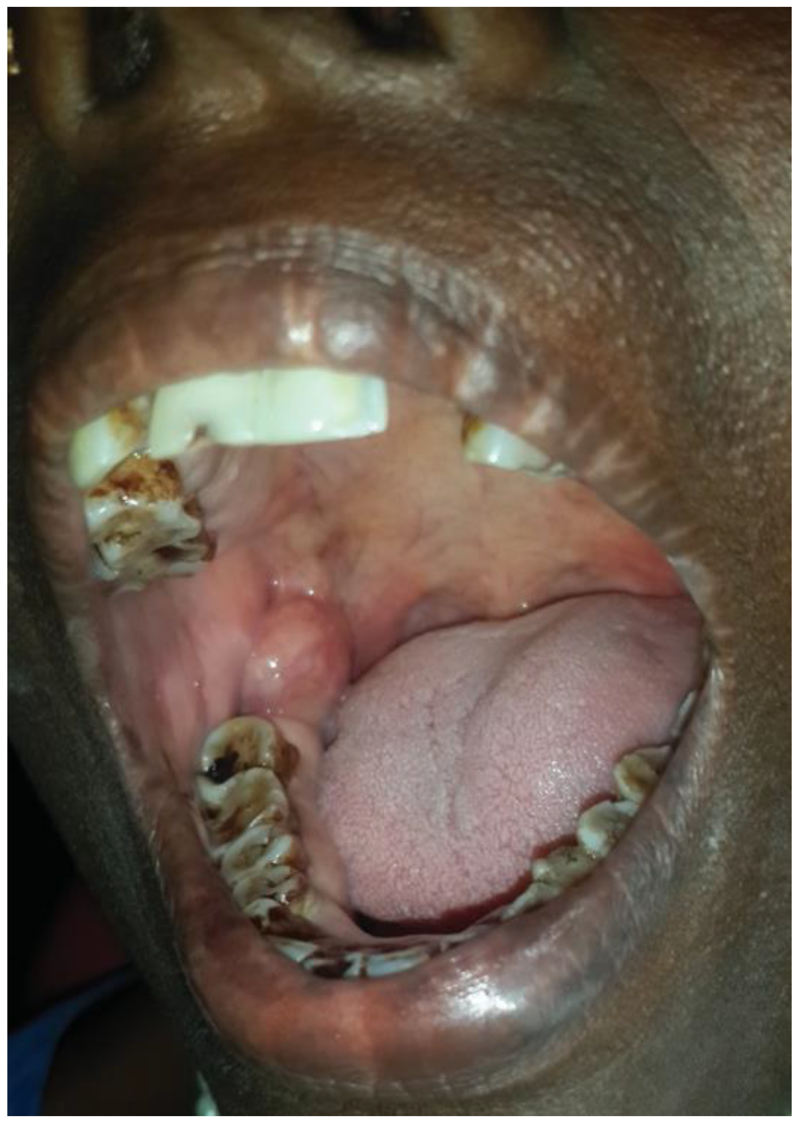
Figure 1. Preoperative view depicting intraoral mass in right retromolar region.
Cmtr 10 00025 g001
Figure 2. Postoperative excised specimen.
Cmtr 10 00025 g002
Figure 3. High power view (hematoxylin and eosin, ×40) showing tubules lined by luminal epithelial cells and basal myoepithelial cells surrounded by a fibrous stroma.
Cmtr 10 00025 g003
Figure 4. Low power view (hematoxylin and eosin, ×10) shows squamous mucosa and an underlying well-encapsulated neoplasm showing tubular, acinar, and trabecular pattern.
Cmtr 10 00025 g004
Figure 5. High power view (hematoxylin and eosin, ×40) showing uniform basaloid cells forming tubules with intraluminal secretions. No atypia or mitosis evident.
Cmtr 10 00025 g005
Figure 6. Scanner view (hematoxylin and eosin, ×5) showing benign cells forming tubules, glands and nests with distinct peripheral nuclear palisading.
Cmtr 10 00025 g006