Article

The Development of New Nanocomposite Polytetrafluoroethylene/Fe2O3 NPs to Prevent Bacterial Contamination in Meat Industry

Prokhorov General Physics Institute of the Russian Academy of Sciences, 38 Vavilova St., 119991 Moscow, Russia
*
Author to whom correspondence should be addressed.
Polymers 2022, 14(22), 4880; https://doi.org/10.3390/polym14224880
Submission received: 8 October 2022 / Revised: 8 November 2022 / Accepted: 10 November 2022 / Published: 12 November 2022
(This article belongs to the Special Issue Polymers in Food Science)

Abstract

:
The bacterial contamination of cutting boards and other equipment in the meat processing industry is one of the key reasons for reducing the shelf life and consumer properties of products. There are two ways to solve this problem. The first option is to create coatings with increased strength in order to prevent the formation of micro damages that are favorable for bacterial growth. The second possibility is to create materials with antimicrobial properties. The use of polytetrafluoroethylene (PTFE) coatings with the addition of metal oxide nanoparticles will allow to the achieving of both strength and bacteriostatic effects at the same time. In the present study, a new coating based on PTFE and Fe2O3 nanoparticles was developed. Fe2O3 nanoparticles were synthesized by laser ablation in water and transferred into acetone using the developed procedures. An acetone-based colloidal solution was mixed with a PTFE-based varnish. Composites with concentrations of Fe2O3 nanoparticles from 0.001–0.1% were synthesized. We studied the effect of the obtained material on the generation of ROS (hydrogen peroxide and hydroxyl radicals), 8-oxoguanine, and long-lived active forms of proteins. It was found that PTFE did not affect the generation of all the studied compounds, and the addition of Fe2O3 nanoparticles increased the generation of H2O2 and hydroxyl radicals by up to 6 and 7 times, respectively. The generation of 8-oxoguanine and long-lived reactive protein species in the presence of PTFE/Fe2O3 NPs at 0.1% increased by 2 and 3 times, respectively. The bacteriostatic and cytotoxic effects of the developed material were studied. PTFE with the addition of Fe2O3 nanoparticles, at a concentration of 0.001% or more, inhibited the growth of E. coli by 2–5 times compared to the control or PTFE without NPs. At the same time, PTFE, even with the addition of 0.1% Fe2O3 nanoparticles, did not significantly impact the survival of eukaryotic cells. It was assumed that the resulting composite material could be used to cover cutting boards and other polymeric surfaces in the meat processing industry.

1. Introduction

Meat deboning is one of the stages of raw meat processing, during which muscle, connective, and adipose tissue, that is, meat itself, is separated from the bone content. Deboning is performed by hand or with special equipment, while the meat is usually on cutting boards during deboning. During the use of cutting boards, microdamages accumulate on the surface of the cutting boards [1]. In these microdamages, favorable conditions are created for the growth and development of pathogenic microorganisms, including the formation of bacterial communities. The development of bacteria on cutting boards leads to biological contamination of meat products and a decrease in its shelf life and deterioration of its organoleptic properties [2]. Unfortunately, antibiotic-resistant strains are often found among these bacterial strains [3,4]. Gastrointestinal infections are an important global health problem [5]. Over 70,000 cases of hospitalizations with severe intestinal infections are predicted annually in the United States alone, of which over 1800 cases are fatal [6]. There are two principal approaches in order to protect the surface of cutting boards from bacterial contamination. The first approach is the creation of self-healing and/or ultra-strong materials to prevent the formation of microdamages favorable for bacterial growth. There are data in the literature on the dependence of the degree of bacterial contamination of S. enterica on the cutting board material. In the case of using a more durable material (glass), bacterial contamination occurred in 10% cases, and in the case of a softer material, i.e., plastic or wood, bacterial growth was observed in 40 and 60% of cases [4]. The second approach is to create a material with antibacterial action to suppress the growth of already-attached microbes.
Polytetrafluoroethylene (PTFE, Teflon, fluoroplast) is a fluoropolymer consisting of tetrafluoroethylene monomers described with the formula …–(CF2-CF2)n-… [7]. PTFE has a number of interesting properties such as high thermal conductivity and mechanical strength, dielectric properties, a resistance to a wide range of chemical agents, etc., [8,9]. These properties make PTFE an ideal material for a wide variety of applications such as mechanical engineering, the electronic and chemical industries, the space industry, the pharmaceutical industry, the manufacturing of implants, air and water filtration and the meat processing industry [10,11,12,13,14,15,16,17,18]. PTFE has pronounced hydrophobic properties, the ability to self-lubricate, and is self-healing. These properties make it possible to create self-healing coatings based on PTFE [19,20,21]. It was assumed in this study that the use of PTFE coatings would increase the mechanical strength of cutting boards and solve the problem of microdamages. The use of nanoparticles of metals and their oxides is one of the promising ways to combat microorganisms, including those resistant to antibiotics [22]. Iron oxide nanoparticles have high toxicity against bacteria and relatively low cytotoxicity against animal and human cells [23].
Previously, combined materials based on metal oxide NPs, including Fe2O3 and polymer matrices using the example of borosiloxane and PLGA, were obtained [24,25,26]. These materials have shown excellent antibacterial properties. Due to its well-known bioinertness and lack of biodegradability, fluoroplast is a good candidate for a matrix for the manufacture of stable antibacterial composite materials. A fundamental possibility for creating materials based on PTFE and metal oxide NPs using Ag2O as an example was described earlier [27]. Fe2O3 was chosen in this study because it is a common iron oxide, and it is cheaper to obtain. A polymer varnish was chosen as a PTFE-containing base. It was assumed that it would completely fill in the irregularities of the working surface and existing microdamages. An important requirement is the smoothness of the surface after processing.
Summarizing the above, the aim of this study was to obtain a new composite coating based on PTFE and Fe2O3 nanoparticles (PTFE/Fe2O3 NPs) and to study its physical properties, surface features, its influence on the generation of reactive oxygen species, namely 8-oxoguanine in DNA and long-lived reactive protein species (LRPS), its bacteriostatic activity, and its cytotoxicity against eukaryotic cells.
For the first time, a composite coating based on PTFE polymer and Fe2O3 NPs was obtained, and its high adhesion to surfaces made of damaged fluoroplastic was shown. We found for the first time that the PTFE/Fe2O3 NP composite coating caused an increase in the generation of ROS, 8-oxoguanine in DNA in vitro, and LRPS, that it had a strong bacteriostatic effect, and did not affect the number of viable fibroblasts in the primary culture.

2. Materials and Methods

2.1. Preparation of Composite PTFE/Fe2O3 NPs

Fe2O3 nanoparticles were obtained by laser ablation in water using a Nd:YAG laser [28]. Chemically pure iron Fe 99.9% (Sigma Aldrich, Burlington, MA, USA) was used as a target. The resulting Fe2O3 nanoparticles were precipitated three times with centrifugation followed by dilution in acetone. The resulting Fe2O3 NPs colloid in acetone was mixed with a LF-32LN PTFE-based varnish, 3V-00001283 (Plastpolymer-Prom, St. Petersburg, Russia). The mixture was stored in glass jars until use. Fe2O3 NPs were added at concentrations of 0.001, 0.01, or 0.1% (m/m).
To study the possibility of leveling damage and adhesion to the surface, we applied a composite coating to the area of the fluoroplast sample damaged during operation. The coating was dried for 48 h. The area of damaged fluoroplasts was photographed before and after coating.
In experiments on the effect of PTFE/Fe2O3 NPs composite material on the generation of ROS, the formation of 8-oxoguanine in DNA, the formation of LRPS, as well as evaluating its bacteriostatic effect, coatings containing Fe2O3 NPs at concentrations of 0.001–0.1% or PTFE without NPs were used. To study the cytotoxicity of the coating in relation to primary cultures of fibroblasts, the maximum of the considered concentrations of Fe2O3 NPs (0.1%) was used. Coatings (V = 500 μL) were applied to the surfaces of round coverslip glasses with a diameter of 25 mm and dried for 48 h in a fume hood. The final thickness of the dried coating was no more than 200 μm. For biological studies, samples of coatings on glasses were sterilized by soaking in 70% ethanol for 3 h. For microbiological studies, samples of coatings were removed from glasses in the form of films with an area of ~20 cm2. To study cytotoxicity, coating samples were dried on glass slides under sterile conditions.

2.2. Physical Properties Assay

The main characteristics of the obtained nanoparticles were studied, namely their size, ζ-potential, absorption spectrum, and shape. The size was estimated using DLS methods with Zetasizer Ultra Red Label (Malvern Panalytical, Malvern, Worcestershire, UK) and TEM with a Libra 200 FE HR electron microscope (Carl Zeiss, Oberkochen, Germany). ζ-potential was measured using Zetasizer Ultra Red Label (Malvern Panalytical, Malvern, Worcestershire, UK). Absorption spectra in the UV–visible region were recorded using a Cintra 4040 spectrometer (GBC Scientific Equipment, Braeside, VIC, Australia). The surface structure of the PTFE/Fe2O3 NPs composite was examined using atomic force microscopy (AFM) using SII Nanopics 2100 (KLA-Tencor, Milpitas, CA, USA). The distribution of Fe2O3 nanoparticles inside the composite was evaluated with modulation interference microscopy (MIM) using MIM-32 (Amphora Lab, Korolyov, Moscow region, Russia). A more detailed description of research methods can be found in earlier research studies [26,29,30].

2.3. Measurement of Reactive Oxygen Species Concentration

The chemiluminescent method of the luminol–p-iodophenol–horseradish peroxidase system was used to estimate the concentration of formed H2O2. Chemiluminescence was recorded using a highly sensitive chemiluminometer, Biotoks-7A (Engineering Center—Ecology, Moscow, Russia). The calibration and registration procedure was carried out according to the protocols described in more detail earlier. Films with an area of 20 cm2 from a composite material containing various concentrations of Fe2O3 NPs in compositions from 0–0.1 wt.% were placed in polypropylene vials for 3 h at 40 °C. In the “Control” group, the experiment was carried out without coating sample. After incubation in 20 mL of water, 1 mL of a previously prepared “counting solution” was added to the sample. This solution contained 1 mM Tris–HCl buffer, pH 8.5, 50 µM p-iodophenol, 50 µM luminol, and 10 nM horseradish peroxidase. The sensitivity of this method made it possible to determine H2O2 at a concentration of <1 nM. [31] The concentration of OH radicals formed in aqueous solutions was determined by reaction with coumarin-3-carboxylic acid (CCA), the product of which was hydroxycoumarin-3-carboxylic acid (7-OH-CCA). 7-OH-CCA is a fluorescent probe that quantifies OH radicals. Briefly, 0.2 M PBS (pH 7.4) was added to a solution of CCA in water (0.5 mM, pH 3.6). Next, samples of the composite material containing various concentrations of Fe2O3 NPs in compositions from 0–0.1 wt.% were added to the vials. In the “Control” group, the experiment was carried out without a coating sample. Next, polypropylene vials with samples and reagents were heated in a thermostat at temperature of 80.0 ± 0.1 °C for 2 h. Fluorescence of 7-OH-CCA was recorded using a JASCO 8300 fluorimeter (JASCO, Hachioji, Japan) at λex = 400 nm (excitation wavelength) and λem = 450 nm (emission wavelength). Commercial 7-OH-CCA (Sigma-Aldrich, Burlington, MA, USA) was used for calibration [32].

2.4. Measurement of 8-Oxoguanine and Long-Lived Reactive Protein Species Concentration

A non-competitive enzyme-linked immunosorbent assay (ELISA) was used to quantify 8-oxoguanine in DNA using monoclonal antibodies specific for 8-oxoguanine (anti-8-OG antibodies). DNA samples (350 μg/mL) were denatured by boiling in a water bath for 5 min and subsequently cooled on ice for 3–4 min. Aliquots (42 μL) were applied to the bottom of the wells of the ELISA plates. The DNA was immobilized using a simple adsorption procedure with incubation for 3 h at 80 °C until the solution became completely dry. Non-specific adsorption sites were blocked with 300 μL of a solution containing 1% skimmed milk powder in 0.15 M Tris-HCl buffer, with pH 8.7 and 0.15 M NaCl. Next, the plates were incubated at room temperature overnight (14–18 h). The formation of an antigen–antibody complex with antibodies against ti-8-OG (at a dilution of 1:2000) was carried out in a blocking solution (100 µL/well) by incubation for 3 h at 37 °C. The samples were washed twice (300 µL/well) with 50 mM Tris–HCl buffer (pH 8.7) and 0.15 M NaCl with 0.1% Triton X-100 after 20 min incubation. Next, a complex with a conjugate (anti-mouse immunoglobulin labeled with horseradish peroxidase (1:1000)) was formed by incubation for 1.5 h at 37 °C in a blocking solution (80 µL/well). Then, the wells were washed 3 times as described above. Next, a chromogenic substrate containing 18.2 mM ABTS and hydrogen peroxide (2.6 mM) in 75 mM citrate buffer, pH 4.2 (100 µL/well), was added to each well. Reactions were stopped by adding an equal volume of 1.5 mM NaN3 in 0.1 M citrate buffer (pH 4.3) upon reaching the correct color. The optical density of the samples was measured on a plate photometer Titertek Multiscan (Flow laboratories, McLean, VA, USA) at λ = 405 nm.
The chemiluminescent method is effective and sensitive for determining free radical reactions. The interaction of radicals is accompanied by the release of energy in the form of emitted light quanta [33]. In this case, the interaction of radicals releases energy, which is emitted in the form of light quanta. Chemiluminescence was recorded using a highly sensitive chemiluminometer, Biotoks-7A (Engineering Center—Ecology, Moscow, Russia). The samples were heated to a temperature of 45 °C within 2 h. Measurements were carried out in the dark at room temperature in 20 mL plastic polypropylene vials (Beckman, Brea, CA, USA). All samples were stored in the dark at room temperature for 30 min after exposure. As a control, protein solutions without preheating were used. A more detailed description of the method is presented in the published article by Sharapov et al. [34].

2.5. Antimicrobial Avtivity Assay

The antibacterial effect of composite films with an area of ~20 mm2 with different concentrations of Fe2O3 NPs was studied against the Gram-negative bacterium Escherichia coli. Samples were pre-styled by incubation in 70% ethanol for 3 h. Next, each sample was placed in a sterile wrap with 5 mL of liquid LB nutrient medium (LenReaktiv, Moscow, Russia). A known equal number of E. coli-colony-forming units (CFU) was added to each hoop with or without an image (control). The number of CFUs was pre-determined with a counting chamber and these were used for further calibration. Hoops with samples and LB and E. coli medium were covered with film and incubated for 24 h at 37 °C, ~150 rpm in an incubator shaker (Biosan, Riga, Latvia). The number of bacteria was estimated from the optical density at 600 nm of the suspension using a UV5Nano Excellence drop spectrometer (Mettler Toledo, Columbus, OH, USA). The value of the measured optical density was recalculated into the number of CFUs per unit volume using the previously constructed calibration [25,35,36].

2.6. Assay of the Influence on Animal Cells Viability

The study was performed on a primary culture of murine fibroblasts (BALB/c males weighing 22–25 g), which were isolated and cultivated according to a standard protocol [37]. All experiments with laboratory animals were carried out in accordance with the regulatory legal act of the Ministry of Health of the Russian Federation №199-n “On approval of the rules of good laboratory practice” and the international legal norms specified in the European Convention ETS №123 “On the protection of vertebrate animals used for experiments or for other scientific purposes”. Round coverslips were coated by PTFE with the addition of 0.1% Fe2O3 nanoparticles or PTFE without nanoparticles and were dried for 24 h in a fume hood. Cells were seeded on prepared slides and uncoated slides (control) and cultured in DMED/F12 medium supplemented with 10% FBS, 2 mM L-glutamine, and antibiotics (Sigma Aldrich, Burlington, MA, USA) for 72 h at 37 °C and 5% CO2. After incubation, cells were stained with fluorescent dyes Hoechst33342 and PI (Sigma Aldrich, Burlington, MA, USA) and were analyzed using a DMI6000 fluorescent microscope (Leica, Wetzlar, Germany) [38,39,40].

2.7. Statistic

All data are presented as means ± standard errors. Statistical significance was assessed using the Mann–Whitney test. In each variant, the experiments were performed with at least three independent repetitions.

3. Results

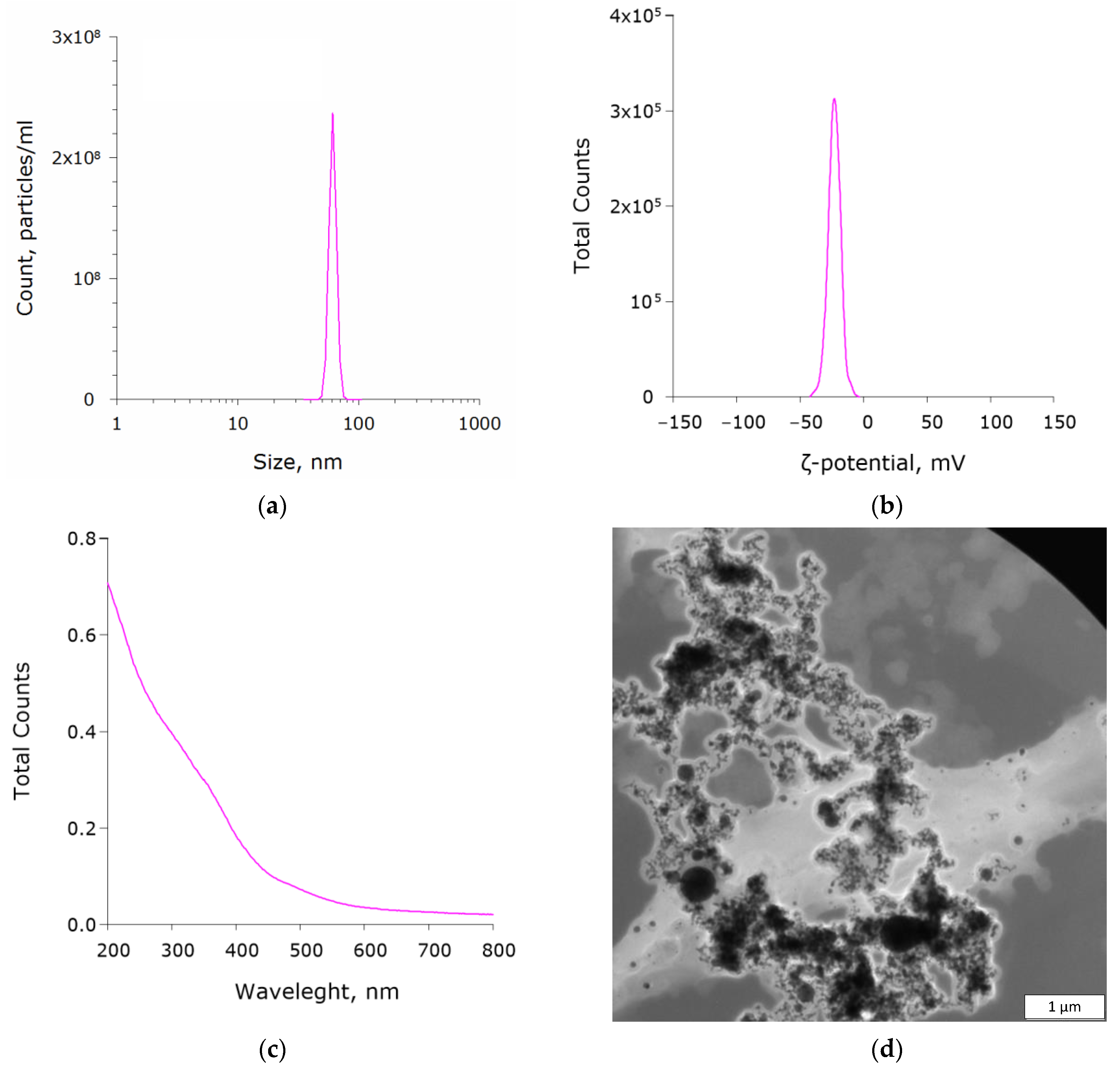
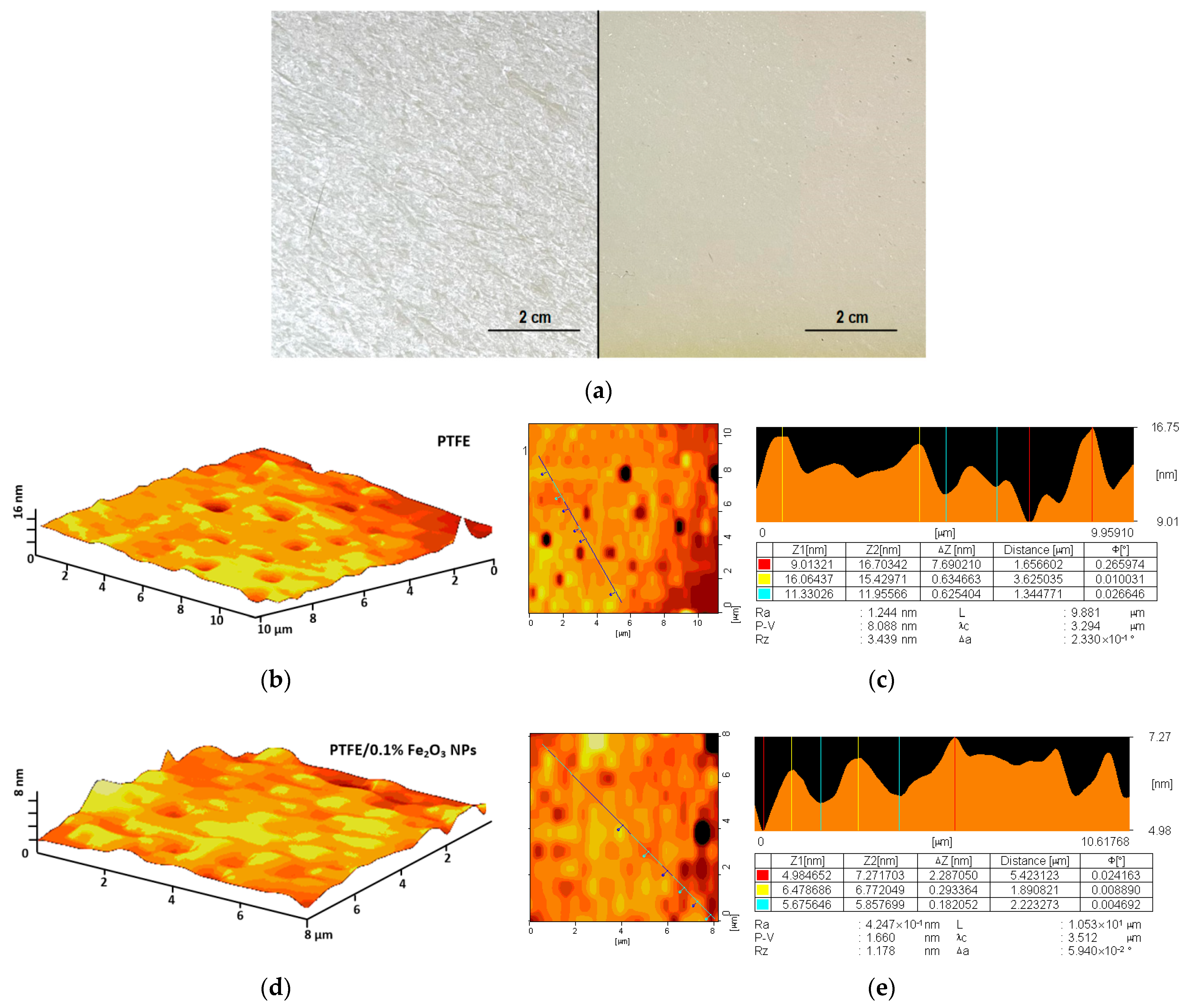
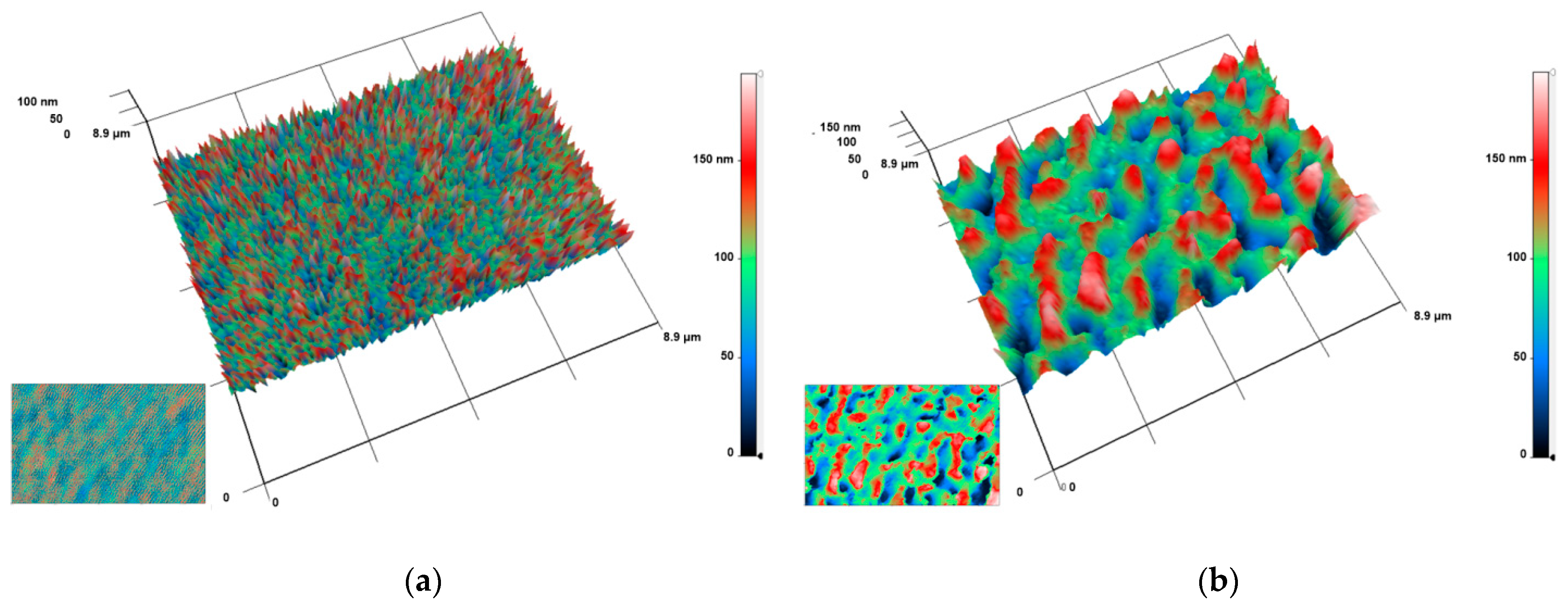
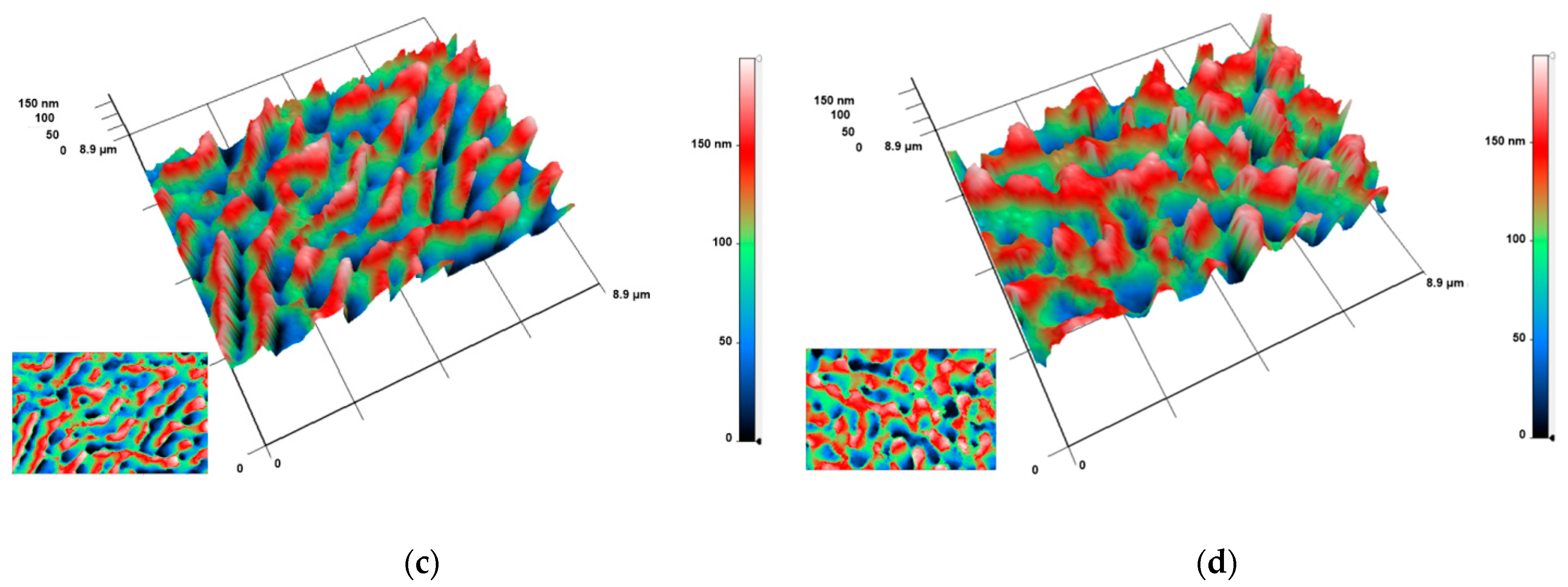
3.1. Physical Properties

According to the DLS data, the size distribution of the synthesized Fe2O3 nanoparticles was symmetrical. The size ranged from 43 to 87 nm with an average value of ~61 nm (Figure 1a). The ζ-potential values ranged from −69 to −7 mV with a mean value of −25 mV at colloid pH 6.7 ± 0.1 (Figure 1b). The absorption spectrum clearly showed absorption at wavelengths <400 nm, which was characteristic of Fe2O3 nanoparticles (Figure 1c). According to the electron microscopy, the size distribution of the nanoparticles was more heterogeneous, and the shape was spherical (Figure 1d). Virtually no aggregates of nanoparticles were found.
We found that the composite coating perfectly adhered to the damaged surface of the PTFE, covering all visible damage (Figure 2a). When trying to mechanically damage the surface 48 h after coating, there were no visible violations of the integrity of the coating and the underlying layer. At the next stage of this research, the surface of the composite material was studied using the AFM method, and the distribution of nanoparticles inside the polymer matrix was estimated. The atomic force microscopy of the PTFE surface without the addition of nanoparticles showed the absence of irregularities and inhomogeneities (Figure 2b). The change in the relief of the PTFE surface did not exceed 10 nm (Figure 2c). The surface of the PTFE/0.1% Fe2O3 composite material was also free from cracks, wrinkles, and other damages and defects. (Figure 2d). The change in the surface relief of PTFE/0.1% Fe2O3 also did not exceed 10 nm (Figure 2e).
The distribution of nanoparticles inside the composite material was studied using the modulation-interference microscopy method, which made it possible to distinguish the objects with different values of the refractive index. In examining the thickness of PTFE without the addition of NPs, no pronounced inhomogeneities were found (Figure 3a). When Fe2O3 nanoparticles were added to PTFE at a concentration of 0.001%, inhomogeneities were found in the sample (Figure 3b). These inhomogeneities had a length of 0.25–1.0 μm and a width of 0.20–0.25 μm. The largest value of the phase difference was 150–160 nm. In the case of the PTFE composites with Fe2O3 nanoparticles at concentrations of 0.01 and 0.1%, a similar pattern was observed (Figure 3c,d), though the size of the inhomogeneities increased with the increase in the nanoparticle concentration. At a concentration of Fe2O3 nanoparticles of 0.01%, the length and width of the inhomogeneities were 0.5–2 and 0.2–0.25 μm, respectively. The phase difference was 150–170 nm (Figure 3c). At the maximum concentration of Fe2O3 nanoparticles of 0.1%, the length, width, and phase difference of the inhomogeneities were 0.5–2, 0.25–0.5 μm, and 150–170 nm, respectively (Figure 3d).

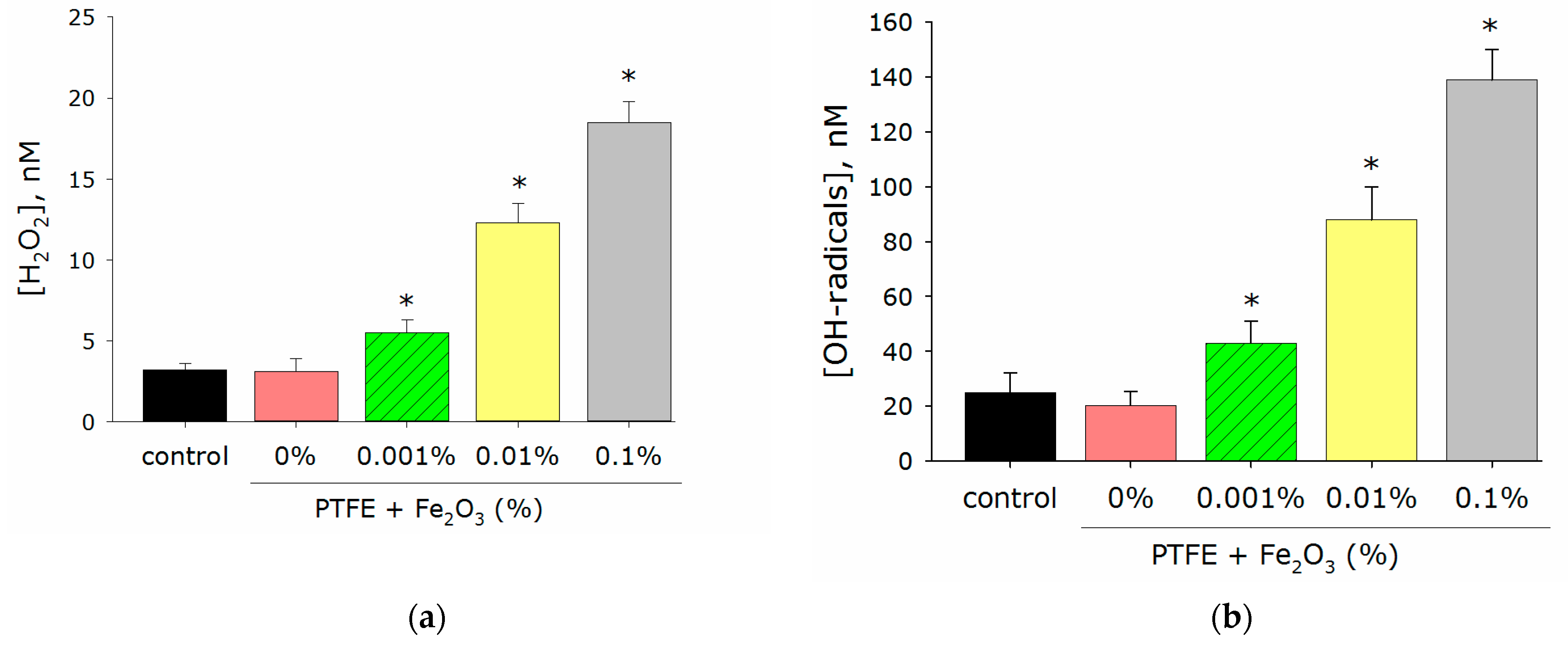
3.2. Generation of Reactive Oxigen Species Concentration

An imbalance between the processes leading to the accumulation of ROS and the ability to remove the excess ROS due to antioxidant systems can lead to an increase in the concentration of ROS and a state of “oxidative stress”, which is dangerous for eukaryotic cells [41]. Therefore, it was very important that the developed materials did not cause excessive production of ROS. Considering the above, the effect of the PTFE/Fe2O3 NPs composite material on the generation of ROS was investigated using H2O2 and hydroxyl radicals as an example (Figure 4b). The concentration of H2O2 in the case of PTFE without nanoparticles did not differ from the control (Figure 4a). A variant of the PTFE/Fe2O3 NPs composite material with a nanoparticle concentration of 0.001% increased the H2O2 concentration by almost 2 times compared to the control. In the presence of PTFE/Fe2O3 NPs composites with nanoparticle concentrations of 0.01% and 0.1%, an increase in the H2O2 concentration by 4 and 6 times, respectively, was recorded. The PTFE polymer without nanoparticles did not change the concentration of hydroxyl radicals compared to the control (Figure 4b). In the presence of a PTFE/Fe2O3 NPs composite with nanoparticle concentrations of 0.001, 0.01, and 0.1%, an increase in the concentration of hydroxyl radicals by 2, 4, and 7 times, respectively, was observed.

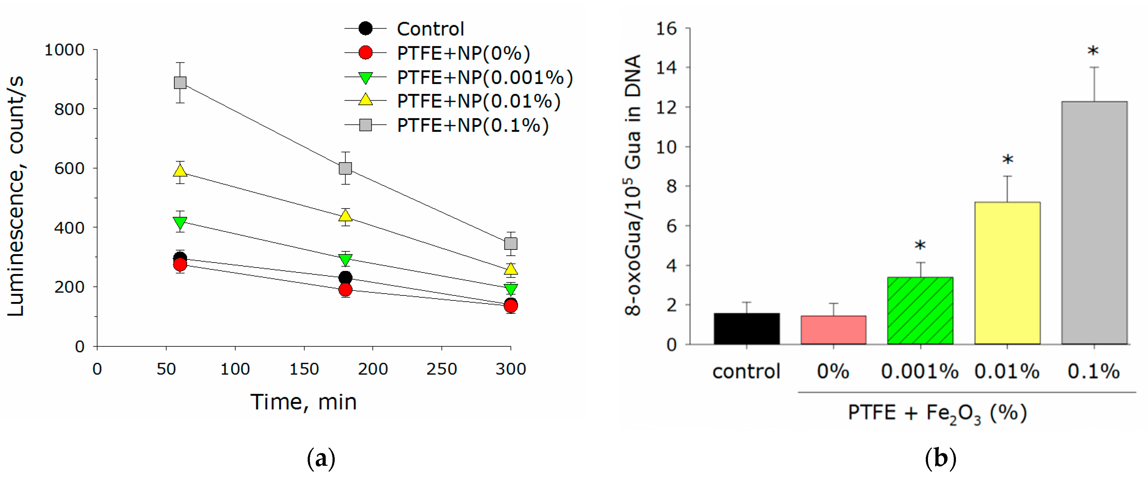
3.3. Generation of 8-Oxoguanine and Long-Lived Reactive Protein Species

At the next stage of the study, the effect of the developed composite material on the generation of markers of oxidative damage to the biological polymers was evaluated. In the case of DNA, this was 8-oxoguanine; in the case of proteins, it was long-lived reactive protein species (LRPS).
The PTFE coating without nanoparticles did not change the production of LRPS and 8-oxoguanine compared to control (Figure 5a,b). The PTFE composite with a concentration of Fe2O3 nanoparticles of 0.001% increased the concentration of LRPS by 25% compared to the control. The addition of Fe2O3 nanoparticles at a concentration of 0.01 and 0.1% to the composite material increased the generation of LRPS by 2 and 3 times, respectively, compared to the control (Figure 5a). The addition of 0.001% Fe2O3 nanoparticles to the composite material doubled the concentration of 8-oxoguanine compared to the control. PTFE/Fe2O3 composites with nanoparticle concentrations of 0.01 and 0.1% Fe2O3 NPs increased the production of 8-oxoguanine by 4 and 6 times compared to the control, respectively (Figure 5b).

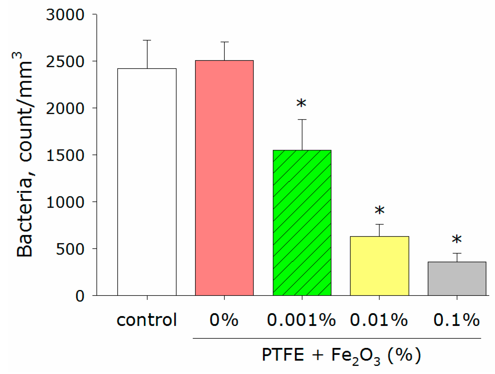
3.4. Antimicrobial Activity

The PTFE polymer without the addition of nanoparticles did not affect the bacterial growth rate compared to the control (Figure 6). The addition of Fe2O3 NPs to the PTFE significantly reduced the number of bacteria in the sample. The effect was dose-dependent. PTFE with a Fe2O3 NP concentration of 0.001% reduced the number of bacteria by ~40%, whereas the PTFE with an Fe2O3 NP concentration of 0.1% reduced the number of bacteria by more than 85% compared to the control.

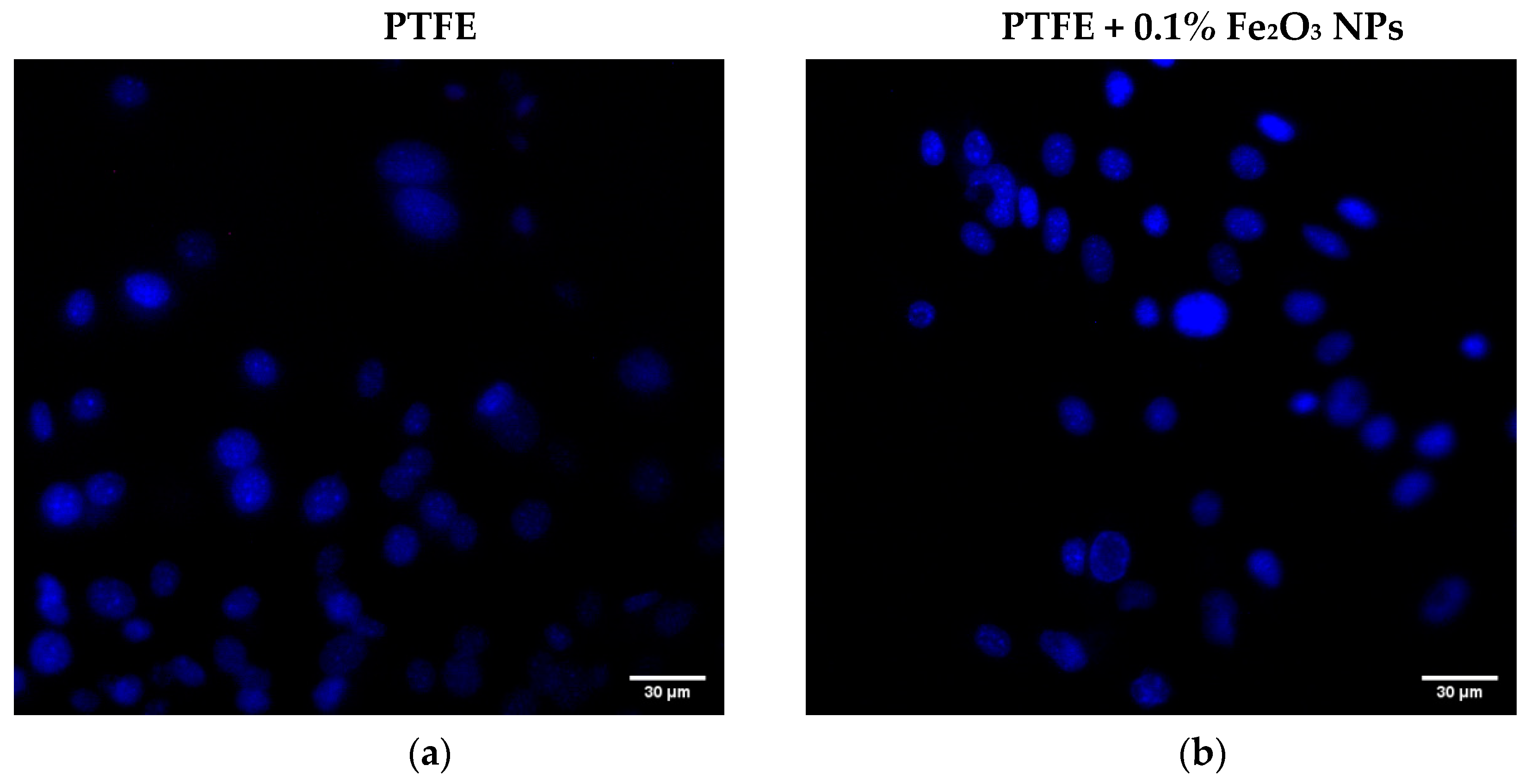
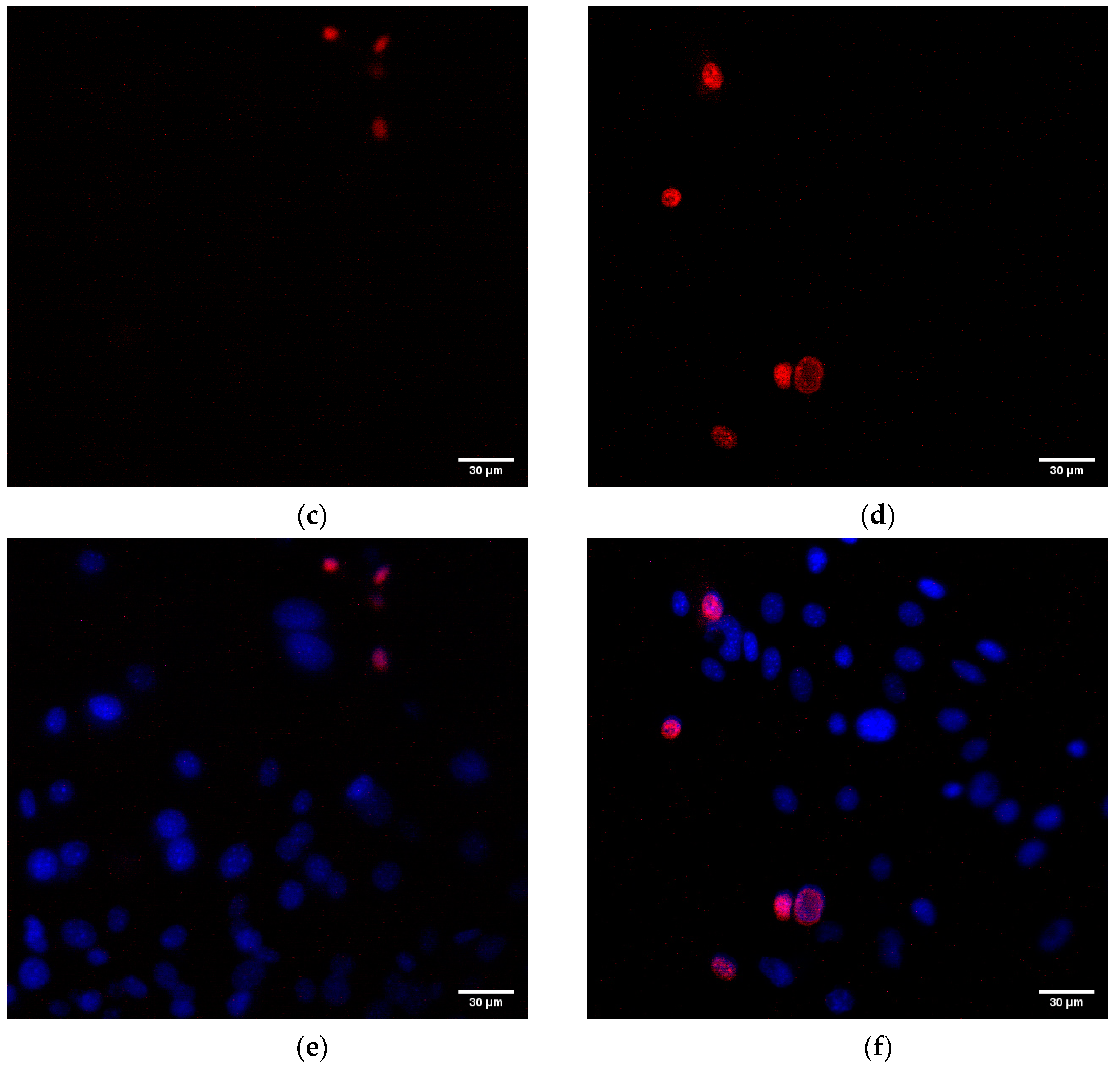
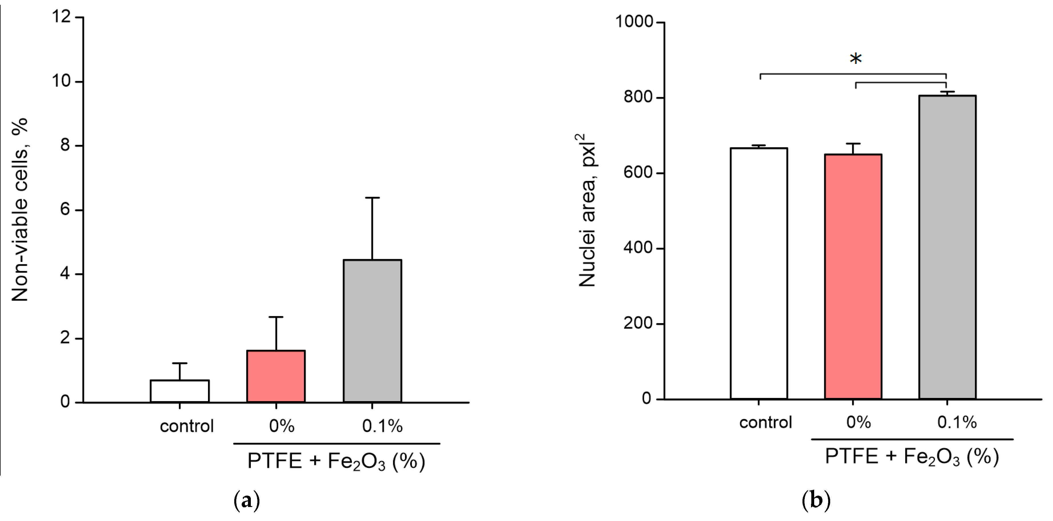
3.5. Influence on the Animal Cells Viability

At the last stage of this research, the effect of the obtained coatings on the viability of animal cells was studied using murine fibroblasts as an example. As criteria for evaluation, the proportion of dead cells and the size of the nucleus were chosen. The proportion of dead cells was defined as the ratio of the number of dead cells whose nuclei were stained with PI (Figure 7c,d) to the total number of cells whose nuclei were stained with Hoechst33342 only (Figure 7a,b).
Examples of staining with both dyes are shown in Figure 7e,f. The number of dead cells in the control was 0.7 ± 0.5% (Figure 8a). The average value of the proportion of dead cells on the PTFE without nanoparticles was 1.8 ± 1.0%, however, statistically significant differences in the proportion of dead cells between the PTFE and control were not found. The proportion of dead cells on the PTFE/Fe2O3 NPs 0.1% composite was 4.5 ± 1.9%. Despite the significant difference in the mean values, statistical significance between the proportions of dead cells during cultivation on PTFE/Fe2O3 NPs 0.1%, PTFE without nanoparticles, and/or the control was not found.
The mean nuclear area in the control was 665 ± 7 pxl2 (Figure 8b). When cultured on PTFE without nanoparticles, the cell nuclei had a size comparable to the control, i.e., 650 ± 28 pxl2. During cultivation on the PTFE/Fe2O3 NPs 0.1% composite, an increase in the average nucleus area up to 806 ± 11 pxl2 was found, which was statistically different from the control and cultivation on PTFE.

4. Discussion

The aim of this study was to obtain a new composite material of PTFE/Fe2O3 NPs, to study its physical properties and surface features, its influence on the generation of reactive oxygen species, 8-oxoguanine in DNA, and long-lived active protein species (LRPS), its bacteriostatic activity, and its cytotoxicity against eukaryotic cells.
In earlier studies, materials based on different polymer matrices (borosiloxane and PLGA) and nanoparticles of ZnO and Ag2O oxides were obtained [35,42]. In the present study, we developed a new composite coating based on PTFE and Fe2O3 NPs. Fe2O3 nanoparticles for the fabrication of the material were obtained by laser ablation in water. The resulting nanoparticles were about 60 nm in size (Figure 1a) and were spherical in shape (Figure 1d). These data were consistent with the literature data [43,44]. The ζ-potential values of the synthesized nanoparticles ranged from −69 to −7 mV with an average value of −25 mV at pH 6.7 ± 0.1 (Figure 1b). The obtained values of the ζ-potential indicated the satisfactory stability of the aqueous colloids synthesized with Fe2O3 NPs [45].
After obtaining the PTFE/Fe2O3 NP composite, its surface was characterized by the AFM method (Figure 2), and the distribution of NPs in the thickness of the polymer matrix was also estimated (Figure 3). On the surface of the PTFE without NPs, noticeable microdamages or defects (cracks, scales, etc.) were not found (Figure 2a,b). The surface of the PTFE/Fe2O3 NP composite was also smooth and free from microdamages (Figure 2c,d). Using the MIM method, it was found that the nanoparticles formed aggregates or clusters in the thickness of the polymer matrix of the PTFE/Fe2O3 NP composite (Figure 3b–d), which was consistent with data obtained in the earlier studies of the research team on polymer composites with nanoparticles of oxides of other metals [36].
There are several ways to additionally increase the strength of PTFE coatings such as adding selenium microparticles to the polymer matrix or creating composites with complex compositions, such as polyoxymethylene/glass fiber/polytetrafluoroethylene and polytetrafluoroethylene/polysulfone [46,47,48]. The inclusion of graphene, poly-p-phenyleneterephthalamide fibers, or glass fibers in the PTFE matrix makes it possible to modify the mechanical properties of PTFE (impact and tensile strength, hardness) [49]. The possibility of increasing the strength of composites of epoxy resins and PTFE by adding TiO2 powder to the matrix has been reported [50]. The search for methods to improve the physical properties of the developed composites is a task of further research.
The formation of moderate amounts of ROS normally occurs in any aerobic cell. In addition, ROS play an important role in the regulation of proliferation, differentiation, and migration of eukaryotic cells [51]. Excess ROS is normally removed during the functioning of antioxidant systems. Excessive generation of ROS, often caused by external causes, can lead to a state of “oxidative stress” that is dangerous for the integrity and functioning of cells [23,52,53]. At the cellular level, it can lead to mutagenesis, protein inactivation, lipid peroxidation, etc., [54]. At the level of the organism, “oxidative stress” is associated with carcinogenesis, genotoxic effects, and aging [41]. An important role in triggering “oxidative stress” is played by hydrogen peroxide (the most stable ROS) and hydroxyl radicals (the most reactive ROS) [31,55,56]. In addition to ROS, there are other inducers and markers of “oxidative stress” such as 8-oxoguanine, which is formed during the oxidative modification of DNA, and LRPS, which are formed during protein modification. LRPS themselves can be sources of new secondary radicals [57]. Considering all this, the ability of the developed PTFE/Fe2O3 NPs composite material to influence the generation of ROS using the examples of H2O2 and hydroxyl radicals, 8-oxoguanine, and LRPS was tested. It was found that the PTFE did not affect the generation of all the tested compounds (Figure 4 and Figure 5). The composite material of PTFE/Fe2O3 NPs increased ROS generation compared to the control (Figure 4 and Figure 5). However, the amount of ROS generated was negligible and should not cause oxidative stress [58].
The coating based on PTFE/Fe2O3 NPs showed a significant bacteriostatic effect (Figure 6) at a Fe2O3 NPs concentration of 0.1%, which corresponded approximately to 10 µg/mL. It was found that the PTFE without nanoparticles did not affect bacterial growth. Therefore, the bacteriostatic effect was provided by the Fe2O3 NPs. Several mechanisms of the antibacterial action of metal oxide nanoparticles have been described in the literature. The first mechanism is the direct disruption of the integrity of bacterial cell walls [59,60,61,62,63]. The second mechanism is the realizing of metal ions (in our case Fe2+), which bind to the SH groups of bacterial proteins. This leads to protein inactivation, an imbalance between the formation of ROS and the functioning of antioxidant systems, which can cause "oxidative stress", and the disruption of the functioning of ATPases [22,64,65,66,67,68,69,70]. The third mechanism is the generation of ROS as a result of the Fenton reaction or similar reactions with the participation of Fe2+ cations released from NPs [71,72]. The fourth mechanism is photocatalysis or the enhancement of ROS generation in the presence of light [73]. The fifth mechanism is the inhibition of DNA replication and genotoxic effects [48,74]. The sixth mechanism is unique for the NPs of some iron oxides with magnetic properties. When an external alternating magnetic field is applied, iron oxide NPs can cause the heating and dissociation of bacterial biofilms and the destruction of individual cells [75,76]. However, these magnetic properties are characteristic of Fe3O4; therefore, in our case, the implementation of this mechanism was extremely unlikely. Using the AFM and MIM methods, we showed that NPs were distributed in the thickness of the polymer matrix (Figure 2 and Figure 3). Based on this, we could assume that the probable mechanisms of the bacteriostatic action of the Fe2O3 NPs in our case were those that are due to the release of Fe2+ from NPs (the second and third mechanisms). However, the participation of the whole NPs in the bacteriostatic activity of the PTFE/Fe2O3 NPs composite coating could not be completely ruled out. It is described in the literature that the release of NP clusters from a polymer matrix is one of the mechanisms of the antibacterial action of nanocomposites [77]. It can be assumed that the prolonged bacteriostatic effects may have been associated with the gradual release of NPs or ions from the composite coating. It should be noted that the Fe2O3 could “protrude” from the polymer matrix in part of the NPs by an insignificant amount (less than 5–10 nm above the surface level, Figure 2), so the “contact” mechanism (first) of antibacterial activity could not be completely excluded. Clarification of the mechanisms of the antibacterial activity of PTFE/Fe2O3 NPs is a task for further research. An evaluation of the degree and duration of the release of Fe2O3 NPs from the polymer matrix during use, the dynamics of the generation of reactive species, and the antibacterial action of the released Fe2O3 NPs is planned to be studied in the future.
A comparison of these results with the data of earlier research is shown in Table 1. The antibacterial activity of iron oxide NPs depends on the degree of oxidation of iron in the oxide and the size of the NPs. NPs with large sizes exhibit lower antibacterial activity than smaller ones [78,79,80]. The nanoparticles obtained in these particular experiments had a size of about 50–70 nm and an MIC of 10 μg/mL, which was consistent with the literature data [79,81]. Fe2O3 NPs have a more pronounced antibacterial activity than Fe3O4 NPs [60,81,82]. The elemental composition of the nanoparticles obtained in this study by laser ablation in water was established by the research team earlier and corresponded to Fe2O3 [83]. These results were consistent with the data on the higher toxicity of Fe2O3 nanoparticles than Fe3O4 nanoparticles against bacteria. Combinations of Fe2O3 nanoparticles with nanoparticles of other metals and their oxides and polymer materials are considered as ways to improve antibacterial properties or reduce cytotoxicity against animal and human cells [84,85,86]. In this case, the antibacterial effect largely depends on the type of polymer used. For example, the addition of chitosan gives a more pronounced bacteriostatic effect than carbon nanotubes or antibiotics [85,86,87]. There are few published studies on the use of metal oxide nanoparticles in a PTFE-based matrix, and these are devoted to the study of the antimicrobial properties of Ag nanoparticles [27,88]. The concentrations of Ag nanoparticles at which the growth of microorganisms is inhibited (5–50 mg/mL) are, strictly speaking, the only ones used from the relevant works [27,88]; therefore, their MICs can be much lower than the indicated values. Previously, a composite material based on PLGA and Fe2O3 NPs was obtained. It also showed excellent antibacterial properties with an MIC of 10 μg/mL [83]. It is noteworthy that the MIC ~10 μm/mL did not depend on the type of polymer matrix (PLGA or PTFE) [83]. Based on the data of the present and earlier studies, it can be concluded that the inclusion of metal oxide nanoparticles inside the polymer matrix is a more effective way of improving the antimicrobial properties of materials than coating the individual nanoparticles with conjugates [27,83,86,87].
The literature describes other approaches to create antibacterial coatings for cutting boards with increased strength, in particular, the reconciliation of food-safe oil coatings [12,89]. These coatings make it possible to effectively fill microdamages and reduce the attachment of bacteria to the surface of cutting boards [12,89,90]. However, the application process of food-safe oil coatings is multi-stage. We made an attempt to create a coating that can be applied in one stage. PTFE was chosen by us because it describes a possible application of PTFE in the food industry. In particular, PTFE has shown itself well as a material for reducing adhesion to the working surfaces of units at milk processing plants [12,91]. It has also been shown that the adhesion of bacteria to working surfaces made of PTFE is reduced compared to other materials [92]. However, in our case, the pure PTFE did not have a bacteriostatic effect, which indicated that the antibacterial effect was realized through Fe2O3 NP-dependent mechanisms.
At the last stage of the study, the developed composite material was tested for its cytotoxicity against eukaryotic cells using mouse fibroblasts as an example (Figure 4 and Figure 5). A significant difference was found in the toxicity of the PTFE composite with 0.1% Fe2O3 NPs against bacteria (85% cell death) (Figure 6) and animal cells (no significant increase in the number of dead cells) (Figure 8a). Such a difference in toxicity could be explained by the more advanced structure of the repair and antioxidant defense systems in eukaryotic cells compared to prokaryotes [93,94,95,96]. Despite the absence of an increase in the number of dead cells, the PTFE/Fe2O3 NP composite material could not be called completely inert with respect to eukaryotic cells. An increase in the size of cell nuclei compared to the control with the PTFE without NPs was found (Figure 8b), which may indicate the onset of cell death processes and was consistent with the literature data on the cytotoxic effect of Fe2O3 NPs [97,98]. It should be noted that the literature data on the cytotoxicity of Fe2O3 NPs are ambiguous. On the one hand, some authors report a very low cytotoxicity of Fe2O3 NPs [66,99]. On the other hand, some works describe a clear cytotoxic effect of Fe2O3 NPs on epithelial cells (human lungs, HUVEC, hepatocytes, cell lines BEAS-2B and A549, etc.) due to the development of "oxidative stress", protein aggregation, impaired glucose metabolism, the excessive activation of the PI3K/Art-signaling pathway, and Caspase-3-dependent apoptosis [100,101,102,103,104]. The toxicity of Fe2O3 NPs in vivo has also been reported, with a predominance of kidney and liver damage [100]. Our results indicated a very low cytotoxicity of Fe2O3 NPs (Figure 8a); however, it may increase with prolonged use of the material, which was indirectly evidenced by an increase in the size of nuclei (Figure 8b). The received data about the count of dead cells (Figure 8) were consistent with the literature data about the good biocompatibility of Fe2O3 NPs. The search for the optimal concentration of Fe2O3 NPs and/or a new type of conjugates for NPs (glycol, proteins (BSA), or peptides (Arg-Gly-Asp) [105,106,107]) to ensure a balance between the high bacteriostatic activity of the developed composite material and the absence of toxicity against eukaryotic cells is a task for further research in this direction.

5. Conclusions

A new coating based on PTFE and Fe2O3 NPs was successfully developed in this study. The obtained coating had high adhesion to cutting boards made from PTFE and demonstrated a high surface quality (no defects at the microscopic level). The PTFE/Fe2O3 NP coating increased the generation of H2O2, 8-oxoguanine, and LRPS. The effect of the composite coating on reactive species generation depended on dose of Fe2O3 NPs in the PTFE matrix. The PTFE/Fe2O3 NP composites with NP concentrations of 0.001–0.1% inhibited bacterial growth, wherein the PTFE/Fe2O3 NPs (0.1% NPs) had no cytotoxicity against animal cells. These properties make coatings based on PTFE and Fe2O3 NPs a promising candidate as a protective coating for cutting boards in the meat processing industry.

Author Contributions

Conceptualization, D.A.S. and S.V.G.; methodology, A.V.S.; software, M.E.A.; validation, A.S.B.; formal analysis, D.V.Y.; investigation, D.A.S., I.V.B., D.E.B., M.E.A. and A.V.S.; writing—original draft preparation, D.A.S.; writing—review and editing, S.V.G.; visualization, D.A.S.; project administration, S.V.G. All authors have read and agreed to the published version of the manuscript.

Funding

This work was supported by a grant of the Ministry of Science and Higher Education of the Russian Federation (075-15-2022-315) for the organization and development of a world-class research center “Photonics”.

Institutional Review Board Statement

The animal study protocol was approved by the regulatory legal act of the laboratory of public health of the Russian Federation No. 199-n “General cases of application of the rules of good practice, international legal standards for use in appropriate conditions” ETS No. 123 “On the protection of vertebrate animals, and scientific research”.

Informed Consent Statement

Not applicable.

Data Availability Statement

The raw data supporting the conclusions of this article will be made available by the authors, without undue reservation.

Acknowledgments

The authors thank the Shared Use Center of the GPI RAS.

Conflicts of Interest

The authors declare no conflict of interest.

References

  1. Sekoai, P.T.; Feng, S.; Zhou, W.; Ngan, W.Y.; Pu, Y.; Yao, Y.; Pan, J.; Habimana, O. Insights into the Microbiological Safety of Wooden Cutting Boards Used for Meat Processing in Hong Kong's Wet Markets: A Focus on Food-Contact Surfaces, Cross-Contamination and the Efficacy of Traditional Hygiene Practices. Microorganisms 2020, 8, 579. [Google Scholar] [CrossRef] [PubMed]
  2. Wang, R. Biofilms and Meat Safety: A Mini-Review. J. Food Prot. 2018, 82, 120–127. [Google Scholar] [CrossRef]
  3. Lo, M.Y.; Ngan, W.Y.; Tsun, S.M.; Hsing, H.L.; Lau, K.T.; Hung, H.P.; Chan, S.L.; Lai, Y.Y.; Yao, Y.; Pu, Y.; et al. A Field Study Into Hong Kong’s Wet Markets: Raised Questions Into the Hygienic Maintenance of Meat Contact Surfaces and the Dissemination of Microorganisms Associated With Nosocomial Infections. Front. Microbiol. 2019, 10, 2618. [Google Scholar] [CrossRef] [PubMed]
  4. Dantas, S.T.A.; Rossi, B.F.; Bonsaglia, E.C.R.; Castilho, I.G.; Hernandes, R.T.; Fernandes, A.J.; Rall, V.L.M. Cross-Contamination and Biofilm Formation by Salmonella enterica Serovar Enteritidis on Various Cutting Boards. Foodborne Pathog. Dis. 2018, 15, 81–85. [Google Scholar] [CrossRef] [PubMed] [Green Version]
  5. Srey, S.; Jahid, I.K.; Ha, S.-D. Biofilm formation in food industries: A food safety concern. Food Control. 2013, 31, 572–585. [Google Scholar] [CrossRef]
  6. Scallan, E.; Griffin, P.M.; Angulo, F.J.; Tauxe, R.V.; Hoekstra, R.M. Foodborne illness acquired in the United States—Unspecified agents. Emerg. Infect. Dis. 2011, 17, 16–22. [Google Scholar] [CrossRef] [PubMed]
  7. Aspell CPolymer science VRGowariker, N.V. Viswanathan and Jayadev Sreedhar, Halsted Press (John Wiley & Sons), New York, 1986. pp. xv + 505, ISBN 0-470-20322-6. Br. Polym. J. 1988, 20, 88. [Google Scholar] [CrossRef]
  8. Dhanumalayan, E.; Joshi, G.M. Performance properties and applications of polytetrafluoroethylene (PTFE)—A review. Adv. Compos. Hybrid Mater. 2018, 1, 247–268. [Google Scholar] [CrossRef]
  9. Burkarter, E.; Saul, C.K.; Thomazi, F.; Cruz, N.C.; Roman, L.S.; Schreiner, W.H. Superhydrophobic electrosprayed PTFE. Surf. Coat. Technol. 2007, 202, 194–198. [Google Scholar] [CrossRef]
  10. Dearn, K.D.; Hoskins, T.J.; Petrov, D.G.; Reynolds, S.C.; Banks, R. Applications of dry film lubricants for polymer gears. Wear 2013, 298–299, 99–108. [Google Scholar] [CrossRef]
  11. Mccook, N.L.; Burris, D.L.; Dickrell, P.L.; Sawyer, W.G. Cryogenic Friction Behavior of PTFE based Solid Lubricant Composites. Tribol. Lett. 2005, 20, 109–113. [Google Scholar] [CrossRef] [Green Version]
  12. Barish, J.A.; Goddard, J.M. Anti-fouling surface modified stainless steel for food processing. Food Bioprod. Processing 2013, 91, 352–361. [Google Scholar] [CrossRef]
  13. O’brien, M.; Baxendale, I.R.; Ley, S.V. Flow Ozonolysis Using a Semipermeable Teflon AF-2400 Membrane To Effect Gas−Liquid Contact. Org. Lett. 2010, 12, 1596–1598. [Google Scholar] [CrossRef]
  14. Teo, A.J.T.; Mishra, A.; Park, I.; Kim, Y.-J.; Park, W.-T.; Yoon, Y.-J. Polymeric Biomaterials for Medical Implants and Devices. ACS Biomater. Sci. Eng. 2016, 2, 454–472. [Google Scholar] [CrossRef] [PubMed]
  15. Greene, J.F.; Preger, Y.; Stahl, S.S.; Root, T.W. PTFE-Membrane Flow Reactor for Aerobic Oxidation Reactions and Its Application to Alcohol Oxidation. Org. Process Res. Dev. 2015, 19, 858–864. [Google Scholar] [CrossRef]
  16. Rodriguez-Lopez, J.; Alpuche-Aviles, M.A.; Bard, A.J. Selective Insulation with Poly(tetrafluoroethylene) of Substrate Electrodes for Electrochemical Background Reduction in Scanning Electrochemical Microscopy. Anal. Chem. 2008, 80, 1813–1818. [Google Scholar] [CrossRef]
  17. Feng, S.; Zhong, Z.; Zhang, F.; Wang, Y.; Xing, W. Amphiphobic Polytetrafluoroethylene Membranes for Efficient Organic Aerosol Removal. ACS Appl. Mater. Interfaces 2016, 8, 8773–8781. [Google Scholar] [CrossRef]
  18. Rondinella, A.; Andreatta, F.; Turrin, D.; Fedrizzi, L. Degradation Mechanisms Occurring in PTFE-Based Coatings Employed in Food-Processing Applications. Coatings 2021, 11, 1419. [Google Scholar] [CrossRef]
  19. Chao, Q.; Meng, L.; Shuxian, C. Anti-icing characteristics of PTFE super hydrophobic coating on titanium alloy surface. J. Alloys Compd. 2021, 860, 157907. [Google Scholar] [CrossRef]
  20. Song, W.; Wang, S.; Lu, Y.; Zhang, X.; Xia, Z. Friction behavior of PTFE-coated Si3N4/TiC ceramics fabricated by spray technique under dry friction. Ceram. Int. 2021, 47, 7487–7496. [Google Scholar] [CrossRef]
  21. Wu, X.; Yang, F.; Gan, J.; Zhao, W.; Wu, Y. A flower-like waterborne coating with self-cleaning, self-repairing properties for superhydrophobic applications. J. Mater. Res. Technol. 2021, 14, 1820–1829. [Google Scholar] [CrossRef]
  22. Gudkov, S.V.; Burmistrov, D.E.; Smirnova, V.V.; Semenova, A.A.; Lisitsyn, A.B. A Mini Review of Antibacterial Properties of Al2O3 Nanoparticles. Nanomaterials 2022, 12, 2635. [Google Scholar] [CrossRef] [PubMed]
  23. Gudkov, S.V.; Burmistrov, D.E.; Serov, D.A.; Rebezov, M.B.; Semenova, A.A.; Lisitsyn, A.B. Do Iron Oxide Nanoparticles Have Significant Antibacterial Properties? Antibiotics 2021, 10, 884. [Google Scholar] [CrossRef]
  24. Chausov, D.N.; Burmistrov, D.E.; Kurilov, A.D.; Bunkin, N.F.; Astashev, M.E.; Simakin, A.V.; Vedunova, M.V.; Gudkov, S.V. New Organosilicon Composite Based on Borosiloxane and Zinc Oxide Nanoparticles Inhibits Bacterial Growth, but Does Not Have a Toxic Effect on the Development of Animal Eukaryotic Cells. Materials 2021, 14, 6281. [Google Scholar] [CrossRef]
  25. Gudkov, S.V.; Simakin, A.V.; Sarimov, R.M.; Kurilov, A.D.; Chausov, D.N. Novel Biocompatible with Animal Cells Composite Material Based on Organosilicon Polymers and Fullerenes with Light-Induced Bacteriostatic Properties. Nanomaterials 2021, 11, 2804. [Google Scholar] [CrossRef] [PubMed]
  26. Smirnova, V.V.; Chausov, D.N.; Serov, D.A.; Kozlov, V.A.; Ivashkin, P.I.; Pishchalnikov, R.Y.; Uvarov, O.V.; Vedunova, M.V.; Semenova, A.A.; Lisitsyn, A.B.; et al. A Novel Biodegradable Composite Polymer Material Based on PLGA and Silver Oxide Nanoparticles with Unique Physicochemical Properties and Biocompatibility with Mammalian Cells. Materials 2021, 14, 6915. [Google Scholar] [CrossRef] [PubMed]
  27. Zhang, S.; Liang, X.; Gadd, G.M.; Zhao, Q. A sol–gel based silver nanoparticle/polytetrafluorethylene (AgNP/PTFE) coating with enhanced antibacterial and anti-corrosive properties. Appl. Surf. Sci. 2021, 535, 147675. [Google Scholar] [CrossRef]
  28. Gudkov, S.V.; Baimler, I.V.; Uvarov, O.V.; Smirnova, V.V.; Volkov, M.Y.; Semenova, A.A.; Lisitsyn, A.B. Influence of the Concentration of Fe and Cu Nanoparticles on the Dynamics of the Size Distribution of Nanoparticles. Front. Phys. 2020, 8, 578. [Google Scholar] [CrossRef]
  29. Sarimov, R.M.; Nagaev, E.I.; Matveyeva, T.A.; Binhi, V.N.; Burmistrov, D.E.; Serov, D.A.; Astashev, M.E.; Simakin, A.V.; Uvarov, O.V.; Khabatova, V.V.; et al. Investigation of Aggregation and Disaggregation of Self-Assembling Nano-Sized Clusters Consisting of Individual Iron Oxide Nanoparticles upon Interaction with HEWL Protein Molecules. Nanomaterials 2022, 12, 3960. [Google Scholar] [CrossRef]
  30. Ivanyuk, V.V.; Shkirin, A.V.; Belosludtsev, K.N.; Dubinin, M.V.; Kozlov, V.A.; Bunkin, N.F.; Dorokhov, A.S.; Gudkov, S.V. Influence of Fluoropolymer Film Modified With Nanoscale Photoluminophor on Growth and Development of Plants. Front. Phys. 2020, 8, 616040. [Google Scholar] [CrossRef]
  31. Gudkov, S.V.; Lyakhov, G.A.; Pustovoy, V.I.; Shcherbakov, I.A. Influence of Mechanical Effects on the Hydrogen Peroxide Concentration in Aqueous Solutions. Phys. Wave Phenom. 2019, 27, 141–144. [Google Scholar] [CrossRef]
  32. Gudkov, S.V.; Simakin, A.V.; Sevostyanov, M.A.; Konushkin, S.V.; Losertová, M.; Ivannikov, A.Y.; Kolmakov, A.G.; Izmailov, A.Y. Manufacturing and study of mechanical properties, structure and compatibility with biological objects of plates and wire from new Ti-25Nb-13Ta-5Zr alloy. Metals 2020, 10, 1584. [Google Scholar] [CrossRef]
  33. Gudkov, S.; Garmash, S.; Shtarkman, I.; Chernikov, A.; Karp, O.; Bruskov, V. Long-lived protein radicals induced by X-ray irradiation are the source of reactive oxygen species in aqueous medium. Biochem. Bioph. Rep. 2010, 430, 1. [Google Scholar] [CrossRef]
  34. Sharapov, M.; Novoselov, V.; Penkov, N.; Fesenko, E.; Vedunova, M.; Bruskov, V.; Gudkov, S. Protective and adaptogenic role of peroxiredoxin 2 (Prx2) in neutralization of oxidative stress induced by ionizing radiation. Free. Radic. Biol. Med. 2019, 134, 76–86. [Google Scholar] [CrossRef] [PubMed]
  35. Chausov, D.N.; Smirnova, V.V.; Burmistrov, D.E.; Sarimov, R.M.; Kurilov, A.D.; Astashev, M.E.; Uvarov, O.V.; Dubinin, M.V.; Kozlov, V.A.; Vedunova, M.V.; et al. Synthesis of a Novel, Biocompatible and Bacteriostatic Borosiloxane Composition with Silver Oxide Nanoparticles. Materials 2022, 15, 527. [Google Scholar] [CrossRef]
  36. Astashev, M.E.; Sarimov, R.M.; Serov, D.A.; Matveeva, T.A.; Simakin, A.V.; Ignatenko, D.N.; Burmistrov, D.E.; Smirnova, V.V.; Kurilov, A.D.; Mashchenko, V.I.; et al. Antibacterial behavior of organosilicon composite with nano aluminum oxide without influencing animal cells. React. Funct. Polym. 2022, 170, 105143. [Google Scholar] [CrossRef]
  37. Seluanov, A.; Vaidya, A.; Gorbunova, V. Establishing primary adult fibroblast cultures from rodents. J. Vis. Exp. 2010, 44, 2033. [Google Scholar] [CrossRef] [Green Version]
  38. Sevost’yanov, M.A.; Nasakina, E.O.; Baikin, A.S.; Sergienko, K.V.; Konushkin, S.V.; Kaplan, M.A.; Seregin, A.V.; Leonov, A.V.; Kozlov, V.A.; Shkirin, A.V.; et al. Biocompatibility of new materials based on nano-structured nitinol with titanium and tantalum composite surface layers: Experimental analysis in vitro and in vivo. J. Mater. Science. Mater. Med. 2018, 29, 33. [Google Scholar] [CrossRef]
  39. Kaplan, M.A.; Sergienko, K.V.; Kolmakova, A.A.; Konushkin, S.V.; Baikin, A.S.; Kolmakov, A.G.; Sevostyanov, M.A.; Kulikov, A.V.; Ivanov, V.E.; Belosludtsev, K.N.; et al. Development of a Biocompatible PLGA Polymers Capable to Release Thrombolytic Enzyme Prourokinase. J. Biomater. Sci. Polym. Ed. 2020, 31, 1405–1420. [Google Scholar] [CrossRef]
  40. Starinets, V.S.; Serov, D.A.; Penkov, N.V.; Belosludtseva, N.V.; Dubinin, M.V.; Belosludtsev, K.N. Alisporivir Normalizes Mitochondrial Function of Primary Mouse Lung Endothelial Cells Under Conditions of Hyperglycemia. Biochem. Mosc. 2022, 87, 605–616. [Google Scholar] [CrossRef]
  41. Klaunig, J.E.; Kamendulis, L.M.; Hocevar, B.A. Oxidative Stress and Oxidative Damage in Carcinogenesis. Toxicol. Pathol. 2009, 38, 96–109. [Google Scholar] [CrossRef] [PubMed] [Green Version]
  42. Burmistrov, D.E.; Simakin, A.V.; Smirnova, V.V.; Uvarov, O.V.; Ivashkin, P.I.; Kucherov, R.N.; Ivanov, V.E.; Bruskov, V.I.; Sevostyanov, M.A.; Baikin, A.S.; et al. Bacteriostatic and Cytotoxic Properties of Composite Material Based on ZnO Nanoparticles in PLGA Obtained by Low Temperature Method. Polymers 2022, 14, 49. [Google Scholar] [CrossRef] [PubMed]
  43. Amutha, S.; Sridhar, S. Green synthesis of magnetic iron oxide nanoparticle using leaves of Glycosmis mauritiana and their antibacterial activity against human pathogens. J. Innov. Pharm. Biol. Sci. 2018, 5, 22–26. [Google Scholar]
  44. Gabrielyan, L.; Hakobyan, L.; Hovhannisyan, A.; Trchounian, A. Effects of iron oxide (Fe3O4) nanoparticles on Escherichia coli antibiotic-resistant strains. J. Appl. Microbiol. 2019, 126, 1108–1116. [Google Scholar] [CrossRef] [PubMed]
  45. Kumar, A.; Dixit, C.K. 3—Methods for characterization of nanoparticles. In Advances in Nanomedicine for the Delivery of Therapeutic Nucleic Acids; Woodhead Publishing: Sawston, UK, 2017; pp. 43–58. Available online: https://www.sciencedirect.com/science/article/pii/B9780081005576000031 (accessed on 22 September 2022). [CrossRef]
  46. Martins, S.A.; Dias, F.W.; Nunes, L.S.; Borges, L.A.; D’almeida, J.R. Mechanical Characterization of Silica Reinforced-PTFE Matrix Composites. Procedia Eng. 2011, 10, 2651–2656. [Google Scholar] [CrossRef] [Green Version]
  47. Singh, J.S.K.; Ching, Y.C.; Liu, S.; Ching, K.Y.; Razali, S.; Gan, S.N. Effects of PTFE Micro-Particles on the Fiber-Matrix Interface of Polyoxymethylene/Glass Fiber/Polytetrafluoroethylene Composites. Materials 2018, 11, 2164. [Google Scholar] [CrossRef] [Green Version]
  48. Khayet, M.; Wang, R. Mixed Matrix Polytetrafluoroethylene/Polysulfone Electrospun Nanofibrous Membranes for Water Desalination by Membrane Distillation. ACS Appl. Mater. Interfaces 2018, 10, 24275–24287. [Google Scholar] [CrossRef]
  49. Xu, H.X.; Yuan, X.H.; Zhu, E.B.; Li, S.L.; Chen, L.; Han, Z.R. Comparison of Mechanical and Frictional Properties of PTFE Matrix Composites Reinforced by Different Fibers and Graphite. Key Eng. Mater. 2014, 575, 203–208. [Google Scholar]
  50. Ibrahim, N.H.; Romli, A.Z.; Ibrahim, N.N.I.N. Hardness optimization of epoxy filled PTFE with/without TiO2 filler. IOP Conf. Ser. Mater. Sci. Eng. 2020, 839, 012002. [Google Scholar] [CrossRef]
  51. Wegner, A.M.; Haudenschild, D.R. NADPH oxidases in bone and cartilage homeostasis and disease: A promising therapeutic target. J. Orthop. Res. Off. Publ. Orthop. Res. Soc. 2020, 38, 2104–2112. [Google Scholar] [CrossRef]
  52. Shcherbakov, I.A.; Baimler, I.V.; Gudkov, S.V.; Lyakhov, G.A.; Mikhailova, G.N.; Pustovoy, V.I.; Sarimov, R.M.; Simakin, A.V.; Troitsky, A.V. Influence of a Constant Magnetic Field on Some Properties of Water Solutions. Dokl. Phys. 2020, 65, 273–275. [Google Scholar] [CrossRef]
  53. Menshchikova, E.B.; Kozhin, P.M.; Chechushkov, A.V.; Khrapova, M.V.; Zenkov, N.K. The Oral Delivery of Water-Soluble Phenol TS-13 Ameliorates Granuloma Formation in an In Vivo Model of Tuberculous Granulomatous Inflammation. Oxidative Med. Cell. Longev. 2021, 2021, 6652775. [Google Scholar] [CrossRef] [PubMed]
  54. Cadet, J.; Douki, T.; Gasparutto, D.; Ravanat, J.-L. Oxidative damage to DNA: Formation, measurement and biochemical features. Mutat. Res. Fundam. Mol. Mech. Mutagenesis 2003, 531, 5–23. [Google Scholar] [CrossRef] [PubMed]
  55. Sharapov, M.G.; Gordeeva, A.E.; Goncharov, R.G.; Tikhonova, I.V.; Ravin, V.K.; Temnov, A.A.; Fesenko, E.E.; Novoselov, V.I. The Effect of Exogenous Peroxiredoxin 6 on the State of Mesenteric Vessels and the Small Intestine in Ischemia–Reperfusion Injury. Biophysics 2017, 62, 998–1008. [Google Scholar] [CrossRef]
  56. Winterbourn, C.C. Toxicity of iron and hydrogen peroxide: The Fenton reaction. Toxicol. Lett. 1995, 82–83, 969–974. [Google Scholar] [CrossRef]
  57. Burmistrov, D.E.; Yanykin, D.V.; Paskhin, M.O.; Nagaev, E.V.; Efimov, A.D.; Kaziev, A.V.; Ageychenkov, D.G.; Gudkov, S.V. Additive Production of a Material Based on an Acrylic Polymer with a Nanoscale Layer of Zno Nanorods Deposited Using a Direct Current Magnetron Discharge: Morphology, Photoconversion Properties, and Biosafety. Materials 2021, 14, 6586. [Google Scholar] [CrossRef] [PubMed]
  58. Sies, H.; Jones, D.P. Reactive oxygen species (ROS) as pleiotropic physiological signalling agents. Nat. Reviews. Mol. Cell Biol. 2020, 21, 363–383. [Google Scholar] [CrossRef] [PubMed]
  59. Gabrielyan, L.; Badalyan, H.; Gevorgyan, V.; Trchounian, A. Comparable antibacterial effects and action mechanisms of silver and iron oxide nanoparticles on Escherichia coli and Salmonella typhimurium. Sci. Rep. 2020, 10, 13145. [Google Scholar] [CrossRef] [PubMed]
  60. Gabrielyan, L.; Hovhannisyan, A.; Gevorgyan, V.; Ananyan, M.; Trchounian, A. Antibacterial effects of iron oxide (Fe3O4) nanoparticles: Distinguishing concentration-dependent effects with different bacterial cells growth and membrane-associated mechanisms. Appl. Microbiol. Biotechnol. 2019, 103, 2773–2782. [Google Scholar] [CrossRef]
  61. Sousa, C.; Sequeira, D.; Kolen’ko, Y.V.; Pinto, I.M.; Petrovykh, D.Y. Analytical Protocols for Separation and Electron Microscopy of Nanoparticles Interacting with Bacterial Cells. Anal. Chem. 2015, 87, 4641–4648. [Google Scholar] [CrossRef] [Green Version]
  62. Arakha, M.; Pal, S.; Samantarrai, D.; Panigrahi, T.K.; Mallick, B.C.; Pramanik, K.; Mallick, B.; Jha, S. Antimicrobial activity of iron oxide nanoparticle upon modulation of nanoparticle-bacteria interface. Sci. Rep. 2015, 5, 14813. [Google Scholar] [CrossRef] [PubMed]
  63. Gudkov, S.V.; Serov, D.A.; Astashev, M.E.; Semenova, A.A.; Lisitsyn, A.B. Ag2O Nanoparticles as a Candidate for Antimicrobial Compounds of the New Generation. Pharmaceuticals 2022, 15, 968. [Google Scholar] [CrossRef] [PubMed]
  64. Liu, Y.; He, L.; Mustapha, A.; Li, H.; Hu, Z.Q.; Lin, M. Antibacterial activities of zinc oxide nanoparticles against Escherichia coli O157:H7. J. Appl. Microbiol. 2009, 107, 1193–1201. [Google Scholar] [CrossRef] [PubMed]
  65. Saha, R.K.; Debanath, M.K.; Paul, B.; Medhi, S.; Saikia, E. Antibacterial and nonlinear dynamical analysis of flower and hexagon-shaped ZnO microstructures. Sci. Rep. 2020, 10, 2598. [Google Scholar] [CrossRef] [PubMed] [Green Version]
  66. Sihem, L.; Hanine, D.; Faiza, B. Antibacterial Activity of α-Fe2O3 and α-Fe2O3@Ag Nanoparticles Prepared by Urtica Leaf Extract. Nanotechnologies Russ. 2020, 15, 198–203. [Google Scholar] [CrossRef]
  67. Shkodenko, L.; Kassirov, I.; Koshel, E. Metal Oxide Nanoparticles Against Bacterial Biofilms: Perspectives and Limitations. Microorganisms 2020, 8, 1545. [Google Scholar] [CrossRef]
  68. Kudrinskiy, A.A.; Ivanov, A.Y.; Kulakovskaya, E.V.; Klimov, A.I.; Zherebin, P.M.; Khodarev, D.V.; Le, A.-T.; Tam, L.T.; Lisichkin, G.V.; Krutyakov, Y.A. The Mode of Action of Silver and Silver Halides Nanoparticles against Saccharomyces cerevisiae Cells. J. Nanoparticles 2014, 2014, 568635. [Google Scholar] [CrossRef] [Green Version]
  69. Sirelkhatim, A.; Mahmud, S.; Seeni, A.; Kaus, N.H.M.; Ann, L.C.; Bakhori, S.K.M.; Hasan, H.; Mohamad, D. Review on Zinc Oxide Nanoparticles: Antibacterial Activity and Toxicity Mechanism. Nano-Micro Lett. 2015, 7, 219–242. [Google Scholar] [CrossRef] [Green Version]
  70. Xie, Y.; He, Y.; Irwin, P.L.; Jin, T.; Shi, X. Antibacterial activity and mechanism of action of zinc oxide nanoparticles against Campylobacter jejuni. Appl. Environ. Microbiol. 2011, 77, 2325–2331. [Google Scholar] [CrossRef] [Green Version]
  71. Fenton, H.J.H. Oxidation of Tartaric Acid in Presence of Iron. J. Chem. Soc. Trans. 1894, 65, 899–910. [Google Scholar] [CrossRef] [Green Version]
  72. Abou-Gamra, Z.M. Kinetic and Thermodynamic Study for Fenton-Like Oxidation of Amaranth Red Dye. Adv. Chem. Eng. Sci. 2014, 4, 285–291. [Google Scholar] [CrossRef]
  73. Maji, S.K.; Mukherjee, N.; Mondal, A.; Adhikary, B. Synthesis, characterization and photocatalytic activity of α-Fe2O3 nanoparticles. Polyhedron 2012, 33, 145–149. [Google Scholar] [CrossRef]
  74. Ansari, M.O.; Parveen, N.; Ahmad, M.F.; Wani, A.L.; Afrin, S.; Rahman, Y.; Jameel, S.; Khan, Y.A.; Siddique, H.R.; Tabish, M.; et al. Evaluation of DNA interaction, genotoxicity and oxidative stress induced by iron oxide nanoparticles both in vitro and in vivo: Attenuation by thymoquinone. Sci. Rep. 2019, 9, 6912. [Google Scholar] [CrossRef] [PubMed] [Green Version]
  75. Kolen’ko, Y.V.; Bañobre-López, M.; Rodríguez-Abreu, C.; Carbó-Argibay, E.; Deepak, F.L.; Petrovykh, D.Y.; Cerqueira, M.F.; Kamali, S.; Kovnir, K.; Shtansky, D.V.; et al. High-Temperature Magnetism as a Probe for Structural and Compositional Uniformity in Ligand-Capped Magnetite Nanoparticles. J. Phys. Chem. C 2014, 118, 28322–28329. [Google Scholar] [CrossRef] [PubMed]
  76. Li, W.; Wei, W.; Wu, X.; Zhao, Y.; Dai, H. The antibacterial and antibiofilm activities of mesoporous hollow Fe3O4 nanoparticles in an alternating magnetic field. Biomater. Sci. 2020, 8, 4492–4507. [Google Scholar] [CrossRef] [PubMed]
  77. Dimitrakellis, P.; Kaprou, G.D.; Papavieros, G.; Mastellos, D.C.; Constantoudis, V.; Tserepi, A.; Gogolides, E. Enhanced antibacterial activity of ZnO-PMMA nanocomposites by selective plasma etching in atmospheric pressure. Micro Nano Eng. 2021, 13, 100098. [Google Scholar] [CrossRef]
  78. Ismail, R.A.; Sulaiman, G.M.; Abdulrahman, S.A.; Marzoog, T.R. Antibacterial activity of magnetic iron oxide nanoparticles synthesized by laser ablation in liquid. Mater. Sci. Eng. C 2015, 53, 286–297. [Google Scholar] [CrossRef]
  79. Saqib, S.; Munis, M.F.H.; Zaman, W.; Ullah, F.; Shah, S.N.; Ayaz, A.; Farooq, M.; Bahadur, S. Synthesis, characterization and use of iron oxide nano particles for antibacterial activity. Microsc. Res. Tech. 2019, 82, 415–420. [Google Scholar] [CrossRef]
  80. Bashir, M.; Ali, S.; Farrukh, M.A. Green synthesis of Fe2O3 nanoparticles from orange peel extract and a study of its antibacterial activity. J. Korean Phys. Soc. 2020, 76, 848–854. [Google Scholar] [CrossRef]
  81. Prabhu, Y.; Rao, K.V.; Kumari, B.S.; Kumar, V.S.S.; Pavani, T. Synthesis of Fe3O4 nanoparticles and its antibacterial application. Int. Nano Lett. 2015, 5, 85–92. [Google Scholar] [CrossRef]
  82. Al-Kattan, A.; Grojo, D.; Drouet, C.; Mouskeftaras, A.; Delaporte, P.; Casanova, A.; Robin, J.D.; Magdinier, F.; Alloncle, P.; Constantinescu, C. Short-Pulse Lasers: A Versatile Tool in Creating Novel Nano-/Micro-Structures and Compositional Analysis for Healthcare and Wellbeing Challenges. Nanomaterials 2021, 11, 712. [Google Scholar] [CrossRef] [PubMed]
  83. Gudkov, S.V.; Burmistrov, D.E.; Lednev, V.N.; Simakin, A.V.; Uvarov, O.V.; Kucherov, R.N.; Ivashkin, P.I.; Dorokhov, A.S.; Izmailov, A.Y. Biosafety Construction Composite Based on Iron Oxide Nanoparticles and PLGA. Inventions 2022, 7, 61. [Google Scholar] [CrossRef]
  84. Bhushan, M.; Kumar, Y.; Periyasamy, L.; Viswanath, A.K. Antibacterial applications of α-Fe2O3/Co3O4 nanocomposites and study of their structural, optical, magnetic and cytotoxic characteristics. Appl. Nanosci. 2018, 8, 137–153. [Google Scholar] [CrossRef] [Green Version]
  85. Bhattacharya, P.; Neogi, S. Gentamicin coated iron oxide nanoparticles as novel antibacterial agents. Mater. Res. Express 2017, 4, 095005. [Google Scholar] [CrossRef]
  86. Nehra, P.; Chauhan, R.; Garg, N.; Verma, K. Antibacterial and antifungal activity of chitosan coated iron oxide nanoparticles. Br. J. Biomed. Sci. 2018, 75, 13–18. [Google Scholar] [CrossRef] [PubMed]
  87. Khashan, K.S.; Sulaiman, G.M.; Mahdi, R. Preparation of iron oxide nanoparticles-decorated carbon nanotube using laser ablation in liquid and their antimicrobial activity. Artif. Cells Nanomed. Biotechnol. 2017, 45, 1699–1709. [Google Scholar] [CrossRef] [PubMed] [Green Version]
  88. Zhang, S.; Wang, L.; Liang, X.; Vorstius, J.; Keatch, R.; Corner, G.; Nabi, G.; Davidson, F.; Gadd, G.M.; Zhao, Q. Enhanced Antibacterial and Antiadhesive Activities of Silver-PTFE Nanocomposite Coating for Urinary Catheters. ACS Biomater. Sci. Eng. 2019, 5, 2804–2814. [Google Scholar] [CrossRef]
  89. Maccallum, N.; Howell, C.; Kim, P.; Sun, D.; Friedlander, R.; Ranisau, J.; Ahanotu, O.; Lin, J.J.; Vena, A.; Hatton, B.; et al. Liquid-Infused Silicone As a Biofouling-Free Medical Material. ACS Biomater. Sci. Eng. 2015, 1, 43–51. [Google Scholar] [CrossRef]
  90. Awad, T.S.; Asker, D.; Hatton, B.D. Food-Safe Modification of Stainless Steel Food-Processing Surfaces to Reduce Bacterial Biofilms. ACS Appl. Mater. Interfaces 2018, 10, 22902–22912. [Google Scholar] [CrossRef]
  91. Huang, K.; Goddard, J.M. Influence of fluid milk product composition on fouling and cleaning of Ni–PTFE modified stainless steel heat exchanger surfaces. J. Food Eng. 2015, 158, 22–29. [Google Scholar] [CrossRef]
  92. Zore, A.; Bezek, K.; Jevšnik, M.; Abram, A.; Runko, V.; Slišković, I.; Raspor, P.; Kovačević, D.; Bohinc, K. Bacterial adhesion rate on food grade ceramics and Teflon as kitchen worktop surfaces. Int. J. Food Microbiol. 2020, 332, 108764. [Google Scholar] [CrossRef] [PubMed]
  93. Li, D.; Shen, M.; Xia, J.; Shi, X. Recent developments of cancer nanomedicines based on ultrasmall iron oxide nanoparticles and nanoclusters. Nanomedicine 2021, 16, 609–612. [Google Scholar] [CrossRef] [PubMed]
  94. Shields, H.J.; Traa, A.; Van Raamsdonk, J.M. Beneficial and Detrimental Effects of Reactive Oxygen Species on Lifespan: A Comprehensive Review of Comparative and Experimental Studies. Front. Cell Dev. Biol. 2021, 9, 628157. [Google Scholar] [CrossRef] [PubMed]
  95. Aguirre, J.; Ríos-Momberg, M.; Hewitt, D.; Hansberg, W. Reactive oxygen species and development in microbial eukaryotes. Trends Microbiol. 2005, 13, 111–118. [Google Scholar] [CrossRef] [PubMed]
  96. Branzei, D.; Foiani, M. Regulation of DNA repair throughout the cell cycle. Nat. Rev. Mol. Cell Biol. 2008, 9, 297–308. [Google Scholar] [CrossRef]
  97. Eidet, J.R.; Pasovic, L.; Maria, R.; Jackson, C.J.; Utheim, T.P. Objective assessment of changes in nuclear morphology and cell distribution following induction of apoptosis. Diagn. Pathol. 2014, 9, 92. [Google Scholar] [CrossRef] [PubMed] [Green Version]
  98. Filippi-Chiela, E.C.; Oliveira, M.M.; Jurkovski, B.; Callegari-Jacques, S.M.; Da Silva, V.D.; Lenz, G. Nuclear morphometric analysis (NMA): Screening of senescence, apoptosis and nuclear irregularities. PLoS ONE 2012, 7, e0042522. [Google Scholar] [CrossRef] [Green Version]
  99. Saliani, M.; Jalal, R.; Goharshadi, E.K. Effects of pH and temperature on antibacterial activity of zinc oxide nanofluid against Escherichia coli O157: H7 and Staphylococcus aureus. Jundishapur J. Microbiol. 2015, 8, 17115. [Google Scholar] [CrossRef] [Green Version]
  100. Hanini, A.; Schmitt, A.; Kacem, K.; Chau, F.; Ammar, S.; Gavard, J. Evaluation of iron oxide nanoparticle biocompatibility. Int. J. Nanomed. 2011, 6, 787–794. [Google Scholar] [CrossRef] [Green Version]
  101. Arias, L.S.; Pessan, J.P.; Vieira, A.P.M.; Lima, T.M.T.; Delbem, A.C.B.; Monteiro, D.R. Iron Oxide Nanoparticles for Biomedical Applications: A Perspective on Synthesis, Drugs, Antimicrobial Activity, and Toxicity. Antibiotics 2018, 7, 46. [Google Scholar] [CrossRef] [Green Version]
  102. Yarjanli, Z.; Ghaedi, K.; Esmaeili, A.; Rahgozar, S.; Zarrabi, A. Iron oxide nanoparticles may damage to the neural tissue through iron accumulation, oxidative stress, and protein aggregation. BMC Neurosci. 2017, 18, 51. [Google Scholar] [CrossRef] [PubMed]
  103. Sarkar, A.; Sil, P.C. Iron oxide nanoparticles mediated cytotoxicity via PI3K/AKT pathway: Role of quercetin. Food Chem. Toxicol. Int. J. Publ. Br. Ind. Biol. Res. Assoc. 2014, 71, 106–115. [Google Scholar] [CrossRef]
  104. Lai, X.; Wei, Y.; Zhao, H.; Chen, S.; Bu, X.; Lu, F.; Qu, D.; Yao, L.; Zheng, J.; Zhang, J. The effect of Fe2O3 and ZnO nanoparticles on cytotoxicity and glucose metabolism in lung epithelial cells. J. Appl. Toxicol. JAT 2015, 35, 651–664. [Google Scholar] [CrossRef] [PubMed]
  105. Pelaz, B.; Del Pino, P.; Maffre, P.; Hartmann, R.; Gallego, M.; Rivera-Fernández, S.; De La Fuente, J.M.; Nienhaus, G.U.; Parak, W.J. Surface Functionalization of Nanoparticles with Polyethylene Glycol: Effects on Protein Adsorption and Cellular Uptake. ACS Nano 2015, 9, 6996–7008. [Google Scholar] [CrossRef] [PubMed]
  106. Yu, S.; Perálvarez-Marín, A.; Minelli, C.; Faraudo, J.; Roig, A.; Laromaine, A. Albumin-coated SPIONs: An experimental and theoretical evaluation of protein conformation, binding affinity and competition with serum proteins. Nanoscale 2016, 8, 14393–14405. [Google Scholar] [CrossRef] [PubMed] [Green Version]
  107. Liu, X.; Yang, X.L.; Hu, Q.; Liu, M.S.; Peng, T.; Xu, W.F.; Huang, Q.H.; Li, B.; Liao, X.L. Arg-Gly-Asp Peptide-Functionalized Superparamagnetic γ-Fe2O3 Nanoparticles Enhance Osteoblast Migration In Vitro. J. Nanosci. Nanotechnol. 2020, 20, 6173–6179. [Google Scholar] [CrossRef]
Figure 1. Physical characteristics of Fe2O3 NPs: (a) size distribution histogram; (b) ζ-potential distribution histogram; (c) UV–visible absorbance spectrum; (d) TEM image (scale bar—1 μm).
Polymers 14 04880 g001
Figure 2. Example of applying a PTFE-based coating to a damaged surface of a fluoroplast before (left) and after (right) coating (a); AFM images of composite material surface: (b) 3D reconstruction of PTFE without NPs; (c) result of analysis of PTFE selected area (shown as a diagonal line in the figure) without NPs surface; (d) 3D reconstruction of PTFE/0.1% Fe2O3 NPs; (e) result of analysis of PTFE/0.1% Fe2O3 NPs surface. The results of the analysis in panels (c) and (e) are presented in the form of profiles and a table with data on the depth of the asperities in the sample.
Polymers 14 04880 g002
Figure 3. The results of the analysis of the obtained materials by MIM method: (a) PTFE without the addition of nanoparticles; (b) PTFE with the addition of 0.001% Fe2O3 nanoparticles; (c) PTFE with the addition of 0.01% Fe2O3 nanoparticles; (d) PTFE with the addition of 0.1% Fe2O3 nanoparticles. The images are presented as 3D reconstructions, where the abscissa and ordinate axes correspond to the real distance in μm. The Oz axis displays the phase difference in nm (the larger the phase difference, the higher the value on the Oz axis). Coloring is a pseudo-color. The initial data on the spatial distribution of the phase difference in the analyzed sample, used to construct 3D reconstructions, are shown in the lower left corners of each panel.
Polymers 14 04880 g003aPolymers 14 04880 g003b
Figure 4. Effect of PTFE/Fe2O3 NPs on generation of free radicals: (a) hydrogen peroxide (2 h, 40 °C); (b) OH radials (2 h, 80 °C). Data are presented as means ± standard errors. *—p < 0.05 vs. control, Mann–Whitney test (n = 3).
Polymers 14 04880 g004
Figure 5. Effect of PTFE/Fe2O3 NPs on generation of free radicals: (a) LRPS (2 h, 40 °C); (b) 8-oxoguanine (8-oxoGua) in DNA in vitro (2 h, 45 °C). Data are presented as means ± standard errors. *—p < 0.05 vs. control, Mann–Whitney test (n = 3).
Polymers 14 04880 g005
Figure 6. Examination of antibacterial activity of composite PTFE/Fe2O3 NPs against E. coli. Data are presented as means ± standard errors. *—p < 0.05 vs. control, Mann–Whitney test (n = 3).
Polymers 14 04880 g006
Figure 7. Examples of fluorescent microscopy images of cells during cytotoxicity study: (a) Hoechst (blue stain) image of cells on PTFE; (b) Hoechst image of cells on PTFE + 0.1% Fe2O3 NPs; (c) PI (red stain) of cells on PTFE; (d) PI of cells on PTFE + 0.1% Fe2O3 NPs; (e) merged image of cells on PTFE; (f) merged image of cells on PTFE + 0.1% Fe2O3 NPs. Scale bar is 30 μm. Magnification ×200, image resolution 1024 × 1024 pxl2.
Polymers 14 04880 g007aPolymers 14 04880 g007b
Figure 8. Cytotoxicity of composite PTFE/Fe2O3 NPs: (a) percentage of non-viable cells, where the amount of all analyzed cells was taken as 100%; (b) average nuclei size of cells. Data are presented as means ± standard errors. *—p < 0.05, Mann–Whitney test (n = 3).
Polymers 14 04880 g008
Table 1. The literature data on the antibacterial activity of composites of nanoparticles and polymer matrixes.
Synthesis MethodCompositionSizeMICMicroorganismEffectRef
1Laser ablation in dimethyl formamide or sodium dodecyl sulphateα-Fe2O350–1104.25 mg/mLE. coli,
P. aeruginosa,
S. aureus,
S. marcescens
BS 1[78]
2Co-precipitation methodFe2O325–4010–50 µg/mLE. coli,
S. aureus,
S. dysentery
BS[79]
3Co-precipitation methodFe2O3~500.5 mg/mLB. subtilis,
E. coli,
P. aeruginosa,
S. aureus
BS[80]
4Wet chemical methodFe3O433–4025–100 µg/mLE. coli,
P. vulgaris,
S. aureus,
Xanthomonas sp.
BS[81]
5Co-precipitation methodFe3O46–932–128 μg/mLE. coli,
L. monocytogenes,
P. aeruginosa,
S. marcescens
BS[82]
6Co-precipitation methodFe3O410.64 ± 4.7350–500 µg/mLE. coli,
E. hirae
BS[60]
7Co-precipitation methodα-Fe2O3,
ZnO/α-Fe2O3
~30400–800 µg/mLB. subtilis,
E. coli,
S. aureus,
S. typhimurium
BS[84]
8Co-precipitation methodFe2O3, FeO, coated with gentamicin10–15200 µg/mLB. subtilis,
E. coli,
P. aeruginosa,
S. aureus
BC 2[85]
10Co-precipitation methodFe3O4 coated with chitozan~1130–40 μg/mLA. niger,
B. subtilis,
C. albicans,
E. coli,
F. solani
BS[86]
11Laser ablation in waterFe2O3 NPs/carbon nanotubes6–7400–800 μg/mLE. coli,
K. pneumonia,
S. aureus
BS[87]
12ElectrolysisAg NPs/PTFE150~5 mg/mLE. coli,
S. aureus
BC[88]
13Electrolysis + sol–gel methodAg NPs/PTFE500~50 mg/mLE. coliBC[27]
14Laser ablation in waterFe2O3/PLGA50 nm10 μg/mLE. coliBS[83]
15Laser ablation in waterFe2O3/PTFE60 nm10 μg/mLE. coliBSCurrent study
1 BS—bacteriostatic activity; 2 BC—bactericidal activity.
Publisher’s Note: MDPI stays neutral with regard to jurisdictional claims in published maps and institutional affiliations.

