Cyberknife Radiosurgery for Prostate Cancer after Abdominoperineal Resection (CYRANO): The Combined Computer Tomography and Electromagnetic Navigation Guided Transperineal Fiducial Markers Implantation Technique

 ,
,  , ,
, ,  , , , ,
  and
and
Abstract
:1. Introduction
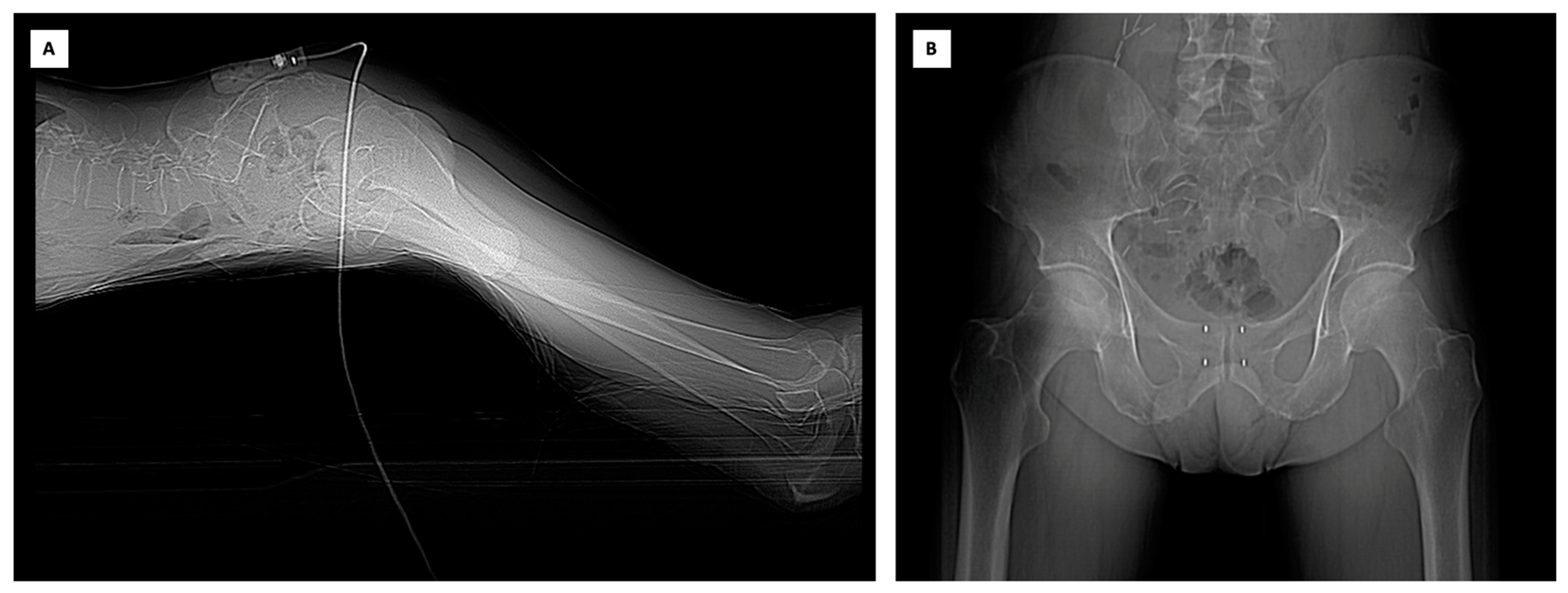
2. Case Report
3. Discussion
Author Contributions
Funding
Institutional Review Board Statement
Informed Consent Statement
Data Availability Statement
Acknowledgments
Conflicts of Interest
References
- Sung, H.; Ferlay, J.; Siegel, R.L.; Laversanne, M.; Soerjomataram, I.; Jemal, A.; Bray, F. Global Cancer Statistics 2020: Globocan Estimates of Incidence and Mortality Worldwide for 36 Cancers in 185 Countries. CA Cancer J. Clin. 2021, 71, 209–249. [Google Scholar] [CrossRef]
- Siegel, R.L.; Miller, K.D.; Fuchs, H.E.; Jemal, A. Cancer Statistics, 2022. CA Cancer J. Clin. 2022, 72, 7–33. [Google Scholar] [CrossRef] [PubMed]
- Mottet, N.; Van Den Bergh, R.C.N.; Briers, E.; Van Den Broeck, T.; Cumberbatch, M.G.; De Santis, M.; Fanti, S.; Fossati, N.; Gandaglia, G.; Gillessen, S.; et al. EAU-EANM-ESTRO-ESUR-SIOG Guidelines on Prostate Cancer—2020 Update. Part 1: Screening, Diagnosis, and Local Treatment with Curative Intent. Eur. Urol. 2021, 79, 243–262. [Google Scholar] [CrossRef] [PubMed]
- De Bari, B.; Arcangeli, S.; Ciardo, D.; Mazzola, R.; Alongi, F.; Russi, E.G.; Santoni, R.; Magrini, S.M.; Jereczek-Fossa, B.A. Extreme Hypofractionation for Early Prostate Cancer: Biology Meets Technology. Cancer Treat. Rev. 2016, 50, 48–60. [Google Scholar] [CrossRef]
- Widmark, A.; Gunnlaugsson, A.; Beckman, L.; Thellenberg-Karlsson, C.; Hoyer, M.; Lagerlund, M.; Kindblom, J.; Ginman, C.; Johansson, B.; Björnlinger, K.; et al. Ultra-Hypofractionated versus Conventionally Fractionated Radiotherapy for Prostate Cancer: 5-Year Outcomes of the HYPO-RT-PC Randomised, Non-Inferiority, Phase 3 Trial. Lancet 2019, 394, 385–395. [Google Scholar] [CrossRef]
- Brand, D.H.; Tree, A.C.; Ostler, P.; Van Der Voet, H.; Loblaw, A.; Chu, W.; Ford, D.; Tolan, S.; Jain, S.; Martin, A.; et al. Intensity-Modulated Fractionated Radiotherapy versus Stereotactic Body Radiotherapy for Prostate Cancer (PACE-B): Acute Toxicity Findings from an International, Randomised, Open-Label, Phase 3, Non-Inferiority Trial. Lancet Oncol. 2019, 20, 1531–1543. [Google Scholar] [CrossRef]
- Höcht, S.; Aebersold, D.M.; Albrecht, C.; Böhmer, D.; Flentje, M.; Ganswindt, U.; Hölscher, T.; Martin, T.; Sedlmayer, F.; Wenz, F.; et al. Hypofractionated Radiotherapy for Localized Prostate Cancer. Strahlenther. Onkol. 2017, 193, 1–12. [Google Scholar] [CrossRef]
- Tree, A.C.; Ostler, P.; Van Der Voet, H.; Chu, W.; Loblaw, A.; Ford, D.; Tolan, S.; Jain, S.; Martin, A.; Staffurth, J.; et al. Intensity-Modulated Radiotherapy versus Stereotactic Body Radiotherapy for Prostate Cancer (PACE-B): 2-Year Toxicity Results from an Open-Label, Randomised, Phase 3, Non-Inferiority Trial. Lancet Oncol. 2022, 23, 1308–1320. [Google Scholar] [CrossRef]
- Lukka, H.R.; Pugh, S.L.; Bruner, D.W.; Bahary, J.-P.; Lawton, C.A.F.; Efstathiou, J.A.; Kudchadker, R.J.; Ponsky, L.E.; Seaward, S.A.; Dayes, I.S.; et al. Patient Reported Outcomes in NRG Oncology RTOG 0938, Evaluating Two Ultrahypofractionated Regimens for Prostate Cancer. Int. J. Radiat. Oncol. Biol. Phys. 2018, 102, 287–295. [Google Scholar] [CrossRef]
- Ito, M.; Yoshioka, Y.; Takase, Y.; Suzuki, J.; Takahashi, H.; Minami, Y.; Sakuragi, A.; Oshima, Y.; Okuda, T.; Suzuki, K. Stereotactic Body Radiation Therapy for Prostate Cancer: A Study Comparing 3-Year Genitourinary Toxicity between CyberKnife and Volumetric-Modulated Arc Therapy by Propensity Score Analysis. Radiat. Oncol. 2023, 18, 39. [Google Scholar] [CrossRef]
- Scobioala, S.; Kittel, C.; Elsayad, K.; Kroeger, K.; Oertel, M.; Samhouri, L.; Haverkamp, U.; Eich, H.T. A Treatment Planning Study Comparing IMRT Techniques and Cyber Knife for Stereotactic Body Radiotherapy of Low-Risk Prostate Carcinoma. Radiat. Oncol. 2019, 14, 143. [Google Scholar] [CrossRef]
- White, E.; Boswell, W.; Whang, G.; Mandelin, P.; Mbbs, V.D. CT-Guided Fiducial Marker Placement for Stereotactic Radiosurgery. Appl. Radiol. 2012, 40, 24. [Google Scholar] [CrossRef]
- Holmes, O.E.; Gratton, J.; Szanto, J.; Vandervoort, E.; Doody, J.; Henderson, E.; Morgan, S.C.; O’Sullivan, J.; Malone, S. Reducing Errors in Prostate Tracking with an Improved Fiducial Implantation Protocol for CyberKnife Based Stereotactic Body Radiotherapy (SBRT). J. Radiosurg. SBRT 2018, 5, 217–227. [Google Scholar] [PubMed]
- Zlevor, A.M.; Kisting, M.A.; Couillard, A.B.; Rossebo, A.E.; Szczykutowicz, T.P.; Mao, L.; White, J.K.; Hartung, M.P.; Gettle, L.M.; Hinshaw, J.L.; et al. Percutaneous CT-Guided Abdominal and Pelvic Biopsies: Comparison of an Electromagnetic Navigation System and CT Fluoroscopy. J. Vasc. Interv. Radiol. 2023, 34, 910–918. [Google Scholar] [CrossRef]
- Appelbaum, L.; Sosna, J.; Nissenbaum, Y.; Benshtein, A.; Goldberg, S.N. Electromagnetic Navigation System for CT-Guided Biopsy of Small Lesions. Am. J. Roentgenol. 2011, 196, 1194–1200. [Google Scholar] [CrossRef]
- Kim, S.; Kim, N.; Chung, S.; Kim, J.; Hyun, I.; Choi, J.; Lee, H. Diagnostic Accuracy and Safety of Electromagnetic Navigation Transthoracic Needle Biopsy under Moderate Sedation for the Diagnosis of Peripheral Pulmonary Lesions. Transl. Lung Cancer Res. 2023, 12, 1496–1505. [Google Scholar] [CrossRef] [PubMed]
- Meyer, L.A.; Broaddus, R.R.; Lu, K.H. Endometrial Cancer and Lynch Syndrome: Clinical and Pathologic Considerations. Cancer Control 2009, 16, 14–22. [Google Scholar] [CrossRef] [PubMed]
- Nakamura, K.; Banno, K.; Yanokura, M.; Iida, M.; Adachi, M.; Masuda, K.; Ueki, A.; Kobayashi, Y.; Nomura, H.; Hirasawa, A.; et al. Features of Ovarian Cancer in Lynch Syndrome (Review). Mol. Clin. Oncol. 2014, 2, 909–916. [Google Scholar] [CrossRef]
- Lim, A.; Rao, P.; Matin, S.F. Lynch Syndrome and Urologic Malignancies: A Contemporary Review. Curr. Opin. Urol. 2019, 29, 357–363. [Google Scholar] [CrossRef]
- Nagy, R.; Sweet, K.; Eng, C. Highly Penetrant Hereditary Cancer Syndromes. Oncogene 2004, 23, 6445–6470. [Google Scholar] [CrossRef]
- Hampel, H.; Frankel, W.L.; Martin, E.; Arnold, M.; Khanduja, K.; Kuebler, P.; Nakagawa, H.; Sotamaa, K.; Prior, T.W.; Westman, J.; et al. Screening for the Lynch Syndrome (Hereditary Nonpolyposis Colorectal Cancer). N. Engl. J. Med. 2005, 352, 1851–1860. [Google Scholar] [CrossRef]
- Sun, M.; Moquet, J.; Ellender, M.; Bouffler, S.; Badie, C.; Baldwin-Cleland, R.; Monahan, K.; Latchford, A.; Lloyd, D.; Clark, S.; et al. Potential Risks Associated with the Use of Ionizing Radiation for Imaging and Treatment of Colorectal Cancer in Lynch Syndrome Patients. Fam. Cancer 2023, 22, 61–70. [Google Scholar] [CrossRef]
- Marvaso, G.; Gugliandolo, S.G.; Bellerba, F.; Gandini, S.; Corrao, G.; Volpe, S.; Rojas, D.P.; Riva, G.; Zerini, D.; Pepa, M.; et al. Phase II Prospective Trial “Give Me Five” Short-Term High Precision Radiotherapy for Early Prostate Cancer with Simultaneous Boost to the Dominant Intraprostatic Lesion: The Impact of Toxicity on Quality of Life (AIRC IG-13218). Med. Oncol. 2020, 37, 74. [Google Scholar] [CrossRef]
- Nieder, C.; Andratschke, N.H.; Grosu, A.L. Increasing Frequency of Reirradiation Studies in Radiation Oncology: Systematic Review of Highly Cited Articles. Am. J. Cancer Res. 2013, 3, 152–158. [Google Scholar]
- Nieder, C.; Willmann, J.; Andratschke, N.H. Prospective Randomized Clinical Studies Involving Reirradiation: Update of a Systematic Review. Strahlenther. Onkol. 2023, 199, 787–797. [Google Scholar] [CrossRef] [PubMed]
- Jereczek-Fossa, B.A.; Beltramo, G.; Fariselli, L.; Fodor, C.; Santoro, L.; Vavassori, A.; Zerini, D.; Gherardi, F.; Ascione, C.; Bossi-Zanetti, I.; et al. Robotic Image-Guided Stereotactic Radiotherapy, for Isolated Recurrent Primary, Lymph Node or Metastatic Prostate Cancer. Int. J. Radiat. Oncol. Biol. Phys. 2012, 82, 889–897. [Google Scholar] [CrossRef]
- Ahn, S.J.; Lee, J.M.; Lee, D.H.; Lee, S.M.; Yoon, J.-H.; Kim, Y.J.; Lee, J.-H.; Yu, S.J.; Han, J.K. Real-Time US-CT/MR Fusion Imaging for Percutaneous Radiofrequency Ablation of Hepatocellular Carcinoma. J. Hepatol. 2017, 66, 347–354. [Google Scholar] [CrossRef]
- Mauri, G.; Cova, L.; De Beni, S.; Ierace, T.; Tondolo, T.; Cerri, A.; Goldberg, S.N.; Solbiati, L. Real-Time US-CT/MRI Image Fusion for Guidance of Thermal Ablation of Liver Tumors Undetectable with US: Results in 295 Cases. Cardiovasc. Interv. Radiol. 2015, 38, 143–151. [Google Scholar] [CrossRef] [PubMed]
- Bruners, P.; Penzkofer, T.; Nagel, M.; Elfring, R.; Gronloh, N.; Schmitz-Rode, T.; Günther, R.W.; Mahnken, A.H. Electromagnetic Tracking for CT-Guided Spine Interventions: Phantom, Ex-Vivo and in-Vivo Results. Eur. Radiol. 2009, 19, 990–994. [Google Scholar] [CrossRef]
- Wallace, M.J.; Gupta, S.; Hicks, M.E. Out-of-Plane Computed-Tomography-Guided Biopsy Using a Magnetic-Field-Based Navigation System. Cardiovasc. Interv. Radiol. 2006, 29, 108–113. [Google Scholar] [CrossRef] [PubMed]
- Rouchy, R.; Moreau-Gaudry, A.; Chipon, E.; Aubry, S.; Pazart, L.; Lapuyade, B.; Durand, M.; Hajjam, M.; Pottier, S.; Renard, B.; et al. Evaluation of the Clinical Benefit of an Electromagnetic Navigation System for CT-Guided Interventional Radiology Procedures in the Thoraco-Abdominal Region Compared with Conventional CT Guidance (CTNAV II): Study Protocol for a Randomised Controlled Trial. Trials 2017, 18, 306. [Google Scholar] [CrossRef] [PubMed]
- Thariat, J.; Trimaud, R.; Angellier, G.; Caullery, M.; Amiel, J.; Bondiau, P.-Y.; Gerard, J.-P. Innovative Image-Guided CyberKnife ® Stereotactic Radiotherapy for Bladder Cancer. BJR 2010, 83, e118–e121. [Google Scholar] [CrossRef]
- Dagoglu, N.; Nedea, E.; Poylin, V.; Nagle, D.; Mahadevan, A. Post Operative Stereotactic Radiosurgery for Positive or Close Margins after Preoperative Chemoradiation and Surgery for Rectal Cancer. J. Gastrointest. Oncol. 2016, 7, 315–320. [Google Scholar] [CrossRef] [PubMed]


| Dose Target | Goal | Results |
|---|---|---|
| PTV-DIL (40 Gy) | ||
| D0.03cc | <116% | 117.00% |
| D98% | 100.50% | |
| D95% | >95% | 101.30% |
| D2% | <114% | 103.40% |
| D50% (Median) | 106.30% | |
| PTV-P (36.25 Gy) | ||
| D 0.03cc | <114% | 115.30% |
| D98% | 95.20% | |
| D95% | >95% | 97.10% |
| D2% | <104% | 110.9% |
| D50% (Median) | 103.70% |
Disclaimer/Publisher’s Note: The statements, opinions and data contained in all publications are solely those of the individual author(s) and contributor(s) and not of MDPI and/or the editor(s). MDPI and/or the editor(s) disclaim responsibility for any injury to people or property resulting from any ideas, methods, instructions or products referred to in the content. |
© 2023 by the authors. Licensee MDPI, Basel, Switzerland. This article is an open access article distributed under the terms and conditions of the Creative Commons Attribution (CC BY) license (https://creativecommons.org/licenses/by/4.0/).
Share and Cite
Vavassori, A.; Mauri, G.; Mazzola, G.C.; Mastroleo, F.; Bonomo, G.; Durante, S.; Zerini, D.; Marvaso, G.; Corrao, G.; Ferrari, E.D.; et al. Cyberknife Radiosurgery for Prostate Cancer after Abdominoperineal Resection (CYRANO): The Combined Computer Tomography and Electromagnetic Navigation Guided Transperineal Fiducial Markers Implantation Technique. Curr. Oncol. 2023, 30, 7926-7935. https://doi.org/10.3390/curroncol30090576
Vavassori A, Mauri G, Mazzola GC, Mastroleo F, Bonomo G, Durante S, Zerini D, Marvaso G, Corrao G, Ferrari ED, et al. Cyberknife Radiosurgery for Prostate Cancer after Abdominoperineal Resection (CYRANO): The Combined Computer Tomography and Electromagnetic Navigation Guided Transperineal Fiducial Markers Implantation Technique. Current Oncology. 2023; 30(9):7926-7935. https://doi.org/10.3390/curroncol30090576
Chicago/Turabian StyleVavassori, Andrea, Giovanni Mauri, Giovanni Carlo Mazzola, Federico Mastroleo, Guido Bonomo, Stefano Durante, Dario Zerini, Giulia Marvaso, Giulia Corrao, Elettra Dorotea Ferrari, and et al. 2023. "Cyberknife Radiosurgery for Prostate Cancer after Abdominoperineal Resection (CYRANO): The Combined Computer Tomography and Electromagnetic Navigation Guided Transperineal Fiducial Markers Implantation Technique" Current Oncology 30, no. 9: 7926-7935. https://doi.org/10.3390/curroncol30090576
APA StyleVavassori, A., Mauri, G., Mazzola, G. C., Mastroleo, F., Bonomo, G., Durante, S., Zerini, D., Marvaso, G., Corrao, G., Ferrari, E. D., Rondi, E., Vigorito, S., Cattani, F., Orsi, F., & Jereczek-Fossa, B. A. (2023). Cyberknife Radiosurgery for Prostate Cancer after Abdominoperineal Resection (CYRANO): The Combined Computer Tomography and Electromagnetic Navigation Guided Transperineal Fiducial Markers Implantation Technique. Current Oncology, 30(9), 7926-7935. https://doi.org/10.3390/curroncol30090576