Next Article in Journal
Adverse Events Associated with Anti-EGFR Therapies for the Treatment of Metastatic Colorectal Cancer
Previous Article in Journal
Optimizing the Management of Advanced Non-small-Cell Lung Cancer: A Personal View
 
 
Current Oncology is published by MDPI from Volume 28 Issue 1 (2021). Previous articles were published by another publisher in Open Access under a CC-BY (or CC-BY-NC-ND) licence, and they are hosted by MDPI on mdpi.com as a courtesy and upon agreement with Multimed Inc..
Article

The Predictive Value of Pre-treatment Inflammatory Markers in Advanced Non-small-Cell Lung Cancer

1
Division of Pulmonary Diseases, Sir Mortimer B. Davis Jewish General Hospital, 3755 Cote St-Catherine, E731, Montreal, QC, Canada
2
Department of Oncology, Cancer Nutrition Rehabilitation Program, McGill University, Montreal, QC, Canada
3
Department of Oncology, Sir Mortimer B. Davis Jewish General Hospital, Montreal, QC, Canada
*
Author to whom correspondence should be addressed.
Curr. Oncol. 2010, 17(4), 52-58; https://doi.org/10.3747/co.v17i4.567
Submission received: 1 August 2010 / Revised: 1 August 2010 / Accepted: 1 August 2010 / Published: 1 August 2010

Abstract

:
Background: Accurate prediction of outcome in advanced nonsmall-cell lung cancer (NSCLC) remains challenging. Even within the same stage and treatment group, survival and response to treatment vary. We set out to determine the predictive value of inflammatory markers C-reactive protein (CRP) and white blood cells (WBCs) in patients with advanced NSCLC. Patients and Methods: Patients were assigned a prognostic index (PI): 0 for CRP 10 mg/L or less and WBCs 11×109/L or less, 1 if one of the two markers was elevated, and 2 if both markers were elevated. We then used chest computed tomography (CT) imaging to evaluate response after 2 cycles of chemotherapy treatment. Results: Of 134 patients, 46 had a PI of 0; 60, a PI of 1; and 28, a PI of 2. Disease progressed in 41 patients. Progression was significantly more frequent among patients with a PI of 2 (p = 0.008). Median survival was 20.0 months for the PI 0 group, 10.4 months for the PI 1 group, and 7.9 months for the PI 2 group (p < 0.001). The PI was the only significant prognostic factor for survival even after adjustment for performance status, smoking, and weight loss (hazard ratio: 1.57; 95% confidence interval: 1.2 to 2.14; p = 0.004). Conclusions: Inflammatory state correlates significantly with both chemotherapy response and survival in stage IV NSCLC. The PI may provide additional guidance for therapeutic decision-making.

1. INTRODUCTION

Patients with NSCLC have heterogeneous tumour and host biology that leads to wide predictive and prognostic variance. To improve medical decision-making and to deliver the most suitable treatment, predictive and prognostic factors are used to divide the NSCLC population into subgroups. Most studies have focused on the clinical characteristics of tumour and host, such as disease stage and performance status [1]. However, even within the same stage and with a similar performance status, response to treatment and survival varies from patient to patient.
Basic scientists have clearly demonstrated the importance of molecular and cellular pathways linking cancer and inflammation [2,3,4,5]. Many blood markers, including acute-phase C-reactive protein (CRP), were identified [5,6,7,8,9,10,11,12,13]. The CRP induced by a cytokine consortium in which interleukin-6 (IL-6) is the dominant partner was shown to be an important adverse survival determinant—independent of stage and performance status—in advanced cancer, including non-small-cell lung cancer (NSCLC) 10,14,15.
In an effort to further refine prognostic accuracy, a variety of indices based on inflammatory markers have been proposed [10,14,15]. The Glasgow Prognostic Score (GPS) was shown to be superior to a subjective assessment of Eastern Cooperative Oncology Group (ECOG) performance status (PS) in predicting the survival of patients with advanced NSCLC [16]. However, little is known about the effect of underlying inflammation on the response of NSCLC to chemotherapy [17].
In the present study, we evaluated the association between baseline systemic inflammation and two outcome measures in advanced NSCLC: freedom from progression in response to chemotherapy, and survival.

2. PATIENTS AND METHODS

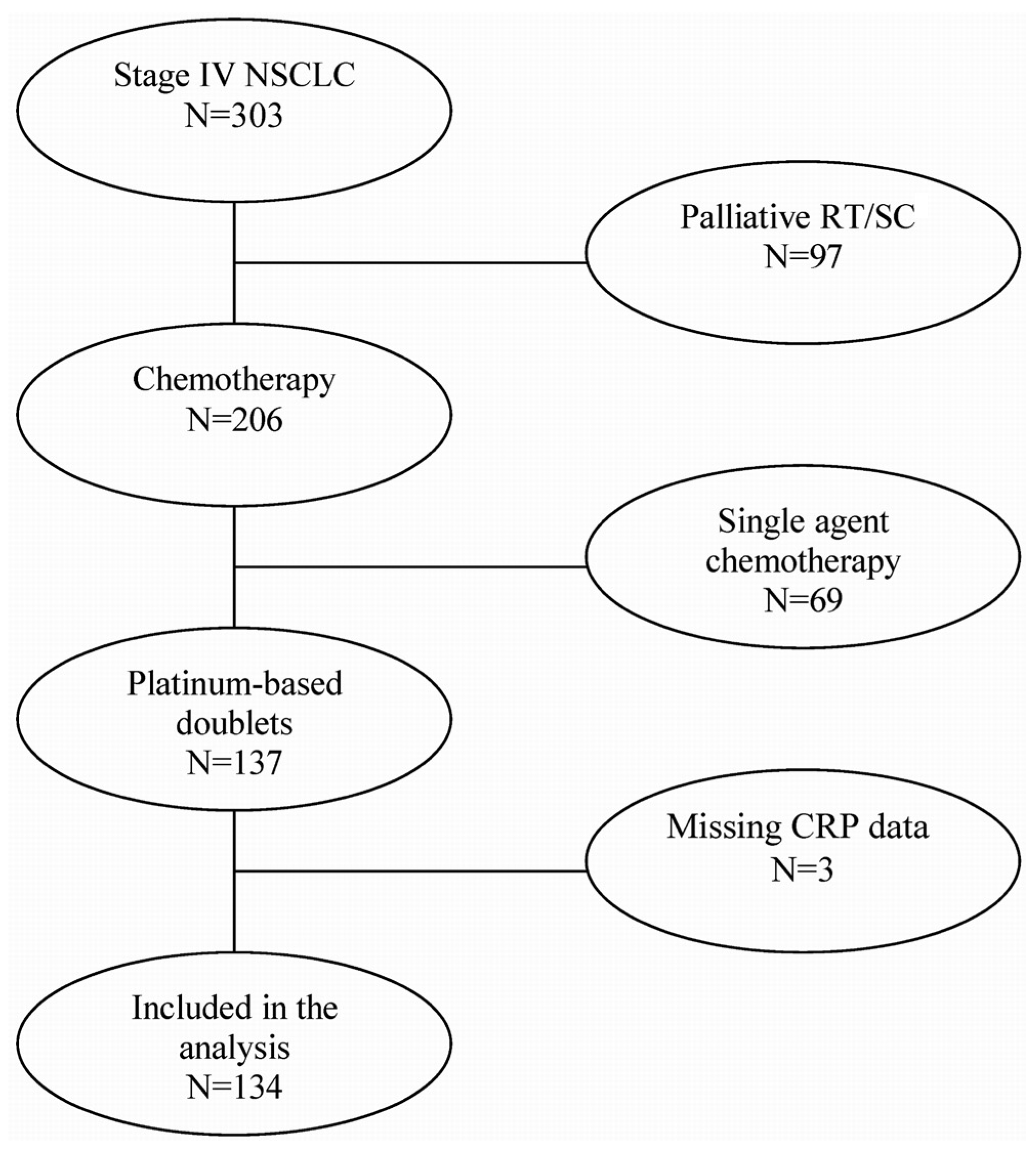
2.1. Participant Identification

This prospective study was conducted between January 1, 2005, and January 10, 2009, in a cohort of consecutive patients with newly diagnosed stage IV (per the 7th edition of the International Association for the Study of Lung Cancer staging system) NSCLC who had been treated with 2 cycles of platinumbased double-agent chemotherapy.
Computed tomography (CT) imaging of chest and abdomen, followed by metabolic imaging using positron-emission tomography, was used for staging. The particular chemotherapy regimen prescribed was chosen according to protocols used at the Jewish General Hospital, Montreal, Quebec. Possibilities included cisplatin or carboplatin in combination with paclitaxel, gemcitabine, vinorelbine, docetaxel, or etoposide. The choice of the treatment regimen was at the discretion of the treating physician.
The study protocol was approved by the Institutional Review Board at the Jewish General Hospital, and all patients provided informed consent.

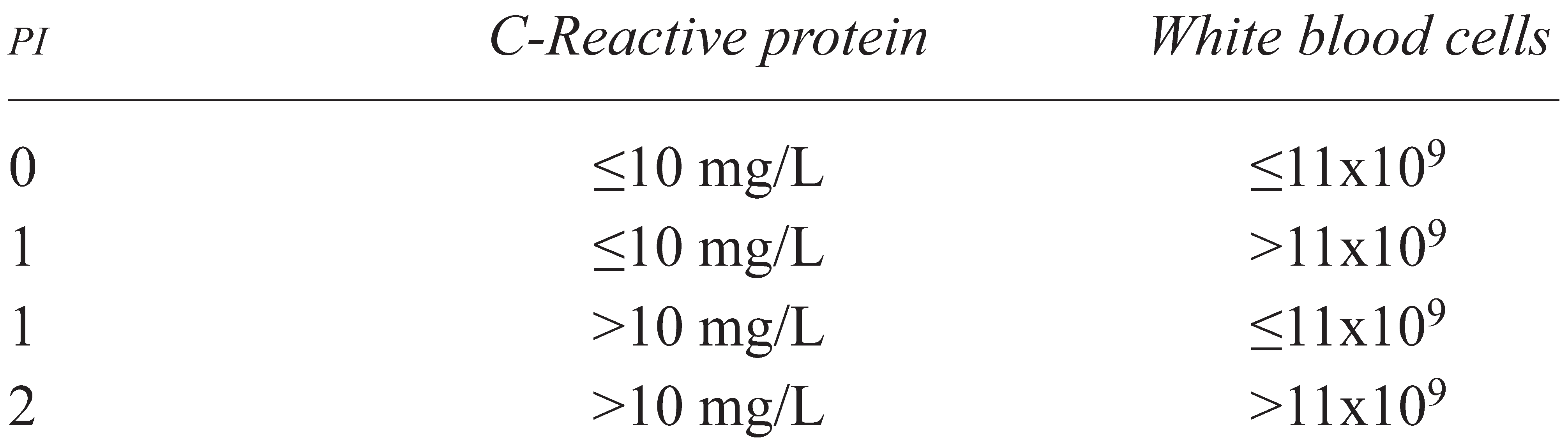
2.2. Prognostic Index

Standard techniques were used to determine CRP, albumin, white blood cell count (WBCs), hemoglobin, and lactate dehydrogenase. A particle-enhanced turbidimetric assay was used to measure CRP [18]. The upper limits of the normal ranges for CRP (10 mg/L) and for WBCs (11×109/L) were used as cut-off points [19].
Based on previous work by our group, the CRP and WBC results were used to categorize patients into 3 different “prognostic index” (PI) groups (Table 1) [6]:
  • PI 0 for CRP 10 mg/L or less and WBCs 11×109/L or less,
  • PI 1 if one of the two markers was elevated, and
  • PI 2 if both markers were elevated.

2.3. Response to Chemotherapy

Response to chemotherapy was evaluated by chest CT after 2 cycles of chemotherapy. The response was categorized as progressive disease or freedom from progression. Freedom from progression was defined as an absence of objective progression; it included patients with either a complete response, a partial response, or stable disease according to the Response Evaluation Criteria in Solid Tumors (RECIST) [20].

2.4. Statistical Analysis
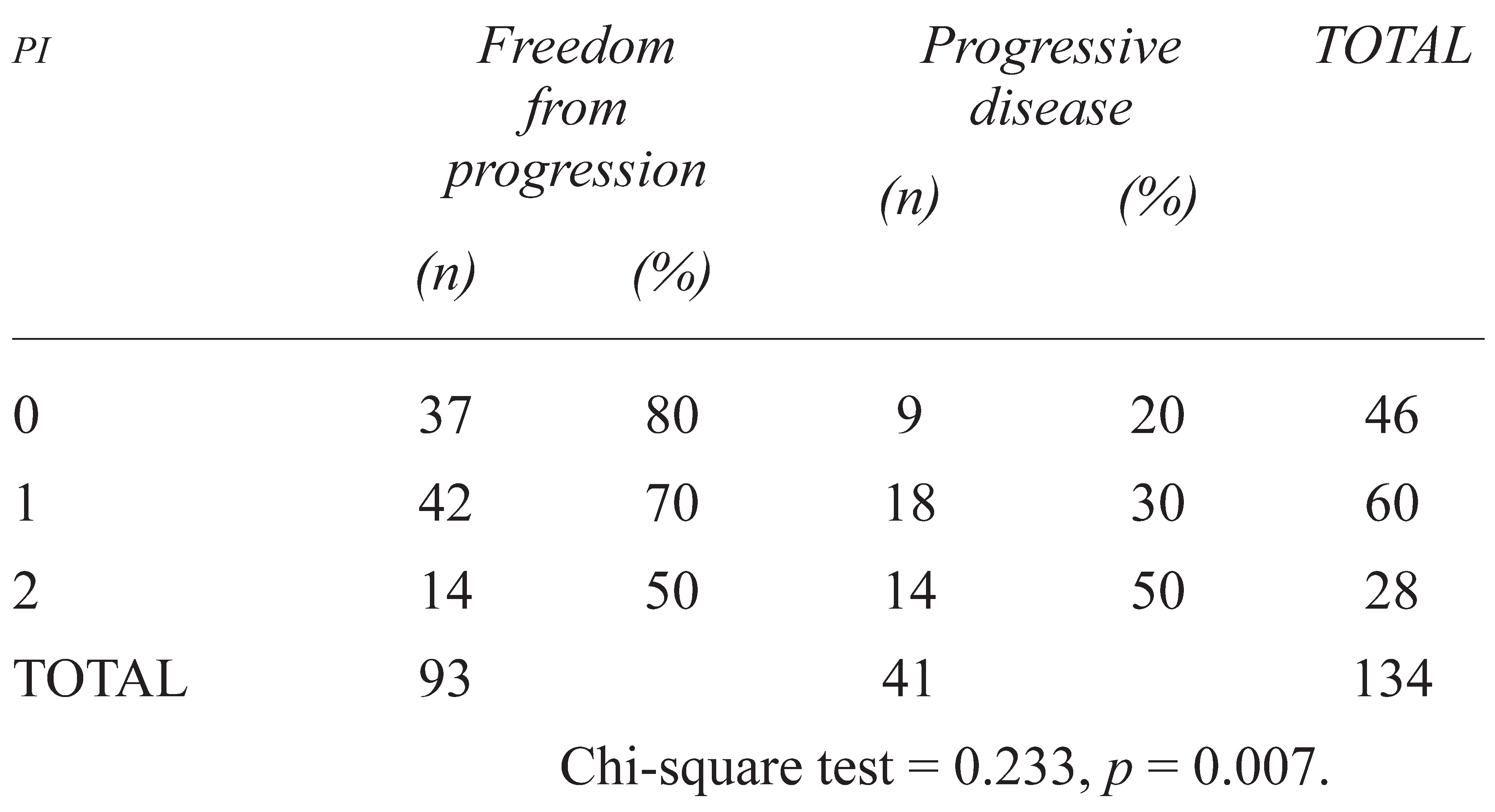
The primary outcomes of the study were the response to 2 cycles of platinum-based chemotherapy (as defined earlier) and overall survival. The association between PI and freedom from progression was investigated by chi-square test. Using t-tests and chi-square tests as appropriate, baseline blood parameters and the covariates of interest (age, sex, PS, smoking history, stage, and weight loss at first presentation) for the 3 PI groups were compared to confirm the homogeneity of the groups. Contingency tables and the chi-square test were used for categorical variables; t-tests were used for continuous variables.
Overall survival was defined as the time in months from diagnosis to the end of the study. Patients alive at the time of the last observation (March 30, 2009) were censored. Survival analysis was performed using the Kaplan–Meier method. Differences were assessed using a log-rank test. A Cox proportional hazards model was used to estimate the predictive power of the PI. The potential baseline prognostic factors included in the model were age, sex, CRP, hemoglobin, weight loss, smoking status (smokers and ex-smokers vs. never-smokers), ECOG PS, and PI. The likelihood ratio was used to test the contribution of each variable to the model when added last (that is, after adjustment for all of the other covariates).
All statistical analyses were carried out using the SPSS software package (version 14.0: SPSS, Chicago, IL, U.S.A.). All statistical tests were twotailed. Statistical significance was accepted at p values of 0.05 or less.

3. RESULTS

The study identified 303 patients with stage IV NSCLC. Of those 303 patients, 206 received chemotherapy and 97 were offered palliative radiation or supportive care (Figure 1). Of the 206 patients given chemotherapy, 69 were treated with a single agent and 137 received double-agent chemotherapy. In 3 patients, CRP data were missing. The remaining 134 patients who had received 2 cycles of therapy and whose records had no missing data were analyzed.
Table 2 shows the characteristics of the patient cohort. Mean age was 62 years (range: 31–83 years). Most had adenocarcinoma, a good PS, and no weight loss. Sites of metastases included lung, lymph nodes, bone, liver, and adrenal glands. The group included 65 ex-smokers (48%) who had quit more than 1 year before diagnosis, 40 patients (30%) who were still smoking at the time of diagnosis, and 29 never-smokers (22%). The most common treatment regimens were carboplatin–gemcitabine and carboplatin–paclitaxel.
Most patients had normal hemoglobin, albumin, and lactate dehydrogenase levels. The median CRP concentration before chemotherapy was 13.2 mg/L. In 77 patients (57%), CRP exceeded 10 mg/L. Median WBCs were 9.4×10⁹/L. Before chemotherapy, WBCs were elevated in 39 patients (29%).
We calculated the PI for each patient based on binary CRP and WBC values. In 46 patients (34%), the PI was 0; in 60 (45%), the PI was 1; and in 28 (21%), the PI was 2. We observed no association of PI with age or ECOG PS (Table 3); however, significantly more men, smokers and ex-smokers, and patients with a weight loss of 5% or more were in the PI 2 group.
After 2 cycles of chemotherapy, 41 patients (31%) were found to have progressive disease. The PI and progression (Table 4) were weakly positively correlated (χ2 = 0.233; p = 0.007). In the logistic regression analysis, the PI was the only significant predictor of progression. Age, sex, ECOG PS, cancer stage, and smoking status were nonsignificant (Table 5).
Median follow-up was 10.3 months (range: 2.69–41.87 months). At the time of censoring, 86 patients (64%) had died. Overall median survival was 11.9 months [95% confidence interval (CI): 9.9 to 13.8 months]. Median survival for the PI 0 group was 20.0 months (95% CI: 9.50 to 30.5 months); for the PI 1 group, it was 10.4 months (95% CI: 8.3 to 12.5 months); and for the PI 2 group, it was 7.9 months (95% CI: 4.2 to 11.7 months; p < 0.001; Figure 2).
Univariate analysis identified CRP and WBCs as significant prognostic factors in addition to PS. We observed no interaction between CRP and WBCs. When CRP and WBCs were combined into the PI, the effect was even stronger.
In a multivariate analysis, using the backward logistic regression method in a Cox model with all the variables of interest included, only the PI retained significance (hazard ratio: 1.57; 95% CI: 1.2 to 2.14; p = 0.004). The hazard for death was 2.5 for PI 1 and 3.9 for PI 2.

4. DISCUSSION

To our knowledge, this is the first prospective study aimed at evaluating the predictive value of pretherapeutic inflammatory markers in NSCLC.
We found that, combined into a PI, elevated plasma CRP and WBCs at the time of NSCLC diagnosis are independently associated with disease progression in response to 2 cycles of first-line treatment and with shorter overall survival in advanced NSCLC. A lower PI score was associated with a reduction in the probability of disease progression after chemotherapy. The hazard for death was elevated by a factor of 2.5 for the PI 1 group and by a factor of 3.9 for the PI 2 group as compared with the PI 0 group. The development of the PI provides improved prediction accuracy for progression after 2 cycles of first-line treatment.
Despite advances in the accuracy of clinical staging, predictive and prognostic modeling for patients with advanced NSCLC is still not satisfactory [1]. A search is underway for molecular factors that might account for variation in tumour biology and its effect on survival [3,21,22,23,24]. It is now becoming clear that the tumour microenvironment, which is largely orchestrated by inflammatory cells, is an indispensable participant in the neoplastic process [25]. This environment promotes proliferation, survival, and migration of tumour cells and results in increased tumour aggressiveness. Some lung cancer cell lines have been shown to produce IL-6. Indeed, there is evidence that IL-6 is strongly related to CRP and that CRP, a routinely available and well-standardized clinical laboratory measurement, is a useful surrogate measure of tumour biology in patients with NSCLC 14.
An increased serum CRP level has been recognized as an ominous prognostic factor for several malignant tumours, including lung cancer [9,11,26,27,28,29,30,31]. In a prospective cohort study, Siemes et al. hypothesized that CRP level and CRP gene variation are associated with an altered risk of colorectal, lung, breast, and prostate cancer [32]. In addition, CRP is known to modulate both innate and adaptive immunity [33,34].
Other biologic factors such as baseline leucocytes and polynuclear cells have been less frequently studied in cancer patients in terms of their significance as predictive factors. A few studies demonstrated that in addition to PS, WBCs are an independent prognostic factor for poor survival in advanced NSCLC [35,36,37,38,39].
These insights are encouraging new anti-inflammatory therapeutic approaches to cancer treatment [6,40]. However, the association between elevated inflammatory markers and chemotherapy response has not been clearly established [17,41].
The success of chemotherapy in esophageal and prostate cancer may relate to CRP status [27,29,42,43]. Wilop et al. correlated changes in CRP over time with response to treatment in lung cancer patients receiving chemotherapy [44]. Those authors reported that normalization of CRP was associated with a low risk for progression, but that patients with an increase in CRP of more than 25% showed progressive disease in most cases. However, their retrospective study was underpowered, given the small sample size and lack of control for stage and PS.
Several prognostic indices based on inflammatory factors have recently been described in NSCLC [11,12,45]. The most widely recognized is the GPS, which uses a combination of CRP and albumin [46]. When compared with PS and WBCs, GPS was superior in predicting survival [47]. However, we found that serum albumin at diagnosis was occasionally low in our population and that adding WBCs to CRP resulted in superior stratification of the resulting subgroups.
There are several possible explanations to account for the association between CRP and chemotherapy response. Chronic inflammation results in excessive cell proliferation and activation of a cascade of cellular actions that can lead to induction of irreversible DNA damage and subsequent tumour progression [48]. Chronic inflammation might also be responsible for activation, in peripheral leucocytes, of messenger RNAs for nuclear factor κB inhibitor and for the effect of that inhibitor on cytochrome systems [49,50]. A tumour-associated inflammatory response involving CRP and IL-6 downregulates the CYP3A4 gene and correlates with suppression of drug metabolism [51,52,53], resulting in increased toxicity during chemotherapy.
Incorporated into a PI, CRP and WBCs identified 3 subgroups of stage IV NSCLC patients with distinct risks of progression and death. This information might be pertinent to treatment decision-making and might also influence a revised staging system. If the objective is prognostic stratification, then using anatomic features alone for staging is no longer sufficient.
The major strength of the present study is the uniformity of the patient population with respect to diagnosis, stage, and treatment. The study is limited by the relatively small cohort and the weak correlation between PI and freedom from tumour progression.

5. CONCLUSIONS

Based on widely available, inexpensive, and easyto-perform measurements, we developed a novel PI. The proposed PI may improve the accuracy of predictions of freedom from progression and of survival in response to first-line treatment. It may also provide information to complement other prognostic models, such as those based on gene profiling. The biologic markers that form the PI could be incorporated into the more traditional anatomic methods of staging.

Acknowledgments

The work presented here was supported by the Mona Zavalkoff Fund for Pulmonary Oncology, by an unrestricted grant from Sanofi–Aventis, and by the Angel Ball.

Conflicts of Interest

All authors declare that there are no financial conflicts of interest.

References

  1. Brundage, M.D.; Davies, D.; Mackillop, W.J. Prognostic factors in non-small cell lung cancer: A decade of progress. Chest 2002, 122, 1037–1057. [Google Scholar] [CrossRef] [PubMed]
  2. Apetoh, L.; Tesniere, A.; Ghiringhelli, F.; Kroemer, G.; Zitvogel, L. Molecular interactions between dying tumor cells and the innate immune system determine the efficacy of conventional anticancer therapies. Cancer Res 2008, 68, 4026–4030. [Google Scholar] [CrossRef]
  3. Deans, C.; Rose-Zerilli, M.; Wigmore, S.; et al. Host cytokine genotype is related to adverse prognosis and systemic inflammation in gastro-oesophageal cancer. Ann Surg Oncol 2007, 14, 329–339. [Google Scholar] [CrossRef]
  4. Hilmy, M.; Campbell, R.; Bartlett, J.M.; McNicol, A.M.; Underwood, M.A.; McMillan, D.C. The relationship between the systemic inflammatory response, tumour proliferative activity, T-lymphocytic infiltration and COX-2 expression and survival in patients with transitional cell carcinoma of the urinary bladder. Br J Cancer 2006, 95, 1234–1238. [Google Scholar] [CrossRef] [PubMed]
  5. Watine, J. Comments on: Haemostatic abnormalities in lung cancer: Prognostic implications, Buccheri et al., Eur J Cancer, 33, pp. 50–55, 1997. Eur J Cancer 1998, 34, 430. [Google Scholar]
  6. Aggarwal, B.B.; Vijayalekshmi, R.V.; Sung, B. Targeting inflammatory pathways for prevention and therapy of cancer: Short-term friend, long-term foe. Clin Cancer Res 2009, 15, 425–440. [Google Scholar] [CrossRef] [PubMed]
  7. Bonnefoy, M.; Ayzac, L.; Ingenbleek, Y.; Kostka, T.; Boisson, R.C.; Bienvenu, J. Usefulness of the prognostic inflammatory and nutritional index (PINI) in hospitalized elderly patients. Int J Vitam Nutr Res 1998, 68, 189–195. [Google Scholar]
  8. Brown, D.J.; Milroy, R.; Preston, T.; McMillan, D.C. The relationship between an inflammation-based prognostic score (Glasgow Prognostic Score) and changes in serum biochemical variables in patients with advanced lung and gastrointestinal cancer. J Clin Pathol 2007, 60, 705–708. [Google Scholar] [CrossRef]
  9. Casamassima, A.; Picciariello, M.; Quaranta, M.; et al. C-Reactive protein: A biomarker of survival in patients with metastatic renal cell carcinoma treated with subcutaneous interleukin-2 base immunotherapy. J Urol 2005, 173, 52–55. [Google Scholar] [CrossRef]
  10. Clinchy, B.; Fransson, A.; Druvefors, B.; et al. Preoperative interleukin-6 production by mononuclear blood cells predicts survival after radical surgery for colorectal carcinoma. Cancer 2007, 109, 1742–1749. [Google Scholar] [CrossRef]
  11. Kelly, L.; White, S.; Stone, P.C. The B12/CRP index as a simple prognostic indicator in patients with advanced cancer: A confirmatory study. Ann Oncol 2007, 18, 1395–1399. [Google Scholar] [CrossRef]
  12. MacDonald, N.; Kasymjanova, G.; Dobson, S.; et al. Prognostic value of baseline inflammatory markers in inoperable non-small cell lung cancer (NSCLC) [abstract 17035]. Proc Am Soc Clin Oncol 2006, 24. Available online: www.asco.org/ASCOv2/Meetings/Abstracts?&vmview=abst_detail_view&confID=40&abstractID=31987 (accessed on 10 June 2010).
  13. Zitvogel, L.; Apetoh, L.; Ghiringhelli, F.; André, F.; Tesniere, A.; Kroemer, G. The anticancer immune response: Indispensable for therapeutic success? J Clin Invest 2008, 118, 1991–2001. [Google Scholar] [CrossRef]
  14. McKeown, D.J.; Brown, D.J.; Kelly, A.; Wallace, A.M.; McMillan, D.C. The relationship between circulating concentrations of C-reactive protein, inflammatory cytokines and cytokine receptors in patients with non-small-cell lung cancer. Br J Cancer 2004, 91, 1993–1995. [Google Scholar] [CrossRef]
  15. Heikkila, K.; Harris, R.; Lowe, G.; et al. Associations of circulating C-reactive protein and interleukin-6 with cancer risk: Findings from two prospective cohorts and a meta-analysis. Cancer Causes Control 2009, 20, 15–26. [Google Scholar] [CrossRef]
  16. Forrest, L.M.; McMillan, D.C.; McArdle, C.S.; Angerson, W.J.; Dunlop, D.J. Evaluation of cumulative prognostic scores based on the systemic inflammatory response in patients with inoperable non-small-cell lung cancer. Br J Cancer 2003, 89, 1028–1030. [Google Scholar] [CrossRef]
  17. Koch, A.; Fohlin, H.; Sorenson, S. Prognostic significance of C-reactive protein and smoking in patients with advanced non-small cell lung cancer treated with first-line palliative chemotherapy. J Thorac Oncol 2009, 4, 326–332. [Google Scholar] [CrossRef]
  18. Schwartz, M.W.; Schifreen, R.S.; Gorman, E.; Tuhy, P.M.; Bienvenu, J.; Warkentin, D.L. Development and performance of a fully automated method for assay of C-reactive protein in the aca discrete clinical analyzer. Clin Chem 1988, 34, 1646–1649. [Google Scholar] [CrossRef]
  19. Jewish General Hospital, Department of Diagnostic Medicine. Normal Reference Range Table; Jewish General Hospital: Montreal, QC, 2008. [Google Scholar]
  20. Julka, P.K.; Doval, D.C.; Gupta, S.; Rath, G.K. Response assessment in solid tumours: A comparison of WHO, SWOG and RECIST guidelines. Br J Radiol 2008, 81, 444–449. [Google Scholar] [CrossRef]
  21. Armaiz–Pena, G.N.; Lutgendorf, S.K.; Cole, S.W.; Sood, A.K. Neuroendocrine modulation of cancer progression. Brain Behav Immun 2009, 23, 10–15. [Google Scholar] [CrossRef]
  22. Apetoh, L.; Obeid, M.; Tesniere, A.; et al. Immunogenic chemotherapy: Discovery of a critical protein through proteomic analyses of tumor cells. Cancer Genomics Proteomics 2007, 4, 65–70. [Google Scholar]
  23. Ullrich, E.; Ménard, C.; Flament, C.; et al. Dendritic cells and innate defense against tumor cells. Cytokine Growth Factor Rev 2008, 19, 79–92. [Google Scholar] [CrossRef]
  24. Zitvogel, L.; Apetoh, L.; Ghiringhelli, F.; Kroemer, G. Immunological aspects of cancer chemotherapy. Nat Rev Immunol 2008, 8, 59–73. [Google Scholar] [CrossRef]
  25. Colotta, F.; Allavena, P.; Sica, A.; Garlanda, C.; Mantovani, A. Cancer-related inflammation, the seventh hallmark of cancer: Links to genetic instability. Carcinogenesis 2009, 7, 1073–1081. [Google Scholar] [CrossRef]
  26. Geissbühler, P.; Mermillod, B.; Rapin, C.H. Elevated serum vitamin B12 levels associated with CRP as a predictive factor of mortality in palliative care cancer patients: A prospective study over five years. J Pain Symptom Manage 2000, 20, 93–103. [Google Scholar] [CrossRef]
  27. Guillem, P.; Triboulet, J.P. Elevated serum levels of C-reactive protein are indicative of a poor prognosis in patients with esophageal cancer. Dis Esophagus 2005, 18, 146–150. [Google Scholar] [CrossRef]
  28. Karakiewicz, P.I.; Hutterer, G.C.; Trinh, Q.D.; et al. C-Reactive protein is an informative predictor of renal cell carcinoma– specific mortality. Cancer 2007, 110, 1241–1247. [Google Scholar] [CrossRef]
  29. Graff, J.; Lalani, A.S.; Lee, S.; et al. C-Reactive protein as a prognostic marker for men with androgen-independent prostate cancer (AIPC): Results from the ASCENT trial [abstract 5074]. Proc Am Soc Clin Oncol 2007, 25. Available online: www.asco.org/ASCOv2/Meetings/Abstracts?&vmview=abst_detail_view&confID=47&abstractID=31092 (accessed on 10 June 2010).
  30. Suh, S.Y.; Ahn, H.Y. A prospective study on C-reactive protein as a prognostic factor for survival time of terminally ill cancer patients. Support Care Cancer 2007, 15, 613–620. [Google Scholar] [CrossRef]
  31. Hara, M.; Matsuzaki, Y.; Shimuzu, T.; et al. Preoperative serum C-reactive protein level in non-small cell lung cancer. Anticancer Res 2007, 27, 3001–3004. [Google Scholar]
  32. Siemes, C.; Visser, L.E.; Coebergh, J.W.; et al. C-Reactive protein levels, variation in the C-reactive protein gene, and cancer risk: The Rotterdam Study. J Clin Oncol 2006, 24, 5216–5222. [Google Scholar] [CrossRef]
  33. Du Clos, T.W.; Mold, C. C-Reactive protein: An activator of innate immunity and a modulator of adaptive immunity. Immunol Res 2004, 30, 261–277. [Google Scholar] [CrossRef]
  34. Peisajovich, A.; Marnell, L.; Mold, C.; Du Clos, T.W. C-Reactive protein at the interface between innate immunity and inflammation. Expert Rev Clin Immunol 2008, 4, 379–390. [Google Scholar] [CrossRef] [PubMed]
  35. Panek, B.; Chyczewska, E.; Ossolińska, M.; Naumnik, W.; Izycki–Herman, T.; Korniluk, M. Evaluation of some functions of polymorphonuclear granulocytes in lung cancer patients during chemotherapy [Polish]. Pneumonol Alergol Pol 2005, 73, 167–171. [Google Scholar] [CrossRef] [PubMed]
  36. Teramukai, S.; Kitano, T.; Kishida, Y.; et al. Pretreatment neutrophil count as an independent prognostic factor in advanced non-small-cell lung cancer: An analysis of Japan Multinational Trial Organisation LC00-03. Eur J Cancer 2009, 5, 1950–1958. [Google Scholar] [CrossRef] [PubMed]
  37. Tibaldi, C.; Vasile, E.; Bernardini, I.; Orlandini, C.; Andreuccetti, M.; Falcone, A. Baseline elevated leukocyte count in peripheral blood is associated with poor survival in patients with advanced non-small cell lung cancer: A prognostic model. J Cancer Res Clin Oncol 2008, 134, 1143–1149. [Google Scholar] [CrossRef]
  38. Kato, K.; Hitsuda, Y.; Kawasaki, Y.; et al. The value of serum C-reactive protein as a survival determinant in patients with advanced non-small-cell lung cancer [Japanese]. Nihon Kokyuki Gakkai Zasshi 2000, 38, 575–580. [Google Scholar]
  39. Mignot, G.; Ullrich, E.; Bonmort, M.; et al. The critical role of IL-15 in the antitumour effects mediated by the combination therapy imatinib and IL-2. J Immunol 2008, 180, 6477–6483. [Google Scholar] [CrossRef]
  40. MacDonald, N. Cancer cachexia and targeting chronic inflammation: A unified approach to cancer treatment and palliative/supportive care. J Support Oncol 2007, 5, 157–162. [Google Scholar]
  41. Wilop, S.; Crysandt, M.; Bendel, M.; Mahnken, A.H.; Osieka, R.; Jost, E. Correlation of C-reactive protein with survival and radiographic response to first-line platinum-based chemotherapy in advanced non-small cell lung cancer. Onkologie 2008, 31, 665–670. [Google Scholar] [CrossRef]
  42. Beer, T.M.; Ryan, C.W.; Venner, P.M.; et al. Double-blinded randomized study of high-dose calcitriol plus docetaxel compared with placebo plus docetaxel in androgen-independent prostate cancer: A report from the ASCENT investigators. J Clin Oncol 2007, 25, 669–674. [Google Scholar] [CrossRef]
  43. Qin, T.J.; An, G.L.; Zhao, X.H.; et al. Combined treatment of oxaliplatin and capecitabine in patients with metastatic esophageal squamous cell cancer. World J Gastroenterol 2009, 15, 871–876. [Google Scholar] [CrossRef]
  44. Al Murri, A.M.; Bartlett, J.M.; Canney, P.A.; Doughty, J.C.; Wilson, C.; McMillan, D.C. C. Evaluation of an inflammation-based prognostic score (GPS) in patients with metastatic breast cancer. Br J Cancer 2006, 94, 227–230. [Google Scholar] [CrossRef] [PubMed]
  45. Forrest, L.M.; McMillan, D.C.; McArdle, C.S.; Angerson, W.J.; Dagg, K.; Scott, H.R. A prospective longitudinal study of performance status, an inflammation-based score (GPS) and survival in patients with inoperable non-small-cell lung cancer. Br J Cancer 2005, 92, 1834–1836. [Google Scholar] [CrossRef]
  46. Ackerman, W.E., 3rd; Zhang, J.M. Serum hs-CRP as a useful marker for predicting the efficacy of lumbar epidural steroid injections on pain relief in patients with lumbar disc herniations. J Ky Med Assoc 2006, 104, 295–299. [Google Scholar] [PubMed]
  47. Forrest, L.M.; McMillan, D.C.; McArdle, C.S.; Angerson, W.J.; Dunlop, D.J. Comparison of an inflammation-based prognostic score (GPS) with performance status (ECOG) in patients receiving platinum-based chemotherapy for inoperable non-small-cell lung cancer. Br J Cancer 2004, 90, 1704–1706. [Google Scholar] [CrossRef] [PubMed]
  48. Taieb, J.; Chaput, N.; Ménard, C.; et al. A novel dendritic cell subset involved in tumor immunosurveillance. Nat Med 2006, 12, 214–219. [Google Scholar] [CrossRef]
  49. Bisoendial, R.J.; Birjmohun, R.S.; Akdim, F.; et al. C-Reactive protein elicits white blood cell activation in humans. Am J Med 2009, 122, 582.e1–582.e9. [Google Scholar] [CrossRef]
  50. Assenat, E.; Gerbal–Chaloin, S.; Maurel, P.; Vilarem, M.J.; Pascussi, J.M. Is nuclear factor kappa-Β the missing link between inflammation, cancer and alteration in hepatic drug metabolism in patients with cancer? Eur J Cancer 2006, 42, 785–792. [Google Scholar] [CrossRef]
  51. Robertson, G.R.; Liddle, C.; Clarke, S.J. Inflammation and altered drug clearance in cancer: Transcriptional repression of a human CYP3A4 transgene in tumor-bearing mice. Clin Pharmacol Ther 2008, 83, 894–897. [Google Scholar] [CrossRef]
  52. Kacevska, M.; Robertson, G.R.; Clarke, S.J.; Liddle, C. Inflammation and CYP3A4-mediated drug metabolism in advanced cancer: Impact and implications for chemotherapeutic drug dosing. Expert Opin Drug Metab Toxicol 2008, 4, 137–149. [Google Scholar] [CrossRef]
  53. Sharma, R.; Kacevska, M.; London, R.; Clarke, S.J.; Liddle, C.; Robertson, G. Downregulation of drug transport and metabolism in mice bearing extra-hepatic malignancies. Br J Cancer 2008, 98, 91–97. [Google Scholar] [CrossRef]
Figure 1. Cohort organization chart. NSCLC = non-small-cell lung cancer; RT = radiotherapy; SC = supportive care; CRP = C-reactive protein. 
Curroncol 17 00052 g001
Figure 2. Kaplan–Meier survival curves based on the prognostic index (PI). 
Curroncol 17 00052 g002
Table 1. Definition of the prognostic index (PI).
Curroncol 17 00052 i001
Table 2. Clinical characteristics of the study patients.
Curroncol 17 00052 i002
ECOG = Eastern Cooperative Oncology Group.
Table 3. Clinical difference among the prognostic index (PI) groups.
Curroncol 17 00052 i003
ECOG = Eastern Cooperative Oncology Group.
Table 4. Correlation of prognostic index (PI) with disease progression.
Curroncol 17 00052 i004
Table 5. Factors affecting rate of progression.
Curroncol 17 00052 i005
a y intercept. ECOG = Eastern Cooperative Oncology Group.

