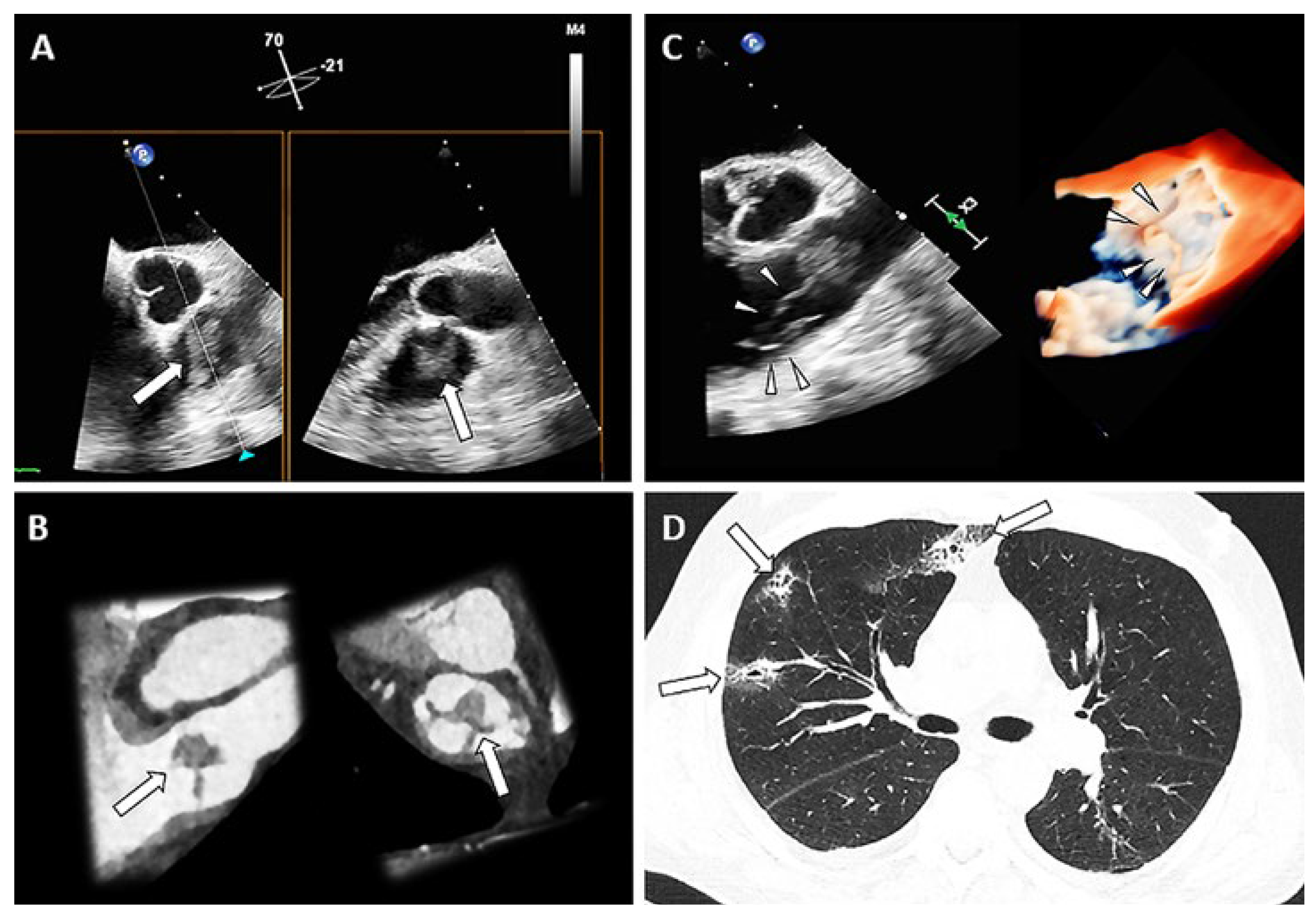
A 74-year-old male patient presented with malaise, fever, chills, and palpitations for the past five days. On admission, he was afebrile, hypotensive and tachycardic. Laboratory testing showed: leucocytosis of 20.1 Giga/l (ref. 2.6– 7.8 Giga/l), C-reactive protein of 278 mg/l (ref. <5 mg/l), procalcitonin of 4.64 µg/l (ref. <0.5 µg/l), increased d-dimer level of 29 372 ng/ml (ref. <500 ng/l). Transthoracic echocardiography revealed a normal ejection fraction with D-shaping and estimated systolic pulmonary arterial pressure of 65 mm Hg; no signs of endocarditis were detected. Due to the signs of right ventricular overload and severely impaired renal function at this time lung perfusion scintigraphy (SPECT/CT) was performed, which showed multiple small consolidations, suggesting septic emboli (
Figure 1, arrows, panel D). The suspicion of right heart endocarditis triggered transoesophageal echocardiography, which exhibited masses on all cusps of the pulmonary valve, also depicted in a follow-up CT (arrows, panel A and B, corresponding planes). Furthermore, a large (19 × 12 mm), mobile, ribbon-shaped vegetation was protruding into the right ventricular outflow tract (arrow heads, panel C, 2-dimensional and 3-dimensional; 2D: video 1, 3D: video 2); the function of the pulmonary valve was normal. All other valves were free of vegetations. Blood cultures showed growth of Staphylococcus aureus. After interdisciplinary discussion and in line with guideline recommendations, conservative treatment with flucloxacillin was started.
Isolated pulmonary valve endocarditis is the rarest form of endocarditis, accounting for less than 2% of cases. Predisposing factors include intravenous drug abuse, immunosuppression, alcoholism, and catheter-related infections. However, as in our case up to 28% of patients do not have any predisposing factors. The patient was discharged in good condition.
Disclosure statements
No financial support and no other potential conflict of interest relevant to this article was reported. None of the authors has received any compensation for this article. Prof. Kobza received institutional grants from Abbott, Biosense-Webster, Biotronik, Boston, Medtronic and Sis Medical. Dr Stämpfli received travel grants, speaker fees, consulting fees, proctoring from Alnylam, Amgen, AstraZeneca, Bayer, Bristol-Myers Squibb, Fumedica, Novartis, Pfizer, Takeda.
© 2023 by the author. Attribution - Non-Commercial - NoDerivatives 4.0.


