Next Article in Journal
Schweizer Preis für Angiologie 2022/Prix Suisse d’Angiologie 2022
Previous Article in Journal
The Zurich Postgraduate Course in Heart Failure Now in Its Fifth Edition
 
 
cardiovascmed-logo
Article Menu

Article Menu

Cardiovascular Medicine is published by MDPI from Volume 28 Issue 1 (2025). Previous articles were published by another publisher in Open Access under a CC-BY (or CC-BY-NC-ND) licence, and they are hosted by MDPI on mdpi.com as a courtesy and upon agreement with Editores Medicorum Helveticorum (EMH).
Review

Extracorporeal Life Support Use in Cardiac and Circulatory Failure: A Summary of Recently Published S3 Guidelines

by
Omer Dzemal
1,
Christoph Starck
2,
Lukas Wessel
3,
Oliver Miera
4,
Karl Werdan
5,
Marion Burckhardt
6,
Ralf Muellenbach
7,
Rolf Jaksties
8,
Florian Schmidt
9,
Karsten Wiebe
10,
Christof Schmid
11,
Stefan Kluge
12,
Kevin Pilarczyk
13,
Nils Haake
13,
Thomas Schaible
14,
Andreas Flemmer
15,
Stefan Klotz
16,
Alexander Assmann
17,
Uwe Janssens
18,
Matthias Lubnow
19,
Susanne Herber-Jonat
15,
Markus Ferrari
20,
Dirk Buchwald
21,
Stephan Ensminger
22,
York Zausig
23,
Andreas Beckmann
24,
Mark Rosenberg
25,
Malte Kelm
26,
Marcus Hennersdorf
27,
Christiane Hartog
28,
Stefan Fischer
29,
Ardawan Rastan
30,
Daniel Zimpfer
31,
Andreas Fründ
32,
Sven Maier
33,
Elfriede Ruttmann-Ulmer
34,
Heinrich Groesdonk
35,
Christian Schlensak
36,
Monika Nothacker
37,
Michael Buerke
38,
Harald Köditz
39,
Guido Michels
40,
Lars Krüger
41 and
Udo Boeken
17
add Show full author list remove Hide full author list
1
Department of Cardiac Surgery, City Hospital of Zurich – Site Triemli, Zurich, Switzerland
2
German Heart Centre, Department of Cardiothoracic and Vascular Surgery, Berlin, German
3
Department of Paediatric Surgery, University Hospital Mannheim, Mannheim, Germany
4
Department of Congenital Heart Disease- Paediatric Cardiology, German Heart Centre Berlin, Berlin, Germany
5
Clinic for Internal Medicine III, University Hospital Halle (Saale), Martin-Luther University Halle-Wittenberg, Halle-Wittenberg, Germany
6
Department of Health Sciences and Management; Baden-Wuerttemberg Cooperative State University (DHBW), Stuttgart, Germany
7
Department of Anaesthesiology and Critical Care Medicine, Campus Kassel of the University of Southampton, Kassel, Germany
8
German Heart Foundation, Frankfurt, Germany
9
Department of Paediatric Cardiology and Intensive Care Medicine, Medical School Hannover, Hannover, Germany
10
Department of Cardiothoracic Surgery, Münster University Hospital, Münster, Germany
11
Department of Cardiothoracic Surgery, University Medical Centre Regensburg, Regensburg, Germany
12
Klinik für Intensivmedizin, Universitätsklinikum Hamburg-Eppendorf, Hamburg, Germany
13
Imland Hospital Rendsburg, Department for Intensive Care Medicine, Rendsburg, Schleswig-Holstein, Germany
14
Department of Neonatology, University Children's Hospital Mannheim, University of Heidelberg, Mannheim, Germany
15
Division of Neonatology, Dr. v. Hauner Children’s Hospital and Perinatal Centre Munich - Grosshadern, LMU Munich, München, Germany
16
Department of Cardiac Surgery, Segeberger Kliniken, Bad Segeberg, Germany
17
Department of Cardiac Surgery, Heinrich-Heine-University Medical School, Duesseldorf, Germany
18
Medical Clinic and Medical Intensive Care Medicine, St Antonius Hospital, Eschweiler, Germany
19
Department of Internal Medicine II, University Hospital Regensburg, Regensburg, Germany
20
HSK, Clinic of Internal Medicine I, Helios-Kliniken, Wiesbaden, Germany
21
Department of Cardiothoracic Surgery, University Hospital Bergmannsheil, Ruhr-University, Bochum, Germany
22
Department of Cardiac and Thoracic Vascular Surgery, University Hospital of Schleswig-Holstein, Lübeck, Germany
23
Department of Anaesthesiology and Operative Intensive Care Medicine, Aschaffenburg-Alzenau Hospital, Aschaffenburg, Bavaria, Germany
24
German Society for Thoracic and Cardiovascular Surgery, Langenbeck-Virchow-Haus, Berlin, Germany
25
Klinikum Aschaffenburg-Alzenau, Medizinische Klinik 1, Aschaffenburg, Germany
26
Department of Cardiology, Pulmonology and Vascular Medicine, Heinrich-Heine- University Medical School, Duesseldorf, Germany
27
Department of Cardiology, Pneumology, Angiology and Internal Intensive Care Medicine, SLK-Kliniken Heilbronn, Heilbronn, Germany
28
Department of Anaesthesiology and Operative Intensive Care Medicine, Charité, Humboldt-Universität zu Berlin, and Berlin Institute of Health, Berlin, Germany
29
Department of Thoracic Surgery and Lung Support, Ibbenbueren General Hospital, Ibbenbueren, Germany
30
Department of Cardiac and Vascular Thoracic Surgery, Philipps-University Hospital Marburg, Marburg, Germany
31
Department of Cardiac Surgery, Medical University of Vienna, Wien, Austria
32
Department of Physiotherapy, Heart- and Diabetes Centre NRW, Ruhr-University, Bochum, Germany
33
Department of Cardiovascular Surgery, Heart Centre Freiburg University, Freiburg, Germany
34
Department of Cardiac Surgery, Medical University of Innsbruck, Innsbruck, Austria
35
Department of Intensive Care Medicine, Helios Clinic Erfurt, Erfurt, Germany
36
Department of Cardio-thoracic and Vascular Surgery, University of Tübingen, Tübingen, Germany
37
Institute for Medical Knowledge Management, Association of the Scientific Medical Societies (AWMF), Universität Marburg, Marburg, Germany
38
Department of Cardiology, Angiology and Internal Intensive Care Medicine, St. Marienkrankenhaus Siegen, Siegen, Germany
39
Medical University Children’s Hospital, Hannover, Germany
40
Department of Acute and Emergency Care, St Antonius Hospital Eschweiler, Eschweiler, Germany
41
Division of Thoracic and Cardiovascular Surgery, Heart- and Diabetes Centre NRW, Ruhr-University, Bochum, Germany
Cardiovasc. Med. 2022, 25(6), 165; https://doi.org/10.4414/cvm.2022.02234
Submission received: 1 August 2022 / Revised: 1 September 2022 / Accepted: 1 October 2022 / Published: 1 November 2022

Introduction

Extracorporeal life support (ECLS) represents a widely accepted treatment modality for patients with cardiac and/or respiratory failure failing to respond to conventional medical therapy. Through the establishment of a modified cardiopulmonary bypass circuit, ECLS provides a mechanism for temporary cardiac support and gas exchange, allowing patients to recover from existing life-threatening cardiac and/or lung disease. This article summarises the current recommendations for ECLS therapy in adults, which are based on the recently published S3 guidelines entitled “Extracorporeal circulation (ECLS / extracorporeal membrane oxygenation = ECMO), use in cardiac and circulatory failure” (Boeken et al. The Thoracic and Cardiovascular Surgeon. In press).

Guidelines

A great number of guidelines have been issued in recent years by the association of the scientific medical societies (Arbeitsgemeinschaft der Wissenschaftlichen Medizinischen Fachgesellschaften, AWMF). Medical societies involved in the publication of the current S3 guideline concerning ECLS use in cardiac and circulatory failure are listed in Table 1. The level of evidence and the strength of recommendations of ECLS management presented in the current manuscript is high. Detailed description of all S3 guidelines can be found on the AWMF website (www.awmf.org/leitlinien/detail/ll/011-021.html) and were published recently by Boeken and associates (The Thoracic and Cardiovascular Surgeon, in press).

Staffing issues

A multidisciplinary ECLS team should initiate the ECLS (indication and implantation) in adult patients. The implantation should ideally be in an ECLS centre with sufficient expertise by an ECLS team with appropriate skills in terms of implantation [1,2]. According to the current literature no minimum number of implantations per year can be defined in order to achieve sufficient therapeutic success with the ECLS. However an implantation rate of at least 20 ECLS per year should be aimed for [3,4,5]. For the ECLS implantation, a standardised procedure adapted to local conditions should be available in written form [6]. For the ECLS initiation, a specific minimum of medical equipment and facilities should be provided [2,7].
ECLS therapy is to be performed in a centre with a full range of intensive care treatment options in a standardised, multidisciplinary and multimodal approach under the guidance of a specialist experienced in the field of ECLS with an additional qualification in intensive care medicine [2,8]. Further medical specialists as listed in Table 2 should be involved for the management of potential ECLS complications [2,8].

Staff training and continuing education

The processes of initiation, further care, training and employee qualification of the multiprofessional ECLS team should be coordinated by the medical director of the ECLS programme, depending on the institutional structure [2,7,8].
A multiprofessional team specially trained in the ECLS therapy process should carry out the multimodal therapy in that field in the intensive care unit (ICU) [7]. Continuing education of the multiprofessional team in the ICU should take place regularly according to a defined internal curriculum. The training requirement depends on the centre-specific ECLS volume and the individual experience of the medical stuff [2].

ECLS circuit and cannulation site

Only centrifugal pumps should be used for ECLS. Heparin-coated components should preferably be used. The selection of the arterial cannulation site should be based on the patient’s individual circumstances. In adults, either peripheral (femoral artery) or central (subclavian artery; ascending aorta) cannulation can be performed.

Patient ICU care and monitoring

Depending on the medical and nursing effort, in the multidisciplinary approach the patient to nurse ratio in the ICU should be determined individually from shift to shift. The patient’s individual nursing care in the ICU should be ensured [8]. In addition to medical and nursing treatment, technical checks of the ECLS system should be carried out at least once a day by a perfusionist.
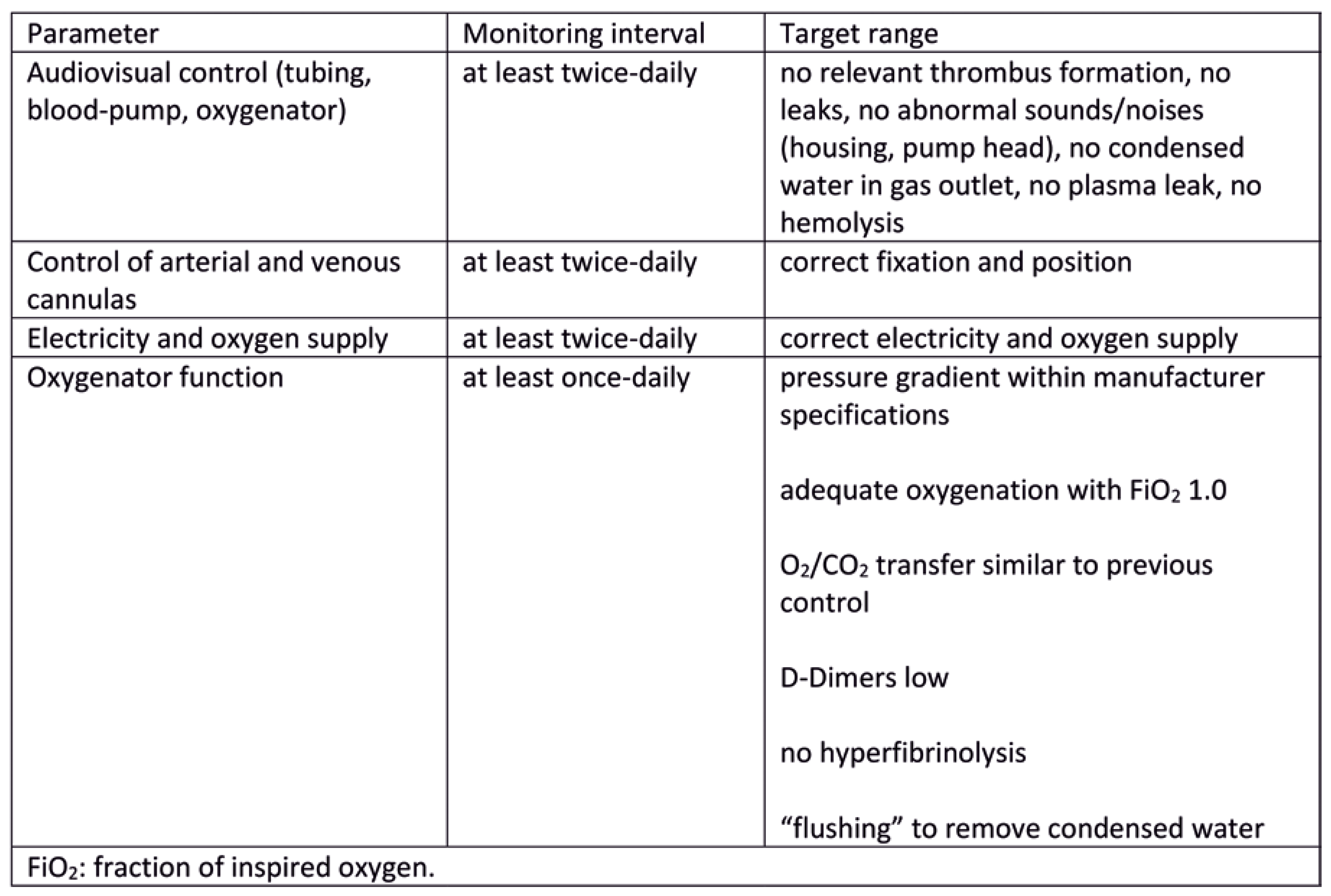
Perfusion, haemodynamics, cardiac unloading, oxygenation, anticoagulation and the functionality of the ECLS system should be continuously monitored in patients on ECLS therapy [6,9,10]. (Figure 1 and Figure 2). Thus a safety net for early detection of possible complication can be established [11,12,13,14,15,16,17,18].
Every extracorporeal circulation system requires anticoagulation because of contact activation of blood coagulation. Anticoagulation is routinely performed with systemic administration of heparin except in cases of heparin-induced thrombocytopenia, where alternative agents such as argatroban, bivalirudin and lepirudin should be used. Coagulation can be monitored with the use of parameters such as partial thromboplastin time (PTT), activated clotting time (ACT), factor Xa or even thromboelastography.
In patients cannulated in a femoral axis, adequate oxygenation should be monitored by measuring the peripheral oxygen saturation in the right upper extremity and through blood gas analysis of blood samples from arteries of the right upper extremity.
A basic clinical-neurological examination should be carried out daily and the pupillary reflex should be checked several times a day.
A basic clinical-neurological examination should be carried out daily and the pupillary reflex should be checked several times a day. Due to the lack of reliable data, no additional apparatus-based method for routine neurological monitoring can be recommended. In the case of femoral arterial cannulation, a distal perfusion line should be placed to avoid distal limb ischaemia. In the case of peripheral ECLS cannulation, the arterial cannula should be preferably placed contralaterally to the venous cannula.

Management of ventricular distension and central hypoxia

In the case of left ventricular distension, the left ventricle should be actively unloaded after conservative measures have been exhausted. Various techniques have been described for left ventricular unloading, including open surgical placement of a left ventricular vent, percutaneous left atrial venting, transseptal drainage, intra-aortic balloon pump (IABP) and microaxial blood pump placement in the left ventricle. Retrospective studies have shown that the additional use of a microaxial blood pump (Impella®) placed in the left ventricle was associated with a lower 30-day and 1-year mortality compared with the use of ECLS alone without an increase in complication rates. Even though the additional use of Impella during ECLS has shown an improvement in outcomes, further prospective studies should be conducted to confirm the results of the previous retrospective studies [19,20,21].
Central hypoxia (watershed or Harlequin syndrome) under ECLS therapy with femoral arterial cannulation should be treated immediately after the diagnosis has been established. The following measures are suitable:
  • Arterial cannulation of right axillary artery.
  • Insertion of another cannula (e.g., via the right internal jugular vein) and change of circuit configuration to veno-arteriovenous (V-AV)
  • Insertion of another venous drainage cannula and change of the configuration to VV-A
  • Change from peripheral to central cannulation.

ECLS weaning and explantation

According to the current literature, there is no evidence for levosimendan therapy as part of ECLS weaning [22]. The following criteria should be evaluated before ECLS weaning is initiated according to the standardised protocol:
  • Pulsatile arterial blood pressure and evidence of biventricular contractility on echocardiography
  • Mean arterial blood pressure >60 mm Hg
  • Mixed venous oxygen saturation (SvO2) ≥ 65% (central venous oxygen saturation [ScvO2] ≥ 60%)
  • Lactate values ≤ 2 mmol/l or falling
  • Vasopressor/inotropic dosage low or falling
  • Sufficient pulmonary oxygenation (Horowitz-index or ratio of arterial oxygen partial pressure (PaO2 in mmHg) to fraction of inspired oxygen (FiO2) > 200 mm Hg) / CO2 elimination performance under lung-protective ventilation
  • Compensated end organ functions, especially liver function
In addition, criteria 1–7 should be met with a low ECLS flow (<2.0 l/min) and with a low gas flow (<2 l/min) before ECLS explantation [23,24]. Pulsatile blood pressure serves as an indication of presence of biventricular contraction, which can be precisely quantified with the help of echocardiography. Furthermore, several retrospective studies have shown that echocardiographic parameters of left and right ventricular function, specifically left ventricular ejection fraction (LVEF), tricuspid annular plane systolic excursion (TAPSE), mitral lateral annular systolic velocity (s’), right ventricle (RV) s’ and left ventricular outflow tract (LVOT) velocity time integral (VTI) are predictors of successful weaning from ECLS [25,26,27]. Due to the complexity of the clinical conditions underlying vasopressor/inotropic therapy, it is difficult to define a target vasopressor/inotropic dose, as a prerequisite for initiating ECLS weaning. However, some orientating doses could be <0.1–0.2 μg/kg/min noradrenaline, <0.1–0.2 μg/kg/min adrenaline, <6 μg/kg/min dobutamine and <2.0 mg/h milrinone [28]. Even though the above-mentioned criteria for weaning should be evaluated before initiating weaning from ECLS, further decision to proceed with weaning should be based on the individual judgement of the medical team involved in the patient’s treatment and after taking into consideration the patient’s general health condition.
During the ECLS weaning process, arterial and central venous blood gas analysis to monitor oxygenation, CO2 elimination and circulatory function should be performed about 30 minutes after the ECLS blood flow has been reduced [9,10].
An additional mechanical circulatory support system, such as Impella or IABP, should not be routinely implanted during ongoing ECLS treatment (including weaning) [29,30,31,32,33,34].
Therapy limitations in ECLS patients should be made as a patient-centered decision with the interprofessional treatment team, taking medical and ethical aspects into account. Such a scenario exists when the desired therapy goal cannot be achieved or the therapy goal is not desired by the patient.
In intensive care patients in the early phase after explantation of an ECLS system, perfusion, haemodynamics (with invasive arterial blood pressure measurement) and oxygenation should be continuously monitored.
Echocardiography should be performed shortly after the ECLS explantation and daily thereafter (early phase after the explantation).
In the early phase after decannulation of a peripherally implanted ECLS system, the cannulation sites should be examined clinically at least once a day.
An ultrasound examination of the cannulated vessels should also be carried out routinely after decannulation.

Normal ward care

As part of the care of patients after ECLS therapy on the normal ward, attention should be given to cardiac deterioration signs and the consequences of cannulationrelated complications (infection, thrombosis or ischaemia).

Rehabilitation and follow-up

After ECLS therapy, patients should be rehabilitated in an inpatient setting. Patients should have regular and long-term cardiological follow-up examinations, and depending on the complexity of the underlying disease, in an interdisciplinary special outpatient department.
Omer Dzemali
Heart Centre Triemli Department of Cardiac Surgery Birmensdorferstrasse 497 CH-8063 Zürich
omer.dzemali[at]stadtspital.ch

Disclosure Statement

No financial support and no other potential conflict of interest relevant to this article was reported.

References

  1. Michels, G.; Wengenmayer, T.; Hagl, C.; Dohmen, C.; Böttiger, B.W.; Bauersachs, J.; Markewitz, A.; Bauer, A.; Gräsner, J.-T.; Pfister, R.; et al. Empfehlungen zur extrakorporalen kardiopulmonalen Reanimation (eCPR). Med. Klin.-Intensiv. und Notfallmedizin 2018, 113, 478–486. [Google Scholar] [CrossRef]
  2. Abrams, D.; Garan, A.R.; Abdelbary, A.; Bacchetta, M.; Bartlett, R.H.; Beck, J.; Belohlavek, J.; Chen, Y.-S.; Fan, E.; Ferguson, N.D.; et al. Position paper for the organization of ECMO programs for cardiac failure in adults. Intensiv. Care Med. 2018, 44, 717–729. [Google Scholar] [CrossRef]
  3. McCarthy, F.H.; McDermott, K.M.; Spragan, D.; Hoedt, A.; Kini, V.; Atluri, P.; Gaffey, A.; Szeto, W.Y.; Acker, M.A.; Desai, N.D. Unconventional Volume-Outcome Associations in Adult Extracorporeal Membrane Oxygenation in the United States. Ann. Thorac. Surg. 2016, 102, 489–495. [Google Scholar] [CrossRef]
  4. Barbaro, R.P.; Odetola, F.O.; Kidwell, K.M.; Paden, M.L.; Bartlett, R.H.; Davis, M.M.; Annich, G.M. Association of Hospital-Level Volume of Extracorporeal Membrane Oxygenation Cases and Mortality. Analysis of the Extracorporeal Life Support Organization Registry. Am. J. Respir. Crit. Care Med. 2015, 191, 894–901. [Google Scholar] [CrossRef]
  5. Huesch, M.D. Volume–Outcome Relationships in Extracorporeal Membrane Oxygenation: Retrospective Analysis of Administrative Data From Pennsylvania, 2007–2015. Asaio J. 2018, 64, 450–457. [Google Scholar] [CrossRef] [PubMed]
  6. Extracorporeal Life Support Organization. ELSO Guidelines for ECMO Centers. 2014. Available online: https://www.elso.org/Portals/0/IGD/Archive/FileManager/faf3f6a3c7cusersshyerdocumentselsoguidelinesecmocentersv1.8.pdf (accessed on 9 March 2020).
  7. Michels, G.; Wengenmayer, T.; Hagl, C.; Dohmen, C.; Böttiger, B.W.; Bauersachs, J.; Markewitz, A.; Bauer, A.; Gräsner, J.-T.; Pfister, R.; et al. Empfehlungen zur extrakorporalen kardiopulmonalen Reanimation (eCPR). Med. Klin.-Intensiv. und Notfallmedizin 2018, 113, 478–486. [Google Scholar] [CrossRef]
  8. Combes, A.; Brodie, D.; Bartlett, R.; Brochard, L.; Brower, R.; Conrad, S.; De Backer, D.; Fan, E.; Ferguson, N.; Fortenberry, J.; et al. Position Paper for the Organization of Extracorporeal Membrane Oxygenation Programs for Acute Respiratory Failure in Adult Patients. Am. J. Respir. Crit. Care Med. 2014, 190, 488–496. [Google Scholar] [CrossRef] [PubMed]
  9. Beckmann, A.; Benk, C.; Beyersdorf, F.; Haimerl, G.; Merkle, F.; Mestres, C.; Pepper, J.; Wahba, A. Position article for the use of extracorporeal life support in adult patients. Eur. J. Cardio-Thoracic Surg. 2011, 40, 676–681. [Google Scholar] [CrossRef] [PubMed]
  10. Pichler, P.; Antretter, H.; Dünser, M.; Eschertzhuber, S.; Gottardi, R.; Heinz, G.; Pölzl, G.; Pretsch, I.; Rajek, A.; Wasler, A.; et al. Positionspapier der Österreichischen Kardiologischen Gesellschaft zum Einsatz der extrakorporalen Membranoxygenation (ECMO) bei Erwachsenen kardiologischen Patienten. Med. Klin.-Intensiv. und Notfallmedizin 2015, 110, 407–420. [Google Scholar] [CrossRef]
  11. Richardson, A. (.C.; Schmidt, M.; Bailey, M.; Pellegrino, V.A.; Rycus, P.T.; Pilcher, D.V. ECMO Cardio-Pulmonary Resuscitation (ECPR), trends in survival from an international multicentre cohort study over 12-years. Resuscitation 2017, 112, 34–40. [Google Scholar] [CrossRef]
  12. Lorusso, R.; Barili, F.; Di Mauro, M.; Gelsomino, S.; Parise, O.; Rycus, P.T.; Maessen, J.; Mueller, T.; Muellenbach, R.; Belohlavek, J.; et al. In-Hospital Neurologic Complications in Adult Patients Undergoing Venoarterial Extracorporeal Membrane Oxygenation: Results From the Extracorporeal Life Support Organization Registry. Crit. Care Med. 2016, 44, e964–e972. [Google Scholar] [CrossRef]
  13. Bizzarro, M.J.; Conrad, S.A.; Kaufman, D.A.; Rycus, P. Infections acquired during extracorporeal membrane oxygenation in neonates, children, and adults*. Pediatr. Crit. Care Med. 2011, 12, 277–281. [Google Scholar] [CrossRef] [PubMed]
  14. Vogel, A.M.; Lew, D.F.; Kao, L.S.; Lally, K.P. Defining risk for infectious complications on extracorporeal life support. J. Pediatr. Surg. 2011, 46, 2260–2264. [Google Scholar] [CrossRef]
  15. Dalton, H.J.; Garcia-Filion, P.; Holubkov, R.; Moler, F.W.; Shanley, T.; Heidemann, S.; Meert, K.; Berg, R.A.; Berger, J.; Carcillo, J.; et al. Association of Bleeding and Thrombosis With Outcome in Extracorporeal Life Support*. Pediatr. Crit. Care Med. 2015, 16, 167–174. [Google Scholar] [CrossRef]
  16. Polito, A.; Barrett, C.S.; Wypij, D.; Rycus, P.T.; Netto, R.; Cogo, P.E.; Thiagarajan, R.R. Neurologic complications in neonates supported with extracorporeal membrane oxygenation. An analysis of ELSO registry data. Intensiv. Care Med. 2013, 39, 1594–1601. [Google Scholar] [CrossRef]
  17. Werho, D.K.; Pasquali, S.K.; Yu, S.; Donohue, J.; Annich, G.M.; Thiagarajan, R.R.; Hirsch-Romano, J.C.; Gaies, M. Epidemiology of Stroke in Pediatric Cardiac Surgical Patients Supported With Extracorporeal Membrane Oxygenation. Ann. Thorac. Surg. 2015, 100, 1751–1757. [Google Scholar] [CrossRef] [PubMed]
  18. Wightman, A.M.; Bradford, M.C.; Symons, J.; Brogan, T.V. Impact of Kidney Disease on Survival in Neonatal Extracorporeal Life Support. Pediatr. Crit. Care Med. 2015, 16, 576–582. [Google Scholar] [CrossRef] [PubMed]
  19. Patel, S.M.; Lipinski, J.; Al-Kindi, S.G.; Patel, T.; Saric, P.; Li, J.; Nadeem, F.; Ladas, T.; Alaiti, A.; Phillips, A.; et al. Simultaneous Venoarterial Extracorporeal Membrane Oxygenation and Percutaneous Left Ventricular Decompression Therapy with Impella Is Associated with Improved Outcomes in Refractory Cardiogenic Shock. Asaio J. 2019, 65, 21–28. [Google Scholar] [CrossRef]
  20. Pappalardo, F.; Schulte, C.; Pieri, M.; Schrage, B.; Contri, R.; Soeffker, G.; et al. Concomitant implantation of Impella on top of veno-arterial extracorporeal membrane oxygenation may improve survival of patients with cardiogenic shock. Eur. J. Heart Fail. 2017. Available online: http://onlinelibrary.wiley.com/o/cochrane/clcentral/articles/778/CN-01245778/frame.html. [CrossRef]
  21. Schmack, B.; Seppelt, P.; Weymann, A.; Alt, C.; Farag, M.; Arif, R.; Doesch, A.O.; Raake, P.W.; Kallenbach, K.; Mansur, A.; et al. Extracorporeal life support with left ventricular decompression—improved survival in severe cardiogenic shock: results from a retrospective study. PeerJ 2017, 5, e3813–e3813. [Google Scholar] [CrossRef]
  22. Distelmaier, K.; Roth, C.; Schrutka, L.; Binder, C.; Steinlechner, B.; Heinz, G.; Lang, I.M.; Maurer, G.; Koinig, H.; Niessner, A.; et al. Beneficial effects of levosimendan on survival in patients undergoing extracorporeal membrane oxygenation after cardiovascular surgery. Br. J. Anaesth. 2016, 117, 52–58. [Google Scholar] [CrossRef]
  23. Smith, M.; Vukomanovic, A.; Brodie, D.; Thiagarajan, R.; Rycus, P.; Buscher, H. Duration of veno-arterial extracorporeal life support (VA ECMO) and outcome: an analysis of the Extracorporeal Life Support Organization (ELSO) registry. Crit. Care 2017, 21, 1–9. [Google Scholar] [CrossRef]
  24. Extracorporeal Life Support Organization. Ultrasound Guidance for Extra-corporeal Membrane OxygenationGeneral Guidelines. 2015. [Google Scholar]
  25. Aissaoui, N.; Luyt, C.-E.; Leprince, P.; Trouillet, J.-L.; Léger, P.; Pavie, A.; Diebold, B.; Chastre, J.; Combes, A. Predictors of successful extracorporeal membrane oxygenation (ECMO) weaning after assistance for refractory cardiogenic shock. Intensiv. Care Med. 2011, 37, 1738–1745. [Google Scholar] [CrossRef]
  26. Akin, S.; Miranda, D.d.R.; Caliskan, K.; Soliman, O.I.; Guven, G.; Struijs, A.; van Thiel, R.J.; Jewbali, L.S.; Lima, A.; Gommers, D.; et al. Functional evaluation of sublingual microcirculation indicates successful weaning from VA-ECMO in cardiogenic shock. Crit. Care 2017, 21, 265–265. [Google Scholar] [CrossRef]
  27. Cavarocchi, N.C.; Pitcher, H.T.; Yang, Q.; Karbowski, P.; Miessau, J.; Hastings, H.M.; Hirose, H. Weaning of extracorporeal membrane oxygenation using continuous hemodynamic transesophageal echocardiography. J. Thorac. Cardiovasc. Surg. 2013, 146, 1474–1479. [Google Scholar] [CrossRef] [PubMed]
  28. Gaies, M.G.; Jeffries, H.E.; Niebler, R.A.; Pasquali, S.K.; Donohue, J.E.; Yu, S.; et al. Vasoactive-inotropic score is associated with outcome after infant cardiac surgery: an analysis from the Pediatric Cardiac Critical Care Consortium and Virtual PICU System Registries. Pediatr. Crit. Care Med. 2014, 15, 529–537. [Google Scholar] [CrossRef]
  29. Ro, S.K.; Kim, J.B.; Jung, S.H.; Choo, S.J.; Chung, C.H.; Lee, J.W. Extracorporeal life support for cardiogenic shock: influence of concomitant intra-aortic balloon counterpulsation. Eur. J. Cardio-Thoracic Surg. 2014, 46, 186–192. [Google Scholar] [CrossRef]
  30. Lin, L.-Y.; Liao, C.-W.; Wang, C.-H.; Chi, N.-H.; Yu, H.-Y.; Chou, N.-K.; Hwang, J.-J.; Lin, J.-L.; Chiang, F.-T.; Chen, Y.-S. Effects of Additional Intra-aortic Balloon Counter-Pulsation Therapy to Cardiogenic Shock Patients Supported by Extra-corporeal Membranous Oxygenation. Sci. Rep. 2016, 6, 1–8. [Google Scholar] [CrossRef] [PubMed]
  31. Aso, S.; Matsui, H.; Fushimi, K.; Yasunaga, H. The Effect of Intraaortic Balloon Pumping Under Venoarterial Extracorporeal Membrane Oxygenation on Mortality of Cardiogenic Patients: An Analysis Using a Nationwide Inpatient Database. Crit. Care Med. 2016, 44, 1974–1979. [Google Scholar] [CrossRef]
  32. Pappalardo, F.; Schulte, C.; Pieri, M.; Schrage, B.; Contri, R.; Soeffker, G.; et al. Concomitant implantation of Impella® on top of veno-arterial extracorporeal membrane oxygenation may improve survival of patients with cardiogenic shock. Eur. J. Heart Fail. 2017, 19, 404–412. [Google Scholar] [CrossRef]
  33. Park, T.K.; Yang, J.H.; Choi, S.-H.; Bin Song, Y.; Hahn, J.-Y.; Choi, J.-H.; Sung, K.; Lee, Y.T.; Gwon, H.-C. Clinical impact of intra-aortic balloon pump during extracorporeal life support in patients with acute myocardial infarction complicated by cardiogenic shock. BMC Anesthesiol. 2014, 14, 27–27. [Google Scholar] [CrossRef] [PubMed]
  34. Bréchot, N.; Demondion, P.; Santi, F.; Lebreton, G.; Pham, T.; Dalakidis, A.; Gambotti, L.; Luyt, C.-E.; Schmidt, M.; Hekimian, G.; et al. Intra-aortic balloon pump protects against hydrostatic pulmonary oedema during peripheral venoarterial-extracorporeal membrane oxygenation. Eur. Hear. Journal. Acute Cardiovasc. Care 2017, 7, 62–69. [Google Scholar] [CrossRef] [PubMed]
Figure 1. Patient-related monitoring parameters during extracorporeal life support (ECLS) therapy.
Cardiovascmed 25 00165 g001
Figure 2. Monitoring parameters of the extracorporeal life support (ECLS) system.
Cardiovascmed 25 00165 g002
Table 1. Medical societies involved in the publication of current S3 guidelines concerning extracorporeal life support (ECLS) use in cardiac and circulatory failure.
Deutsche Gesellschaft für Thorax-, Herz- und Gefäßchirurgie e.V. (DGTHG)
Deutsche Gesellschaft für Anästhesiologie und Intensivmedizin e.V. (DGAI)
Deutsche Interdisziplinäre Vereinigung für Intensiv- und Notfallmedizin (DIVI)
Deutsche Gesellschaft für Kardiologie - Herz- und Kreislaufforschung e.V. (DGK)
Gesellschaft für Neonatologie und pädiatrische Intensivmedizin e.V. (GNPI)
Deutsche Gesellschaft für Pädiatrische Kardiologie e.V. (DGPK)
Deutsche Gesellschaft für Internistische Intensivmedizin und Notfallmedizin (DGIIN)
Deutsche Gesellschaft für Innere Medizin e.V. (DGIM)
Deutsche Gesellschaft für Kinder- und Jugendmedizin e.V. (DGKJ)
Deutsche Gesellschaft für Kinderchirurgie (DGKCH)
Deutsche Gesellschaft für Thoraxchirurgie (DGT)
Deutsche Gesellschaft für Fachkrankenpflege und Funktionsdienste e. V. (DGF)
Schweizerische Gesellschaft für Herz- und thorakale Gefässchirurgie (SGHC)
Österreichische Gesellschaft für Thorax- und Herzchirurgie (ÖGTHC)
Deutsche Herzstiftung e.V.
Deutscher Verband für Physiotherapie (ZVK)
Akademie für Ethik in der Medizin (AEM)
Deutsche Gesellschaft für Kardiotechnik e.V. (DGfK)
Table 2. Disciplines that should be available for the management of extracorporeal life support (ECLS) therapy.
ECLS initiationECLS
continuation
Cardiac surgeryXX
CardiologyXX
AnaesthesiologyXX
Intensive care medicineXX
Neurology X
Neurosurgery X
General surgery X
Vascular surgery X
Angiology X
Radiology X
Haematology X
Gastroenterology X
Nephrology X
Pulmonology X
Ethics committee X