Next Article in Journal
Author’s Reply
Previous Article in Journal
Prof. Daniel Wagner a éTé Nommé Professeur Ordinaire de L’UNIL
 
 
Cardiovascular Medicine is published by MDPI from Volume 28 Issue 1 (2025). Previous articles were published by another publisher in Open Access under a CC-BY (or CC-BY-NC-ND) licence, and they are hosted by MDPI on mdpi.com as a courtesy and upon agreement with Editores Medicorum Helveticorum (EMH).
Interesting Images

Loss of Biventricular Pacing: What’s the Problem?

by
Sylvie Cochard
* and
Jürg Schläpfer
Departement of Cardiology, Centre Hospitalier Universitaire Vaudois, Lausanne, Switzerland
*
Author to whom correspondence should be addressed.
Cardiovasc. Med. 2017, 20(3), 74; https://doi.org/10.4414/cvm.2017.00469
Submission received: 15 December 2016 / Revised: 15 January 2017 / Accepted: 15 February 2017 / Published: 15 March 2017

Case presentation

A 74-year-old man with a long history of ischaemic dilated cardiomyopathy and a severely depressed left ventricular ejection fraction (LVEF) of 15% went to the hospital because of dyspnoea and epigastric discomfort for six days, triggered by an emotional event.
Seven days before, he had a scheduled ambulatory cardiological assessment, including a clinical and biological work up, interrogation of his cardioverter resynchronisation therapy-defibrillator device (CRT-D), which showed biventricular pacing of 99% without any arrhythmia, and echocardiography; the global clinical situation was considered stable under both maximal medical and resynchronisation therapies.
The day after, the patient suered a violent emotional shock (the death of his beloved cat), which was followed by epigastric discomfort and progressive dyspnoea. He waited a week before the present consultation. Clinically the patient was normotensive and normocardic; he showed signs of hypoperfusion with cold extremities and discreet marbling on legs. Lung auscultation was normal. The rest of the physical examination was unremarkable.
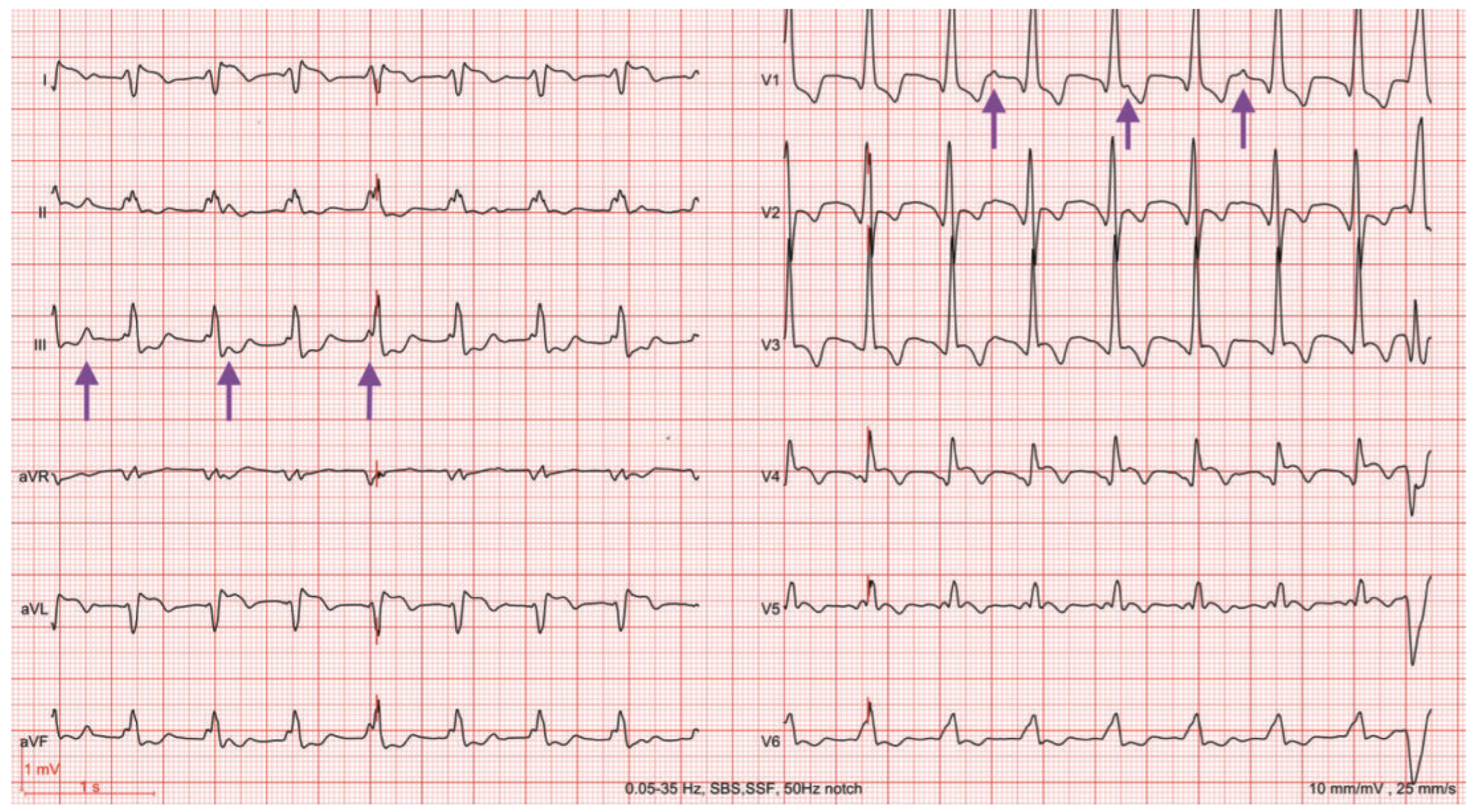
The initial ECG is shown in Figure 1 and Figure 2.

Question

What is the problem on the ECG?

Commentary

The ECG shows a regular wide-complex (QRS duration of 160 ms), normocardiac rhythm (95/min) with an atypical right bundle-branch block morphology (qR in leads V1 to V4) and right axis deviation and. Positive P waves without any relation with QRS complexes can easily be seen in lead V1, indicating ventricular dissociation; these P waves are also positive in lead I and in inferior leads and negative in aVR suggesting a sinus node origin (arrows). All these features are diagnostic of a slow ventricular tachycardia (VT) [1,2]. The last visible QRS complex is a ventricular premature beat and a red mark is superimposed to the fifth QRS complex in the extremity leads and to the second QRS complex in precordial leads.
The laboratory workup showed increased N-terminal pro-B-type natriuretic peptide (NT-proBNP) to twice the reference value. A transthoracic echocardiogram confirmed the pre-existing poor LVEF. Interrogation of the CRT-D showed incessant episodes of nonsustained and sustained VT for six days and, accordingly, depressed biventricular pacing to 70%. The arrhythmias were classified as ventricular sense episodes by the device and not as VT because of the slow frequency of 95 bpm, which was below the programmed VT detection rate of 128 bpm (470 ms). The VT was successfully overdriven with an accelerated resynchronised pacing rate of 1OO bpm.
The patient underwent an electrophysiology study that could not induce any ventricular arrhythmia: local abnormal ventricular activities were diusely eliminated and a line around a large anterior infarct scar was ablated.
After the procedure, the ECG showed a resynchronised paced rhythm at 75 bpm (Figure 3). After a long discussion, destination therapy was accepted by the patient.
This case reminds us that a patient with a CRT-D may present with prolonged unrecognised slow VT leading to a loss of resynchronisation and haemodynamic instability [3]. In these situations, an ECG is mandatory as simple pulse check by the patient or the physician will be confusing.
Indeed, VT above the tachycardia detection interval may be associated with clinical symptoms such as angina, palpitations, heart failure and syncope [4,5]. Independent predictors of slow VTs are impaired left ventricular function, inducible and spontaneous monomorphic VTs, and class III anti-arrhythmic drugs (amiodarone).
This case also reminds us that a violent emotional event may trigger ventricular arrhythmias and be the cause of a sudden cardiac death.

Conflicts of Interest

No financial support and no other potential conflict of interest relevant to this article was reported.

References

  1. Brugada, P.; Brugada, J.; Mont, L.; Smeets, J.; Andries, E.W. A new approach to the di erential diagnosis of a regular tachycardia with a wide QRS complex. Circulation. 1991, 83, 1649–1659. [Google Scholar] [CrossRef] [PubMed]
  2. Vereckei, A.; Duray, G.; Szenasi, G.; Altenose, G.T.; Miller, J.M. New algorithm using only lead aVR for di erential diagnosis of wide QRS complex tachycardia. Heart Rhythm 2008, 5, 89–98. [Google Scholar] [CrossRef] [PubMed]
  3. Kutyifa, V.; Klein, H.U.; Wang, P.J.; McNitt, S.; Polonsky, B.; Zima, E.; Merkely, B.; Moss, A.J.; Zareba, W. Clinical significance of ventricular tachyarrhythmias in patients treated with CRT-D. Heart Rhythm. 2013, 10, 943–950. [Google Scholar] [CrossRef] [PubMed]
  4. Bänsch, D.; Castrucci, M.; Böcker, D.; Breithardt, G.; Block, M. Ventricular tachycardias above initially programmed tachycardia detection interval in patients with implantable cardioverter-defibrillators: incidence, prediction and significance. J Am Coll Cardiol 2000, 36, 557–565. [Google Scholar] [CrossRef] [PubMed]
  5. Israel, C.W.; Burmistrava, T. Optimal tachycardia programming in ICDs: Recommendations in the post-MADIT-RIT era. Herzschrittmacherther Elektrophysiol. 2016, 27, 163–170. [Google Scholar] [CrossRef] [PubMed]
Figure 1. Standard 12-lead ECG at admission.
Cardiovascmed 20 00074 g001
Figure 2. Standard 12-lead ECG at admission showing regular wide-complex (QRS 160 ms), normocardiac rhythm with an atypical right bundle-branch block morphology (qR in leads V1 to V4) and right axis deviation. Sinus P waves are indicated with purple arrows. See text for details.
Cardiovascmed 20 00074 g002
Figure 3. Standard 12-lead ECG showing a paced rhythm with ventricular resynchronisation; note the wide QRS (240 ms) despite resynchronisation.
Cardiovascmed 20 00074 g003