Next Article in Journal
Wolff-Parkinson-White Syndrome and Diverticulosis of the Heart?
Previous Article in Journal
René A. Lerch Steps down as Editor-in-Chief
 
 
Cardiovascular Medicine is published by MDPI from Volume 28 Issue 1 (2025). Previous articles were published by another publisher in Open Access under a CC-BY (or CC-BY-NC-ND) licence, and they are hosted by MDPI on mdpi.com as a courtesy and upon agreement with Editores Medicorum Helveticorum (EMH).
Case Report

A Patient with Arrhythmias and Infective Cardiac Disease

by
Giuseppe Cocco
1,* and
Philipp Amiet
2
1
Cardiology Office, CH-4310 Rheinfelden, Switzerland
2
Medical Office, CH-4310 Rheinfelden, Switzerland
*
Author to whom correspondence should be addressed.
Cardiovasc. Med. 2016, 19(1), 28; https://doi.org/10.4414/cvm.2016.00386
Submission received: 20 October 2015 / Revised: 20 November 2015 / Accepted: 20 December 2015 / Published: 20 January 2016

Abstract

In western countries patients with a Trypanosoma cruzi infection and with Chagas cardiomyopathy are rare. We report the case of a patient with Chagas cardiomyopathy in Switzerland. Methods: The family history was consistent with a possible T. cruzi infection. The patient came from Central America. The patient had symptoms of arrhythmia and congestive heart failure. ECG detected a complex arrhythmia, with brady-/tachycardic episodes, Atrioventricular and bundle-branch blocks, paroxysmal atrial fibrillation and complex supraventricular and ventricular premature beats. Echocardiography detected a dilated, hypokinetic left ventricle with moderately reduced left ventricular ejection fraction and severe diastolic dysfunction. NT-proBNP (N-terminal of the prohormone brain natriuretic peptide) was highly elevated. The patient had also symptoms of gastrointestinal Chagas disease. The diagnosis of T. cruzi infection was confirmed by IgG serological testing with an ELISA test and PCR assessment. Therapy: An implantable cardioverter defibrillator device was implanted and the patient was dismissed under medical treatment with amiodarone, perindopril, rivaroxaban, pantoprazole and benznidazole. In the follow-up, amiodarone was substituted with metoprolol retard and perindopril with candesartan. Congestive heart failure increased. The patient was treated with torasemide, low-dose spironolactone, metoprolol retard, valsartan/ sacubitril, rivaroxaban and pantoprazole and signs and symptoms of heart failure were controlled. Conclusion: The chronic parasitic T. cruzi infection may be asymptomatic. However, after decades a highly arrhythmogenic cardiomyopathy occurs in up to 30% of patients. Gastrointestinal Chagas disease is less frequent. The majority of infections in Europe are found in persons who lived in Latin America. European cases of Chagas disease are rare and underrecognised. Suggestions: Persons who lived in Latin America may have been infected with T. cruzi. If there is a pertinent anamnesis, these persons should be checked for cardiac arrhythmias and dysfunction and also for gastrointestinal pathologies. Early assessment of these pathologies may allow treatment before the Chagas disease induces irreversible damage. There is no specific therapy for Chagas disease, but current empirical therapy allows a better prognosis.

Introduction

In western countries Trypanosoma cruzi infection with secondary Chagas cardiomyopathy is unusual. The most frequent aetiologies of cardiac arrhythmias are related to atherosclerosis, valvular cardiac pathologies and genetic mutations. In 2013, a patient with Chagas cardiomyopathy was described in the USA [1]. We report a patient with a similar pathology.

Patient

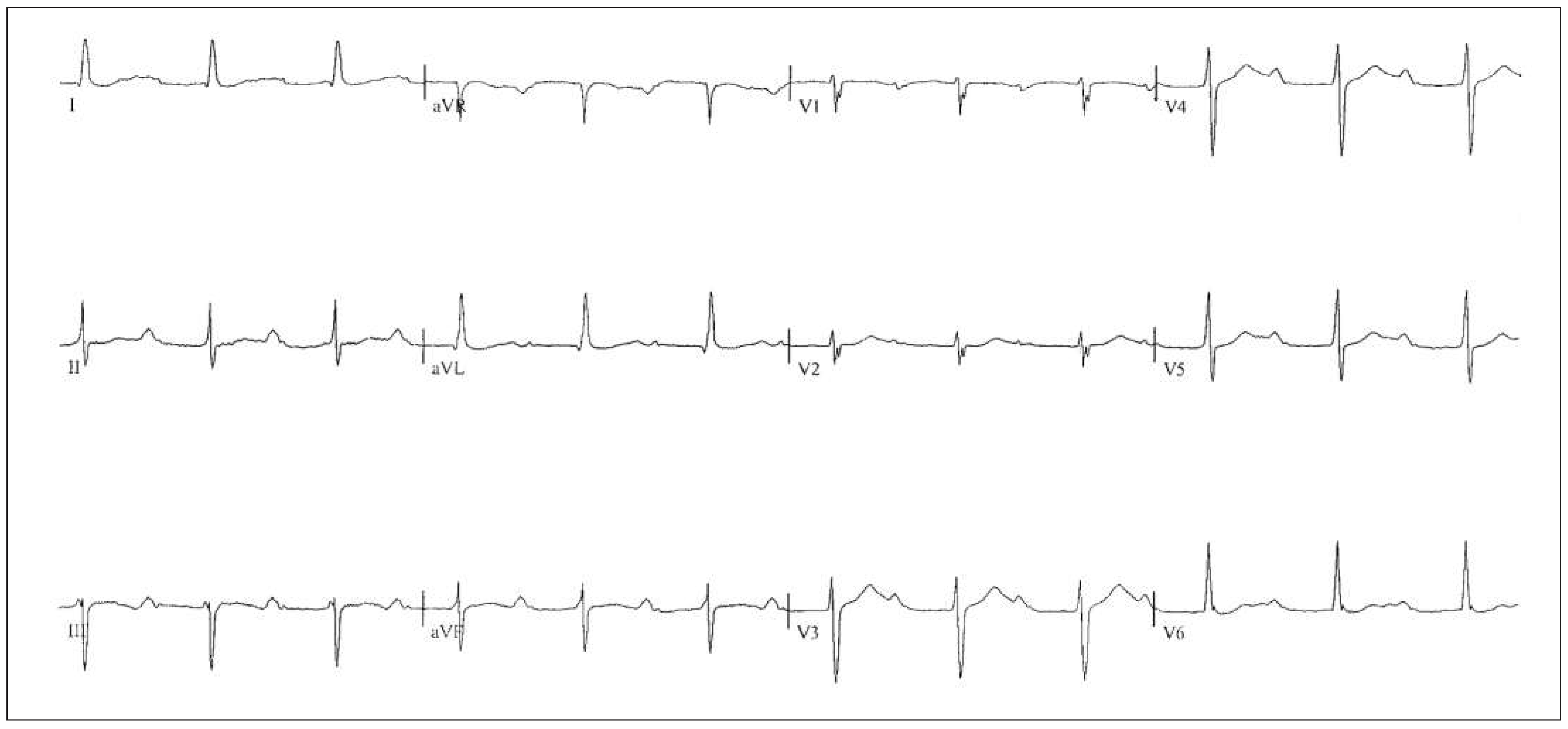
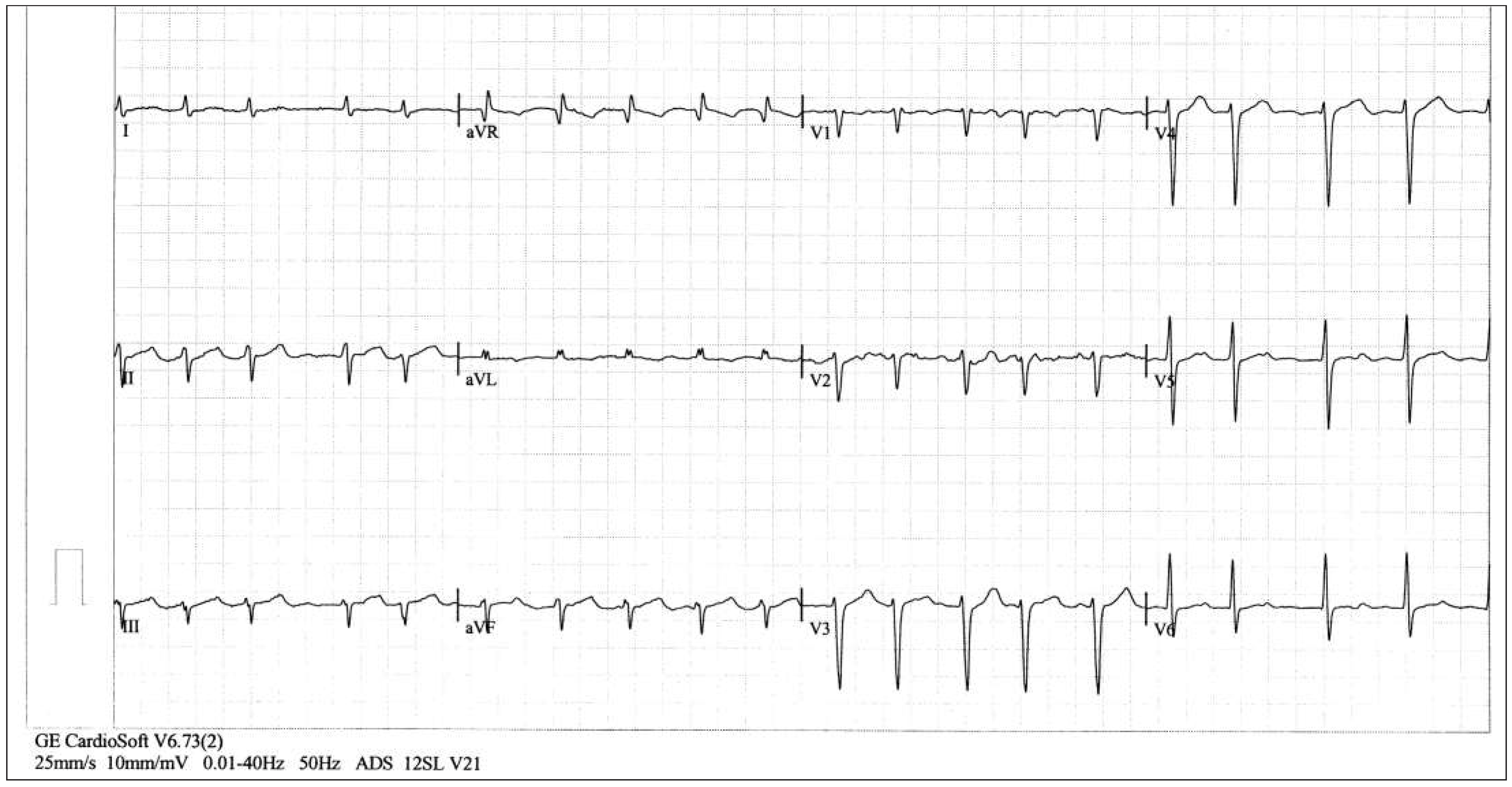
A 58-year-old man was born and lived in rural areas of Central America until, at the age of 23, he immigrated to Switzerland. His parents (mother at the age of 50, father at the age of 54 years) died because of a cardiac pathology with symptomatic arrhythmias. The patient did not know the medical diagnosis. A 52-year-old sister suffers from symptomatic arrhythmias and chronic constipation. The patient drank alcohol rarely and for more than 30 years had smoked about 20 cigarettes/ day. In 2012, an ECG (Figure 1), recorded prior to minor surgery, showed a regular sinus bradycardia (46 to 50 bpm), an incomplete right bundle-branch block (RBBB) and a long QTc-interval (474 ms). In 2013, the patient reported moderate dyspnoea and frequent dizziness. An ECG (Figure 2) showed a first-degree atrioventricular (AV) block Mobitz 1 (PR 256 ms) and delayed right ventricular conduction. Fatigue and dizziness increased, and the patient reported palpitations, reflux and epigastric distress. He came for evaluation.
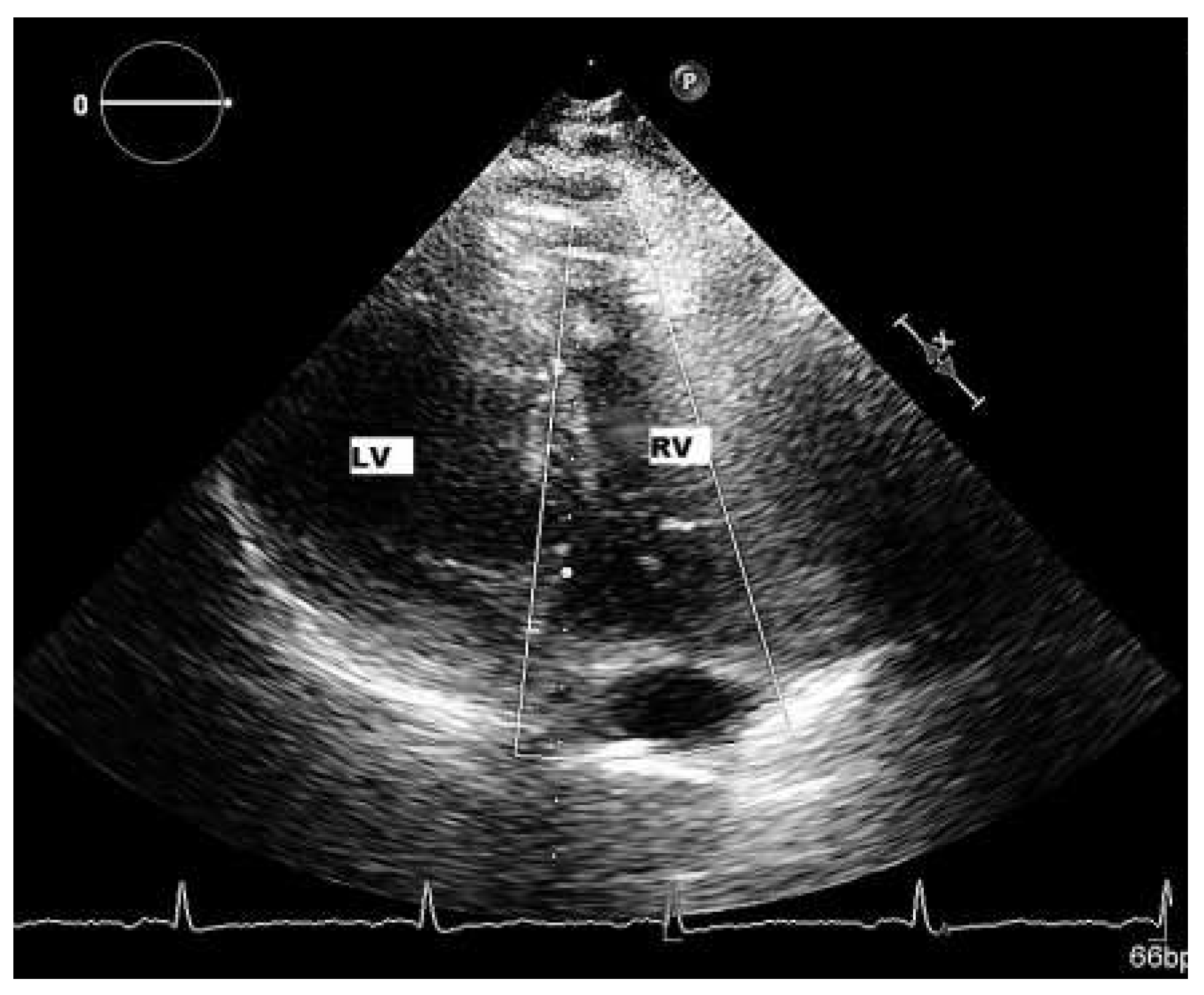
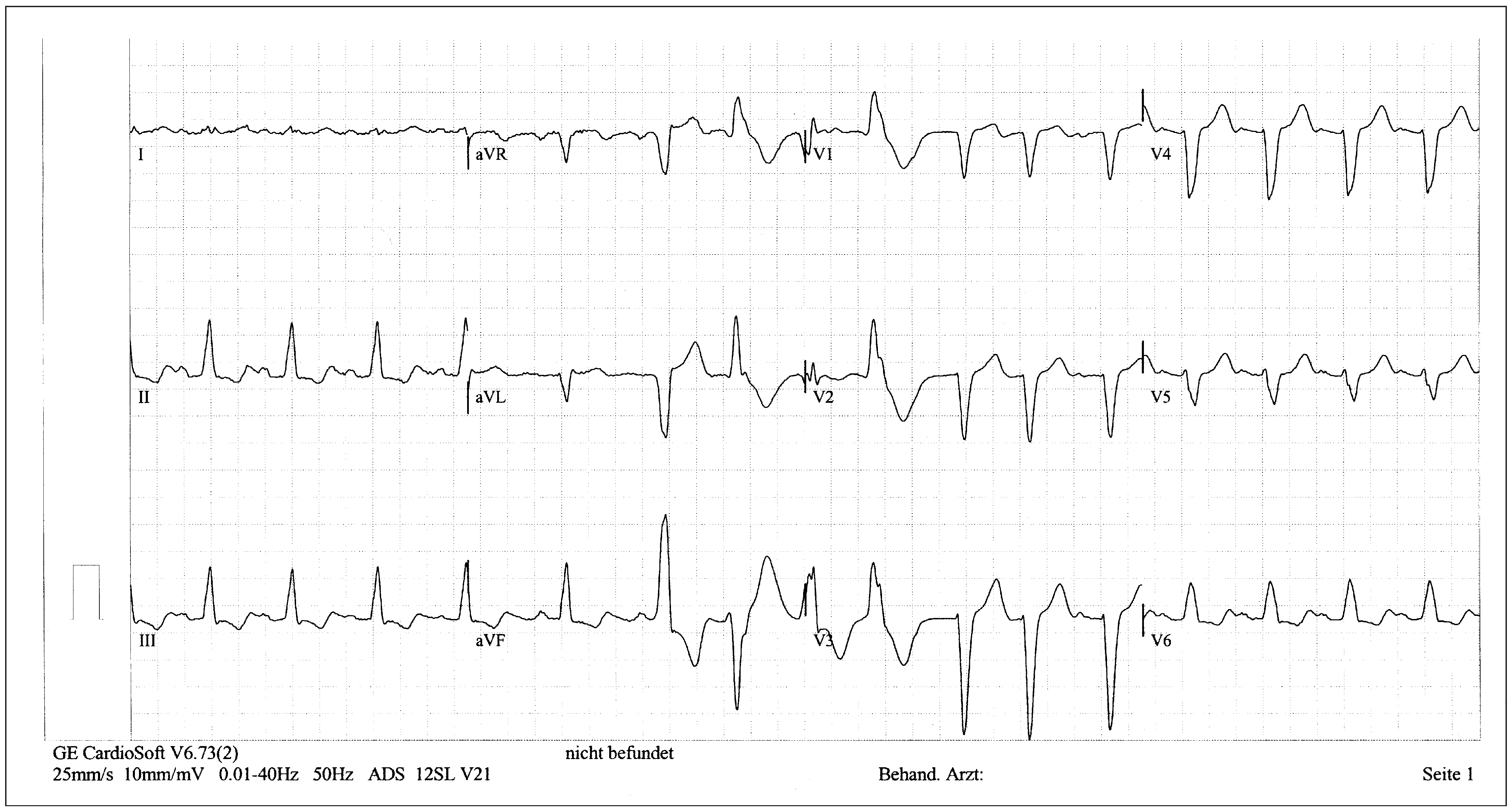
Findings: The general status was moderately reduced, the weight was normal. The blood pressure was 138/ 78 mm Hg. The lungs were clear. Palpation of the epigastrium elicited mild pain. The first and second heart sounds had normal tone but there was a nonrespiratory sinus arrhythmia with many premature beats; first to second degree systolic murmurs were heard over all valves. Haematological and chemistry laboratory tests showed that troponin T and D-dimer values were normal but N-terminal of the prohormone brain natriuretic peptide (NT-proBNP) was highly increased (3 387 pg/ml). ECG (Figure 3) showed sinus tachycardia (100 bpm), frequent supraventricular premature beats with wide QRS complexes, and a complete left bundle-branch block (LBBB) with a QTc of 536 ms. ECG monitoring (Figure 4) showed sinus bradycardia alternating with sinus tachycardia, a second-degree AV block Mobitz 2, and a slightly prolonged QTc. A 24-hour dynamic ECG detected many episodes of firstand second-degree AV block, 35.5% complex supraventricular premature beats with wide QRS complexes and partial compensatory pauses and 18.3% complex ventricular premature beats. Frequent episodes (one is shown in Figure 4) of paroxysmal atrial fibrillation lasting to >40 QRS complexes were detected. Echocardiography demonstrated traces of mitral, tricuspid and pulmonary insufficiency; the left ventricle (Figure 5) was hypokinetic, with round morphology, and moderately dilated. The maximal left ventricular ejection fraction (LVEF) was 32% and the relaxation was pathological (pseudonormalisation, E/E1 25). The patient was hospitalised for possible Chagas cardiomyopathy.
Hospital data: Ventriculo-coronarography showed normal coronary arteries, the left ventricle was dilated with increased end-diastolic pressure and diffusely hypokinetic; the systolic ejection fraction was <30%. Gastroscopy detected motility disorders and mild achalasia of the oesophagus (attributable to gastrointestinal Chagas disease). The diagnosis of chronic T. cruzi infection was confirmed by IgG serological testing with an enzyme-linked immunosorbent assay and polymerase chain-reaction assessment. A Medtronic EnTrust implantable cardioverter defibrillator (ICD) device was implanted with a VVI stimulation modus. The patient was discharged under amiodarone, perindopril, rivaroxaban and pantoprazole therapy. A consultant for tropical medicine added benznidazole.

Six-month follow-up

In the first 2 months the ICD interrupted two episodes of sustained ventricular tachycardia. A 48-hour dynamic ECG detected recurring (6 out of 48 hours) pacemaker stimulation on need because of bradycardia (rate from 38 to 52 bpm), frequent first- and second-degree AV-block Mobitz type, 20.4% complex supraventricular and 23.1% complex premature ventricular beats. Amiodarone was discontinued because of subclinical thyroid hyperfunction and was replaced by metoprolol retard. The patient developed dry cough and perindopril was replaced with candesartan.

Sixteen-month follow-up

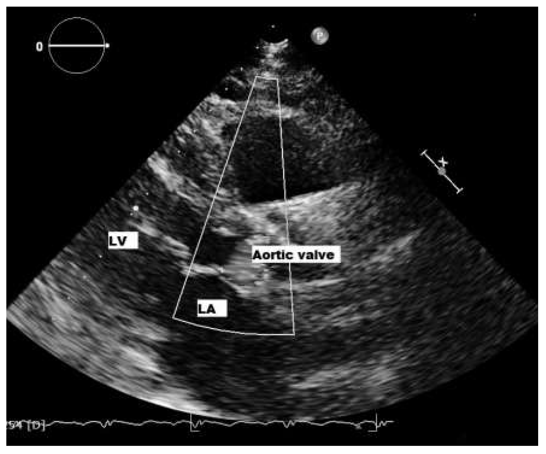
The patient complained of recurring moderate dyspnoea and pitting leg oedema. NT-proBNP was increased (1 312 pg/ml) and glomerular function was slightly decreased (MDRD estimated glomerular filtration rate 61 ml/min/1.73 m2). The ECG was unchanged. Echocardiography (mediocre quality, recorded in a half-sitting position, because the patient could not keep the supine left-lateral position) showed an enlarged, diffusely hypokinetic left ventricle with a maximal LVEF of 35%; the relaxation was moderately pathological (E/A 04, E/E1 20). Therapy with metoprolol retard, candesartan, rivaroxaban and pantoprazole were unchanged, and torasemide and low-dose spironolactone were added. The use of digoxin was considered and rejected because of the complex arrhythmia. Benznidazole was stopped: the trypanocidal drug had been prescribed because it had been shown to be effective in reducing serum parasite detection, and it was hypothesised that it would positively affect the cardiomyopathy [2]. Unfortunately, a recent paper confirmed that benznidazole significantly reduces serum parasite detection but showed that it does not improve cardiac function in patients with chronic T. cruzi infection [3]. Under the new therapy leg oedema disappeared and dyspnoea decreased, but a few months later dyspnoea increased again. Candesartan was replaced with valsartan/sacubitril. The patient is doing well. At the last follow-up NT-proBNP was 623 pg/ml. A 48-hour dynamic ECG showed a marked reduction in supraventricular and ventricular premature beats (10.2 and 11.1%, respectively) and ventricular tachycardia was not detected. Intermittent pacemaker rhythm was detected because of recurring sinus bradycardia. Echocardiography showed a marginally enlarged left ventricle (Figure 7) with a LVEF of 52% and slightly impaired diastolic function (E/A 0.9, E/E1 16).
Figure 6. Echocardiography (taken in semi-orthostatic position): enlarged, hypokinetic left ventricle.
Cardiovascmed 19 00028 g006
Figure 7. Echocardiography shows a slightly enlarged left ventricle with normal contraction.
Cardiovascmed 19 00028 g007
In summary, it is likely that patient’s parents died because of Chagas cardiomyopathy. His sister may have Chagas disease. A Brazilian study [4] identified six independent prognostic factors for Chagas cardiomyopathy: NYHA class III–IV (5 points), cardiomegaly on radiography (5 points), left ventricular dysfunction on echocardiography (3 points), nonsustained ventricular tachycardia on 24-hour dynamic ECG (3 points), low QRS voltage on ECG (2 points), and male sex (2 points). Patients are categorised as low (0 to 6 points), intermediate (7 to 11 points) and high risk (12 to 20 points). The 10-year mortality rates were 10%, 44% and 84%, respectively. Our patient had 18 points and was at high risk. Unfortunately, benznidazole was found to be unable to stop the progression of the cardiomyopathy in chronic Chagas disease. The long-term prognosis of Chagas cardiomyopathy remains poor.

Infected European patients

As in our case, infected European Chagas patients are people who have lived in Latin-America countries where T. cruzi infection is endemic [1,2]. Direct assessments of prevalence of Chagas disease in Europe have been restricted to small-scale surveys in populations chosen because of an anticipated high risk (e.g., Latin American immigrants with nonischaemic heart disease) [2]. Because Chagas disease is rare in Europe, women at risk for congenital transmission to their infants are rarely screened and in patients cardiac and gastrointestinal pathologies are often diagnosed at a late stage [2].

Diagnosis of Chagas disease

The first step for diagnosing Chagas disease is the history. Patients who have lived in countries with endemic T. cruzi infection and who complain of cardiacand, less often, gastrointestinal symptoms. These patients need regular follow-up for arrhythmias (dynamic ECG) and/or cardiac dysfunction (echocardiography). In the asymptomatic phase of Chagas cardiomyopathy subtle changes can be detected [4,5,6] and allow early treatment. Also, in patients who complain of gastrointestinal symptoms, gastroenterological diagnostics are required and allow empirical therapy.

Aetiology

Chagas disease may occur in all countries in persons who have lived in Latin America. The disease is infectious and caused by the protozoan parasite T. cruzi. Vectorborne transmission is limited to Central and South America and a few areas of North America [1,2,6,7,8,9,10,11]. The protozoan parasite is transmitted through infected faeces of the triatomine vector, inoculated through a bite [1,2,3,4,7,8,9,10,11]. Oral transmission occurs through an intact mucous membrane of the mammalian host [2,11]. Other infection routes include transfusion, organ and bone marrow transplantation, and congenital transmission [2,6,7,8,9,10,11]. Outbreaks attributed to contaminated food or water have been reported in northern South America, where transmission cycles involving wild vector populations and mammalian reservoir hosts are prominent [2,3,4,8,9,10].
The incubation period after vectorborne transmission is 1 to 2 weeks [2]. The vast majority of acute infections are undetected. Parasitaemia disappears in 4 to 8 weeks [2,4,8,9,10].
Most infected persons are asymptomatic but are permanently infected [2,3,4,5,6,8,9,10]. Over decades up to 30% of infected patients develop Chagas cardiomyopathy, which is highly arrhythmogenic [1,2,3,4,5,6,8,9,10]. Orally transmitted T. cruzi infection appears to be associated with a higher incidence of myocarditis and death than vectorborne infection [11].

Complications

Chagas cardiomyopathy is highly arrhythmogenic. Many complex arrhythmias appear, such as multiple supraventricular ectopic beats, atrial flutter and/or fibrillation, sinus dysfunction with bradycardias and tachycardias, AV- and bundle-branch blocks, and complex ventricular premature beats with ventricular tachycardia [1,2,3,4,5,6,9,10]. Congestive cardiac failure follows, seldom appearing before the arrhythmias. In countries with endemic Chagas disease, echocardiography is performed in infected persons without cardiac symptoms and several echocardiographic changes have been described [5,6]. These changes are not specific for Chagas cardiomyopathy but indicate that the heart is affected years before the pathology becomes symptomatic.
Gastrointestinal Chagas disease predominantly affects the oesophagus, colon, or both and results from damage to intramural neurons [2,8]. It is less frequent than Chagas cardiomyopathy and is more common in Argentina, Bolivia, Chile, Paraguay, Uruguay, and parts of Brazil than in northern South and Central America [2,8,9,10,11]. It was suggested that this geographic pattern might derive from different genotypes of T. cruzi [2,11,12,13] but, in several studies, no strain differences have been detected between infections with and without gastrointestinal manifestations [8,13,14].

Pathogenesis

Parasite persistence is essential but many questions about the pathogenic factors are still open [2,5,6,7,8,9,10,11,12,13,14]. In the acute phase of infection, inflammatory responses occur with involvement of native immune cells and macrophages, which are activated by interferon-γ and tumour necrosis factor-α.
In the chronic phase, T cell-mediated immunity reduces parasite replication [2,7,8,9,10,14]. However, parasite infection persists and, influenced by both host and parasite factors, plays a predominant role. The antiparasitic reactions are inefficient to eradicate the parasite. The infection is lifelong and induces inflammatory responses with damage in the heart and in the gastrointestinal tract [2,10,15,16].
Since the prevalence of severe Chagas cardiomyopathy has fallen in areas with effective vector control, it was postulated that repeated superinfection sustains the tissue antigen load and stimulates strong inflammatory responses which promote cardiac damage [2,7,8,9,10]. However, it appears that the host’s inflammatory and immune responses might also be an important determining factor for the progression, whereas T. cruzi strain virulence and tissue tropism are possible contributory factors [2,12,13,14,15,16,17].

Therapy

Progress has been made in the past 5 years toward improving the treatment of Chagas disease [2]. Rarely Chagas infection may be recognized in the early phase and the trypanocidal drug benznidazole can be used to reduce the level of parasitaemia of T. cruzi and might be useful to treat acute infections [2]. However, a recent study has demonstrated that therapy with benznidazole has no detectable effects in patients with established Chagas cardiomyopathy [3]. T. cruzi infection remains the most important parasitic disease in South, Central and North America, with an estimated disease burden (measured by disability-adjusted life-years) that is 7.5 times as great as that of malaria [18]. These facts show the need of an urgent search for a treatment with high efficacy during the chronic phase.
There is no specific cardiac therapy for Chagas cardiomyopathy. The disease is highly arrhythmogenic. Therefore, bradycardic arrhythmias, AV- and/or bundle-branch blocks and complex ectopic arrhythmias are a proven indication for a pacemaker and/or ICD implantation. Atrial flutter and/or fibrillation are frequent complications and high-risk patients (assessed with the CHA2-DS2-VASc score) need oral anticoagulation. Congestive heart failure is empirically treated as recommended in modern guidelines [19]. As yet there are no trials on the use of valsartan/sacubitril in Chagas cardiomyopathy, but our personal opinion is to use this therapy when Chagas patients develop clear signs of heart failure.
The therapy of gastrointestinal Chagas is also empirical and is adapted to symptoms and signs of the pathology [7].

Acknowledgments

The authors thank Mrs. J. Bugmann for technical assistance and secretarial help.

Disclosure statement

No financial support and no other potential conflict of interest relevant to this article were reported.

References

  1. Fox, M.C.; Lakdawala, N.; Miller, A.L.; Loscalzo, J. A Patient with Syncope. New Engl. J. Med. 2013, 369, 966–972. [Google Scholar] [CrossRef] [PubMed]
  2. Bern, C. Chagas’ Disease. N. Engl. J. Med. 2015, 373, 456–466. [Google Scholar] [CrossRef] [PubMed]
  3. Morillo, C.A.; Marin-Neto, J.A.; Avezum, A.; et al. Randomized Trial of Benznidazole for Chronic Chagas’ Cardiomyopathy. New Engl. J. Med. 2015, 373, 1295–06. [Google Scholar] [CrossRef] [PubMed]
  4. Rassi, A., Jr.; Rassi, A.; Little, W.C.; et al. Development and Validation of a risk Score for Predicting Death in Chagas’ Heart Disease. New Engl. J. Med. 2006, 355, 799–808. [Google Scholar] [CrossRef] [PubMed]
  5. Acquatella, H. Heart Disease in Latin America. Echocardiography in Chagas Heart Disease. Circulation 2007, 115, 1124–1131. [Google Scholar] [CrossRef] [PubMed]
  6. Sabino, E.C.; Ribeiro, A.L.; Salemi, V.M.; et al. Ten-year incidence of Chagas cardiomyopathy among asymptomatic Trypanosoma cruzi-seropositive former blood donors. Circulation 2013, 127, 1105–1115. [Google Scholar] [CrossRef] [PubMed]
  7. Pinazo, M.J.; Cañas, E.; Elizalde, J.I.; et al. Diagnosis, management and treatment of chronic Chagas’ gastrointestinal disease in areas where Trypanosoma cruzi infection is not endemic. Gastroenterol. Hepatol. 2010, 33, 191–200. [Google Scholar] [CrossRef] [PubMed]
  8. Pinto Dias, J.C. Natural history of Chagas disease. Arq. Bras. Cardiol. 1995, 65, 359–366. [Google Scholar] [PubMed]
  9. Rassi AJr Dias, J.C.; Marin-Neto, J.A.; Rassi, A. Challenges and opportunities for primary, secondary, and tertiary prevention of Chagas’ disease. Heart 2009, 95, 524–534. [Google Scholar] [CrossRef] [PubMed]
  10. Rassi AJr Rassi, A.; Marcondes de Rezende, J. American trypanosomiasis (Chagas disease). Infect. Dis. Clin. North. Am. 2012, 26, 275–291. [Google Scholar] [CrossRef] [PubMed]
  11. Shikanai-Yasuda, M.A.; Carvalho, N.B. Oral transmission of Chagas disease. Clin. Infect. Dis. 2012, 54, 845–852. [Google Scholar] [CrossRef] [PubMed]
  12. Miles, M.A.; Cedillos, R.A.; Póvoa, M.M.; et al. Do radically dissimilar Trypanosoma cruzi strains (zymodemes) cause Venezuelan and Brazilian forms of Chagas’ disease? Lancet 1981, 1, 1338–1340. [Google Scholar] [CrossRef] [PubMed]
  13. del Puerto, R.; Nishizawa, J.E.; Kikuchi, M.; et al. Lineage analysis of circulating Trypanosoma cruzi parasites and their association with clinical forms of Chagas disease in Bolivia. PLoS Negl. Trop. Dis. 2010, 4, e687. [Google Scholar] [CrossRef] [PubMed]
  14. Tarleton, L. Parasite persistence in the aetiology of Chagas disease. Int. J. Parasitol. 2001, 31, 550–554. [Google Scholar] [CrossRef] [PubMed]
  15. Machado, F.S.; Dutra, W.O.; Esper, L.; et al. Current understanding of immunity to Trypanosoma cruzi infection and pathogenesis of Chagas disease. Semin. Immunopathol. 2012, 34, 753–770. [Google Scholar] [CrossRef] [PubMed]
  16. Marin-Neto, J.A.; Cunha-Neto, E.; Maciel, B.C.; Simões, M.V. Pathogenesis of chronic Chagas heart disease. Circulation 2007, 115, 1109–1123. [Google Scholar] [CrossRef] [PubMed]
  17. Dutra, W.O.; Menezes, C.A.; Magalhães, L.M.; Gollob, K.J. Immunoregulatory networks in human Chagas disease. Parasite Immunol. 2014, 36, 377–387. [Google Scholar] [CrossRef] [PubMed]
  18. Global burden of disease estimates for 2000–2012; World Health Organization, 2014; Available online: http://www.who.int/ healthinfo/global_burden_disease/estimates/en/ index2.html.
  19. McMurray, J.V.; Adamopoulos, S.; Anker, S.D.; et al. ESC Guidelines for the diagnosis and treatment of acute and chronic heart failure. Eur. Heart J. 2012, 33, 1787–1747. [Google Scholar] [PubMed]
Figure 1. The ECG shows a regular sinus bradycardia (50 bpm), an incomplete right bundle-branch block and a long QTc interval (474 ms).
Cardiovascmed 19 00028 g001
Figure 2. The ECG shows sinus rhythm, first-degree atrioventricular block Mobitz 1 (PR 256 ms) and delayed intraventricular conduction.
Cardiovascmed 19 00028 g002
Figure 3. The ECG shows sinus tachycardia (100 bpm), frequent supraventricular premature beats with wide QRS complexes, and a permanent left bundle-branch block with secondary repolarisation changes.
Cardiovascmed 19 00028 g003
Figure 4. The ECG shows atrial fibrillation with horizontal R axe.
Cardiovascmed 19 00028 g004
Figure 5. Echocardiography shows a dilated, hypokinetic left ventricle with round morphology.
Cardiovascmed 19 00028 g005