Next Article in Journal
Long-Term Follow-Up After Closure of Patent Foramen Ovale
Previous Article in Journal
Wolff-Parkinson-White Syndrome and Diverticulosis of the Heart?
 
 
Cardiovascular Medicine is published by MDPI from Volume 28 Issue 1 (2025). Previous articles were published by another publisher in Open Access under a CC-BY (or CC-BY-NC-ND) licence, and they are hosted by MDPI on mdpi.com as a courtesy and upon agreement with Editores Medicorum Helveticorum (EMH).
Case Report

Rivaroxaban Dissolves Postinfarction Left Ventricular Thrombus

by
Assadullah Azizi
,
Serban Puricel
,
Stéphane Cook
and
Nicolas Brugger
*
Department of Cardiology, University and Hospital Fribourg, CH-1708 Fribourg, Switzerland
*
Author to whom correspondence should be addressed.
Cardiovasc. Med. 2016, 19(1), 25; https://doi.org/10.4414/cvm.2016.00381
Submission received: 20 October 2015 / Revised: 20 November 2015 / Accepted: 20 December 2015 / Published: 20 January 2016

Abstract

Despite early management of ST elevation myocardial infarction, prompt coronary revascularization and potent dual antiplatelet therapy, left ventricular thrombus still remains a dreaded complication especially in anterior STEMI. Little is known about the efficacy and safety of the new oral anticoagulants in the context of post STEMI left ventricular thrombus. We describe the case of a 54 old male Caucasian who developed an apical thrombus, despite a DAPT, 3 days after a triple stenting of the left anterior descending coronary artery, for a subacute STEMI. After one month of treatment using rivaroxaban, dissolution of the thrombus was evident on echocardiography. The present case report is the first to demonstrate left ventricular thrombus dissolution using NOAC in the setting of a subacute STEMI that forces an association with dual antiplatelet therapy. NOACs offer a rapid and more constant anticoagulation than vitamin K antagonists with less food interaction and do not require routine monitoring. For these reasons these molecules are potential good candidates to supplant VKA for the treatment of left ventricular thrombus, but randomized controlled trials are needed to demonstrate the advantages of the NOACs in this setting.

Introduction

In the current era of early management of the patient suffering from ST elevation myocardial infarction (STEMI), including prompt coronary revascularisation with drug-eluting stents and potent dual antiplatelet therapy (DAPT), the incidence of left ventricular thrombus has significantly declined [1,2]. However, some studies still suggest that apical thrombi are detectable in one quarter of patients after anterior STEMIs [3]. In these cases, oral anticoagulation with a vitamin K antagonist (VKA) is considered the standard of care according to the recent guidelines from the European Society of Cardiology (ESC) [4]. The disadvantages of these molecules are mainly a hypercoagulable state during initiation and a labile anticoagulation state (defined as <60% time in the therapeutic range) during steady state. More recently, the novel oral anticoagulants (NOAC) have emerged as an alternative to VKAs for thromboembolic prevention in patients with nonvalvular atrial fibrillation [5,6]. NOACs offer a rapid and more constant anticoagulation with less food interaction and do not require routine monitoring [7]. So far, only a few cases of the use of NOACs to treat left ventricular thrombi have been reported, none of which were in the context of subacute STEMI [8,9,10,11].

Case description

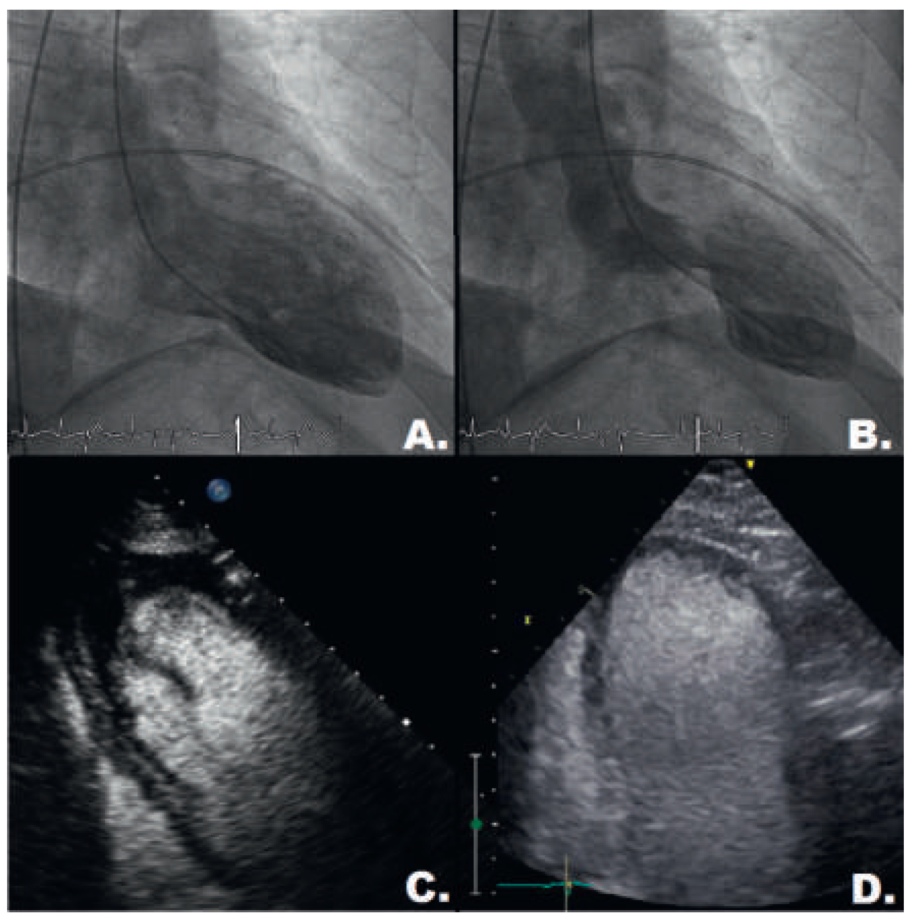
A 54-year-old Caucasian male was admitted with retrosternal pain evolving during the past 2 weeks. The ECG showed a subacute anterior STEMI with marked ST elevation and Q waves in leads V2 to V4. The coronary angiogram revealed a total occlusion of the left anterior descending coronary artery (LAD) in its mid-portion, an ulcerated plaque in its proximal part and an intermediate lesion in its distal portion. The vessel was treated with three newer generation drug-eluting stents (Resolute Onyx™ stents, proximal 3.5 × 12 mm, mid 3.5 × 26 mm and distal LAD 3.0 × 15 mm, Medtronic, Minneapolis, MN, USA) with a good angiographic final result (Figure 1A–F). The left ventricular angiogram demonstrated apical ballooning (Figure 2A, B). DAPT was initiated with acetylsalicylic acid (100 mg/d) and prasugrel (10 mg/d). Fondaparinux (2.5 mg/d) was prescribed for 5 days. According to routine, transthoracic echocardiography was performed 3 days after the percutaneous coronary invention and revealed apical thrombus (Figure 2C), which was not detectible on ventriculography. HAS-BLED score was 1 point.
Consequently the patient was started on rivaroxaban, and prasugrel was switched to clopidogrel (75 mg/d) in order to limit the bleeding risk. At 1 month, echocardiography was repeated and revealed complete dissolution of the thrombus despite persistence of the apical dyskinesia (Figure 2D).

Discussion

Rivaroxaban is an oral inhibitor that binds directly to factor Xa. It is approved for treatment of deep venous thrombosis, pulmonary embolism and nonvalvular atrial fibrillation [5,7,12].
A Japanese team published three cases of left atrial appendage thrombus resolution using rivaroxaban 10 mg/d [13]. Another case report described the growth of a left atrial appendage thrombus despite well-conducted treatment with a VKA, which then disappeared during treatment with rivaroxaban 15 mg/d [14]. To the best of our knowledge, NOACs have so far been successfully used to treat left ventricular thrombi in four published cases. Kaku et al. described a case of thrombus dissolution in a 59-year-old patient suffering from left mid-ventricular obstruction with apical aneurysm formation [8]. Nagamoto et al. reported a case after an ancient myocardial infarction [9]. Padilla Pérez et al. summarised the case of a patient with left ventricular thrombus dissolution in a setting of dilated cardiomyopathy [10]. Nakasuka et al. reported complete apical thrombus disappearance in a case of tachycardiomyopathy [11].
The present case report is the first to demonstrate left ventricular thrombus dissolution using a NOAC (rivaroxaban) in the setting of a subacute STEMI that forced administration of DAPT. In this setting, the European Society of Cardiology has different antiplatelet and anticoagulation recommendations for patients at low risk of bleeding (HAS-BLED score of 0–2) compared with patients at high risk of bleeding (HAS-BLED ≥3) [15]. However, triple therapy using a NOAC could be hazardous and only a few studies have investigated triple therapy including a NOAC in patients suffering from coronary artery disease and nonvalvular atrial fibrillation. In the APPRAISE-2, apixaban was combined with aspirin and clopidogrel in 81% of patients, and led to a significant increase in fatal and intracranial bleeding without clinical benefit [16]. In ATLAS ACS 2, low-dose rivaroxaban (2.5– 5 mg twice daily) was administered with aspirin and clopidogrel in 92% of patients. This was associated with a 16% reduction in the composite efficacy endpoint (cardiovascular death, myocardial infarction and stroke) and a small increase in major bleedings [17].
Risk of bleeding should be utilised in decision-making. The HAS-BLED score was 1 for this patient, who had normal renal function with a creatinine clearance of 83 ml/ min (1 point because of the concomitant use of antiplatelet agents and oral anticoagulation, i.e., a risk of major bleeding of 1.0–1.5% per year). Based on the above described case reports, where rivaroxaban was the most frequently used molecule and always prescribed once a day (never 15 mg twice daily as for the first 3 weeks of the pulmonary embolism therapy), we decided to use rivaroxaban at a dosage of 20 mg/d during a period of 3 months. This permits us to avoid initial quadruple therapy (aspirin, clopidogrel, low-molecular-weight heparin and VKA) until achieving therapeutic anticoagulation.

Conclusion

Short duration rivaroxaban was effective for the treatment of left ventricular thrombus in a patient under DAPT after drug-eluting stent implantation for STEMI, and at low bleeding risk. Randomised controlled trials are urgently needed to confirm these encouraging observational data, to define the optimal dosage of NOACs when associated with DAPT, and to demonstrate that these molecules are effectively good candidates to supplant VKAs in the treatment of post-STEMI left ventricular thrombi.

Disclosure statement

No financial support and no other potential conflict of interest relevant to this article was reported.

References

  1. Solheim, S.; Seljeflot, I.; Lunde, K.; Bjornerheim, R.; Aakhus, S.; Forfang, K.; et al. Frequency of left ventricular thrombus in patients with anterior wall acute myocardial infarction treated with percutaneous coronary intervention and dual antiplatelet therapy. Am. J. Cardiol. 2010, 106, 1197–1200. [Google Scholar] [CrossRef] [PubMed]
  2. Osherov, A.B.; Borovik-Raz, M.; Aronson, D.; Agmon, Y.; Kapeliovich, M.; Kerner, A.; et al. Incidence of early left ventricular thrombus after acute anterior wall myocardial infarction in the primary coronary intervention era. Am. Heart J. 2009, 157, 1074–1080. [Google Scholar] [CrossRef] [PubMed]
  3. Porter, A.; Kandalker, H.; Iakobishvili, Z.; Sagie, A.; Imbar, S.; Battler, A.; et al. Leh ventricular mural thrombus after anterior ST-segmentelevation acute myocardial infarction in the era of aggressive reperfusion therapy—Still a frequent complication. Coron. Artery Dis. 2005, 16, 275–279. [Google Scholar] [CrossRef] [PubMed]
  4. Task Force on the management of STseamiotESoC; Steg, P.G.; James, S.K.; Atar, D.; Badano, L.P.; Blomstrom-Lundqvist, C.; et al. ESC Guidelines for the management of acute myocardial infarction in patients presenting with ST-segment elevation. Eur. Heart J. 2012, 33, 2569–2619. [Google Scholar] [CrossRef] [PubMed]
  5. Camm, A.J.; Lip, G.Y.; De Caterina, R.; Savelieva, I.; Atar, D.; Hohnloser, S.H.; et al. 2012 focused update of the ESC Guidelines for the management of atrial fibrillation: An update of the 2010 ESC Guidelines for the management of atrial fibrillation. Developed with the special contribution of the European Heart Rhythm Association. Eur. Heart J. 2012, 33, 2719–2747. [Google Scholar] [PubMed]
  6. Patel, M.R.; Mahaffey, K.W.; Garg, J.; Pan, G.; Singer, D.E.; Hacke, W.; et al. Rivaroxaban versus warfarin in nonvalvular atrial fibrillation. N. Engl. J. Med. 2011, 365, 883–891. [Google Scholar] [CrossRef] [PubMed]
  7. Harder, S. Pharmacokinetic and pharmacodynamic evaluation of rivaroxaban: Considerations for the treatment of venous thromboembolism. Thromb. J. 2014, 12, 22. [Google Scholar] [CrossRef] [PubMed]
  8. Kaku, B. Intra-cardiac thrombus resolution after anti-coagulation therapy with dabigatran in a patient with mid-ventricular obstructive hypertrophic cardiomyopathy: A case report. J. Med. Case Rep. 2013, 7, 238. [Google Scholar] [CrossRef] [PubMed]
  9. Nagamoto, Y.; Shiomi, T.; Matsuura, T.; Okahara, A.; Takegami, K.; Mine, D.; et al. Resolution of a left ventricular thrombus by the thrombolytic action of dabigatran. Heart Vessels. 2014, 29, 560–562. [Google Scholar] [CrossRef] [PubMed]
  10. Padilla Perez, M.; Salas Bravo, D.; Garcelan Trigo, J.A.; Vazquez Ruiz de Castroviejo, E.; Torres Llergo, J.; Lozano Cabezas, C.; et al. Resolution of left ventricular thrombus by rivaroxaban. Future Cardiol. 2014, 10, 333–336. [Google Scholar] [CrossRef] [PubMed]
  11. Nakasuka, K.; Ito, S.; Noda, T.; Hasuo, T.; Sekimoto, S.; Ohmori, H.; et al. Resolution of left ventricular thrombus secondary to tachycardiainduced heart failure with rivaroxaban. Case Rep. Med. 2014, 2014, 814524. [Google Scholar] [CrossRef] [PubMed]
  12. Agnelli, G.; Gallus, A.; Goldhaber, S.Z.; Haas, S.; Huisman, M.V.; Hull, R.D.; et al. Treatment of proximal deep-vein thrombosis with the oral direct factor Xa inhibitor rivaroxaban (BAY 59-7939): The ODIXaDVT (Oral Direct Factor Xa Inhibitor BAY 59-7939 in Patients With Acute Symptomatic Deep-Vein Thrombosis) study. Circulation 2007, 116, 180–187. [Google Scholar] [CrossRef] [PubMed]
  13. Takasugi, J.; Yamagami, H.; Okata, T.; Toyoda, K.; Nagatsuka, K. Dissolution of the left atrial appendage thrombus with rivaroxaban therapy. Cerebrovasc. Dis. 2013, 36, 322–323. [Google Scholar] [CrossRef] [PubMed]
  14. Hammerstingl, C.; Potzsch, B.; Nickenig, G. Resolution of giant left atrial appendage thrombus with rivaroxaban. Thromb. Haemost. 2013, 109, 583–584. [Google Scholar] [CrossRef] [PubMed]
  15. Windecker, S.; Kolh, P.; Alfonso, F.; Collet, J.P.; Cremer, J.; Falk, V.; et al. 2014 ESC/EACTS Guidelines on myocardial revascularization: The Task Force on Myocardial Revascularization of the European Society of Cardiology (ESC) and the European Association for Cardio-Thoracic Surgery (EACTS)Developed with the special contribution of the European Association of Percutaneous Cardiovascular Interventions (EAPCI). Eur Heart J. 2014, 35, 2541–2619. [Google Scholar] [PubMed]
  16. Alexander, J.H.; Lopes, R.D.; James, S.; Kilaru, R.; He, Y.; Mohan, P.; et al. Apixaban with antiplatelet therapy after acute coronary syndrome. N. Engl. J. Med. 2011, 365, 699–708. [Google Scholar] [CrossRef] [PubMed]
  17. Mega, J.L.; Braunwald, E.; Wiviott, S.D.; Bassand, J.P.; Bhatt, D.L.; Bode, C.; et al. Rivaroxaban in patients with a recent acute coronary syndrome. N. Engl. J. Med. 2012, 366, 9–19. [Google Scholar] [CrossRef] [PubMed]
Figure 1. Coronary angiogram: (A). Occlusion of the mid LAD. (B). Good result after stenting of the mid LAD with a Resolute Onyx 3.5 × 26 mm stent. (C). Significant lesion of the distal LAD. (D). Excellent result after stenting of the distal LAD with a Resolute Onyx 3.0 × 15 mm stent. (E). Ulcerated plaque of the proximal LAD. (F). Good result after stenting of the proximal LAD with a Resolute Onyx 3.5 × 12 mm stent.
Cardiovascmed 19 00025 g001
Figure 2. Left ventricular angiogram: (A). Telediastolic frame. (B). Telesystolic frame with clearly visible apical aneurysm. Echocardiographic findings: (C). At 3 days, left ventricular apical thrombus (T) nicely delineated using contrast (SonoVue®, Bracco, Italy). (D). At 1 month, complete dissolution of the thrombus.
Cardiovascmed 19 00025 g002