Next Article in Journal
Axillary vein Puncture for Device Implantation and Use of 4F Catheter-Delivered Pacing Leads in Children
Previous Article in Journal
Real-Time Contact Force Measurement for Catheter Ablation of Cardiac Arrhythmias
 
 
Cardiovascular Medicine is published by MDPI from Volume 28 Issue 1 (2025). Previous articles were published by another publisher in Open Access under a CC-BY (or CC-BY-NC-ND) licence, and they are hosted by MDPI on mdpi.com as a courtesy and upon agreement with Editores Medicorum Helveticorum (EMH).
Case Report

A Valve-in-Valve Approach for Ebstein’s Anomaly

by
Fabienne Schwitz
1,*,
Peter Wenaweser
2,
Alexander Kadner
1,3,
Kerstin Wustmann
1,
Stephan Windecker
2 and
Markus Schwerzmann
1
1
Centre for Congenital Heart Disease, Department of Cardiology, University Hospital Inselspital, Bern, Switzerland
2
Department of Cardiology, University Hospital Inselspital, Bern, Switzerland
3
Department of Cardiac Surgery, University Hospital Inselspital, Bern, Switzerland
*
Author to whom correspondence should be addressed.
Cardiovasc. Med. 2015, 18(4), 139; https://doi.org/10.4414/cvm.2015.00331 (registering DOI)
Submission received: 22 January 2015 / Revised: 22 February 2015 / Accepted: 22 March 2015 / Published: 22 April 2015

Abstract

A 41-year-old women with Ebstein’s anomaly required tricuspid valve replacement six years ago. At that time a biological prosthesis was implanted. The tricuspid valve prosthesis became severely stenotic within five years. Instead of re-do surgery, the patient underwent a percutaneous procedure. An Edwards SAPIEN 29-mm bioprosthesis was successfully implanted in the stenotic 33-mm stented Perimount bioprosthesis. A periprocedural transoesophageal echocardiography showed a residual transprothetic mean gradient of 1 mm Hg after the procedure. In conclusion, percutaneous valve replacement is an additional option for patients with a degenerated right-sided bioprostheses in atrioventriuclar position, potentially reducing the need for re-do surgery in adults with congenital heart disease.

Introduction: Ebstein’s anomaly

Ebstein’s anomaly is a malformation of the right ventricle and its atrioventricular valve. It is a rather rare congenital defect with an incidence of 1 per 200,000 live births [1]. The anatomical hallmark of this malformation is a downward displacement of the hinge point of the septal and posterior tricuspid leaflets below the annulus due to incomplete delamination of these tricuspid valve leaflets from the underlying myocardium during embryogenesis. Some affected individuals die in utero because of right ventricular dysfunction, others need surgery in early childhood. In a less severe form, Ebstein’s anomaly results in various degrees of tricuspid regurgitation or, rarely, stenosis. Additional interatrial communications are present in 80% to 94% of patients with Ebstein’s anomaly [2] and accessory pathways in approximately 6% to 36% of patients [1]. Medical therapy is used to relieve symptoms of right heart failure or to control arrhythmias, but the majority of patients will require tricuspid valve repair or replacement during their lifetime. The timing of surgical intervention depends on symptoms, exercise performance, heart size, the occurrence of arrhythmias or the presence and consequences of cyanosis due to interatrial right-to-left shunting, eventually complicated by paradoxical embolism [3]. Reconstruction of the tricuspid valve is nowadays the preferred repair technique if feasible. Different surgical techniques are currently used, such as pericardial septal leaflets, cone repairs and leaflet augmentation, in addition to conventional annular plication and sliding leaflet plasties. There are many variants in an Ebsteinoid tricuspid valve, making it important to have more than one repair strategy in a surgical armamentarium [4]. Bioprosthetic valve replacement as opposed to mechanical valve replacement is usually performed in adults not amenable to valvular reconstruction, owing to the perceived lower risk of prosthetic thrombosis and the lack of need for anticoagulation in the absence of atrial arrhythmias. However, bioprosthetic valves are susceptible to leaflet degeneration, necessitating re-do surgery at a given time.

Case report

A mother of twins was diagnosed with Ebstein’s anomaly at the age of 34 years. After an uneventful pregnancy and delivery, she complained about persistent shortness of breath on exercise and undue fatigue. Her symptoms were initially attributed to exhausting family demands. Three years after delivery, she was found to have Ebstein’s anomaly with severe tricuspid regurgitation – the calculated Celemayer index was 0.55 indicating a moderate degree of disease severity. She underwent cardiac surgery two years later as a result of persistent exercise intolerance. She was in New York Heart Association (NYHA) functional class II–III. Reconstruction of the dysplastic tricuspid valve could not be accomplished. A 33-mm stented Perimount bioprosthesis was implanted at the level of the hypothetical tricuspid annulus. The mean gradient after surgery was 3 mm Hg. Exercise capacity improved substantially after surgery to NYHA I–II. After a period of five years without cardiac complaints, the patient noticed a decline in her exercise capacity. At physical examination, an elevated jugular venous pressure was observed. On cardiac auscultation, however, no systolic or diastolic murmer was heard. Transthoracic echocardiography showed a fusion and severely restricted motion of two of the three bioprosthesis leaflets causing a stenosis with a mean gradient of 10 mm Hg at a heart rate of 67 bpm. There was only mild tricuspid regurgitation. Cardiopulmonary exercise testing revealed a decrease in peak oxygen consumption compared with the last examination one year before. The oxygen pulse curve was suggestive of a cardiac limitation as primary mechanism for the impaired exercise capacity.
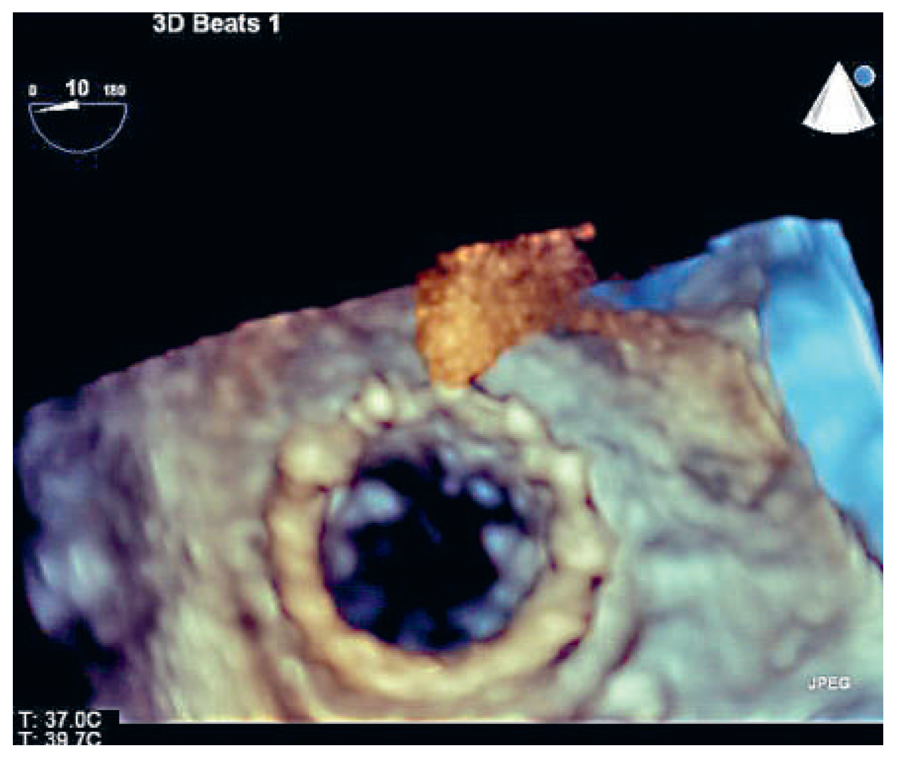
She refused to undergo re-do surgery at this time. The option of a percutaneous valve-in-valve procedure was discussed. On transoesophageal three-dimensional echocardiography, the true internal diameter measured 27 mm in diameter (Figure 1). These dimensions were further confirmed with a cardiac computed tomography (CT) scan, suggesting that a percutaneously deployed 29 mm Edwards SAPIEN bioprosthesis could be anchored within the rigid bioprosthetic annulus.
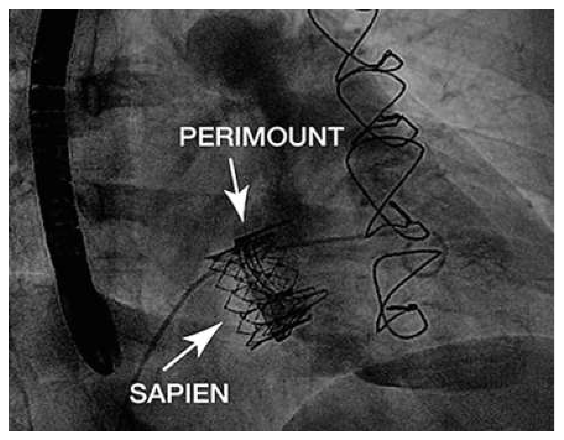
With the aim to delay re-do surgery, the patient decided to opt for a percutaneous tricuspid valve replacement. The procedure was done in a hybrid catheterisation suite with the cardiac surgeon at standby in case the deployed valve embolised into the right ventricle. An Edwards SAPIEN 29 mm bioprosthesis was successfully implanted (Figure 2) via the transfemoral approach and with an Amplatz extra stiff guidewire (placed into the pulmonary artery). The valve was placed with 30%–50% of the stent frame below the tricuspid bioprosthetic valve in order to have an optimal sealing effect. Periprocedural transoesophageal echocardiography showed a reduction of the transvalvular mean gradient from 7 to 1 mm Hg (Figure 3). There was no peri- or transvalvular regurgitation seen after the valve-in-valve implantation. At follow-up three month after intervention, the patient had improved clinically and we measured a mean gradient of 4 mm Hg at a heart rate of 63/min.

Discussion

The last 50 years have witnessed dramatic changes in survival of patients with congenital heart disease. Despite all medical advances, many adults with con-genital heart defects face still multiple re-operations in their lifetime and each operation has inherently an increasingly higher surgical risk [5]. In a survey of patients with congenital heart disease from one of the largest centers worldwide, perioperative mortality constituted 18% of the total mortality burden seen in these patients [6].
In recent years, more and more experience has been gained with transcatheter valve replacement for aortic and pulmonary valves, and in the meantime valve-in-valve procedures have also been established. The Melody transcatheter valve for replacement of degenerated valves in right ventricle-to-pulmonary artery conduits or homografts was the first commercially available percutaneous valve [7]. Currently no established percutaneous options are available to replace a right-sided atrioventricular valve or native pulmonary valve. The absence of stable anchoring structures at the native atrioventricular level makes primary percutaneous tricuspid valve replacement challenging. However, in a patient already having a bioprosthesis in the tricuspid valve position, experience with limited percutaneous valve replacement has recently been gained. The first procedures were carried out using a Melody transcatheter pulmonary valve [8]. An important limitation of the Melody valve, which is sourced from bovine jugular venous valves, is its maximal diameter. Melody valve competence has been tested and recommended only up to a diameter of 22 mm, even though the Melody valve diameter can be expanded to 24 mm. Its advantage is that the delivery system and usual mounting of the valves for pulmonic interventions is designed for an antegrade delivery of the valve, as opposed to the retrograde access routinely used for percutaneous aortic valve replacement. Nevertheless, considering the available sizes of the Edwards SAPIEN valve (up to 29 mm), valves initially developed for an aortic position may become the preferred prostheses for valve-in-valve implantation in the tricuspid position also [9]. The Edwards XT valve represents a suitable device to use for this indication, although the reversed crimping on the balloon needs to be done with care in order to avoid leaflet damage. The correct sizing is performed using the internal diameter dimension of the labelled implanted surgical valve plus CT scan measurements. The size of the SAPIEN valve should include an oversizing of 10%– 20% for safe anchoring. Beyond doubt, percutaneous tricuspid valve-in-valve implantation is still a rare procedure. To our knowledge, this is only the third case report of a percutaneous aortic valve implanted in a tricuspid bioprosthesis in adults with repaired Ebstein’s anomaly [9,10].)

Conclusions

In summary, percutaneous valve-in-valve procedures with large aortic valves have the potential to reduce the surgical burden in adults with Ebstein’s anomaly and degenerated bioprosthetic tricuspid valves. With this in mind, the limitations of early degeneration of biological valves in the tricuspid position in a young patient population can potentially be reduced. However, this new therapeutic option is still an emerging technique and long-term follow-up is unknown.

Disclosures

Honoraria and lecture fees from Edwards Lifesciences (Peter Wenaweser). No other conflicts of interests to disclose.

References

  1. Attenhofer Jost, C.H.; Connolly, H.M.; Dearani, J.A.; Edwards, W.D.; Danielson, G.K. Ebstein’s anomaly. Circulation. 2007, 115, 277–85. [Google Scholar] [CrossRef] [PubMed]
  2. Brickner, M.E.; Hillis, L.D.; Lange, R.A. Congenital heart disease in adults. Second of two parts. N Engl J Med. 2000, 342, 334–42. [Google Scholar] [CrossRef] [PubMed]
  3. Baumgartner, H.; Bonhoeffer, P.; De Groot, N.M.; de Haan, F.; Deanfield, J.E.; Galie, N.; Westby, J. ESC Guidelines for the management of grown-up congenital heart disease (new version 2010). Eur Heart J. 2010, 31, 2915–57. [Google Scholar] [CrossRef] [PubMed]
  4. Van Arsdell, G. Can we modify late functional outcome in Ebstein anomaly by altering surgical strategy? J Am Coll Cardiol. 2008, 52, 467–9. [Google Scholar] [CrossRef] [PubMed][Green Version]
  5. Warnes, C.A. The adult with congenital heart disease: born to be bad? J Am Coll Cardiol. 2000, 46, 1–8. [Google Scholar] [CrossRef] [PubMed]
  6. Oechslin, E.N.; Harrison, D.A.; Connelly, M.S.; Webb, G.D.; Siu, S.C. Mode of death in adults with congenital heart disease. Am J Cardiol. 2000, 86, 1111–6. [Google Scholar] [CrossRef] [PubMed]
  7. Bonhoeffer, P.; Boudjemline, Y.; Saliba, Z.; Merckx, J.; Aggoun, Y.; Bonnet, D.; Kachaner, J. Percutaneous replacement of pulmonary valve in a right-ventricle to pulmonary-artery prosthetic conduit with valve dysfunction. Lancet. 2000, 356, 1403–5. [Google Scholar] [CrossRef] [PubMed]
  8. Roberts, P.A.; Boudjemline, Y.; Cheatham, J.P.; Eicken, A.; Ewert, P.; McElhinney, D.B.; Zahn, E. Percutaneous tricuspid valve replacement in congenital and acquired heart disease. J Am Coll Cardiol. 2011, 58, 117–22. [Google Scholar] [CrossRef] [PubMed]
  9. Hoendermis, E.S.; Douglas, Y.L.; van den Heuvel, A.F. Percutaneous Edwards SAPIEN valve implantation in the tricuspid position: case report and review of literature. EuroIntervention. 2012, 8, 628–33. [Google Scholar] [CrossRef] [PubMed]
  10. Cunnington, C.; Hoschtitzky, J.A.; Hasan, R.; Clarke, B.; Mahadevan, V.S. Percutaneous tricuspid valve-in-valve implantation in Ebstein’s anomaly: One-year follow-up of valve function. Int J Cardiol. 2014, 174, e77–8. [Google Scholar] [CrossRef] [PubMed]
Figure 1. Transoesophageal 3D echocardiography of the stenosed tricuspid valve bioprosthesis, view from the right atrium. The internal diameter measured 27 mm.
Cardiovascmed 18 00139 g001
Figure 2. Right ventricular angiography after the valve-invalve procedure.
Cardiovascmed 18 00139 g002
Figure 3. Transoesophageal 3D echocardiography of the Edwards Sapiens 29 mm valve anchored in the Perimount bioprosthesis.
Cardiovascmed 18 00139 g003