Next Article in Journal
An Unusual Case of Multi-Chamber Thrombi in a Patient with Mitral Valve Repair
Previous Article in Journal
An Elderly Patient Mentally Confused
 
 
Cardiovascular Medicine is published by MDPI from Volume 28 Issue 1 (2025). Previous articles were published by another publisher in Open Access under a CC-BY (or CC-BY-NC-ND) licence, and they are hosted by MDPI on mdpi.com as a courtesy and upon agreement with Editores Medicorum Helveticorum (EMH).
Interesting Images

Left Ventricular Apical Thrombus Many Months After Pericardial Biopsy

by
Niklas F. Ehl
*,
Franziska Rohner
,
Hans Rickli
and
Micha T. Maeder
Division of Cardiology, Kantonsspital, CH-9007 St. Gallen, Switzerland
*
Author to whom correspondence should be addressed.
Cardiovasc. Med. 2013, 16(4), 132; https://doi.org/10.4414/cvm.2013.00123
Submission received: 17 January 2013 / Revised: 17 February 2013 / Accepted: 17 March 2013 / Published: 17 April 2013
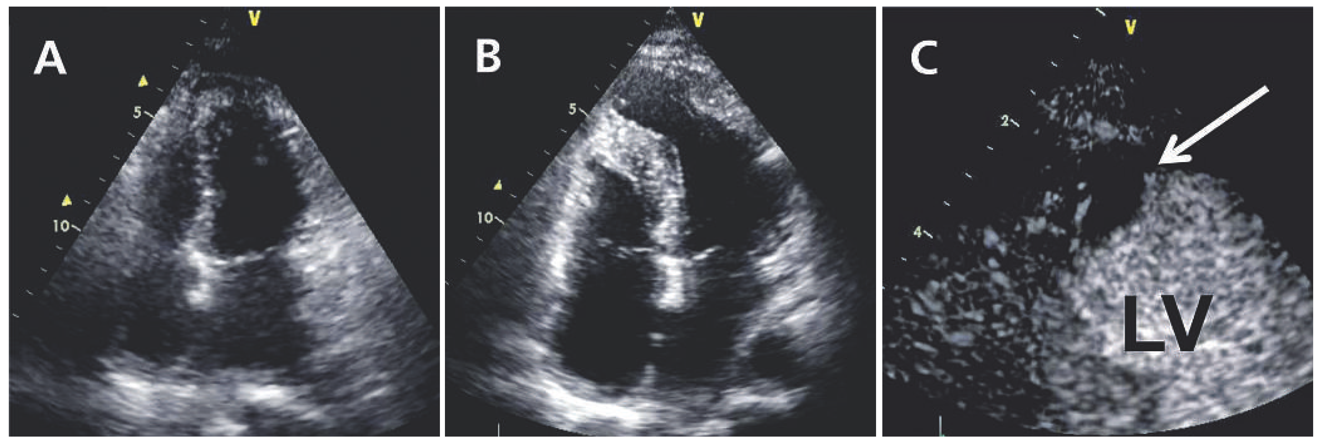
A 39-year-old woman presented with recurrent episodes of acute pericarditis over a four year period. Repeated transthoracic echocardiograms showed normal left ventricular dimensions and wall motion (Figure 1A).
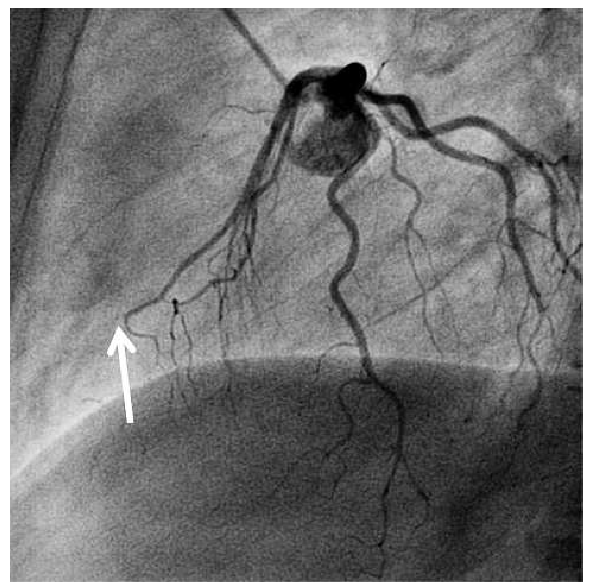
During the last episode, open pericardial biopsy was performed but did not reveal evidence of a specific aetiology. Fifteen months later, the patient was referred because of atypical chest pain, most likely of musculo-skeletal origin. However, the ECG at that time showed Q waves and negative T waves in leads V1 to V4. Transthoracic echocardiography now revealed a left ventricular apical aneurysm (Figure 1B), with a thrombus evident after contrast administration (Figure 1C). Coronary angiography showed smooth coronary arteries with the exception of a totally occluded mid-to-distal left anterior descending artery (LAD, Figure 2). Review of the operation report on the pericardial biopsy procedure revealed the annotation of “accidental perforation of the right ventricle resulting in heavy bleeding”, which was treated by surgical suture. Retrospectively, the LAD rather than the right ventricle must have been injured, and attempts to achieve haemostasis must have resulted in occlusion of the mid-to-distal LAD with subsequent apical infarction with aneurysm and thrombus formation. As evident from the angiogram (Figure 2) the patient’s heart had a somewhat unusual rotation. The more anterior position of the left ventricle and adhesions between the pericardium and epicardium following several episodes of pericarditis may have made the LAD more prone to this unusual complication.

Funding / potential competing interests
No financial support and no other potential conflict of interest relevant to this article were reported.
Figure 1. A. Transthoracic echocardiography (apical four chamber view)—end-systolic still frame after normal contraction of the left ventricular apex. B. Transthoracic echocardiography fifteen months later—end-systolic still frame demonstrating an aneurysm of the left ventricular apex. C. Transthoracic echocardiography showing the apical aneurysm with evidence of a thrombus (arrow) after contrast administration. LV = left ventricle.
Cardiovascmed 16 00132 g001
Figure 2. Coronary angiography (90° left lateral view) showing a totally occluded mid-to-distal LAD (arrow) and otherwise unremarkable coronary arteries.
Cardiovascmed 16 00132 g002