Next Article in Journal
Ake Senning—Pionier der Schweizer Herzchirurgie in Zürich: Zu seinem 10. Todestag und zum 50-Jahr-Jubiläum der Herzchirurgie am UniversitätsSpital Zürich
Previous Article in Journal
Swiss Cooperative Study Acute Coronary Syndromes and Inflammation: A success story supported by the Swiss National Science Foundation
 
 
Cardiovascular Medicine is published by MDPI from Volume 28 Issue 1 (2025). Previous articles were published by another publisher in Open Access under a CC-BY (or CC-BY-NC-ND) licence, and they are hosted by MDPI on mdpi.com as a courtesy and upon agreement with Editores Medicorum Helveticorum (EMH).
Interesting Images

Diastolic Mitral Regurgitation in Complete Atrioventricular Block

by
Alexander Breitenstein
*,
Bernhard Herzog
and
Patric Biaggi
Cardiology, Cardiovascular Centre, University Hospital Zurich, CH-8091 Zurich, Switzerland
*
Author to whom correspondence should be addressed.
Cardiovasc. Med. 2011, 14(5), 163; https://doi.org/10.4414/cvm.2011.01586
Submission received: 25 February 2011 / Revised: 25 March 2011 / Accepted: 25 April 2011 / Published: 25 May 2011

Case report

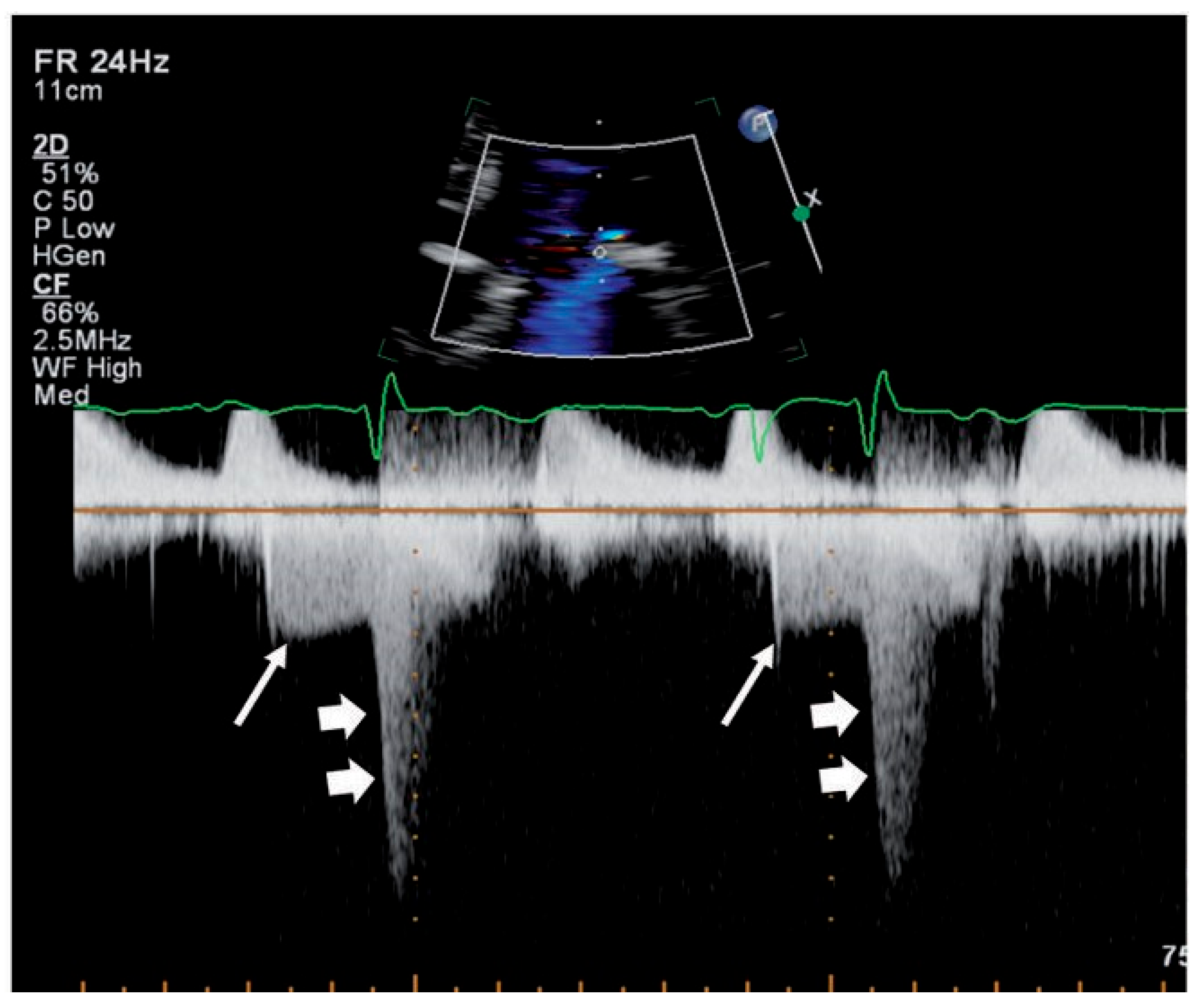
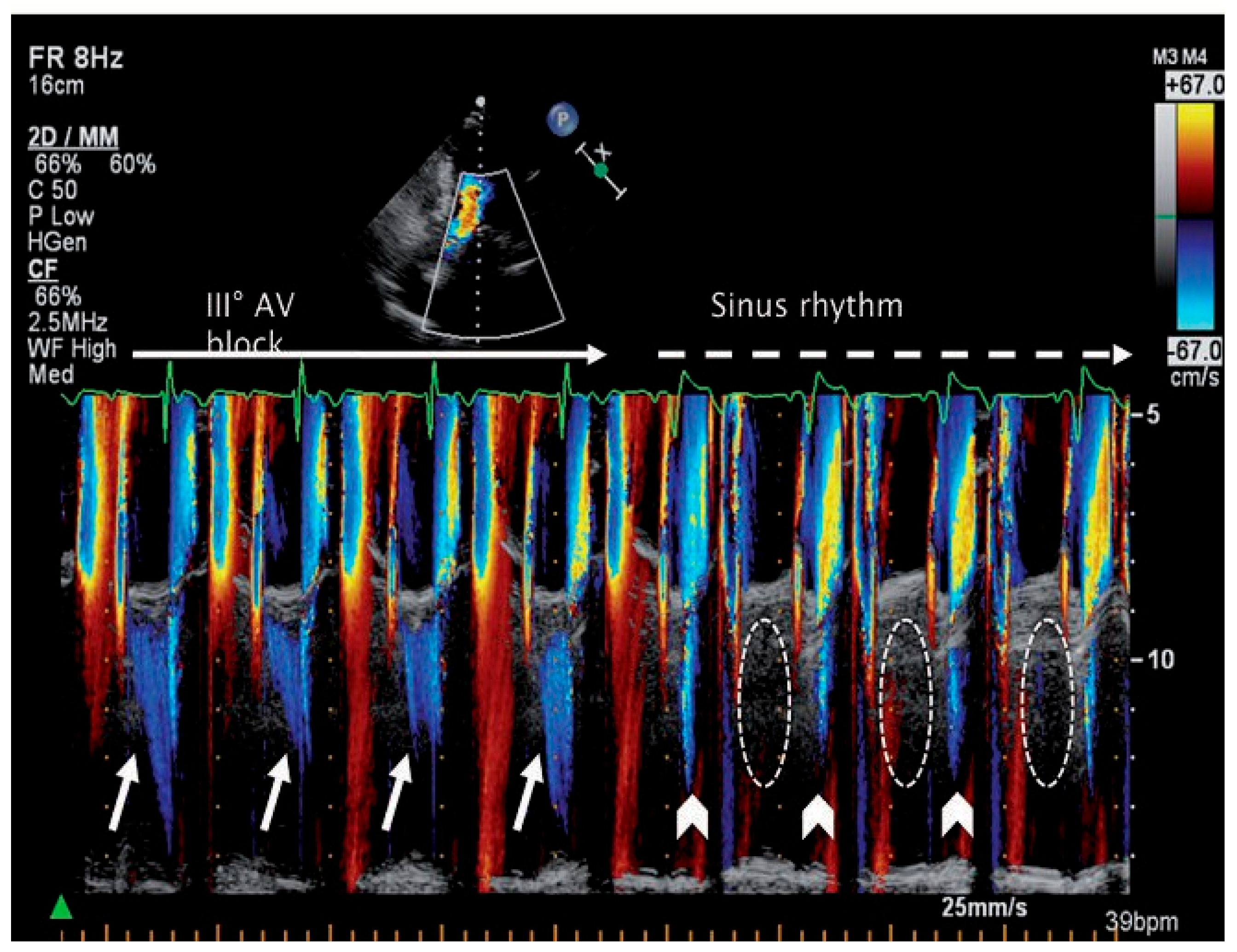
A 75-year-old woman was admitted to our hospital with progressive dyspnea and fever due to an acute, community-acquired pneumonia. On examination, the heart rate was irregular and slow (43 beats per minute), and there was a soft end-diastolic and a somewhat louder systolic murmur over the apex. The 12-lead surface electrocardiogram confirmed the diagnosis of a third-degree atrioventricular (AV) block, most likely due to a pneumonia-associated myocarditis. The transthoracic echocardiogram revealed the presence of a significant diastolic mitral regurgitation (MR) as the reason for the diastolic murmur (Figure 1). The timing of diastolic MR was best demonstrated by continuous-wave Doppler recordings (Figure 2) and colour-Doppler M–mode (Figure 3, left side). During the M-mode acquisition, the patient switched back to SR, and the diastolic MR disappeared (Figure 3, right side).
Diastolic MR is caused by an elevated left ventricular end-diastolic pressure, leading to an inverse pressure gradient across the mitral valve in diastole, and hence to a diastolic MR with a relatively low velocity of 1–2 m/s [1,2]. This condition is frequently met during atrio- ventricular conduction abnormalities of any degree [1], after ventricular premature beats or in patients with a pacemaker and a prolonged PQ interval [3], during atrial flutter, in patients with restrictive cardiomyopathy [4], and in patients with severe aortic regurgitation [2]. The present case illustrates diastolic MR during complete atrioventricular block and emphasises the diagnostic accuracy of continuous-wave Doppler and colour-Doppler M-mode echocardiography.

Funding/potential competing interests:

No financial support and no other potential conflict of interest relevant to this article was reported.

References

  1. Margulescu, A.D.; Vinereanu, D.; Cinteza, M. Diastolic mitral regurgitation in 2:1 atrioventricular block. Echocardiography. 2009, 26, 228–229. [Google Scholar] [CrossRef] [PubMed]
  2. Berger, R.L.; Katz, E.; Tunick, P.; Kronzon, I. The “A-dip” of diastolic mitral regurgitation: an unusual Doppler flow pattern in a patient with severe aortic insufficiency and complete heart block. Eur J Echocardiogr. 2008, 9, 69–71. [Google Scholar] [CrossRef] [PubMed]
  3. Ishikawa, T.; Sumita, S.; Kimura, K.; Kikuchi, M.; Kosuge, M.; Kuji, N.; et al. Prediction of optimal atrioventricular delay in patients with implanted DDD pacemakers. Pacing Clin Electrophysiol. 1999, 22, 1365–1371. [Google Scholar] [CrossRef] [PubMed]
  4. Hatle, L.K.; Appleton, C.P.; Popp, R.L. Differentiation of constrictive pericarditis and restrictive cardiomyopathy by Doppler echocardiography. Circulation. 1989, 79, 357–70. [Google Scholar] [CrossRef] [PubMed]
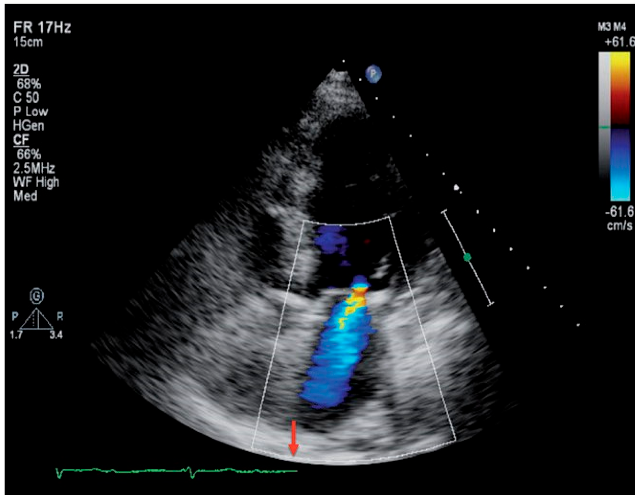
Figure 1. Colour-Doppler image. End-diastolic mitral regurgitation (arrow highlights end-diastole).
Cardiovascmed 14 00163 g001
Figure 2. Continuous-wave Doppler through the mitral valve, demonstrating both diastolic (long arrows) and systolic MR (short arrows).
Cardiovascmed 14 00163 g002
Figure 3. Colour-Doppler M-mode through the mitral valve. During complete atrioventricular (AV) block, diastolic mitral regurgitation is present (long arrows). After conversion into sinus rhythm, minimal systolic MR is noted (arrow heads), while diastolic MR is absent (dashed circles).
Cardiovascmed 14 00163 g003