Next Article in Journal
Kardiale Hybrid-Bildgebung in der Diagnostik der koronaren Herzerkrankung
Previous Article in Journal
Transcatheter Closure of a Large Right Coronary Artery Fistula in a Patient with Bacterial Endocarditis
 
 
Cardiovascular Medicine is published by MDPI from Volume 28 Issue 1 (2025). Previous articles were published by another publisher in Open Access under a CC-BY (or CC-BY-NC-ND) licence, and they are hosted by MDPI on mdpi.com as a courtesy and upon agreement with Editores Medicorum Helveticorum (EMH).
Article

Routine Robotic and Video—Assisted Mitral Valve Repair in Everyday Surgery

by
Aristotelis Panos
1,* and
Patrick O. Myers
2
1
Cardiac Surgery, Rue de l’Athénée 30, CH-1206 Geneva, Switzerland
2
Division of Cardiac Surgery, Brigham and Women’s Hospital & Harvard Medical School, Boston, MA 02125, USA
*
Author to whom correspondence should be addressed.
Cardiovasc. Med. 2011, 14(3), 92; https://doi.org/10.4414/cvm.2011.01573
Submission received: 23 December 2010 / Revised: 23 January 2011 / Accepted: 23 February 2011 / Published: 23 March 2011

Abstract

Objectives: We report our experience with video-assisted minithoracotomy and robotic-enhanced cardiac surgery in the setting of mitral valve repair. Methods: Patients undergoing video-assisted minithoracotomy (n = 74) and robotic-enhanced (n = 15) cardiac surgery were prospectively included between December 2008 and October 2010. The feasibility, safety and echocardiographic follow-up were evaluated. Results: 89 patients with a median age of 48 years (range 24–75) underwent video-assisted or robotic-enhanced cardiac surgery. 74 were operated on through a small anterolateral thoracotomy and fifteen with the da Vinci S robotic system. The procedures performed were lone mitral valve repair in 79 patients, associated with tricuspid valve repair in 10 patients, and mitral valve repair and atrial fibrillation ablation in 20 patients. Two patients required conversion to sternotomy (2.2%). There was no operative mortality. Mean blood transfusion was 1.6 packages/patient. No neurological, vascular or wound complication was observed. Mean CPB and aortic cross clamping were 138 ± 28 min and 114 ± 28 min respectively. Mean ICU stay was 1.5 ± 0.4 days. All the patients had a fast-track extubation (ranging from 3–12 h postoperatively) and 15 patients were extubated in the operating theatre. Bleeding was evaluated to 350 ± 186 mL. Hospital stay was 5 ± 1.3 days. No patient required reoperation. Three patients required reexploration for bleeding and in all of them haemostasis was achieved through the same incision. Follow-up ranged from 2–24 months, during which all of the patients remained with no or trace mitral valve regurgitation in all the mitral repair patients and mild tricuspid valve regurgitation in all the tricuspid repair patients. All the atrial fibrillation ablation patients had a conversion success rate of 45% at 2 years. Conclusion: Minimally invasive mitral valve repair can be safely performed, with excellent and durable results in a wide range of patients in routine surgical settings. Low blood transfusion rate, short hospital stay and avoidance of wound complications are the main advantages of this approach.

Introduction

Video-assisted and robotic-enhanced surgery are emerging fields in cardiac surgery. Although initially more complicated for the surgeon, they have been shown to be less traumatic for the patient and provide faster recovery [1,2]. Dedicated surgical instruments have been developed to fit these approaches. We introduced this approach and decided to perform most of the mitral valve repairs, tricuspid valve repairs, atrial septal defect (ASD) closures and suitable single coronary artery bypasses using this approach, unless it was otherwise contraindicated. We previously reported a subset of these patients who previously underwent tricuspid valve repair [3]. In the current study we present and review our experience with this approach unique in Switzerland.

Methods

Patients and follow-up

Between December 2008 and October 2010, 89 patients were included in the present study. The median age was 48 years (range 24–75). All of the patients were operated on an elective basis and none of them had previously undergone cardiac surgery. From a total of 89 patients (34 females) who underwent robotic or video-assisted procedures, 79 patients had lone mitral valve repair, in 10 patients a tricuspid valve repair was associated and 20 patients had mitral valve repair and atrial fibrillation ablation. The patients were prospectively followed up with transthoracic 2-D echocardiography, before discharge from hospital and then by their referring cardiologist at their discretion. None of the echocardiographic data in this report is based on intraoperative studies. Primary endpoints included feasibility, necessity of conversion to sternotomy, operative mortality and success of valve repair. Secondary endpoints were duration of aortic cross-clamping and cardiopulmonary bypass (CPB), relapse of severe MR, TR and use of blood products in the perioperative period.
Echocardiographic follow-up was performed in all patients. During the follow-up the patients were assessed for clinical improvement and degree of satisfaction with this approach in terms of postoperative pain, return to normal activities and aesthetic result.
A number of these patients were reported on in a prior study focusing on biodegradable tricuspid valve repair [3].

Surgical technique

Standard minimally invasive techniques, as described previously in detail [2], were used. Full normothermic cardiopulmonary bypass was instituted with arterial (DLP arterial cannula, Medtronic Inc., Minneapolis, MN) and venous femoral cannulation (Edwards Lifesciences, Irvine, CA), as well as internal jugular venous cannulation (14F DLP arterial cannula, Medtronic Inc., Minneapolis, MN). For video-assisted surgery, the cardiac operation proceeded through a small right-anterolateral thoracotomy (Figure 1) and through port access for the da Vinci S robotic system (Intuitive Surgical, Sunnyvale, CA). 74 patients were operated on through a small anterolateral thoracotomy and 15 with the da Vinci S robotic system. The robotic system was required for coronary bypasses and for the most complex valve repairs, such as those involving anterior mitral leaflet or bileaflet prolapse requiring chordal transfer or neochordae. On the other hand, video-assisted surgery is less expensive, faster and less cumbersome. Also, the greater cost of robotic surgery is not covered by all insurances. The robotic system was used whenever possible and justified. The main working port is created using a dedicated soft tissue retractor (Edwards Lifesciences, Irvine, CA). Thoracic ports of 5.5, 10.5 and 11.5 mm are introduced in the 5th, 4th and 3rd intercostal spaces respectively on the anterior and mid axillary line. Atrial fibrillation ablation was performed in the first 15 patients with the monopolar Cardioblate radiofrequency system (Medtronic Inc., Minneapolis, MN) and for the remaining 5 patients with the Cobra monopolar radio frequency system (Estech, San Ramon, CA). The valves were repaired according to the appropriate techniques to achieve no or trace regurgitation at the end of the operation with TEE. Rigid Physioring II (Edwards Lifesciences, Irvine, CA) mitral rings were used for the video-assisted mitral repairs and in the robotic patients an annuloplasty Duran An Core band (Medtronic Inc., Minneapolis, MN). These flexible annuloplasty bands were fixed with nitinol clips (U-clip, S18RFRN8, Medtronic Inc., Minneapolis, MN). Flexible rings were used in robotic operations to achieve total endoscopic repair, since the ring can be passed through the chest wall through the thoracoports; this is impossible with rigid rings. At the end of the operation TEE provided adequate removal of air.

Results

There were no operative deaths. Two patients required conversion to sternotomy: one due to the impossibility of externally defibrillating the heart after mitral valve repair, and the second for inadequate exposure with the use of the MitraXS atrial retractor (MitraXS Cardiolife Research, Lovain La Nueve, Belgium). Mean blood transfusion was 1.6 units/patient. No neurological or vascular complications were observed. Two patients presented lymphorrhoea from the right groin incision, on the site of femoral cannulation. Mean CPB and aortic cross clamping were 138 ± 28 min and 114 ± 28 min respectively. All the patients had fast track extubation (ranging from 3–12 h postoperatively) and 15 patients were extubated in the operating theatre. Mean ICU stay was 1.5 ± 0.4 days. Bleeding was evaluated to 350 ± 186 mL. Hospital stay was 5 ± 1.3 days. No patient required reoperation. Three patients required reexploration for bleeding and in all of them haemostasis was achieved through the same incision. No patient required reoperation. Follow-up ranged from 2–24 months, during which all of the patients remained with trace mitral valve regurgitation in all the mitral repair patients and mild tricuspid valve regurgitation in all the tricuspid repair patients. All the atrial fibrillation ablation patients had a conversion to sinus rhythm success rate of 45% at 2 years. All the patients were satisfied with their choice of the minimally invasive approach with respect to postoperative pain and aesthetic result.

Discussion

Cardiac surgery has been traditionally performed through a median sternotomy. However, progress in surgical instrumentation, robotics and perfusion techniques has enabled cardiac surgery to be performed through progressively smaller thoracic incisions or totally endoscopically using robotic technology. Reports from various institutions have shown the efficacy of video-assisted port access and robotic approaches for mitral repair [1,4,5] and tricuspid valve operations [3,6]. Most of these studies reported fewer wound infections, less bleeding, less pain and earlier return to work. Svensson et al. [7] elegantly showed, in a propensity-matched comparison of minimally invasive mitral valve surgery including 1180 patients, that although patients undergoing full sternotomy mitral valve repair were overall sicker than their counterparts undergoing minimally invasive repair, once this difference was controlled for there were no disadvantages to minimally invasive surgery: procedure time was not lengthened, the risks were comparable or lower, transfusions were less frequent, respiratory function was better, early postoperative pain was less marked, length of stay was comparable or shorter, and the results of the repair and long-term survival were comparable [7]. These authors postulated that less perioperative bleeding and fewer blood transfusions were probably due to the less extensive mediastinal dissection required for the minimally invasive approach; that less pain was probably related to less surgical dissection, less spreading of the sternum, and no escalation of tension on the posterior rib head and costovertebral ligaments, because the chest wall is not opened like a trap door; and the better pulmonary function could be explained by the absence of interference with the diaphragm or dissection along it, and improved breathing from less pain. It should be said, however, that a majority of these “minimally invasive” mitral valve repair patients had surgery through a partial sternotomy or limited thoracotomy, and only a minority had robotic or thoracoscopic approaches.
One of the major drawbacks of robotic and thoracoscopic approaches in cardiac surgery is that operative times are longer than with standard sternotomy. This results from working in a much smaller operative field and the significant learning curve associated with these procedures. In the video-assisted approach significant ergonomic and visual limitations persist, particularly in complex repairs such as chordal transfer, sliding valvuloplasty and partial leaflet transposition. Performance of a truly endoscopic mitral repair through thoracic ports requires 3-dimensional visualisation and articulated instrumentation for these complex cases [8]. The da Vinci robotic surgical system enabled surgeons to perform complex mitral repairs through port incisions using optimised high-definition visualisation and fine dexterity [9], despite the lack of haptic feedback. Since this initial experience, further developments in robotic technology, such as the addition of a fourth robotic arm to control an atrial retractor, and imaging improvements permitting magnified and 3-dimensional vision and surrogate feedback that projects “visual tactility”, now enable surgeons to understand tissue deformational characteristics and suture remodelling with tightening [8,10], allowing increasingly complex repairs using these techniques. Sato [11] evaluated 485 on-pump CABG patients in a fasttrack extubation protocol and found that early tracheal extubation can be successfully performed in most patients receiving on-pump CABG. The management of higher-risk patients and efforts to reduce operation time and blood loss are keys to success for early tracheal extubation. Our protocol of fast-track extubation permits better use of medical resources and lower hospital costs without increasing patient morbidity and mortality.
As with any new technology, videoscopic and robotic surgery must prove as effective at repair as conventional approaches. Cohn et al. showed that a minimally invasive approach to the mitral valve through a lower hemisternotomy provided results as good as full sternotomy mitral valve repair, with freedom from reoperation of 92% at 5 years [12]. The Cleveland Clinic group very recently showed, in a propensity-matched retrospective study on 759 patients with degenerative mitral valve disease including 114 right anterior mini-thoracotomies and 261 robotic repairs, that the quality of the repair was similar between matched groups [13]. Nevertheless, particularly in the light of the extremely good long-term results of valve repair for myxomatous mitral regurgitation, future efforts should focus on comparing the effectiveness of these videoscopic and robotic approaches to traditional mitral repair techniques in prospective randomised trials, and investigating the stability of repair over longer-term follow-up.

Conclusion

Videoscopically assisted endoscopic cardiac surgery can be performed safely, with durable results in a wide range of patients in routine surgical settings. However, it involves a stiff learning curve and intensive training. The low blood transfusion rate, short hospital stay, avoidance of wound complications and superior cosmetic result are the main advantages of this approach.

Conflicts of Interest

The authors certify that there is no actual or potential conflict of interest in relation to this article.

References

  1. Modi, P.; Hassan, A.; Chitwood, W.R., Jr. Minimally invasive mitral valve surgery: A systematic review and meta-analysis. Eur J Cardiothorac Surg. 2008, 34, 943–952. [Google Scholar] [CrossRef] [PubMed]
  2. Woo, Y.J.; Rodriguez, E.; Atluri, P.; Chitwood, W.R., Jr. Minimally invasive, robotic, and off-pump mitral valve surgery. Semin Thorac Cardiovasc Surg 2006, 18, 139–147. [Google Scholar] [CrossRef] [PubMed]
  3. Panos, A.; Myers, P.O.; Kalangos, A. Thoracoscopic and robotic tricuspid valve annuloplasty with a biodegradable ring: An initial experience. J Heart Valve Dis. 2010, 19, 201–205. [Google Scholar] [PubMed]
  4. Casselman, F.P.; La Meir, M.; Jeanmart, H.; Mazzarro, E.; Coddens, J.; Van Praet, F.; et al. Endoscopic mitral and tricuspid valve surgery after previous cardiac surgery. Circulation. 2007, 116 (Suppl. 11), I270–275. [Google Scholar] [CrossRef] [PubMed]
  5. Nifong, L.W.; Chitwood, W.R.; Pappas, P.S.; Smith, C.R.; Argenziano, M.; Starnes, V.A.; et al. Robotic mitral valve surgery: A United States multicenter trial. J Thorac Cardiovasc Surg. 2005, 129, 1395–1404. [Google Scholar] [CrossRef] [PubMed]
  6. Lee, T.C.; Desai, B.; Glower, D.D. Results of 141 consecutive minimally invasive tricuspid valve operations: An 11–year experience. Ann Thorac Surg. 2009, 88, 1845–1850. [Google Scholar] [CrossRef] [PubMed]
  7. Svensson, L.G.; Atik, F.A.; Cosgrove, D.M.; Blackstone, E.H.; Rajeswaran, J.; Krishnaswamy, G.; et al. Minimally invasive versus conventional mitral valve surgery: A propensity-matched comparison. J Thorac Cardiovasc Surg. 2010, 139, 926–932 e1-2. [Google Scholar] [CrossRef] [PubMed]
  8. Chitwood, W.R.; Jr Rodriguez, E.; Chu, M.W.; Hassan, A.; Ferguson, T.B.; Vos, P.W.; et al. Robotic mitral valve repairs in 300 patients: A single-center experience. J Thorac Cardiovasc Surg. 2008, 136, 436–441. [Google Scholar] [CrossRef] [PubMed]
  9. Murphy, D.A.; Miller, J.S.; Langford, D.A. Endoscopic robotic mitral valve surgery. J Thorac Cardiovasc Surg. 2007, 133, 1119–1120, author reply 20. [Google Scholar] [CrossRef] [PubMed]
  10. Cheng, W.; Fontana, G.P.; De Robertis, M.A.; Mirocha, J.; Czer, L.S.; Kass, R.M.; et al. Is robotic mitral valve repair a reproducible approach? J Thorac Cardiovasc Surg. 2010, 139, 628–633. [Google Scholar] [CrossRef] [PubMed][Green Version]
  11. Sato, M.; Suenaga, E.; Koga, S.; Matsuyama, S.; Kawasaki, H.; Maki, F. Early tracheal extubation after on-pump coronary artery bypass grafting. Ann Thorac Cardiovasc Surg. 2009, 15, 239–242. [Google Scholar] [PubMed][Green Version]
  12. Greelish, J.P.; Cohn, L.H.; Leacche, M.; Mitchell, M.; Karavas, A.; Fox, J.; Byrne, J.G.; Aranki, S.F.; Couper, G.S. Minimally invasive mitral valve repair suggests earlier operations for mitral valve disease. J Thorac Cardiovasc Surg. 2003, 126, 365–371. [Google Scholar] [CrossRef] [PubMed]
  13. Mihaljevic, T.; Jarrett, C.M.; Gillinov, A.M.; Williams, S.J.; Devilliers, P.A.; Stewart, W.J.; Svensson, L.G.; Sabik, J.F.; Blackstone, E.H. Robotic repair of posterior mitral valve prolapse versus conventional approaches: Potential realized. J Thorac Cardiovasc Surg. 2011, 141, 72–80. [Google Scholar] [PubMed]
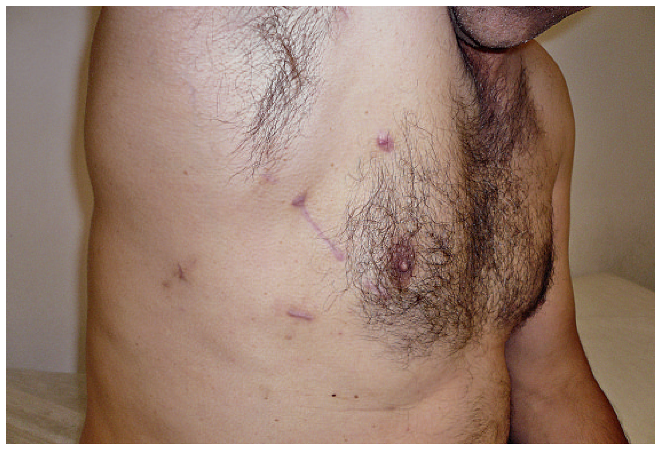
Figure 1. Right anterior thoracotomy (working port) and ports incisions one month postoperatively after mitral valve repair.
Cardiovascmed 14 00092 g001