Next Article in Journal
High-Density Lipoprotein Dysfunction as a New Therapeutic Target
Previous Article in Journal
Recommandations 2011 sur les Bonnes Pratiques dans le Diagnostic et le Traitement des Arythmies par Stimulation Cardiaque et Ablation Percutanée par Cathéter
 
 
Cardiovascular Medicine is published by MDPI from Volume 28 Issue 1 (2025). Previous articles were published by another publisher in Open Access under a CC-BY (or CC-BY-NC-ND) licence, and they are hosted by MDPI on mdpi.com as a courtesy and upon agreement with Editores Medicorum Helveticorum (EMH).
Interesting Images

A Giant Saphenous Vein Graft Aneurysm Compressing the Right Cavities

by
Stéphane Chevallier
1,*,
Etienne Monnard
2 and
Jean-Jacques Goy
1
1
Department of Cardiology, Hôpital Cantonal de Fribourg, Fribourg, Switzerland
2
Department of Radiology, Hôpital Cantonal de Fribourg, Fribourg, Switzerland
*
Author to whom correspondence should be addressed.
Cardiovasc. Med. 2011, 14(2), 69; https://doi.org/10.4414/cvm.2011.01567
Submission received: 23 November 2010 / Revised: 23 December 2010 / Accepted: 23 January 2011 / Published: 23 February 2011

Case report

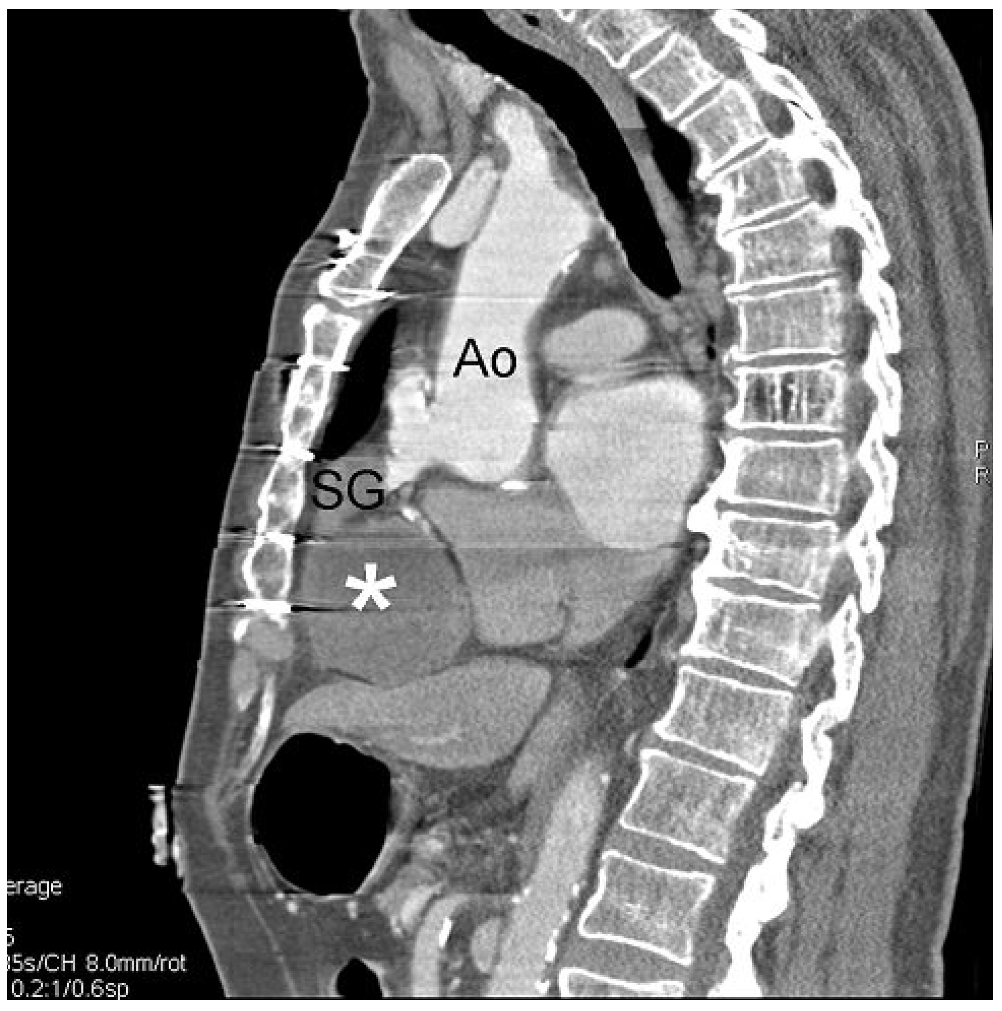
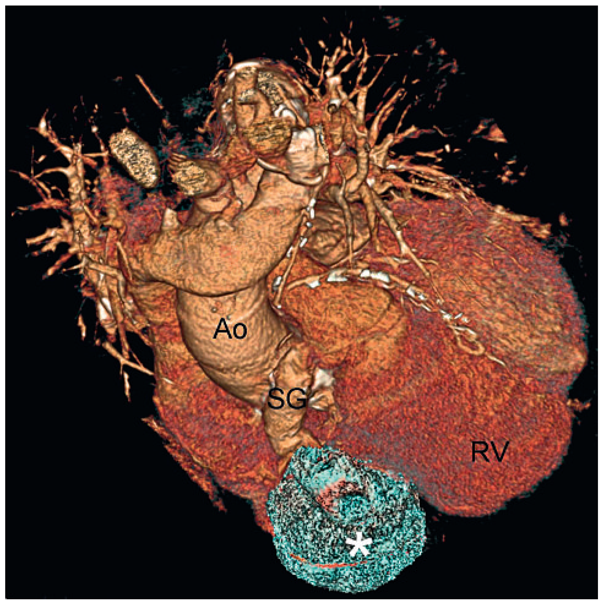
A 86-year-old man was hospitalised because of a cerebral vascular attack. He underwent a triple coronary artery bypass grafting 14 years earlier. He was asymptomatic and received aspirin, beta-blockers, angiotensin receptor inhibitor and statines. Clinical status showed a slight jugular distention but was otherwise normal. On ECG, typical signs of an old inferior myocardial infarction were present. Chest X-ray and blood tests were within the normal values. An echocardiogram was performed as a routine examination following a cerebral attack. Left ventricular function was normal and showed an aneurysm of the basal inferior wall of the left ventricle compatible with the scar of the old infarction. A mass compressing the right cavities at the level of the tricuspid annulus was present in the sub-costal view (Figure 1A) and in a modified apical four chamber view (Figure 1B). A 64-multi-slice cardiac-gated computed tomography (CT) was performed showing a giant, fully thrombosed aneurysm (6 cm diameter) of the saphenous venous graft on the right coronary artery (Figure 2 and Figure 3). As the patient was asymptomatic and the inferior wall fully infarcted, a conservative approach was decided.
An aneurysm of a saphenous graft is a rare complication, but probably underestimated as most of them are asymptomatic. The prevalence is unclear. A mild aneurismal dilatation is found in 14% of bypass grafts 5 to 7 years following surgery, but large dilatation is a rare event with an incidence of less than 1% [1]. The symptoms are mostly atypical with chest pain, and dyspnoea due to compression of the right cavities (oedema, jugular distension) as in the current case [2]. Very few cases have been reported. Cardiac dedicated CT and cardiac MR are the most powerful tools to detect these aneurysms. Coronary angiogram is also useful in evaluating these aneurysms. Surgery is usually recommended to prevent rupture [3] but some patients may be eligible for percutaneous treatment with covered stents [4]. Recently, stenting and coil embolisation has emerged as an alternative to surgery [5].

Conflicts of Interest

The authors certify that there is no actual or potential conflict of interest in relation to this article.

References

  1. Memon, A.Q.; Huang, R.I.; Marcus, F.; Lyndon, X.; Alpert, J. Saphenous vein graft aneurysm. Case report and Review. Cardiol Rev. 2003, 11, 26–34. [Google Scholar] [CrossRef] [PubMed]
  2. Roth, M.; Sprengel, U.; Kraus, B.; Klövekorn, W.P.; Bauer, E.P. Symptomatic aneurysm of a saphenous vein graft with compression of the right atrium. Heart Surg Forum. 1999, 2, 338–340. [Google Scholar]
  3. Nishimura, K.; Nakamura, Y.; Harada, S.; Saiki, M.; Marumoto, A.; Kanaoka, Y.; Nishimura, M. Saphenous vein graft aneurysm after coronary artery bypass grafting. Ann Thorac Cardiovasc Surg. 2009, 15, 61–63. [Google Scholar]
  4. Panetta, C.J.; Schneider, W.; Boller, M.A. Percutaneous management of a long saphenous vein graft aneurysm: a case report and a review of the literature. Cardiol Res Pract. 981292 Epub. 2009. [Google Scholar] [CrossRef] [PubMed]
  5. Chevallier, S.; Cook, S.; Goy, J.J. How should I treat coronary aneurysm. Accepted for publication in Euro Intervention.
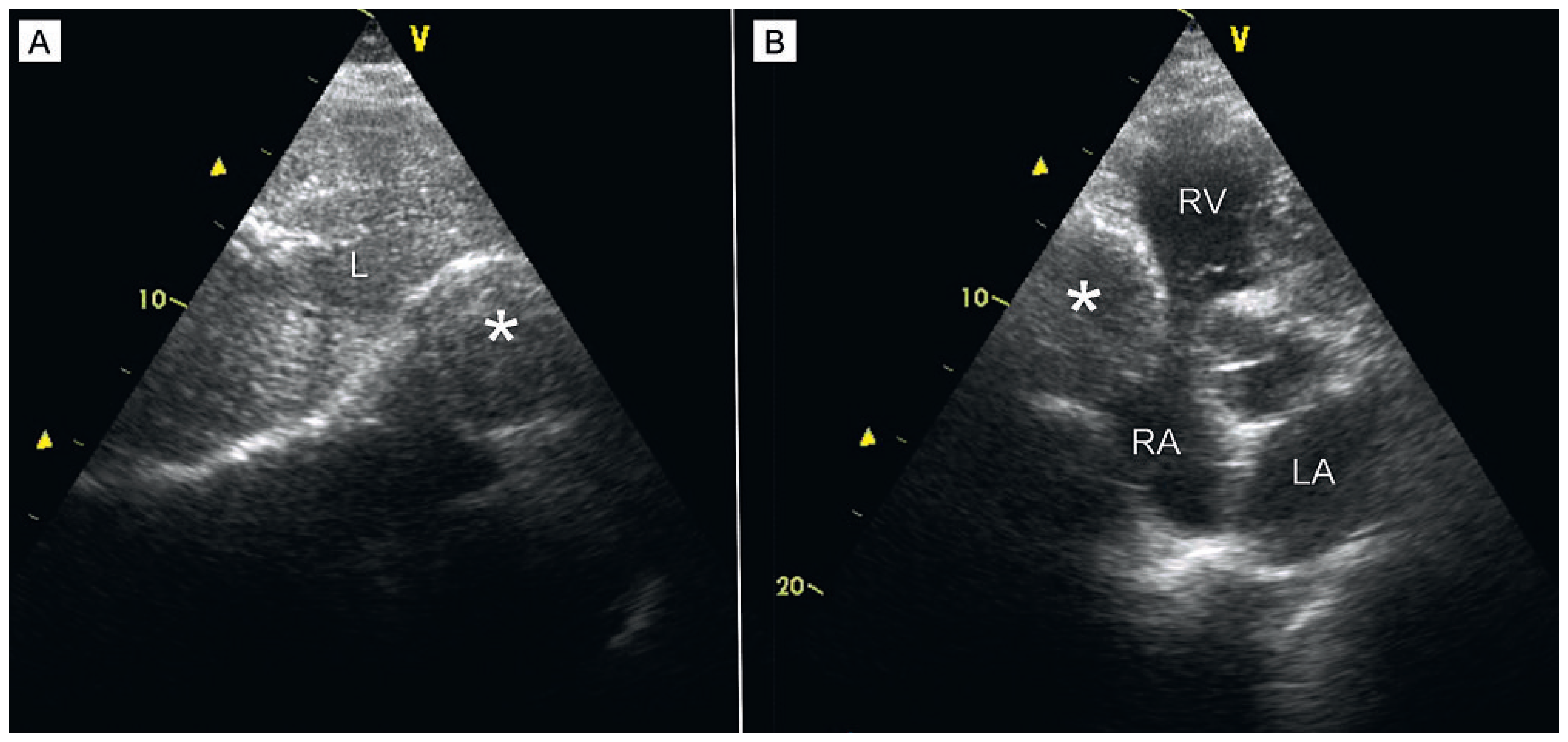
Figure 1. (A) Sub-costal view. A round image (*) between the liver (L) and the right cavities of the heart. (B) Modified four chambers view. The same well-defined round mass (*) is compressing the right cavities maximally at the tricuspid annulus. RV = right ventricle; RA = right atrium; LA = left atrium.
Cardiovascmed 14 00069 g001
Figure 2. Dedicated thoracic computed tomography – sagittal slice. The mass (*) is in the continuity of the saphenous graft (SG) arising from the aorta (Ao).
Cardiovascmed 14 00069 g002
Figure 3. 3-D-computed tomography reconstruction. The mass is a giant aneurysm (*) of a saphenous venous bypass (SG) on the right coronary artery. RV = right ventricle; Ao = ascending aorta.
Cardiovascmed 14 00069 g003