Next Article in Journal
Platinum Reward for Theft, or the Percutaneous Treatment of Coronary Steal Due to a Giant Coronary Artery Fistula
Previous Article in Journal
Blutplättchenhemmer bei Koronarer Herzkrankheit
 
 
Cardiovascular Medicine is published by MDPI from Volume 28 Issue 1 (2025). Previous articles were published by another publisher in Open Access under a CC-BY (or CC-BY-NC-ND) licence, and they are hosted by MDPI on mdpi.com as a courtesy and upon agreement with Editores Medicorum Helveticorum (EMH).
Editorial

Do We Need MR Conditional Pacemakers?

by
Roger Luechinger
1,* and
Firat Duru
2
1
Institute for Biomedical Engineering, University and ETH, CH-8092 Zürich, Switzerland
2
Division of Cardiology, University Hospital of Zurich, CH-8091 Zürich, Switzerland
*
Author to whom correspondence should be addressed.
Cardiovasc. Med. 2010, 13(2), 70; https://doi.org/10.4414/cvm.2010.01477
Submission received: 24 November 2009 / Revised: 24 December 2009 / Accepted: 24 January 2010 / Published: 24 February 2010

Abstract

Magnetic resonance imaging (MRI) is a widely accepted tool for the diagnosis of a variety of disease states. However, due to safety concerns the presence of an implanted cardiac pacemaker is considered to be a contraindication to MRI in most medical centres. The increasing number of implanted pacemakers and the estimated over 50% probability that a pacemaker patient may be a candidate for an MRI increase the need for safe scanning of pacemaker patients. Over the last ten years a major effort has been made to understand the potential risks. The influences from the three electromagnetic fields on pacemakers are versatile and will be summarised. The research in this area has helped to predict the risks of an MRI scan in patients with conventional pacemakers, and has also stimulated pacemaker manufacturers to improve their devices with the goal of providing MR conditional devices. Since autumn 2008 the first approved MR conditional pacemakers have been on the market and other devices are likely to follow this trend. However, the vast majority of devices are still not approved for MRI, a situation which will take several years to change. It is thus important that a solution be also found for these patients. Several studies including over 500 patients with a pressing need for MRI have been performed at different experienced centres. On the basis of those data various organisations in MRI fields have proposed guidelines for MRI in patients who fulfill given requirements. However, at present, nonMRI modalities should be considered, whenever possible, for diagnosis in pacemaker recipients with conventional devices. If other imaging modalities are not adequate, MRI with careful monitoring and preparation for adverse events may be considered only at experienced centres. With the wider availability of MR conditional devices, the risks of MRI are greatly reduced and non-tertiary centres will be able to perform these investigations. In any case, rapid progress in the field of MR conditional pacing is desirable.

Introduction

Magnetic resonance imaging (MRI) is the modality of choice for a wide range of soft tissue pathologies. Since the first live human images reported in 1977, the development of clinical MRI has been rapid. Around 60 million MRI procedures are performed today [1]. On the other hand, the number of implanted pacemakers is increasing, with an estimated 50–75% probability of MRI being indicated in a patient during the lifetime of their device [2].
Various studies have shown that MRI may be hazardous in patients with implanted pacemakers [3,4,5,6,7,8]. The greatest risk posed by MRI in pacemaker patients is the reported lethal consequences in these patients [9,10,11,12]. Six patients have died in Germany [13] but no data are available from other countries. MRI is therefore considered a contraindication for recipients of these devices at most MRI centers. However, there are also reports of safe MRI if this diagnostic technique was an absolute necessity [8,14,15,16,17,18,19,20]. Based on the results of these studies, different organisations have published guidelines stating that MRI may be acceptable for selected patients with pacemakers.

Potential benefits of magnetic resonance imaging in paced patients

In patients with permanent pacemakers MRI may confer major diagnostic benefits since the technique has advantages over other imaging modalities. MRI has now become the imaging modality of choice for all congenital, traumatic, hereditary, vascular, infectious, autoimmune, metabolic, and neoplastic disorders of the central nervous system. It is the imaging modality of choice for further evaluation of musculoskeletal disorders, when physical examination or plain radiography suggests a serious abnormality. Cardiovascular MRI is useful in the evaluation of congenital and acquired diseases of the heart and great vessels, and has been a rapidly advancing area of clinical research [21].

Electromagnetic fields in magnetic resonance imaging

MRI uses three different electromagnetic fields to produce images. The main static magnetic field is used to align protons. This magnetic field is 0.5–3 T strong (~50,000 times stronger than the earth’s magnetic field) in most of the currently available MRI units for clinical use, and is always on. The time-varying magnetic gradient fields, used for spatial localisation, change their strength along different orientations and operate at frequencies up to 100 kHz. Clinical MRI systems have a gradient strength of up to 80 mT/m with a slew rate of up to 200 mT/m/ms. In current systems the usable gradient power is limited by peripheral nerve stimulation. The pulsed RF field, generated by the body-coil or a send/receive head-coil, is used to change the energy state of the protons and elicit MRI signals from tissue. The RF field is switched on and off during measurements. The frequency depends on the main magnetic field with 42 MHz/T (= 21–128 MHz on current clinical systems). The RF amplifiers have a power of up to 25 kW with a duty cycle of <10%.

Effects of magnetic resonance imaging on cardiac pacemakers

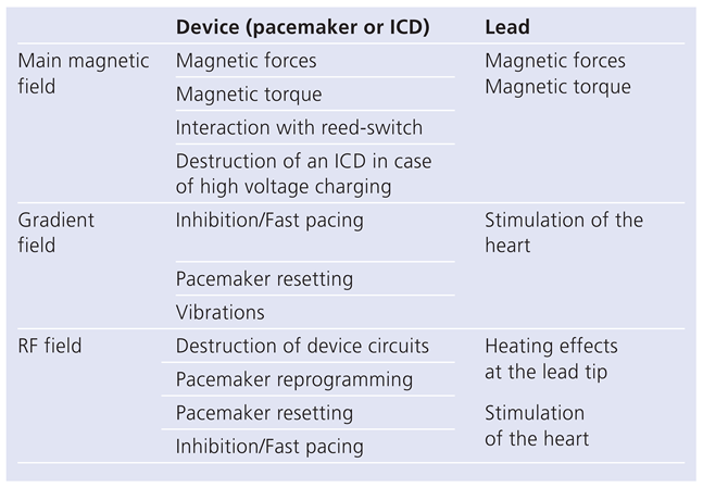
The potential effects of MRI on cardiac pacemakers are multiple (Table 1) [3,4,5,6,22]. MRI may involve the following potential risks for pacemaker patients:
The main magnetic field of the MRI scanner produces force and torque effects; however, with modern pacemakers these effects are not harmful to the patients [23,24]. On the other hand, in strong magnetic fields above 200 mT, the state of the reed switch is unpredictable [25] and the pacemaker may stay in an asynchronous mode or may revert to a synchronous mode. This problem has been solved in some of the newer devices by using semiconductor magnet switches such as hall sensors.
The gradient as well as the RF field may mimic cardiac signals if the pacemaker is in a synchronous mode, and pacing may therefore be inhibited or triggered by MRI. With the gradient field bipolar pacing is safer, but this may not be the case with the RF field, a problem which asynchronous pacing may solve. In worst-case situations the induced voltages from the gradient field and possibly from the RF-field are strong enough to stimulate the heart. This can be prevented by appropriate design of the pacemaker.
Some devices reset as the patient is moved into the strong magnetic field, whereas others do so during imaging while the gradient and RF fields are active [26]. The reset programming may be inadequate for the patient undergoing MRI. Induced voltages in the device will provoke vibrations in the static magnetic field. This effect may be felt by the patient and may even lead to wire fractures inside the device.
High electrical fields of the radiofrequency field will induce currents which will drop over the ohmic resistance of the lead tip-myocardium interface and deposit power, thus possibly destroying parts of the adjacent myocardial tissue. Figure 1 illustrates the potential risk of RF heating. The amount of absorbed RF energy depends on several parameters such as lead position, lead construction, impedance at the lead tip and at the device connector, position within the sending RF coil, type of RF coil, power and duration of the RF pulses and the frequency of the RF field. Since the absorbed power is a resonance effect, small changes in lead configuration may result in a strong increase in heating [27], making testing very challenging. The effects of electrode heating are not detectable by monitoring during MRI. However, an increase in pacing threshold, arrhythmias caused by scar tissue or even myocardial perforation are among the potential concerns long after scanning, but such chronic effects of MRI have not yet been studied.
Image artifacts induced by the pacing system could be a problem for MRI. However, the image artifacts are limited to 5–10 cm around the device. Most pacing leads are non-magnetic and produce limited image artifacts.

Animal and human studies

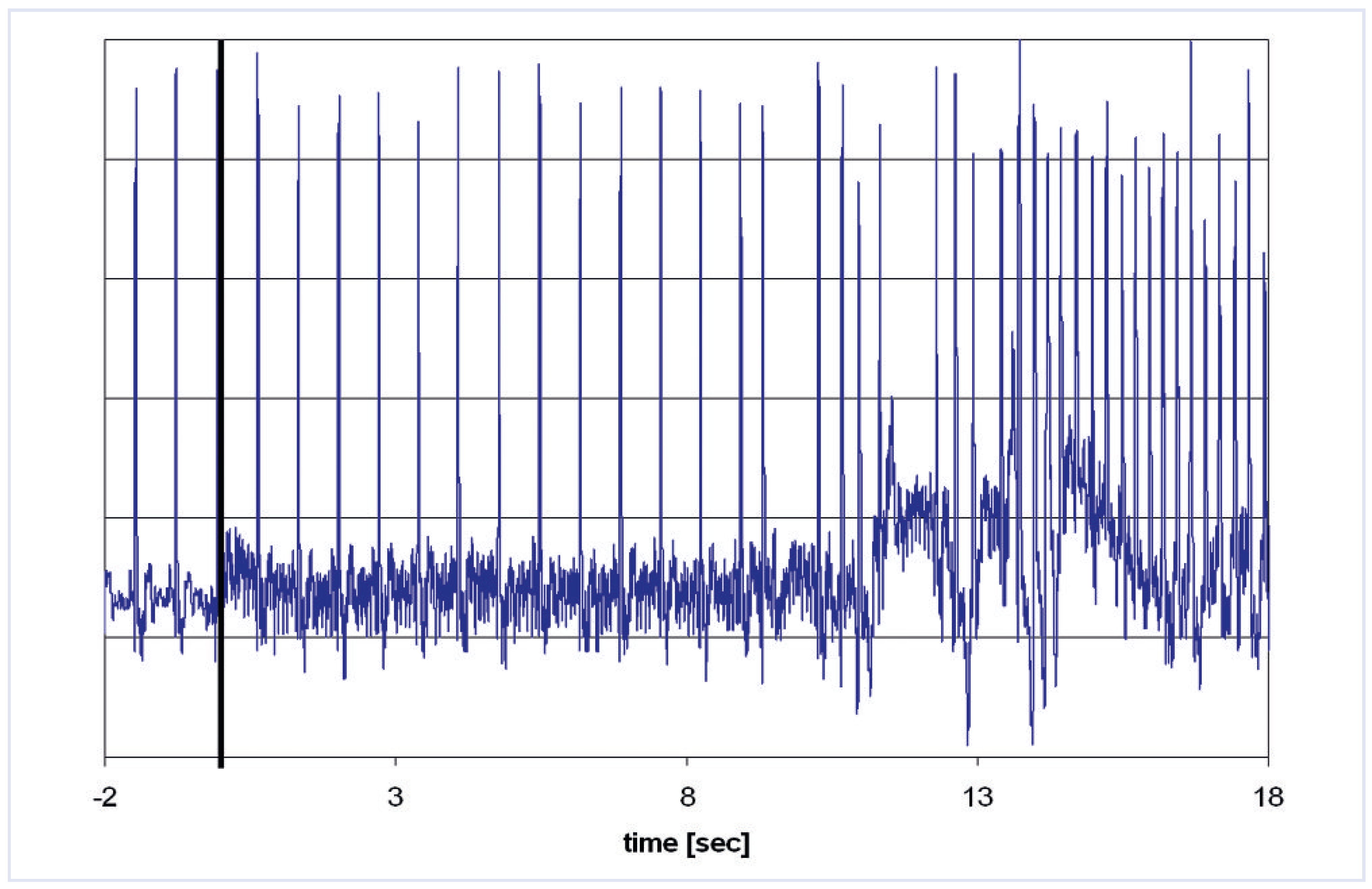
Two published animal studies have shown controversial results. In an acute study by Roguin et al. [28] using a canine model and clinical scans with an SAR of <1.4 W/kg, only minor heating could be observed. In a chronic study [29] using 70 kg pigs allowing lead placement comparable to that in humans, heating of up to 20 °C (SAR up to 3.9 W/kg) at the lead tip could be measured. The lead impedance was increased after MRI. No reproducible histological alteration linked to heating could be found. In one of the animals an arrhythmia could be induced by high SAR levels (Figure 2).
There are a few anecdotal reports of unexpected deaths in patients undergoing MRI [9,10,11,12]. In one case the patient had no escape ventricular rhythm and apparently died from asystole. Another patient developed ventricular fibrillation during the imaging procedure which was not recognised immediately since ECG monitoring was not used [30]. It is likely that many potential complications go unreported in the literature for various reasons (liability, etc.).
A number of human studies have been performed to date in several hundred patients, the majority with limitations to SAR, body regions imaged, type of devices, etc., without severe problems [16,19,26,31,32,33]. Some studies showed pacing threshold increases in some of the patients which could be an indication of RF heating of the heart tissue around the pacing lead tip. In one of these studies a ventricular arrhythmia was induced during scanning in a patient with significant proximal stenosis of the right coronary artery. Thanks to adequate monitoring and the presence of trained personnel, the patient could be cardioverted successfully (private communication with Dr. Claas Philip Nähle, Bonn). The results of anecdotal safe scans of patients are not sufficient to prove safety for MRI examinations in patients with pacemakers.
A safety protocol for imaging of patients with permanent pacemaker and implantable cardioverter-defibrillator (ICD) systems has been proposed by Nazarian et al. in Baltimore [34]. This protocol may be helpful for a systematic approach to such patients. However, the list of devices with previously satisfactory testing needs to be approached with caution.

MRI in patients with conventional pacemakers

Different studies including over 500 safe scans in pacemaker patients under controlled conditions afforded grounds to revise several organisations’ guidelines and change MRI from an absolute to a relative contraindication in pacemaker patients. Revised guidelines of the following societies have been Published: the ACR Blue Ribbon Panel on MR Safety [35], the European Heart Rhythm Association and Working Group on Cardiovascular Magnetic Resonance of the European Society of Cardiology (EuroCMR) [36], the American College of Cardiology Foundation, the North American Society for Cardiac Imaging, and the Society for Cardiovascular Magnetic Resonance [37].
The recommendations may differ slightly on details such as the preferred pacing mode during an MRI investigation, but there is a high degree of agreement on some important points:
-
Consensus by the radiologist and cardiologist on the need for MR imaging without an imaging alternative. The higher risk of pacemaker-dependent patients must be taken into account.
-
Pacemaker patients should only be scanned at experienced centres with expertise in MR imaging and electrophysiology.
-
If abandoned endocardial and/or epicardial leads are present, reconsider the need for MRI; minimise SAR and avoid the thoracic region.
-
A person with expertise in MRI physics and safety should be involved with the scan to ensure optimal planning of the MRI procedure to minimise risk, and consideration should be given to using scanning parameters that are believed to minimise study risk (e.g., lowest RF power levels, weakest/slowest necessary gradient magnetic fields etc.).
-
The pacemaker must be programmed to OFF; i.e., sensing (monitoring)-only mode [OAO, OVO, ODO] or to subthreshold outputs in patients with a reliable intrinsic rhythm. Lead polarity should if possible be programmed to bipolar. Additional diagnostic functions such as magnet response, rate response, ventricular rate regulation and capture management features will need to be disabled. In pacemaker-dependent patients an asynchronous mode should be programmed (VOO or DOO).
-
The patient should be monitored by ECG and pulse oximetry during the entire exam.
-
An advanced cardiac life support (ACLS)-certified physician should be present at the MRI console during the entire examination to monitor the patient and perform basic and ACLS if needed. A crash cart with an external defibrillator must be present at the MR scanner.
-
After completion of the MRI study, the device should be re-interrogated and the parameters reprogrammed to the original settings. Sensing and pacing thresholds should be measured and repeated 1 week and 3 months after the exam.

MR conditional pacemaker

An MR conditional pacemaker can undergo an MRI investigation under conditions provided by the pacemaker manufacturer such as maximum SAR levels, allowed field strengths, excluded body regions, etc. Ideally, devices would even be MR safe. However, this definition implies that the device may be safely exposed to MRI under any future conditions. As long as the pacing systems contain conducting wires, this will never be the case. This is why pacing systems are only termed MR conditional.
The CE mark approval was received for the first MR conditional pacemaker in the fall of 2008 (the Medtronic Enrhythm™ MRI Surescan™ pacemaker). This system has been modified to reduce interactions with MRI electromagnetic fields. After extended invitro and animal testing, a clinical study1 has been performed on over 240 patients worldwide who underwent different MRI scans of the head and the lumbar spine. The first MR imaging was successfully performed on April 10th, 2007 (University Hospital Zurich, Switzerland). The study showed no difference in different pacing parameters pre- and post-MRI compared with the control group. No evidence of clinical (bradycardia or tachycardia), subclinical (pacemaker performance) or technical (pacemaker or lead damage) adverse events was observed in patients receiving MRI.

Future directions for magnetic resonance imaging compatible pacing

The future of MR conditional pacing systems seems to be bright. New MR conditional pacemaker models are likely to follow and it is hoped that restrictions concerning body regions to be imaged will be withdrawn for the future generation of devices. Since the vast majority of the currently implanted leads are conventional leads, these patients may only benefit from new developments if the old leads are removed. However, extracting old leads may also pose significant risks.
The combined use of individual components of previously MR conditional systems (e.g., a pacemaker from one manufacturer and a pacing lead from another) will not necessarily be MR conditional as a combination. Even if only MR conditional devices were implanted in the future, this would not necessarily mean that all patients may be imaged safely. Patients with damaged or abandoned MR conditional leads may not be safely scanned.

Conclusions

The availability of MR conditional pacemakers is an important step in the right direction. However, it is likely to take several years until only MR conditional systems will be implanted. The published guidelines may help in performing MRI in patients with older pacemaker systems, if this imaging modality is an absolute diagnostic necessity.

Conflicts of Interest

Es besteht kein Interessenkonflikt.

References

  1. Sutton, R.; Kanal, E.; Wilkoff, B.L.; Bello, D.; Luechinger, R.; Jenniskens, I.; Hull, M.; Sommer, T. Safety of magnetic resonance imaging of patients with a new Medtronic EnRhythm MRI SureScan pacing system: clinical study design. Trials. 2008, 9, 68. [Google Scholar] [CrossRef]
  2. Kalin, R.; Stanton, M.S. Current clinical issues for MRI scanning of pacemaker and defibrillator patients. Pacing Clin Electrophysiol. 2005, 28, 326–328. [Google Scholar] [CrossRef]
  3. Duru, F.; Luechinger, R.; Scheidegger, M.B.; Luscher, T.F.; Boesiger, P.; Candinas, R. Pacing in magnetic resonance imaging environment: clinical and technical considerations on compatibility. Eur Heart J. 2001, 22, 113–124. [Google Scholar] [CrossRef] [PubMed]
  4. Erlebacher, J.A.; Cahill, P.T.; Pannizzo, F.; Knowles, R.J. Effect of magnetic resonance imaging on DDD pacemakers. Am J Cardiol. 1986, 57, 437–40. [Google Scholar] [CrossRef] [PubMed]
  5. Fetter, J.; Aram, G.; Holmes, D.R., Jr; Gray, J.E.; Hayes, D.L. The effects of nuclear magnetic resonance imagers on external and implantable pulse generators. Pacing Clin Electrophysiol. 1984, 7, 720–727. [Google Scholar] [CrossRef] [PubMed]
  6. Pavlicek, W.; Geisinger, M.; Castle, L.; Borkowski, G.P.; Meaney, T.F.; Bream, B.L.; Gallagher, J.H. The effects of nuclear magnetic resonance on patients with cardiac pacemakers. Radiology. 1983, 147, 149–153. [Google Scholar] [CrossRef]
  7. Bhachu, D.S.; Kanal, E. Implantable pulse generators (pacemakers) and electrodes: safety in the magnetic resonance imaging scanner environment. J Magn Reson Imaging. 2000, 12, 201–204. [Google Scholar] [CrossRef]
  8. Mollerus, M.; Albin, G.; Lipinski, M.; Lucca, J. Ectopy in patients with permanent pacemakers and implantable cardioverter-defibrillators undergoing an MRI scan. Pacing Clin Electrophysiol. 2009, 32, 772–778. [Google Scholar] [CrossRef]
  9. Avery, J.K. Loss Prevention case of the month. Not my responsibility! J Tenn Med Assoc. 1988, 81, 523. [Google Scholar]
  10. Center for Devices and Radiological Health MR Product Reporting Program and Medical Device Report Program. FDA. Available at: http://www.fda.gov/cdrh/ode/primerf6.html. Accessed Nov. 2002.
  11. Schiebler, M.; Kaut-Watson, C.; Williams, D.L. Both sedated and critically ill require monitoring during MRI (survey). Winter MR. 1994, 41–45. [Google Scholar]
  12. Irnich, W. Schrittmacher und Kernspintomographie: Frage der Programmierung oder doch kontraindiziert? (Letter). Deutsches Ärzteblatt. 2002, 11, A692. [Google Scholar]
  13. Irnich, W.; Irnich, B.; Bartsch, C.; Stertmann, W.A.; Gufler, H.; Weiler, G. Do we need pacemakers resistant to magnetic resonance imaging? Europace. 2005, 7, 353–365. [Google Scholar] [CrossRef] [PubMed]
  14. Alagona, P.; Toole, J.C.; Maniscalco, B.S.; Glover, M.U.; Abernathy, G.T.; Prida, X.E. Nuclear magnetic resonance imaging in a patient with a DDD Pacemaker. Pacing Clin Electrophysiol. 1989, 12 4 Pt 1, 619. [Google Scholar] [CrossRef] [PubMed]
  15. Inbar, S.; Larson, J.; Burt, T.; Mafee, M.; Ezri, M.D. Case report: nuclear magnetic resonance imaging in a patient with a pacemaker. Am J Med Sci. 1993, 305, 174–175. [Google Scholar] [CrossRef] [PubMed]
  16. Sommer, T.; Vahlhaus, C.; Lauck, G.; von Smekal, A.; Reinke, M.; Hofer, U.; Block, W.; Traber, F.; Schneider, C.; Gieseke, J.; et al. MR imaging and cardiac pacemakers: in-vitro evaluation and in-vivo studies in 51 patients at 0.5 T. Radiology. 2000, 215, 869–879. [Google Scholar] [CrossRef]
  17. Schmiedel, A.; Yang, A.; Hackenbroch, M.; Hofer, U.; Schild, H.; Sommer, T. MR imaging of the brain at 1.5Tesla and cardiac pacemakers: In vitro evaluation and safe performance of 71 patient exams (abstract). Paper presented at: MAGMA, 2002; Cannes.
  18. Garcia-Bolao, I.; Albaladejo, V.; Benito, A.; Alegria, E.; Zubieta, J.L. Magnetic resonance imaging in a patient with a dual-chamber pacemaker. Acta Cardiol. 1998, 53, 33–35. [Google Scholar]
  19. Gimbel, J.R.; Johnson, D.; Levine, P.A.; Wilkoff, B.L. Safe performance of magnetic resonance imaging on five patients with permanent cardiac pacemakers. Pacing Clin Electrophysiol. 1996, 19, 913–919. [Google Scholar] [CrossRef]
  20. Coman, J.A.; Martin, E.T.; Ramza, B.M.; et al. Pacemaker safety during magnetic resonance imaging at 1.5 Tesla (Abstract). JAm Coll Cardiol. 2001, 37 (Suppl. A), 436A. [Google Scholar]
  21. Schwitter, J. (Ed.) CMR Update, 1st ed.; Zuerich, 2008. [Google Scholar]
  22. Hayes, D.L.; Vlietstra, R.E. Pacemaker malfunction. Ann Intern Med. 1993, 119, 828–835. [Google Scholar] [CrossRef]
  23. Luechinger, R.; Duru, F.; Scheidegger, M.B.; Boesiger, P.; Candinas, R. Force and torque effects of a 1.5-Tesla MRI scanner on cardiac pacemakers and ICDs. Pacing Clin Electrophysiol. 2001, 24, 199–205. [Google Scholar] [CrossRef]
  24. Shellock, F.G.; Tkach, J.A.; Ruggieri, P.M.; Masaryk, T.J. Cardiac pacemakers, ICDs, and loop recorder: evaluation of translational attraction using conventional (“long-bore”) and “short-bore” 1.5- and 3.0-Tesla MR systems. J Cardiovasc Magn Reson. 2003, 5, 387–397. [Google Scholar] [CrossRef]
  25. Luechinger, R.; Duru, F.; Zeijlemaker, V.A.; Scheidegger, M.B.; Boesiger, P.; Candinas, R. Pacemaker reed switch behavior in 0.5, 1.5, and 3.0 Tesla magnetic resonance imaging units: are reed switches always closed in strong magnetic fields? Pacing Clin Electrophysiol. 2002, 25, 1419–23. [Google Scholar] [CrossRef]
  26. Sommer, T.; Naehle, C.P.; Yang, A.; Zeijlemaker, V.; Hackenbroch, M.; Schmiedel, A.; et al. Strategy for safe performance of extrathoracic magnetic resonance imaging at 1.5 tesla in the presence of cardiac pacemakers in non-pacemaker-dependent patients: a prospective study with 115 examinations. Circulation. 2006, 114, 1285–1292. [Google Scholar] [CrossRef] [PubMed]
  27. Nordbeck, P.; Weiss, I.; Ehses, P.; Ritter, O.; Warmuth, M.; Fidler, F.; et al. Measuring RF-induced currents inside implants: Impact of device configuration on MRI safety of cardiac pacemaker leads. Magn Reson Med. 2009, 61, 570–578. [Google Scholar] [CrossRef] [PubMed]
  28. Roguin, A.; Zviman, M.M.; Meininger, G.R.; Rodrigues, E.R.; Dickfeld, T.M.; Bluemke, D.A.; Lardo, A.; Berger, R.D.; Calkins, H.; Halperin, H.R. Modern pacemaker and implantable cardioverter/defibrillator systems can be magnetic resonance imaging safe: in vitro and in vivo assessment of safety and function at 1.5 T. Circulation. 2004, 110, 475–482. [Google Scholar] [CrossRef] [PubMed]
  29. Luechinger, R.; Zeijlemaker, V.A.; Pedersen, E.M.; Mortensen, P.; Falk, E.; Duru, F.; et al. In vivo heating of pacemaker leads during magnetic resonance imaging. Eur Heart J. 2005, 26, 376–383; discussion 325–377. [Google Scholar] [CrossRef]
  30. Pohost, G.M.; Blackwell, G.G.; Shellock, F.G. Safety of patients with medical devices during application of magnetic resonance methods. Ann N Y Acad Sci. 1992, 649, 302–312. [Google Scholar] [CrossRef]
  31. Roguin, A.; Donahue, J.K.; Bomma, C.S.; Bluemke, D.A.; Halperin, H.R. Cardiac magnetic resonance imaging in a patient with implantable cardioverter-defibrillator. Pacing Clin Electrophysiol. 2005, 28, 336–338. [Google Scholar] [CrossRef]
  32. Martin, E.T.; Coman, J.A.; Shellock, F.G.; Pulling, C.C.; Fair, R.; Jenkins, K. Magnetic resonance imaging and cardiac pacemaker safety at 1.5Tesla. J Am Coll Cardiol. 2004, 43, 1315–1324. [Google Scholar] [CrossRef]
  33. Nazarian, S.; Roguin, A.; Zviman, M.M.; Lardo, A.C.; Dickfeld, T.L.; Calkins, H.; Weiss, R.G.; Berger, R.D.; Bluemke, D.A.; Halperin, H.R. Clinical utility and safety of a protocol for noncardiac and cardiac magnetic resonance imaging of patients with permanent pacemakers and implantable-cardioverter defibrillators at 1.5 tesla. Circulation. 2006, 114, 1277–1284. [Google Scholar] [CrossRef]
  34. Nazarian, S.; Halperin, H.R. How to perform magnetic resonance imaging on patients with implantable cardiac arrhythmia devices. Heart Rhythm. 2009, 6, 138–143. [Google Scholar] [CrossRef]
  35. Kanal, E.; Barkovich, A.J.; Bell, C.; Borgstede, J.P.; Bradley, W.G., Jr; Froelich, J.W.; et al. ACR guidance document for safe MR practices: 2007. AJR Am J Roentgenol. 2007, 188, 1447–1474. [Google Scholar] [CrossRef]
  36. Roguin, A.; Schwitter, J.; Vahlhaus, C.; Lombardi, M.; Brugada, J.; Vardas, P.; et al. Magnetic resonance imaging in individuals with cardiovascular implantable electronic devices. Europace. 2008, 10, 336–346. [Google Scholar] [CrossRef]
  37. Levine, G.N.; Gomes, A.S.; Arai, A.E.; Bluemke, D.A.; Flamm, S.D.; Kanal, E.; et al. Safety of magnetic resonance imaging in patients with cardiovascular devices: anAmerican HeartAssociation scientific statement from the Committee on Diagnostic and Interventional Cardiac Catheterization, Council on Clinical Cardiology, and the Council on Cardiovascular Radiology and Intervention: endorsed by the American College of Cardiology Foundation, the North American Society for Cardiac Imaging, and the Society for Cardiovascular Magnetic Resonance. Circulation. 2007, 116, 2878–2891. [Google Scholar]
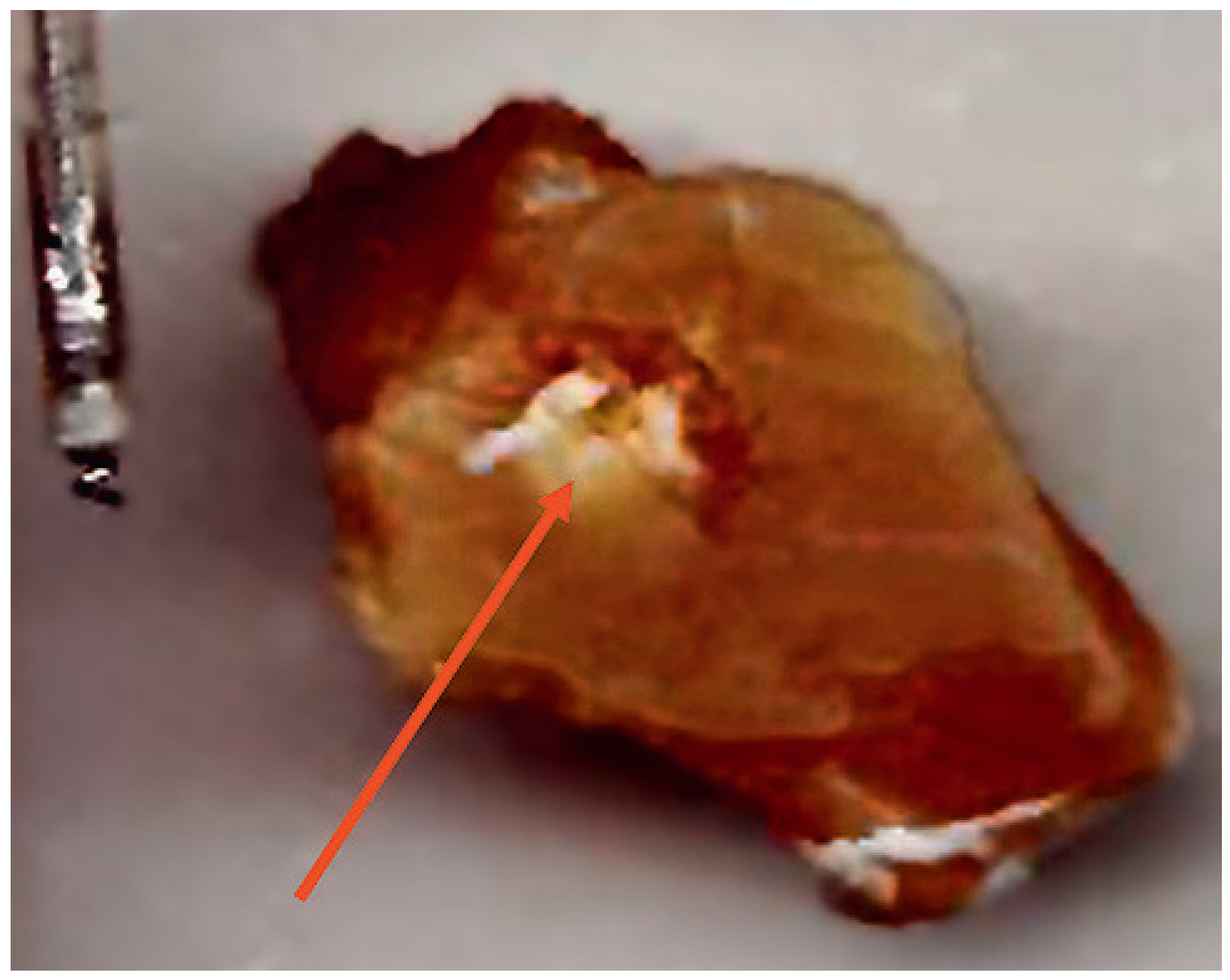
Figure 1. Discoloration of muscle tissue around the lead tip in an in vitro experiment caused by a high RF power MRI scan for 3 min.
Cardiovascmed 13 00070 g001
Figure 2. During an animal experiment using a conventional pacemaker system, a regular sustained ventricular tachycardia at ~240 bpm was observed. A stable rhythm at 85 bpm resumed within a few seconds after the scan was stopped. This illustrates the potential risk of arrhythmia induction in patients with pacemakers and the need for ECG monitoring during scanning (adapted from Luechinger et al. [29]).
Cardiovascmed 13 00070 g002
Table 1. Potential effects of MRI on pacing systems.
Cardiovascmed 13 00070 i001