Next Article in Journal
Peer Reviewers 2008
Previous Article in Journal
Herzrhythmusstörungen in der Klinik: Entscheidungsfindung zur Intervention jenseits existierender Richtlinien
 
 
Cardiovascular Medicine is published by MDPI from Volume 28 Issue 1 (2025). Previous articles were published by another publisher in Open Access under a CC-BY (or CC-BY-NC-ND) licence, and they are hosted by MDPI on mdpi.com as a courtesy and upon agreement with Editores Medicorum Helveticorum (EMH).
Editorial

Emboli Protection Devices in Cardiovascular Medicine

Division of Cardiology, University Hospital of Geneva, CH-1211 Geneva, Switzerland
Cardiovasc. Med. 2009, 12(2), 45; https://doi.org/10.4414/cvm.2009.01390
Submission received: 20 November 2008 / Revised: 20 December 2008 / Accepted: 20 January 2009 / Published: 20 February 2009

Abstract

Emboli protection devices (EPD) have a solid stand in the modern armamentarium of the interventionalist. However, the use of these devices is associated with a broad range of benefits in different cardiovascular territories. The most compelling data for mechanical emboli protection are available for percutaneous interventions of aortocoronary bypass grafts. In this setting, randomised studies have shown that the use of EPD is associated with a significant reduction in major adverse events. Nevertheless, current registry data suggest that the devices are used only in a minority of those cases. Conversely, in acute myocardial infarction, the routine use of EPD was not found to be beneficial. A strong consensus, though not unanimous, supports the use of these devices for carotid artery stenting. With respect to renal stenting, the experience is still at an early stage and the use of EPD cannot be recommended.

Introduction

Distal embolisation of plaque or thrombotic material may occur spontaneously during the process of plaque rupture and subsequent thrombus formation or may be induced by balloon angioplasty or stenting. The detection of this phenomenon, more frequent than previously believed, has been made possible by imaging technologies such as magnetic resonance, myocardial contrast echocardiography, and transcranial Doppler. The linkage between microvascular obstruction and unfavorable long-term clinical outcomes has been established for multiple vascular beds. Distal embolisation during percutaneous revascularisation may be prevented using a pharmacological (i.e., by periprocedural potent platelet inhibition and/or anticoagulation) or a mechanical approach.
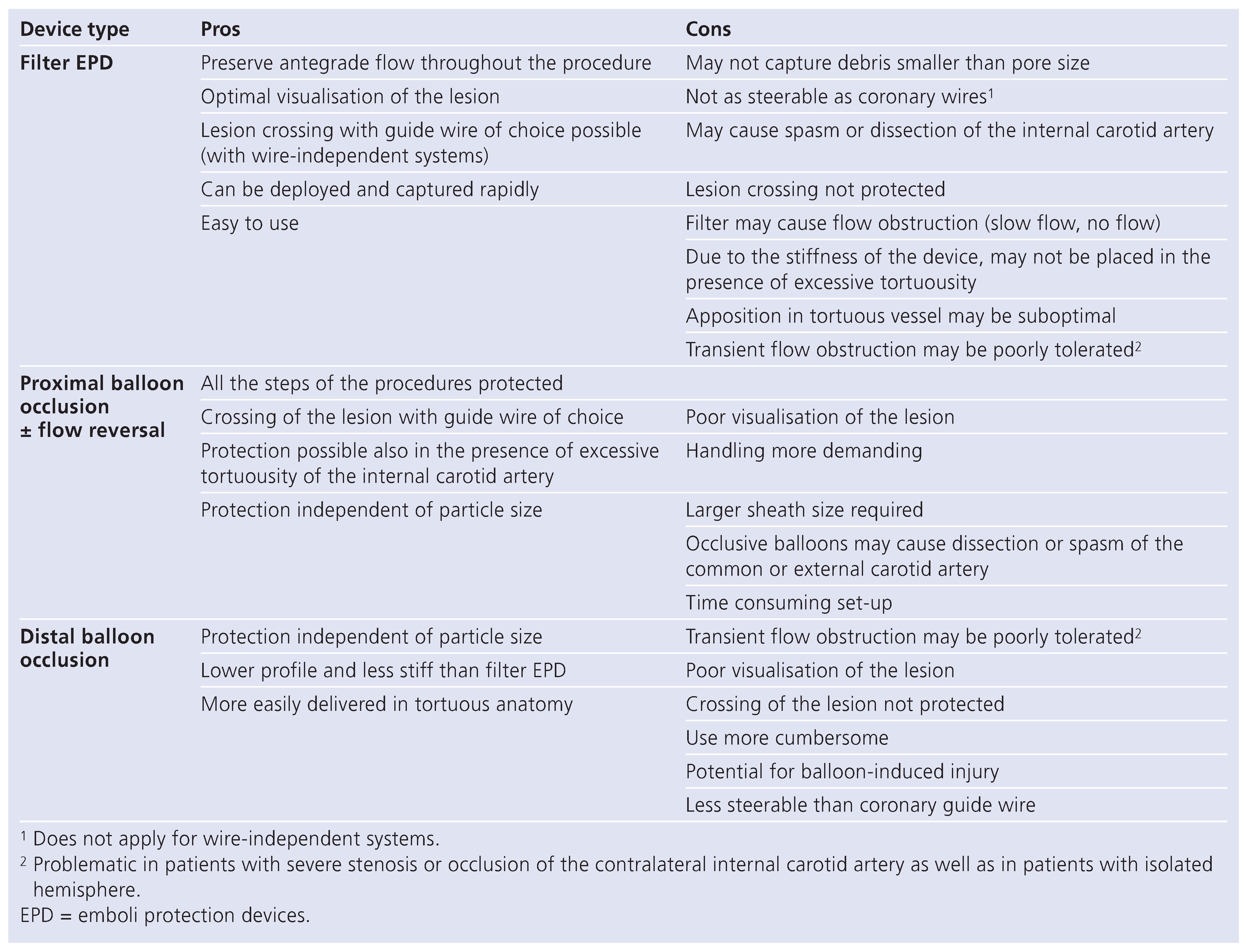
The concept of mechanical emboli protection is based on the placement of a filter device between the lesion treated and the distal vasculature or by a blockage of the blood flow in the vessel – either proximally or distally to the lesion – followed by an aspiration of the blood column prior to allow flow. The different approaches of mechanical emboli protection, in the example of carotid artery stenting, are demonstrated in Figure 1. As a general rule, filter-based emboli protection devices (EPD) are easy to use, allow blood flow throughout the procedure but particles smaller than the pore size (usually 100–150 μm) may reach the distal vasculature. Distal and proximal balloon occlusive devices allow for more complete retrieval of small particles suspended in the blood column at the time of intervention. The disadvantage of these approaches is the potential for ischaemia and the poor visualisation of the lesion (Table 1). Protection devices have been tested – with different degree of success – in various arterial beds including the coronary circulation, aortocoronary bypass grafts, the carotid artery, and the renal artery. The purpose of this manuscript is to briefly review the data supporting the use of EPD in cardiovascular interventions.

Acute myocardial infarction

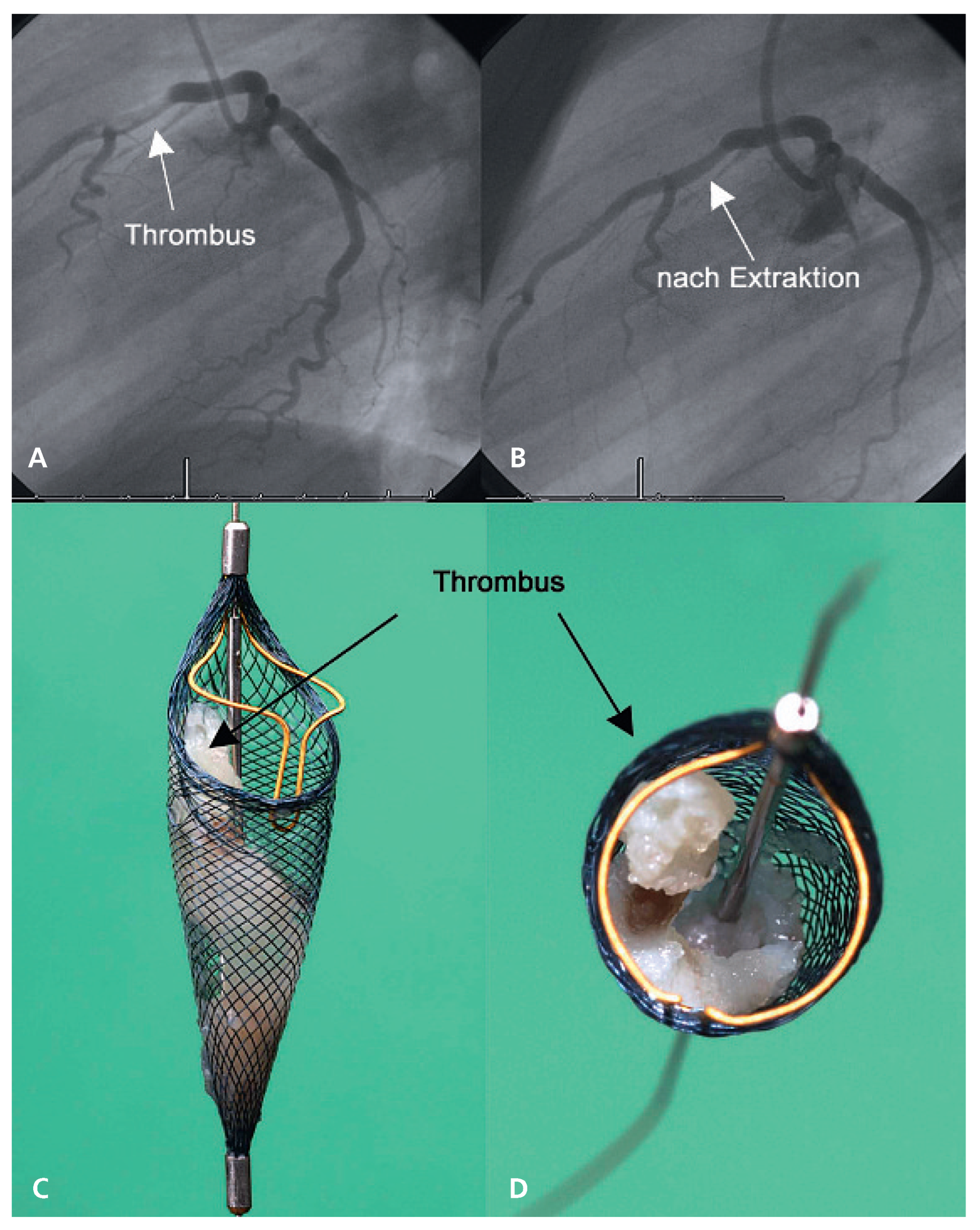
Several small studies have addressed the impact of EPD in the setting of acute myocardial infraction (MI). The largest of them was the EMERALD trial, which randomised 501 patients to conventional treatment or distal balloon protection. No difference was observed in terms of postprocedural ST-segment resolution on ECG, infarct size, or major clinical events at six months [1]. In a meta-analysis of eight randomised studies, for a total of 1467 patients, no difference in terms of post-procedural blood flow normalisation among patients with acute MI treated with protection and those without protection was observed [2]. However, an improvement in myocardial blush grade, suggesting a reduced damage of the microcirculation could be detected. Similarly, in the group treated with protection there was less frequently angiographic evidence of distal embolisation. Nevertheless, these findings were not associated with a survival benefit. In acute MI, the mainstay of distal embolisation prevention is a pharmacologic one based specifically on glycoprotein IIb/IIIa receptor (GP IIb/IIIa) inhibitors. Accordingly, the use of these agents has been associated with improved outcomes in this setting [3]. Adjunctive mechanical emboli protection should be reserved for selected cases with large thrombus burden (Figure 2).

Aortocoronary bypass grafts interventions

Percutaneous coronary intervention (PCI) of saphenous vein grafts (SVG) has been associated with less favorable outcomes compared to procedures involving the native coronary circulation. Acute complications include distal embolisation, “no-reflow”, and higher rate of periprocedural MI. Vein graft and coronary atherosclerosis are to be considered different diseases. Graft atheroma is diffuse, friable, soft, and lipid-rich. The fibrous cap is usually poorly developed or absent and marked calcification is rare [4]. All these features make SVG lesions prone to fragmentation and distal embolisation during PCI.
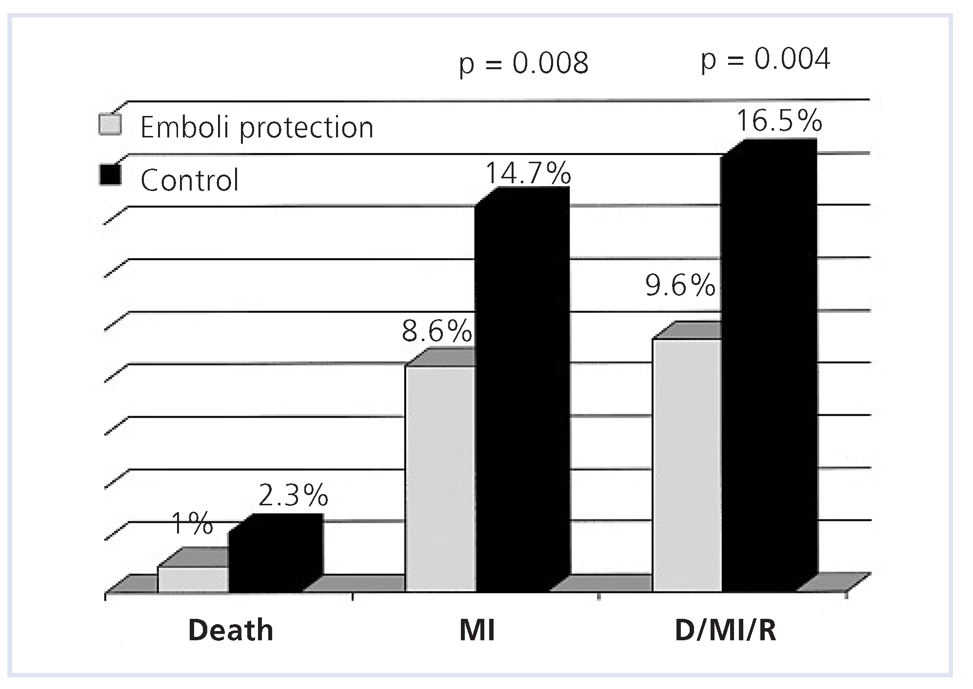
The use of EPD has been a major breakthrough in SVG PCI. A randomised trial enrolling over 800 patients using distal balloon occlusion demonstrated a 42% relative risk reduction of major adverse cardiac events at one month among patients allocated to emboli protection [5]. Most of the benefit was due to a reduction in periprocedural MI (Figure 3). Comparative studies among different type of EPD have followed. Current data suggests that filter devices, distal balloon occlusion devices and proximal occlusion devices may convey similar benefits [6,7]. Conversely, pharmacologic prevention of distal embolisation by means of glycoprotein IIb/IIIa receptor inhibitors was found of no benefit for SVG PCI. Accordingly, a pooled analysis of five large-scale randomised trials including over 600 patients undergoing bypass graft intervention detected no benefit from active treatment compared with placebo [8]. The likely explanation for this failure is that the amount and/or the composition of the material embolised during the procedure may overwhelm the capacity of these agents to protect the distal vasculature. Data from a large United States registry demonstrated that, despite the strong data supporting its use, EPD are currently deployed in <25% of SVG PCI [9]. In summary, mechanical emboli protection should be applied whenever feasible during PCI of SVG while no GP IIb/IIIa blockade is necessary. Importantly, this does not apply for intervention of arterial grafts (no data).

Carotid artery stenting

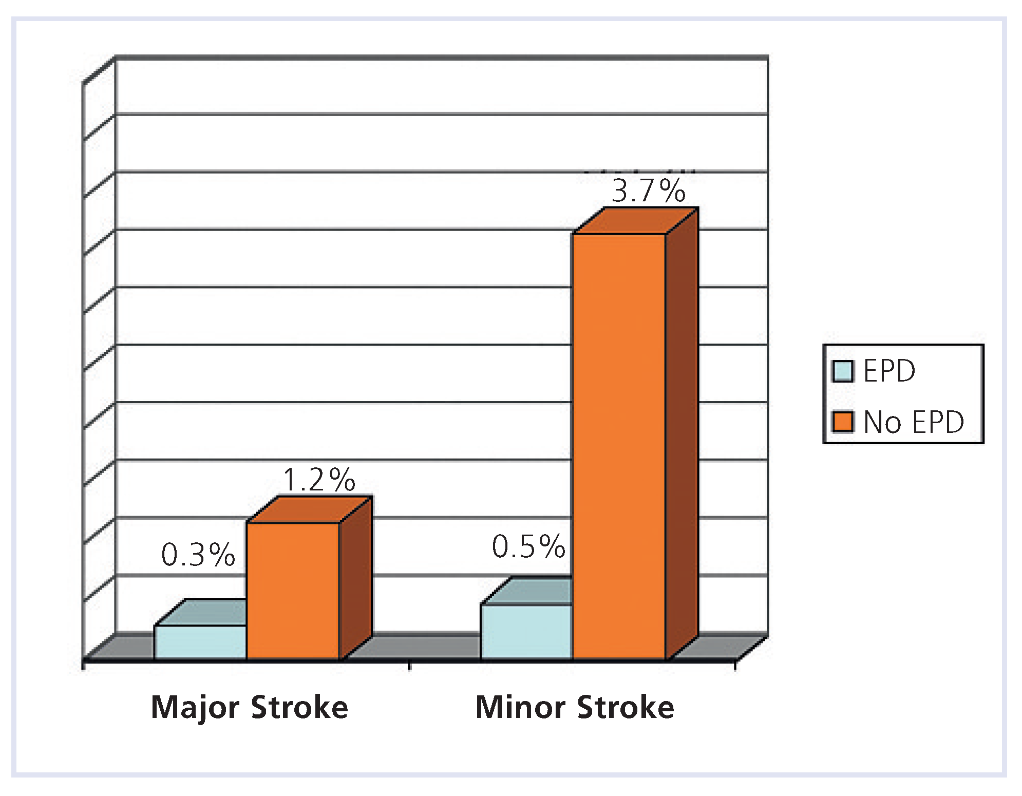
Distal embolisation remains the most feared complication of carotid artery stenting (CAS), although its incidence can be reduced by proper technique, adequate antithrombotic therapy, and the use of EPD. Virtually any step of the procedure may be associated with debris formation. The two most critical steps in this respect are the engagement of the common carotid artery with the guiding catheter or sheath and the postinflation of the stent [10]. The initial need for EPD came from transcranial Doppler studies demonstrating that, though in most cases clinically silent, embolic signals could be detected in virtually all cases of surgical and endovascular carotid revascularisation. Accordingly, new lesions on diffusion-weighted MRI following nonprotected CAS may be documented in over half of the patients [11]. A systematic review including 2357 patients undergoing unprotected CAS and 839 patients stented with adjunctive EPD documented a 30-day death or stroke rate of 5.5% and 1.8% (p <0.001), respectively (Figure 4) [12]. A German prospective CAS registry assessing 1483 procedures detected an in-hospital death or stroke rate of 2.1% among 666 patients undergoing CAS with adjunctive EPD and an event rate of 4.9% among 789 patients treated without EPD. Even after correcting for baseline characteristics the use of EPD was identified as independent protective factor (adjusted OR 0.45; 95% CI: 0.2–0.9; p = 0.026) [13]. Similarly, in a multicenter feasibility trial of CAS performed in 261 patients with and without EPD the oneyear major ipsilateral stroke rate was significantly lower among patients undergoing CAS with adjunctive EPD (0% vs 2.3%, p = 0.05) [14].
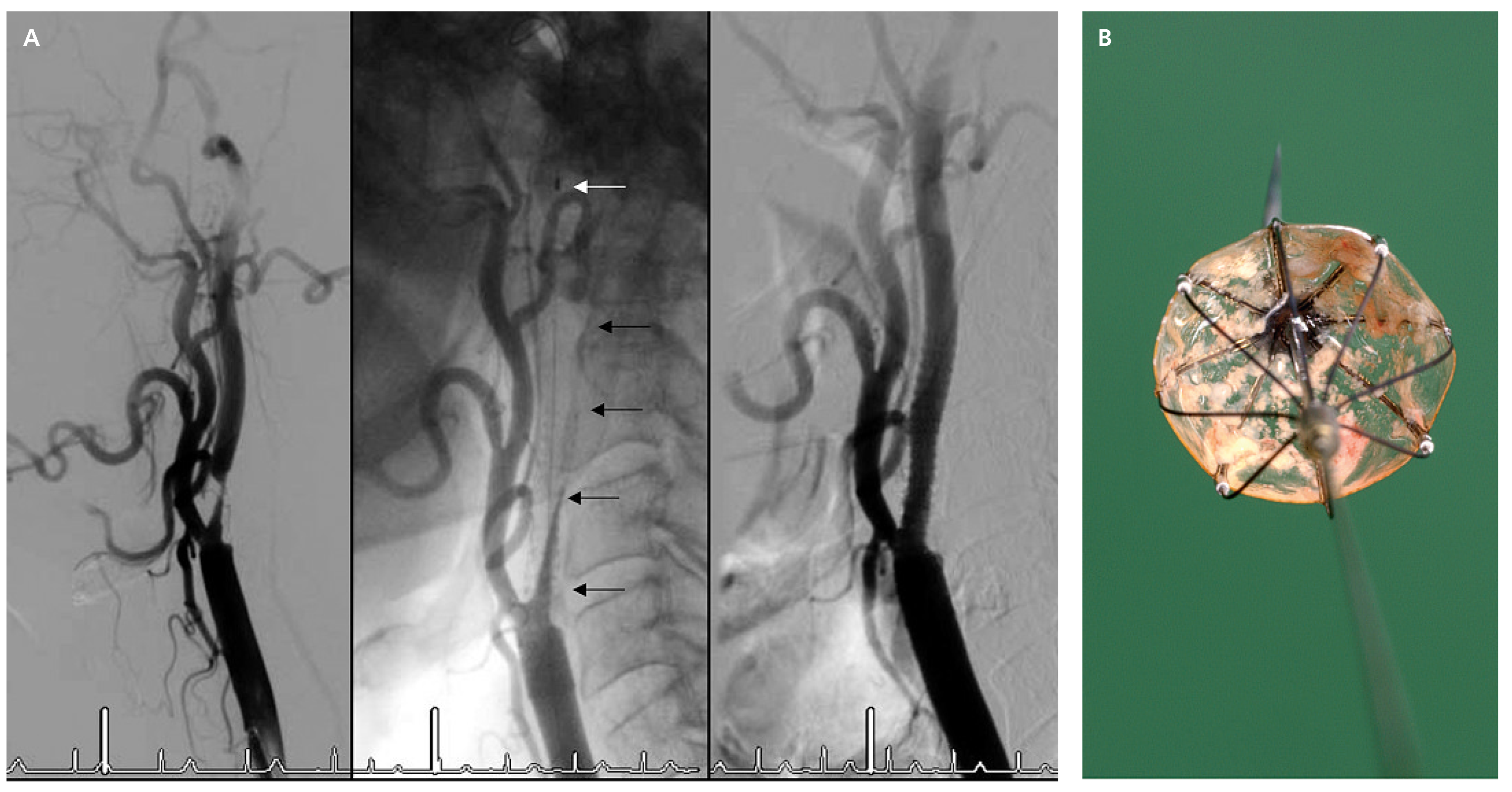
As a possible confounding factor, EPD have been used more recently and therefore likely at a later stage of the operator’s learning curve. Nevertheless, even in large volume centers, the use of EPD positively impacted outcomes. The placement EPD during CAS has been widely embraced, particularly within the community of interventional cardiologists. In our experience, EPD may be utilised in 95% of cases [15]. As correlated of distal embolisation during filter-based CAS, transient flow obstruction in the internal carotid artery may be observed (Figure 5A,B) [16]. The incidence of this phenomenon may differ according to the type of device used [17]. From a clinical perspective, filter EPD and occlusion-based EPD appear to convey similar efficacy [18]. A broadly shared consensus, though not unanimous, supports the use of EPD during CAS despite the lack of randomised trials based on the efficacy data available and the low device-related complication rate (<1%) [19]. In my opinion, CAS should be routinely performed with EPD. If this is not technically feasible, surgery should be considered.

Renal stenting

In order to prevent decline in renal function following renal revascularisation in patients with renal artery stenosis and renal insufficiency, it is currently being explored whether the use of EPD reduce the incidence of periprocedural atheroembolic renal disease (Figure 6). As opposed to the internal carotid artery or aortocoronary bypass grafts, the renal artery is characterised by a short trunk and early branching. These anatomical features limit the ability to protect the distal vasculature with a device. Although protected renal stenting is feasible, its clinical relevance in terms of preservation of renal function remains to be determined [20]. Encouraging appear the results of a French series showing no deterioration in renal function at six months among 45 patients treated under the protection of distal balloon occlusion [21]. Investigators from New Zealand have reported that among 63 patients with renal artery stenosis and renal dysfunction filter-protected renal stenting was associated with stabilisation or improvement of renal function in 97% of cases at six months, whereas in 3% of the patients renal function further declined [22]. Potentially, the use of this device could have deleterious effects including the need for longer catheter manipulations or the administration of a larger amount of contrast medium. In addition, the effects of microembolisation in the kidney are clearly less devastating than in the brain. Until randomised data assessing the efficacy of EPD become available, embolic mechanical protection cannot be advocated during percutaneous renal interventions.

Conclusions

EPD have a solid stand in the modern armamentarium of the interventionalist. However, the use of these devices is associated with a broad range of benefits in different cardiovascular territories. The most compelling data for mechanical emboli protection are available for percutaneous coronary interventions of aortocoronary vein grafts. In this setting, randomised studies have shown that the use of EPD is associated with a significant reduction in major adverse events. Nevertheless, current registry data suggest that the devices are used only in a minority of those cases. A strong consensus, though not unanimous, supports mechanical emboli protection for carotid artery stenting. In acute MI, the routine use of EPD was not found to be beneficial. Nevertheless, these devices may be considered in selected cases with large thrombus burden. With respect to renal stenting, the experience is still at an early stage and the use of these EPD cannot be recommended.

Conflicts of Interest

There is no conflict of interest.

References

  1. Stone, G.W.; Webb, J.; Cox, D.A.; Brodie, B.R.; Qureshi, M.; Kalynych, A.; et al. Distal microcirculatory protection during percutaneous coronary intervention in acute ST-segment elevation myocardial infarction: a randomized controlled trial. JAMA. 2005, 293, 1063–1072. [Google Scholar] [CrossRef] [PubMed]
  2. De Luca, G.; Suryapranata, H.; Stone, G.W.; Antoniucci, D.; Neumann, F.J.; Chiariello, M. Adjunctive mechanical devices to prevent distal embolization in patients undergoing mechanical revascularization for acute myocardial infarction: a meta-analysis of randomized trials. Am Heart J. 2007, 153, 343–353. [Google Scholar] [CrossRef]
  3. Kandzari, D.E.; Hasselblad, V.; Tcheng, J.E.; Stone, G.W.; Califf, R.M.; Kastrati, A.; et al. Improved clinical outcomes with abciximab therapy in acute myocardial infarction: a systematic overview of randomized clinical trials. Am Heart J. 2004, 147, 457–462. [Google Scholar]
  4. Motwani, J.G.; Topol, E.J. Aortocoronary saphenous vein graft disease: pathogenesis, predisposition, and prevention. Circulation. 1998, 97, 916–931. [Google Scholar] [CrossRef]
  5. Baim, D.S.; Wahr, D.; George, B.; Leon, M.B.; Greenberg, J.; Cutlip, D.E.; et al. Randomized trial of a distal embolic protection device during percutaneous intervention of saphenous vein aorto-coronary bypass grafts. Circulation. 2002, 105, 1285–1290. [Google Scholar]
  6. Stone, G.W.; Rogers, C.; Hermiller, J.; Feldman, R.; Hall, P.; Haber, R.; et al. Randomized comparison of distal protection with a filter-based catheter and a balloon occlusion and aspiration system during percutaneous intervention of diseased saphenous vein aorto-coronary bypass grafts. Circulation. 2003, 108, 548–553. [Google Scholar] [PubMed]
  7. Mauri, L.; Cox, D.; Hermiller, J.; Massaro, J.; Wahr, J.; Tay, S.W.; et al. The PROXIMAL trial: proximal protection during saphenous vein graft intervention using the Proxis Embolic Protection System: a randomized, prospective, multicenter clinical trial. J Am Coll Cardiol. 2007, 50, 1442–1449. [Google Scholar]
  8. Roffi, M.; Mukherjee, D.; Chew, D.P.; Bhatt, D.L.; Cho, L.; Robbins, M.A.; et al. Lack of benefit from intravenous platelet glycoprotein IIb/IIIa receptor inhibition as adjunctive treatment for percutaneous interventions of aortocoronary bypass grafts: a pooled analysis of five randomized clinical trials. Circulation. 2002, 106, 3063–3067. [Google Scholar] [PubMed]
  9. Mehta, S.K.; Frutkin, A.D.; Milford-Beland, S.; Klein, L.W.; Shaw, R.E.; Weintraub, W.S.; et al. Utilization of distal embolic protection in saphenous vein graft interventions (an analysis of 19,546 patients in the American College of Cardiology-National Cardiovascular Data Registry). Am J Cardiol. 2007, 100, 1114–1118. [Google Scholar]
  10. Roffi, M.; Abou-Chebl, A.; Mukherjee, D. Principles of Carotid Artery Stenting, 1st ed.; Uni-Med: Bremen, Germany, 2008; In Print. [Google Scholar]
  11. Krapf, H.; Nagele, T.; Kastrup, A.; Buhring, U.; Gronewaller, E.; Skalej, M.; et al. Risk factors for periprocedural complications in carotid artery stenting without filter protection: A serial diffusion-weighted MRI study. J Neurol. 2006, 253, 364–371. [Google Scholar]
  12. Kastrup, A.; Groschel, K.; Krapf, H.; Brehm, B.R.; Dichgans, J.; Schulz, J.B. Early outcome of carotid angioplasty and stenting with and without cerebral protection devices: a systematic review of the literature. Stroke. 2003, 34, 813–819. [Google Scholar]
  13. Zahn, R.; Mark, B.; Niedermaier, N.; Zeymer, U.; Limbourg, P.; Ischinger, T.; et al. Embolic protection devices for carotid artery stenting: better results than stenting without protection? Eur Heart J. 2004, 25, 1550–1558. [Google Scholar] [CrossRef]
  14. Ouriel, K.; Wholey, M.H.; Fayad, P.; Katzen, B.T.; Whitlow, P.; Frentzko, M.; et al. Feasibility trial of carotid stenting with and without an embolus protection device. J Endovasc Ther. 2005, 12, 525–537. [Google Scholar] [CrossRef]
  15. Roffi, M.; Greutmann, M.; Eberli, F.R.; Rainoni, L.; Luscher, T.F.; Amann-Vesti, B.; et al. Starting a carotid artery stenting program is safe. Catheter Cardiovasc Interv. 2008, 71, 469–473. [Google Scholar] [PubMed]
  16. Roffi, M.; Baumgartner, R.W.; Eberli, F.R. Images in cardiology. No reflow during carotid stenting. Heart (British Cardiac Society). 2006, 92, 538. [Google Scholar]
  17. Roffi, M.; Greutmann, M.; Schwarz, U.; Luscher, T.F.; Eberli, F.R.; AmannVesti, B. Flow impairment during protected carotid artery stenting: impact of filter device design. J Endovasc Ther. 2008, 15, 103–109. [Google Scholar] [PubMed]
  18. Zahn, R.; Ischinger, T.; Mark, B.; Gass, S.; Zeymer, U.; Schmalz, W.; et al. Embolic protection devices for carotid artery stenting: is there a difference between filter and distal occlusive devices? J Am Coll Cardiol. 2005, 45, 1769–1774. [Google Scholar] [CrossRef]
  19. Cremonesi, A.; Manetti, R.; Setacci, F.; Setacci, C.; Castriota, F. Protected carotid stenting: clinical advantages and complications of embolic protection devices in 442 consecutive patients. Stroke. 2003, 34, 1936–1941. [Google Scholar] [CrossRef] [PubMed]
  20. Roffi, M. Angioplasty for renal artery stenosis: technical improvements and results. In Electronic Update of the 7th edition of the Textbook Braunwald’s Heart Disease; Zipes, D.P., Libby, P., Bonow, R.O., Braunwald, E., Eds.; Saunders: Philadelphia, 2006. [Google Scholar]
  21. Henry, M.; Klonaris, C.; Henry, I.; Tzetanov, K.; Le Borgne, E.; Foliguet, B.; et al. Protected renal stenting with the PercuSurge GuardWire device: a pilot study. J Endovasc Ther. 2001, 8, 227–237. [Google Scholar] [CrossRef] [PubMed]
  22. Holden, A.; Hill, A.; Jaff, M.R.; Pilmore, H. Renal artery stent revascularization with embolic protection in patients with ischemic nephropathy. Kidney Int. 2006, 70, 948–955. [Google Scholar] [CrossRef][Green Version]
  23. Roffi, M.; Abou-Chebl, A.; Mukherjee, D. Principles of Carotid Artery Stenting, 1st ed.Uni-Med: Bremen, Germany, in Print.[Green Version]
  24. Roffi, M.; Mukherjee, D. Carotid artery disease management. In Manual of Vascular Diseases, 1st ed.; Rajagopalan, S., Mohler, E., Mukherjee, D., Eds.; Lippincott William and Wilkins: New York, NY, USA, 2005. [Google Scholar][Green Version]
  25. Surder, D.; Kucher, N.; Eberli, F.R.; Roffi, M. Intracoronary thrombus in a 26-year-old man. Eur Heart J. 2006, 27, 2631. [Google Scholar] [CrossRef] [PubMed]
Figure 1. Strategies for emboli protection devices in carotid artery stenting. On the left panel is demonstrated a filter device, in the middle a distal balloon occlusive device and in the right panel a proximal occlusive device. (Reproduced from [24]: Roffi M, Mukherjee D. Carotid artery disease management. In: Manual of Vascular Diseases. First edition. Ed. Rajagopalan S, Mohler E, Mukherjee D, editors. New York: Lippincott William and Wilkins; 2005. With kind permission, Copyright© 2009, Lippincott Williams & Wilkins, Baltimore, USA.).
Cardiovascmed 12 00045 g001
Figure 2. A. Coronary angiogram of a 26-year-old smoker with no angiographic evidence of coronary atherosclerosis but a large coronary thrombus in the proximal and mid portion of left anterior descending coronary artery (LAD), partially obstructing flow. B. Despite administration of aspirin, clopidogrel, unfractionated heparin, abciximab, and 20 mg of intracoronary r-tPA the thrombus persisted. C. In an unconventional – off label – way, direct thrombus extraction was performed by introducing a filter protection device into the mid LAD beyond the thrombus. D. The open device was gently pulled back so that the thrombus could be entirely trapped within the filter and then removed using a retrieval sheath. Final angiogram showed complete thrombus removal and no evidence of distal embolisation. E, F The filter contained a large thrombus. (Reproduced from [25]: Surder D, Kucher N, Eberli FR, Roffi M. Intracoronary thrombus in a 26-year-old man. Eur Heart J. 2006;27(22):2631. With kind permission, Copyright © 2009, Oxford University Press, Oxford, UK.).
Cardiovascmed 12 00045 g002
Figure 3. Adverse cardiac events among 801 patients undergoing vein graft interventions randomised to emboli protection (distal balloon occlusion) or no protection. Data extracted from [5]. D = death; MI = myocardial infarction; R = revascularisation.
Cardiovascmed 12 00045 g003
Figure 4. Systematic analysis of the periprocedural ischaemic complications following carotid artery stenting up to the year 2002. Data extracted from [12].
Cardiovascmed 12 00045 g004
Figure 5. A. Carotid artery stenting in a 82-year-old man with 90% symptomatic stenosis of the right internal carotid artery (ICA). The lesion was easily passed with a filter-emboli protection system and the device was deployed in the cervical part of the vessel (left panel). Following balloon post-inflation of the stent, no flow was detectable in the ICA (middle panel). Following aspiration of the carotid blood column the filter was retrieved with good angiographic result locally and intracranially in the absence of symptoms (right panel). B. Inspection of the retrieved filter showed a large amount of debris trapped in the device.
Cardiovascmed 12 00045 g005
Figure 6. Stenting of the left renal artery using a filter emboli protection device. Digital subtraction angiography demonstrates an ostial stenosis (left panel). In the middle panel, the arrow denotes a filter for emboli protection which is in place throughout the procedure and at the same time has guide wire function. The result following stenting is demonstrated in the right panel. (Reproduced from [20]: Roffi M. Angioplasty for renal artery stenosis: technical improvements and results. In: Electronic Update of the 7th edition of the Textbook Braunwald’s Heart Disease. Zipes DP, Libby P, Bonow RO, Braunwald E, editors. Philadelphia: Saunders; 2006. Copyright © 2009, Elsevier, Oxford, UK.).
Cardiovascmed 12 00045 g006
Table 1. Pros and cons of emboli protection devices (after [23]).
Cardiovascmed 12 00045 i001