Next Article in Journal
Flutter Auriculaire 2:1 Simulant un Bloc Atrio-Ventriculaire de Premier Degré
Previous Article in Journal
Le Chaînon Manquant dans le Traitement du Syndrome Coronarien Aigu: L'éducation Thérapeutique
 
 
Cardiovascular Medicine is published by MDPI from Volume 28 Issue 1 (2025). Previous articles were published by another publisher in Open Access under a CC-BY (or CC-BY-NC-ND) licence, and they are hosted by MDPI on mdpi.com as a courtesy and upon agreement with Editores Medicorum Helveticorum (EMH).
Editorial

What Is an Optimal Stent? Biological Requirements of Drug-Eluting Stents

by
Giovanni G. Camici
1,2
1
Cardiovascular Center, University and University Hospital, Zürich Center for Integrative Human Physiology, University of Zürich, CH-8057 Zürich, Switzerland
2
Cardiovascular Research, Physiology Institute, University of Zurich, Winterthurerstrasse 190, CH-8057 Zürich, Switzerland
Cardiovasc. Med. 2008, 11(1), 22; https://doi.org/10.4414/cvm.2008.01301
Submission received: 25 October 2007 / Revised: 25 November 2007 / Accepted: 25 December 2007 / Published: 25 January 2008

Summary

Although drug eluting stents (DES) have been successful in preventing neointima formation thereby decreasing the rates of restenosis, several concerns remain about the incidence of stent thrombosis – a rare complication associated to high mortality. For this reason, much effort is being made in searching for new molecules and approaches to develop innovative DES able to inhibit restenosis as well as decreasing the incidence of late stent thrombosis. Second generation DES have recently entered daily clinical practice but definitive data concerning their efficacy are not available yet. Still, the close relationship between agents used on first generation DES and those employed in second generation DES gives us reason to speculate that more efforts are needed in order to make concrete advances. This review article focuses on the existing problems of currently available DES as well as the innovations needed in order to improve further the outcome of stent implantation. Present time efforts should be directed towards identifying multi-coatings to be applied on inert polymers so as to have targeted approaches for each of the therapeutical requirements of a stented coronary artery. An ideal stent should locally inhibit vascular smooth muscle cells proliferation, enhance endothelial healing through improved endothelial progenitor cells function and ultimately prevent platelets activation. Current technology seems to offer individual alternatives for all these requirements; so the “perfect stent” should be soon available.

Introduction

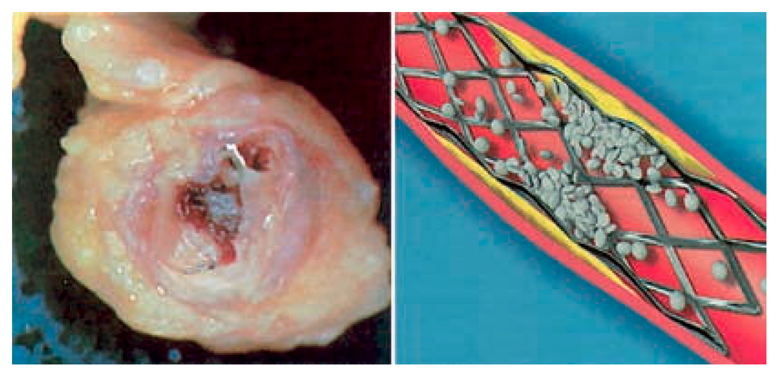
Percutaneous coronary intervention (PCI) is a routine procedure performed to revascularise occluded coronary arteries of patients which suffered an acute myocardial infarction. Following revascularisation, stents – cylinderlike structures designed with the intent of preventing or delaying vessel re-narrowing – are routinely inserted in the vessel. At first, stents were considered as mere physical impediments to prevent restenosis; however in the last decade stents were exploited as true reservoirs for the local release of specific drugs at the site of injury. This novel concept enabled the coating of stents with drugs, such as sirolimus designed to interfere with cell cycle progression to inhibit the excessive proliferation and migration of vascular smooth muscle cells (VSMC) taking place after stent deployment. Drug eluting stents (DES) proved extremely successful in reducing restenosis rates from the 20–30% range, as observed with bare metal stents (BMS) to single digits [1]. Nevertheless, the frequency of stent thromboses (Figure 1) has not decreased as compared to BMS [2].
Cardiovascmed 11 00022 i001
Figure 1. Human artery showing a complete occlusion caused by stent thrombosis (left) and, cartoon depicting early phases of platelet recruitment at the site of the lesion within the stented segment. Website: www.gary ferster.com (right).
Cardiovascmed 11 00022 g001
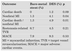
As recently reported at the congress of the American Heart Association, rate of cardiac death and non-fatal myocardial infarctions has unexpectedly increased with the advent of DES as compared to BMS (Table 1).
For this reason, stent thrombosis – a rare but severe complication occurring after stent implantation – remains a major concern in contemporary clinical practice where DES are employed [3]. Such clinical scenario prompted several investigators to seek possible explanations to the lasting occurrence of stent thromboses. Much emphasis has been put on the possible undesired effects caused by the different coatings and eluted agents used in DES [4,5,6].
Limited information are available about the pathogenesis of stent thrombosis [7] however, a recent study identified several key observations, including: (1.) DES show delayed healing as well as reduced reendothelialisation compared with BMS (p = 0.0001); and (2.) other factors associated with late stent thrombosis include local tissue hypersensitivity reactions, and stent specific adverse reactions [8].
DES proved to impair the healing process which normally begins as result of mechanical injury, after stent implantation. Arecent study of 48 matched DES-BMS patients revealed that reendothelialisation occurred in only 56% of the DES cases compared with 90% of the BMS cases. This finding was underlined by higher fibrin depositions which reveal delayed healing caused by inflammation in the DES-treated lesions. In addition, 61% of the DES cases showed signs of late stent thrombosis compared with only 8% of the BMS cases [9] (Figure 2).
Several lines of evidence demonstrated that the observed delay in reendothelialisation may be due to the unspecific nature of the antiproliferative agents eluted by DES. In fact, it is likely that agents released from DES do not only affect proliferation and migration of VSMC, but also that of endothelial cells. This hypothesis was later confirmed by two studies showing that both rapamycin and paclitaxel besides inhibiting VSMC proliferation, also inhibit endothelial cell proliferation and migration [4,10] thus, probably impairing reendothelialisation and leaving the thrombogenic stent struts exposed to circulating platelets and coagulation factors (Figure 2). Although these results may not accurately represent the fate of patients who receive DES and survive, they do show that the natural healing process after DES implantation is not as optimal as originally hypothesised. In consideration of the above, an optimal stent should release agents aimed at suppressing VSMC proliferation without impairing endothelial cells function. Seemingly this could be achieved with existing compounds, such as tacrolimus that differentially affects VSMC and endothelial cells proliferation and thus should be considered for novel combined applications [11]. In addition to sparing endothelial cells proliferation, an optimal stent should be conceived using inert coatings which do not elicit local inflammation as previously illustrated [8] but rather offer a plain surface which diminishes platelet adhesion and fibrinogen binding as compared to BMS [12].
An additional limitation of contemporary DES is represented by the lack of a strategy aimed at enhancing endothelial progenitor cells (EPCs) function. Several studies underlined the fundamental role of EPCs in the process of reendothelialisation taking place following PCI [13,14] hence, current DES should be conceptualised with the intent of stimulating EPCs function. Paradoxically, rapamycin was recently shown to inhibit proliferation, migration, and differentiation of human EPCs in vitro and thus probably accounting at least in part, for the lack of reendothelialisation observed with DES [15,16]. In light of this finding, the decreased rate of reendothelialisation recently reported in DES compared to BMS becomes less surprising [9]. In keeping with this problem, an innovative approach was lately described whereby stents struts are loaded with integrin-binding peptides so as to limit coronary neointima formation and, at the same time accelerate reendothelialisation by attracting EPCs [17]. Results from this study showed enhanced endothelial coverage on stents loaded with integrin-binding peptides at four weeks associated with a significant increase in the early recruitment of infused EPCs [17]. Although long term data on the efficacy of these stents are not available yet, integrin-binding peptides loaded stents may be useful for reducing in-stent restenosis by accelerating reendothelialisation and, may prove a winning formula in the near future.
Thrombosis is principally driven by tissue factor (TF) – the crucial initiator of the coagulation cascade. Intuitively, modern stents should be designed so as to locally reduce the expression of TF protein thus minimising the risks of stent thrombosis. Paradoxically, independent reports recently described that both rapamycin and paclitaxel enhance expression and activity of TF under inflammatory conditions [4,5,6]. This surprising finding represents a real threat especially when considering that agents used on second generation DES ie everolimus and zotarolimus, are structurally very closely related to rapamycin and thus give us reason to expect similar unwanted side effects. Such scenario is surely symptomatic of a lack of sufficient progress.
In this challenging setting, a novel alternative was recently illustrated whereby dimethyl sulfoxide (DMSO) may represent an interesting therapeutic principle for DES. DMSO was shown to inhibit VSMC proliferation and migration while heavily blunting TF expression in endothelial cells, VSMC, and monocytes. Additionally, intraperitoneal application of DMSO in vivo prevented thrombotic occlusion in a mouse model of photochemical carotid artery injury. Such data attract much attention especially in view of a possible additional effect of DMSO as an antiplatelet agent [18]. Consequently, DMSO may hold some of the criteria that agents eluted from modern DES should meet. Nevertheless more insights on higher animal models and humans will be needed before considering possible clinical applications.

Conclusion

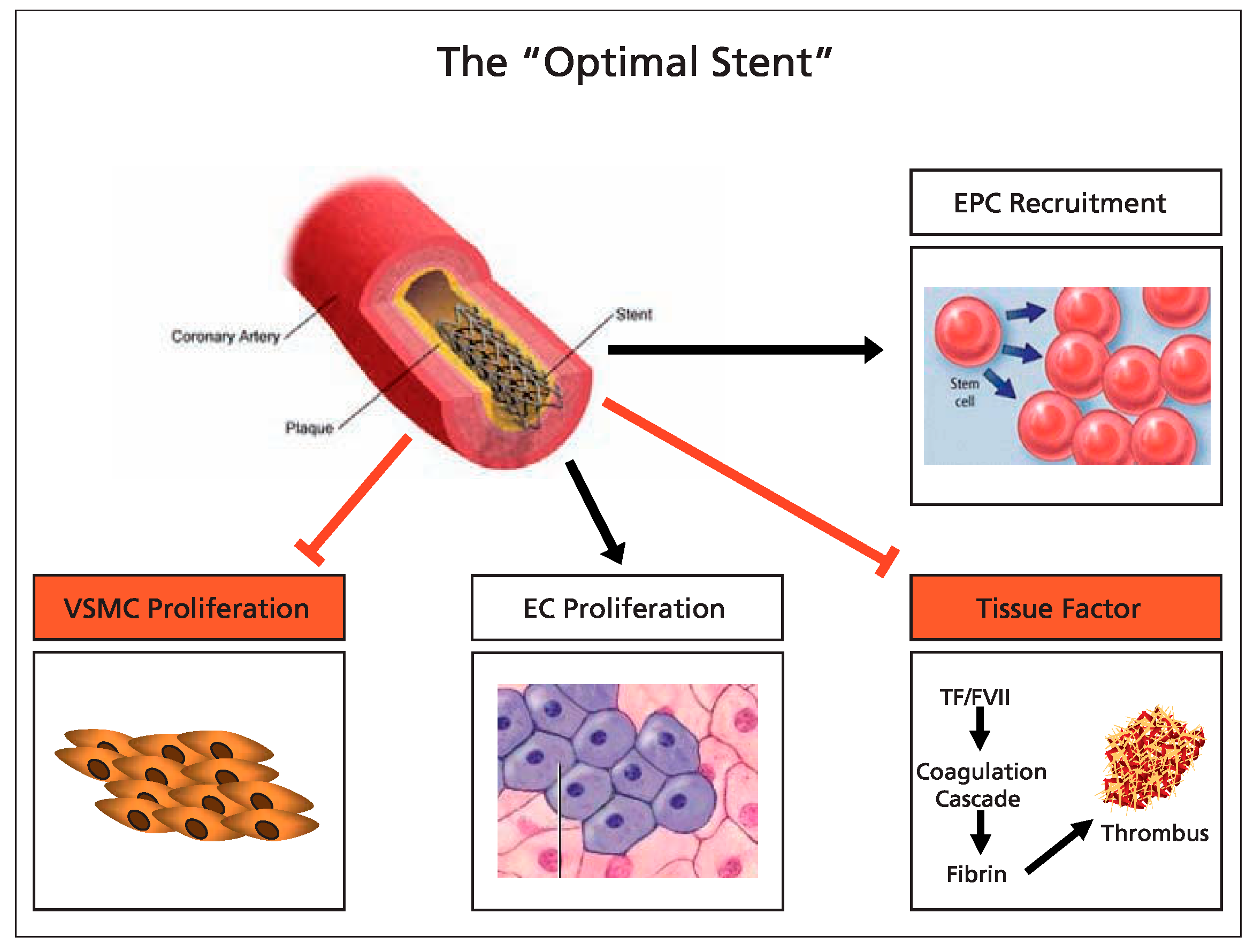
The optimal DES will be coated with a simple-structure neutral polymer which does not elicit local inflammatory responses and does not favour platelet adherence. Additionally, it will inhibit restenosis through a VSMC-specific antiproliferative effect which will spare endothelial cells. More emphasis will be put on the role of EPCs thus, the optimal stent will be coated with peptide-binding integrins [17] or antibodies [19] which will enhance EPCs retrieval thus facilitating reendothelialisation. Ideally, future stents should also target local prothrombotic factors such as TF. In this direction, the optimal stent will be releasing anti-TF agents such as or with similar properties to DMSO which will contribute to maintaining local haemostasis on top of having synergistic effects to antiproliferative agents used on the same stent. The optimal stent will have the “right drug for the right condition” approach and will hence release a multitude of agents which will maintain local homeostasis intact (Figure 3).

References

  1. Steffel, J.; Tanner, F.C. Biological effects of drug-eluting stents in the coronary circulation. Herz. 2007, 32, 268–273. [Google Scholar] [CrossRef] [PubMed]
  2. Iakovou, I.; Schmidt, T.; Bonizzoni, E.; Ge, L.; Sangiorgi, G.M.; Stankovic, G.; et al. Incidence, predictors, and outcome of thrombosis after successful implantation of drug-eluting stents. JAMA. 2005, 293, 2126–2130. [Google Scholar] [CrossRef] [PubMed]
  3. Camenzind, E.; Steg, P.G.; Wijns, W. Stent thrombosis late after implantation of first-generation drug-eluting stents: a cause for concern. Circulation. 2007, 115, 1440–1455, discussion 1455. [Google Scholar] [CrossRef] [PubMed]
  4. Steffel, J.; Latini, R.A.; Akhmedov, A.; Zimmermann, D.; Zimmerling, P.; Luscher, T.F.; et al. Rapamycin, but not FK-506, increases endothelial tissue factor expression: implications for drug-eluting stent design. Circulation. 2005, 112, 2002–2011. [Google Scholar] [CrossRef]
  5. Stahli, B.E.; Camici, G.G.; Steffel, J.; Akhmedov, A.; Shojaati, K.; Graber, M.; et al. Paclitaxel enhances thrombin-induced endothelial tissue factor expression via c-Jun terminal NH2 kinase activation. Circ Res. 2006, 99, 149–155. [Google Scholar] [CrossRef]
  6. Zhu, S.; Viswambharan, H.; Gajanayake, T.; Ming, X.F.; Yang, Z. Sirolimus increases tissue factor expression but not activity in cultured human vascular smooth muscle cells. BMC Cardiovasc Disord. 2005, 5, 22. [Google Scholar] [CrossRef] [PubMed][Green Version]
  7. Holmes, D.R.; Jr, Kereiakes, D.J.; Laskey, W.K.; Colombo, A.; Ellis, S.G.; Henry, T.D.; et al. Thrombosis and drug-eluting stents: an objective appraisal. J Am Coll Cardiol. 2007, 50, 109–118. [Google Scholar] [CrossRef] [PubMed]
  8. Mauri, L.; Hsieh, W.H.; Massaro, J.M.; Ho, K.K.; D’Agostino, R.; Cutlip, D.E. Stent thrombosis in randomized clinical trials of drug-eluting stents. N Engl J Med. 2007, 356, 1020–1029. [Google Scholar] [CrossRef] [PubMed]
  9. Stone, G.W.; Moses, J.W.; Ellis, S.G.; Schofer, J.; Dawkins, K.D.; Morice, M.C.; et al. Safety and efficacy of sirolimus- and paclitaxel-eluting coronary stents. N Engl J Med. 2007, 356, 998–1008. [Google Scholar] [CrossRef] [PubMed]
  10. Parry, T.J.; Brosius, R.; Thyagarajan, R.; Carter, D.; Argentieri, D.; Falotico, R.; et al. Drug-eluting stents: sirolimus and paclitaxel differentially affect cultured cells and injured arteries. Eur J Pharmacol. 2005, 524, 19–29. [Google Scholar] [CrossRef] [PubMed]
  11. Matter, C.M.; Rozenberg, I.; Jaschko, A.; Greutert, H.; Kurz, D.J.; Wnendt, S.; Kuttler, B.; et al. Effects of tacrolimus or sirolimus on proliferation of vascular smooth muscle and endothelial cells. J Cardiovasc Pharmacol. 2006, 48, 286–292. [Google Scholar] [CrossRef] [PubMed]
  12. Windecker, S.; Simon, R.; Lins, M.; Klauss, V.; Eberli, F.R.; Roffi, M.; et al. Randomized comparison of a titanium-nitrideoxide-coated stent with a stainless steel stent for coronary revascularization: the TiNOX trial. Circulation. 2005, 111, 2617–2622. [Google Scholar] [CrossRef] [PubMed]
  13. Griese, D.P.; Ehsan, A.; Melo, L.G.; Kong, D.; Zhang, L.; Mann, M.J.; et al. Isolation and transplantation of autologous circulating endothelial cells into denuded vessels and prosthetic grafts: implications for cell-based vascular therapy. Circulation. 2003, 108, 2710–2715. [Google Scholar] [CrossRef] [PubMed]
  14. Urao, N.; Okigaki, M.; Yamada, H.; Aadachi, Y.; Matsuno, K.; Matsui, A.; et al. Erythropoietin-mobilized endothelial progenitors enhance reendothelialization via Akt-endothelial nitric oxide synthase activation and prevent neointimal hyperplasia. Circ Res. 2006, 98, 1405–1413. [Google Scholar] [CrossRef] [PubMed]
  15. Chen, T.G.; Chen, J.Z.; Wang, X.X. Effects of rapamycin on number activity and eNOS of endothelial progenitor cells from peripheral blood. Cell Prolif. 2006, 39, 117–125. [Google Scholar] [CrossRef] [PubMed]
  16. Butzal, M.; Loges, S.; Schweizer, M.; Fischer, U.; Gehling, U.M.; Hossfeld, D.K.; et al. Rapamycin inhibits proliferation and differentiation of human endothelial progenitor cells in vitro. Exp Cell Res. 2004, 300, 65–71. [Google Scholar] [CrossRef] [PubMed]
  17. Blindt, R.; Vogt, F.; Astafieva, I.; Fach, C.; Hristov, M.; Krott, N.; et al. A novel drug-eluting stent coated with an integrin-binding cyclic Arg-Gly-Asp peptide inhibits neointimal hyperplasia by recruiting endothelial progenitor cells. J Am Coll Cardiol. 2006, 47, 1786–1795. [Google Scholar] [CrossRef] [PubMed]
  18. Dujovny, M.; Rozario, R.; Kossovsky, N.; Diaz, F.G.; Segal, R. Antiplatelet effect of dimethyl sulfoxide, barbiturates, and methyl prednisolone. Ann N Y Acad Sci. 1983, 411, 234–244. [Google Scholar] [CrossRef] [PubMed]
  19. Aoki, J.; Serruys, P.W.; van Beusekom, H.; Ong, A.T.; McFadden, E.P.; Sianos, G.; et al. Endothelial progenitor cell capture by stents coated with antibody against CD34: the HEALINGFIM (Healthy Endothelial Accelerated Lining Inhibits Neointimal Growth-First In Man) Registry. J Am Coll Cardiol. 2005, 45, 1574–1579. [Google Scholar] [CrossRef] [PubMed]
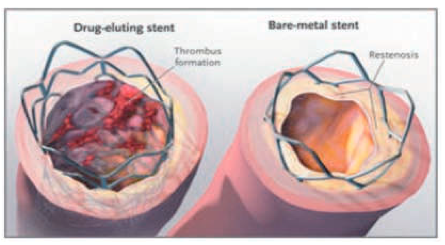
Figure 2. In-growth of tissue may cause BMS to become obstructed, resulting in the need for a second procedure. DES inhibit this process, but uncovered stent struts may be prone to thrombosis after discontinuation of antiplatelet therapy. (Shuchmann M. Debating the risks of drug-eluting stents. N Engl J Med. 2007;356(4):325–8. Copyright© 2008 Massachusetts Medical Society. All rights reserved.)
Cardiovascmed 11 00022 g002
Figure 3. Requisites of the optimal stent. EPC = endothelial progenitor cells; VSMC = vascular smooth muscle cells; EC = endothelial cells.
Cardiovascmed 11 00022 g003
Table 1. Outcome comparison between BMS and DES. (Presented by Pfisterer ME at the American College of Cardiology 2006.)
Cardiovascmed 11 00022 i002